cytosines, but not purines, determine rag … dna structures: ... (high mobility group box 1). ......
TRANSCRIPT

1
CYTOSINES, BUT NOT PURINES, DETERMINE RAG INDUCED BREAKS ON
HETERODUPLEX DNA STRUCTURES: IMPLICATIONS FOR GENOMIC INSTABILITY Abani Kanta Naik1, Michael R. Lieber2 and Sathees C. Raghavan1*
From 1Department of Biochemistry, Indian Institute of Science, Bangalore-560 012, India and 2USC Norris Comprehensive Cancer Ctr., Rm. 5428, Department of Pathology, 1441 Eastlake Ave.,
MC 9176, Los Angeles, CA 90089-9176 Running title: RAG cleavage on heteroduplex DNA occurs at cytosines
Address for correspondence: Sathees C. Raghavan, PhD, Department of Biochemistry, Indian Institute of Science, Bangalore-560 012, India, Ph.+91 80 2293 2674; Fax: 080 2360 0814; E-mail: [email protected]
The sequence-specificity of the RAG complex during V(D)J recombination has been well studied. RAGs can also act as structure-specific nuclease, however, little is known about the mechanism of its action. Here, we show that in addition to DNA structure, sequence dictates the pattern and efficiency of RAG cleavage on altered DNA structures. Cytosine nucleotides are preferentially nicked by RAGs, when present at single-stranded regions of heteroduplex DNA. Although unpaired thymine nucleotides are also nicked, the efficiency is many fold weaker. Induction of single- or double-strand breaks by RAGs depends on the position of cytosines and whether it is present on one or both of the strands. Interestingly, RAGs are unable to induce breaks when adenine or guanine nucleotides are present at single-strand regions. The nucleotide present immediately next to the bubble sequence could also affect RAG cleavage. Hence, we propose “C(d)C(S)C(S)” (d, double-stranded; s, single-stranded) as a consensus sequence for RAG induced breaks at single-/double-strand DNA transitions. Such a consensus sequence motif is useful for explaining RAG cleavage on other types of DNA structures described in the literature. Therefore, the mechanism of RAG cleavage described here could explain facets of chromosomal rearrangements specific to lymphoid tissues leading to genomic instability. The RAG (recombination activating gene) complex, consisting of RAG1 and RAG2, is the nuclease responsible for V(D)J recombination, a physiological process by which immunoglobulin and T-cell receptor diversity is generated. RAGs are normally expressed in B-cells and T-cells (1). During V(D)J recombination the variable (V),
diversity (D) and joining (J) subexons are rearranged. Specific sequences present at the ends of the subexon, called recombination signal sequences (RSS) are recognized by RAGs. Each RSS consists of a conserved heptamer and nonamer, separated by a nonconserved spacer, the length of which designates RSS as a 12-signal or 23-signal. Normally during V(D)J recombination, a 12-signal pairs with a 23-signal with the help of proteins like HMGB1 (high mobility group box 1). The nick induced by RAGs during V(D)J recombination is consistently at 5’ of the heptamers (2-14). The nicked strand is then converted to a hairpin in each V, D and J coding end by a transesterification reaction, leaving each of the signal ends blunt (15). The hairpins are then opened by the Artemis-DNAPKcs complex (16). After cleavage, the RAG complex remains tightly bound to the two signal ends and less tightly bound to the coding end, in a postcleavage complex (17,18). Finally, the complete exon coding for antibody or TCR (T cell receptor) is generated by joining of the broken subexons by nonhomologous DNA endjoining (NHEJ) (19,20).
In the recent past, studies have shown that cryptic RSS sites present elsewhere in the genome can also act as off-target sites for RAG misrecognition, leading to chromosomal translocations in lymphoid cancers such as leukemia (21-24). In addition to its sequence specific endonuclease activities, recent studies have shown that RAG complex can act as a structure-specific nuclease (22). We previously showed that a non-B DNA structure formed at the BCL2 major breakpoint region (MBR) on chromosome 18 involved in t(14;18) translocation in follicular lymphoma can be cleaved by RAGs (25-29). Further, it was also shown that in addition to the BCL2 MBR structure, other non-B
http://www.jbc.org/cgi/doi/10.1074/jbc.M109.089631The latest version is at JBC Papers in Press. Published on January 5, 2010 as Manuscript M109.089631
Copyright 2010 by The American Society for Biochemistry and Molecular Biology, Inc.
by guest on July 11, 2018http://w
ww
.jbc.org/D
ownloaded from

2
DNA structures, such as heteroduplex DNA and heterologous loops are also targets for the RAG complex (30,31). RAG complex can also cleave 3’ overhangs, flap DNA and gap structures (32). All of these studies were done at physiological concentrations of divalent cation, Mg2+. Two independent groups have also shown that RAGs are able to cleave hairpin structures in Mn2+
containing buffer and this was observed to a much lesser extent in Mg2+ (33,34). In a recent study, we found that the most common translocations in early human B cells occur at CpG sites. We proposed that deamination of methylated cytosines at CpG can lead to small unpaired bubble like regions in the genome, which RAGs can cleave to generate breaks (35).
The above studies, therefore, suggest that changes in the normal B-DNA structure could make the region vulnerable to RAGs when present in lymphoid tissues. This could explain why lymphoid cells possess elevated levels of chromosomal translocation and other rearrangements compared to non-lymphoid tissues. Such elevated levels of pathological chromosomal rearrangement leads to the altered expression of critical genes resulting in human lymphoid malignancies such as leukemia and lymphoma (28,36). Since such structural changes in the DNA could lead to elevated levels of single- and double-strand breaks, those could also account for the increased genomic instability. Our previous studies have shown that when acting as a structure-specific nuclease, RAGs recognize and bind to the single-stranded region of heteroduplex DNA, and the efficiency of the cleavage depends on the length of the single-stranded region (28,30,31). However, there are many important questions unanswered. What dictates the specificity of RAG cleavage, when it acts as a structure-specific nuclease? Since, mostly such cleavage on altered DNA structures are pathologic, it is important to know the mechanism which determines whether a given region may be cleaved by RAGs or not. This will also help to understand the mechanism of genomic instability in lymphoid cells. We previously reported occurrence of strand bias, when RAGs cleave on non-B DNA structures (30). Since double-strand break formation needs two independent nicks in close proximity on the DNA, it was not understood how such nicks can lead to
DSBs during chromosomal rearrangements. It has been shown that during sequence-specific cleavage of RSS by RAGs, the coding sequence influences the nicking (37-39). Thus, the role of neighboring sequences, when RAGs act as a structure-specific nuclease, deserves examination.
In the present study we have attempted to understand the mechanism by which RAGs cleave altered DNA structures. Here we report that RAG cleavage on heteroduplex DNA is sequence-dependent. RAGs can cleave heteroduplex DNA only when pyrimidines are present at the single-stranded region. Cytosines are the preferred nucleotides and when present on both strands, RAG cleavage leads to a double-strand break. We further show that the sequence-dependence is also applicable to all other structural alterations studied.
EXPERIMENTAL PROCEDURES
Enzymes, chemicals, and reagents: Chemical reagents were obtained from Sigma Chemical Co. (St. Louis, MO), Amresco (USA) and SRL (India). DNA modifying enzymes were purchased from New England Biolabs (Beverly, MA) and Fermentas (Glen Burnie, MD, USA). Radioisotope-labeled nucleotides were purchased from BRIT (Hyderabad, India). Culture medias were from Sera Laboratory International limited (West Sussex, RH17 5PB UK), FBS and PenStrep were from Gibco BRL (USA). Oligomers: Oligomers were from Sigma Chemical Co. (St. Louis, MO). The sequences of oligomers are shown in Supplementary Table 1. The oligomers were purified using 8-15% denaturing polyacrylamide gel electrophoresis. The complementary oligomers were annealed in 100 mM NaCl and 1 mM EDTA by boiling for 10 min, followed by slow cooling. Hairpin loop forming oligomers were incubated without salt in boiling water and were immediately kept on ice for 30 minutes before use. 5’ end-labeling of oligomers: The 5’ end-labeling of the oligomeric DNA was done using T4 polynucleotide kinase in a buffer containing 20 mM Tris-acetate (pH 7.9), 10 mM magnesium acetate, 50 mM potassium acetate,1 mM DTT and [γ-32P] ATP at 37oC for 1 hr. The labeled substrates were purified using Qiagen quick nucleotide removal kit and stored at -80oC until used.
by guest on July 11, 2018http://w
ww
.jbc.org/D
ownloaded from

3
RAG expression and purification: Both core RAG1 (cRAG1) and core RAG2 (cRAG2) are cloned in vector pEBG in BamHI/NotI site and they are expressed as N-terminal GST fusion proteins under transcriptional control of elongation factor 1α promoter as described earlier (31,39). Expression vectors for mouse cRAG1 (384-1008 aa) and mouse cRAG2 (1-383 aa) were transiently transfected into 293T cells (human embryonic kidney epithelial cells expressing simian virus 40 large tumor (T) antigen) by calcium phosphate precipitation method. Cells were harvested after 48 hr and proteins were purified as described earlier (31,39). Purity was tested on SDS-PAGE and by western blotting (Suppl. Fig. 1A). MBP cRAGs (RAG1, 384-1040 aa; RAG2, 1-383 aa) and full length RAGs (FLRAG1, 1-1040 aa; FLRAG2, 1-527 aa) were purified using mild method as described (40). Briefly, 14 plates of 293T cells were transfected with 10 µg of plasmids each for MBP cRAG1/cRAG2 or cRAG1/FLRAG2 by calcium phosphate method. For MBP FLRAG1/cRAG2 purification, 20 µg FLRAG1 and 10 µg cRAG2 plasmids were used. After 48 hr of transfection, cells were harvested and proteins were purified using amylose resin column (New England Biolabs, MA). Eluted fractions of MBP-RAG proteins were checked by CBB staining (Suppl. Fig. 1B). The activity was checked by site specific nicking on standard RSS. However, due to poor solubility our effort to purify FLRAG1 and FLRAG2 complex were unsuccessful.
Copurified MBP cRAG1 and cRAG2 were fractionated on a Biogel P-100 (Bio-Rad) column equilibrated with 25 mM HEPES (pH 7.4), 150 mM KCl, 10 mM MgCl2, 10% glycerol and 2 mM DTT. Fractions (100 µl) were collected and tested for presence of cRAG1 and cRAG2 proteins by silver staining (Suppl. Fig. 2A). Activity of the fractions were checked by RAG cleavage assay on standard 12-RSS and 6 nt bubble substrates with (C/C)6 sequence. RAG cleavage on oligomeric DNA: Appropriate oligomeric substrates were incubated with RAG proteins for 1 hr at 37°C in a buffer containing 25 mM MOPS, (pH 7.0), 30 mM KCl, 30 mM potassium glutamate, and 5 mM MgCl2 as described earlier (30,31) . In control, RAG reaction buffer alone was used. Reactions were terminated by adding the loading dye containing
formamide and products were resolved on 15% denaturing polyacrylamide gels. The gels were dried and exposed to a PhosphorImager screen, and signal was detected using a Fuji
PhosphorImager FLA9000 (Fuji, Japan). Incubation times used for time-course experiments are indicated in respective figure legends. When RAG cleavage reactions were performed to study DNA double-strand breaks, native dye with glycerol was added to the sample following the cleavage reaction and were loaded onto a 15% native polyacrylamide gel and signals were detected as described above. Each experiment described in the present study was done a minimum of two independent times (independent reaction incubations) with complete agreement.
For quantification of RAG cleavage, Multi Gauge software (v3.0) was used. We first selected a rectangle area covering substrate DNA band in the lane containing no RAG and quantified the intensity. Then we placed the same size rectangle on all the cleaved bands resulting due to RAG activity and quantified. An equal area from elsewhere in the gel where there was no specific band was used as background and was subtracted. We considered no RAG control substrate as 100% and compared with the cleavage product intensities. Suppose we got “x” as the substrate band intensity and “y” as intensity of cleavage products of equal area after background subtraction, the % of cleavage was calculated as, “(y/x)100”. Electrophoretic mobility shift assay: The [γ-32P] ATP labeled bubble substrates were incubated with RAGs in a buffer containing 25 mM MOPS (pH 7.0), 30 mM KCl, 30 mM potassium glutamate, 1 mM MnCl2 and 5 mM MgCl2. In no RAG control reactions, buffer alone was used. A 45 bp double-stranded oligomer (0.1 μM) was used as non-specific DNA. Reaction mixtures were incubated at 37oC for 10 min, and the products were resolved on a 6% native polyacrylamide gel, and signals were detected using a PhosphorImager. P1 nuclease cleavage assay: The substrate DNA containing different types of bubble sequences was incubated with P1 nuclease as described earlier (41). In each experiment, a 5’ end-labeled substrate DNA was tested for P1 nuclease sensitivity by incubating with increasing concentrations (0.001, 0.01 and 0.1 units) of P1
by guest on July 11, 2018http://w
ww
.jbc.org/D
ownloaded from

4
nuclease in a buffer containing 10 mM Tris-HCl (pH 7.9),10 mM MgCl2 ,50 mM NaCl and 1 mM DTT at 37oC for 30 min. Reaction products were then resolved on 12-15% denaturing PAGE and analyzed.
RESULTS Previously we have shown that non-B
DNA structure is a target for RAG cleavage under physiological conditions (22). This was shown in the context of a non-B DNA structure formed at BCL2 MBR (29). Subsequently, we found that a symmetrical bubble or heterologous loop present on an oligomeric DNA could also be cleaved by RAGs (31). During these studies we noted a clear strand bias in the RAG cleavage efficiency between the top and bottom strands of bubble structures (30), which was inexplicable. RAG cleavage efficiency of DNA strands changes with sequence of the bubbles. In order to understand the mechanistic aspects of the strand bias during RAG cleavage on heteroduplex DNA, we synthesized oligomers with different bubble sequences. Preliminary results showed that the efficiency of RAG cleavage on heteroduplex DNA is dependent on the sequence composition of the bubble (data not shown). Based on this, we tested which would be the most favored nucleotide for optimal RAG cleavage of non-B DNA structures. To address this question, we generated oligomeric DNA substrates containing bubble sequences with (A/A)6, (C/C)6, (T/T)6 or (G/G)6 (Fig. 1A). In all cases the length of the double-stranded arms was 15 bp each. Oligomeric DNA, with either top or bottom strands radiolabelled, was incubated with RAGs at 37oC for 1 hr and the products were resolved on a 15% PAGE. Results showed efficient RAG cleavage on both top and bottom strands when the heteroduplex DNA with cytosine bubbles were used (Fig. 1B, lanes 5-8; C). Distinct RAG cleavage was also seen when thymine bubbles were used (Fig. 1B, lanes 9-12), though the efficiency of the cleavage was many fold lower (Fig. 1C). To our surprise, we could not detect any RAG cleavage on heteroduplex DNA, when adenines and guanines were present as bubble sequences (Fig. 1B, lanes 1-4, 13-16; C). Consistent with this, we found that RAG binding also occurs preferentially to cytosine or thymine bubble containing substrates (Suppl. Fig. 3). Further, we confirmed the presence of 6 nt bubbles
in all four heteroduplex DNA substrates by P1 nuclease cleavage assay (data not shown). Hence, our results show that the presence of altered DNA structures alone is not sufficient for RAG cleavage and cytosines are the favored nucleotides for its cleavage on heteroduplex DNA.
Comparable results were also seen when length of the double-stranded arms was increased to 25 from 15 bp while maintaining bubble sequences as (A/A)6, (C/C)6, (T/T)6 or (G/G)6 (Suppl. Fig. 4A-C). These results suggest that irrespective of the length of the flanking region, RAGs can nick heteroduplex-DNA structures, when appropriate sequences are present.
Since we noticed that efficient RAG cleavage on heteroduplex DNA structures was seen only when stretches of cytosines were present, we wondered what would be the status of RAG cleavage when the same sequences are present on a duplex DNA. Results showed that RAGs do not cleave cytosines when present on a duplex DNA (C/G)6, even if they are present in stretches (data not shown). Therefore, the observed cytosine specificity of RAGs is restricted to heteroduplex DNA. RAG cleavage on heteroduplex DNA with cytosine bubbles lead to induction of double-stranded breaks. DNA double-strand breaks are prerequisites for formation of chromosomal translocations and other chromosomal rearrangements. Hence, we wondered whether the RAG induced nicks in the top and bottom strands could contribute to formation of DSBs. In order to test this, the heteroduplex DNA substrates (Fig. 1A) were incubated with RAGs and cleavage products were resolved on a native polyacrylamide gel. Results showed that in the case of bubbles with cytosines, RAG cleavage led to the formation of double-strand breaks (Fig. 1D, lanes 3, 4) suggesting that both strands of the same molecule were cleaved. However, we could not find RAG cleavage on any other substrates including bubbles with thymines (Fig. 1D). Further the identity of the band due to DSB was studied using specific markers as indicated (Fig. 1D, lanes 9-14). Results showed that the DSB observed was due to independent cuts at two different single/double-strand transitions positioned diagonally (Fig. 1D, lanes 4, 10). Hence, it is evident that that when cytosines are present at the single-stranded region on both top and bottom strands of heteroduplex
by guest on July 11, 2018http://w
ww
.jbc.org/D
ownloaded from

5
DNA it can lead to DSBs. Further, we found that RAG cleavage on cytosine bubble could induce DSBs, irrespective of the length of the side arms (Suppl. Fig. 4D). However, in none of the other substrates DSBs could be induced (Suppl. Fig. 4D). Therefore, our study shows that pyrimidines, particularly cytosines are the most favored nucleotides for DSB formation. MBP tagged core and full length RAGs show cytosine specificity on heteroduplex DNA. Since above experiments were performed using GST tagged cRAGs we were interested in testing whether the observed sequence specificity of RAGs hold true when MBP tagged cRAGs or FLRAGs were used (Suppl. Fig. 1B). This is particularly important based on the report that GST may induce dimerization of the target protein, and can have an effect on its properties. To test this, MBP core RAGs were incubated with bubble substrates with (A/A)6, (C/C)6, (T/T)6 or (G/G)6 single-stranded regions (Fig. 1A). Results showed that like GST cRAGs, the cleavage efficiency of MBP cRAGs was also many fold higher when cytosines were present at the bubble region (Fig. 2A, lanes 3, 4). Besides, fractionation of MBP cRAGs on a size exclusion chromatography column indicated that the nuclease activity exhibited by RAGs indeed comigrated with RAG proteins (Suppl. Fig. 2). The cleavage at thymine containing bubble was many fold weaker (Fig. 2A, lanes, 5, 6). Bubbles with adenine or guanine did not show any cleavage even with MBP cRAGs. Comparable results were obtained when full length RAGs (FLRAG1/cRAG2 or cRAG1/FLRAG2) were used (Fig. 2B, C). However, in both combination of full length RAGs the overall efficiency of RAG cleavage was weaker. This suggests that the cytosine preference when RAGs act as a structure specific nuclease is an inherent property of RAGs and that tags did not affect the cleavage property. Prolonged incubation did not alter the sequence preference exhibited by RAGs on heteroduplex DNA. Above experiments were performed using an incubation time of 1 hr. Therefore, we wondered whether increasing the RAG reaction time could change the sequence specificity. Besides, the kinetics of RAG cleavage when it acts as a structure specific nuclease has never been studied. Time course experiments were performed on different heteroduplex DNA substrates
containing (A/A)6, (C/C)6, (T/T)6 or (G/G)6 single-stranded regions (Fig. 1A). Results showed that the observed sequence preference on RAG cleavage remained unaltered irrespective of time of incubation (Fig. 3A, B). Cytosine containing bubble was getting preferentially cleaved while thymine cleavage remained weak (Fig. 3A, lanes 7-12; B, lanes 1-5). Adenine and guanine bubble cleavage was undetectable even on prolonged incubation time (Fig. 3A, lanes 1-6; B, lanes 7-11). Interestingly, we noted an increase in the cleavage efficiency of cytosine with an increase in the incubation time (Fig. 3A, lanes 7-12). This was true even in the case of thymine (Fig. 3B, lanes 1-5). However, as shown previously, RAG cleavage efficiency on RSS remained the same after 60 min (Fig. 3C). Two cytosines present at the double-strand/single-strand junctions are critical for RAG cleavage. Our studies thus far showed that bubbles with six cytosines are efficiently cleaved by RAGs, compared to thymines, adenines or guanines. However, it may be possible that all cytosines may not be important for RAG cleavage. To investigate the minimum number of cytosines required for the RAG cleavage, we synthesized new oligomeric bubble substrates with decreasing number of cytosines at the bubble region by replacing them with guanines (Fig. 4A,I-VII). The bubble region in the antiparallel strand was TTTTTT in all cases. RAG cleavage studies showed that there was no significant difference in the efficiency of RAG cleavage when the number of cytosines was between 3 to 6 (Fig. 4B, lanes 1-8). In these cases, we could see two cleaved products, one at the 15 nt position at the junction of the single strand/double-strand transition (Fig. 4B). The second product, which was weaker in intensity, was due to a cleavage at the first cytosine of the bubble. When the number of cytosine was reduced to 2, the efficiency of RAG cleavage at the junction remained same, however, the cleavage at the first internal cytosine disappeared (Fig. 4B, lanes 9, 10). Interestingly, when the number of cytosines was reduced to 1, cleavage efficiency reduced dramatically (Fig. 4B, lanes 11, 12). When all cytosines were replaced with guanine, RAG cleavage was almost undetectable (Fig. 4B, lanes 13, 14). P1 nuclease analysis confirmed presence of 6 nt bubble region in all the substrates (Suppl. Fig. 5A, B). Thus our
by guest on July 11, 2018http://w
ww
.jbc.org/D
ownloaded from

6
results showed that only two cytosines are critical for RAG cleavage on heteroduplex DNA even when a bubble of 6 nt length was present.
To check the minimum length of the bubble which can be cleaved by RAGs, when sequences at both strands of the bubble are cytosines, we generated substrates containing bubbles with 1-6 nucleotides of cytosines (Fig. 4A, IX-XIV). Results showed that RAGs could cleave the bubble substrates efficiently when the lengths of the bubbles were 2 to 6 nt (Fig. 4C, lanes 5-14). Interestingly, cleavage was weak when the length of the bubble was 1 nt (Fig. 4C, lanes 3, 4). P1 nuclease analysis confirmed bubble region in respective substrates (Suppl. Fig. 5A, C). When a single nucleotide mismatch of C/A was used in the context of CCG/GAC in an oligomeric DNA substrate of 31 bp length, we could detect specific RAG cleavage with low efficiency in one strand (Suppl. Fig. 6A). The cleavage efficiency was better when a single nucleotide mismatch of C/C was used in the context of CCG/GCC in an oligomeric DNA substrate of 31 bp length (Suppl. Fig. 6B, lanes 3, 4). More importantly, a C/C mismatch in this case also led to detectable RAG nicking on both top and bottom strands (Suppl. Fig. 6B, lanes 3-6). These results suggest that the immediate flanking sequence of the mismatch region also affects the efficiency of RAG cleavage. Further, we also tested whether the observed cleavage at the 1 nt mismatch could be influenced by length of the double-stranded arms. To test this, we generated 70 nt oligomer with either CCG/GAC or CCG/GCC 1 nt mismatches. Results showed detectable RAG cleavage on both substrates, although the efficiency was weak (Suppl. Fig. 6C) suggesting that the length of the double-stranded arms did not affect the RAG cleavage efficiency even when a 1 nt mismatch is present.
Based on the above results we tested whether the RAG cleavage at 2 nt cytosine bubbles could lead to DSB formation. Following RAG cleavage of the above substrates (Fig. 4A, VIII-XIV), products were analyzed on a native PAGE. Interestingly, we found that RAGs could induce DSBs when cytosines were present on bubble sequences except in the case of 1 nt bubble (Fig. 4D). The strong band seen below substrates, following treatment with RAGs (Fig. 4D, lanes 6, 8, 10, 12, 14) were identified as the product due to
RAG nicking resulting into a single-strand break (Suppl. Fig. 7). In the case of a 1 nt bubble, the DSB formation was undetectable (Fig. 4D, lanes, 3, 4). Therefore, our results confirm that as low as 2 nt bubble with cytosine could generate DSBs upon cleavage with RAGs. Cytosine preference is seen for RAG cleavage on 3’ overhangs, gaps and hairpin loops. Since we find that RAG cleavage is preferred when cytosines are present on bubble structures, we tested whether similar rule applies for other DNA structures studied in the literature (32). In order to experimentally evaluate the hypothesis, we have generated oligomeric substrates containing gaps, 3’ overhang, or stem loop structures (Fig. 5A). In the case of gap structures, the region corresponding to the gap was synthesized with AAAAAA, CCCCCC, TTTTTT or GGGGGG (Fig. 5A, II-V). In the case of overhangs the 6 nt at the overhang region next to double-stranded DNA was replaced with AAAAAA, CCCCCC, TTTTTT or GGGGGG (Fig. 5A, VI-IX). In the case of stem loops, the loop region was synthesized with AAAAAA, CCCCCC, TTTTTT or GGGGGG sequences (Fig. 5A, X-XIII). A 6 nt bubble substrate containing two cytosines was used as positive control (Fig. 5A, I). In all cases radiolabelled substrate DNA was incubated in RAG reaction buffer containing 5 mM MgCl2 as described earlier. Results showed that efficient RAG cleavage at gap or overhang DNA structures was seen only when CCCCCC was present within the single-stranded regions (Fig. 5B, lanes 5, 6; C, lanes 3,4). In the case of other sequences, we could not find any detectable RAG cleavage under the physiological concentrations of MgCl2 used (Fig. 5B, C). When similar studies were performed using stem loops, we noticed that RAG cleavage was preferred when CCCCCC was present at the single-strand/double-strand transition (Fig. 5D, lanes 3, 4). The cleavage at the thymine loop was weaker (Fig. 5D, lanes 5, 6). Although we did find a band in the stem loop containing GGGGGG, it did not match with the normal cleavage position. These results suggest that the observed RAG cleavage preference in the bubble structures is a general characteristic, and is applicable to other types of DNA structures as well. An immediate single nucleotide mutation altered the cleavage efficiency at bubble sequences. We
by guest on July 11, 2018http://w
ww
.jbc.org/D
ownloaded from
7
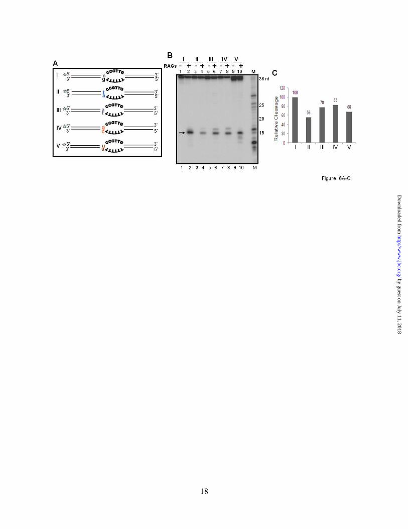
have tested the role of neighboring sequences on RAG cleavage in non-B DNA structures by two different ways. In one of the experiments, RAG cleavage was performed following the swapping of the duplex arms of the heteroduplex DNA and results showed no difference in the cleavage pattern and efficiency (data not shown). Next we tested whether a single nucleotide mutation immediately next to the bubble region can affect the cleavage efficiency as seen in the case of 1 nt bubble described above. A 36 bp oligomeric substrate containing a 6 nt bubble with 2 cytosines immediately next to single stranded region was used for the study (Fig. 6A). Five different single nucleotide mutations at double stranded region immediately flanking the bubble sequence was created by changing C/G of wild type oligomer to T/A, A/T, G/C or U/A (Fig. 6A). To our surprise, we found that C T mutation led to approximately 50% reduction in the RAG cleavage efficiency (Fig. 6B, lanes 1-4, C). It was also observed that C A, C G or C U conversion led to a reduction in the RAG cleavage efficiency, although it was limited (Fig. 6B, C). We also observed a comparable reduction in RAG binding (data not shown). Hence, our results suggest that the nucleotide at double stranded DNA, immediately upstream of the bubble region can affect the RAG cleavage and it is most efficient when cytosine is present.
DISCUSSION In the present study we identify novel
recognition sequences for the RAG complex when acting as a structure-specific nuclease. Further, we show that both structure and sequence features of the heteroduplex DNA are important in determining the pattern and efficiency of RAG cleavage. Sequences of the single-stranded region and immediate neighboring sequences, affect RAG cleavage on heteroduplex DNA. RAGs are well-studied as a sequence-specific nuclease for their role in V(D)J recombination. Its specificity at recombination signal sequences are extensively characterized by different groups (37-39,42-44). Earlier studies by us and others have shown that RAGs can also act as a structure-specific nuclease (22). In the present study we find that although having an altered DNA structure is important for RAG recognition of the bubble containing
sequences, structure alone is not sufficient for its reactivity. Instead we find that the sequence composition of the bubbles dictate the pattern and efficiency of RAG cleavage. We find that cytosines are preferred over thymine for RAG induced single- and double-strand breaks. Although over all cleavage efficiency was comparable between top and bottom strands in respective cases, we found that cleavage at top strand resulted in two bands while it was only one in case of bottom strands. Such a difference in RAG cleavage could be due to differences in the neighboring sequences. However, more studies are required to identify the exact mechanism. The most interesting finding was that when the sequences of the bubbles were purines (adenine or guanine), there was no cleavage at the heteroduplex DNA at all. We also found that when cytosines were present, as small as 2 nt bubbles were sufficient for robust RAG cleavage. However, when nucleotides other than cytosines were present, the number of nucleotides required for optimal cleavage was six (30). Besides, presence of a cytosine in the double-stranded region just upstream of bubble sequence resulted in highest RAG cleavage efficiency. Changing the C to A, or T or G significantly reduced the RAG cleavage efficiency. However, it is important to point out that change of C to U did not change the cleavage efficiency, whereas a C to T conversion, dramatically reduced RAG cleavage efficiency. This is understandable as both cytosine and uracil do not have a methyl group, which thymine possesses.
Based on the above studies, it appears that for optimal RAG cleavage at altered DNA structures, two cytosines close to 5’ end of the heteroduplex region are important. Presence of a cytosine in the duplex DNA next to the bubble is also preferred. Thus, we propose that “C(d)C(S)C(S)” (the subscript “d” denotes double-stranded, while “s” stands for single-stranded DNA) could be a consensus sequence for RAGs to induce single-strand breaks. A consensus sequence for inducing double-strand breaks could be “C(d)C(S)C(S)/C(S)C(S)”. Presence of A or G in place of C in the single-stranded DNA could abolish the RAG cleavage completely. However, a replacement with T could still be cleaved, but with a much lower efficiency. In an earlier study, it has been shown that GC rich sequences are the
by guest on July 11, 2018http://w
ww
.jbc.org/D
ownloaded from
8
most fragile sites in the genome, though no clear consensus sequence was discernable (45). Sequence composition specificity of RAGs on bubble structures reflects a more general property of RAGs. The RAG cleavage at sequences other than RSS sites was first reported for 3’ overhangs, flap DNA structures and gap DNAs, and the authors showed that RAGs could cleave at single-strand/double-strand transitions even in buffers containing Mg2+ (32). In earlier studies, it was also shown that when the heptamer of the RSS was used for RAG cleavage in a single-stranded DNA context, RAGs could cleave at single-strand/double-strand transitions (38). It has also been shown that during V(D)J recombination, the intermediates containing flaps and overhangs could be cleaved by RAGs (46). Later, our own studies have shown that a non-B DNA structure present at the BCL2 MBR sequence could be cleaved by RAGs, which was also extended to other types of DNA structures, like heteroduplexes and heterologous loops (25-31,35). All those experiments were performed in buffers that were close to physiologic conditions in which 5 mM of MgCl2 was used. Hairpins, which are V(D)J recombination intermediates, were also used for testing RAG activity and in two independent studies, it was reported that in the presence Mn2+, RAGs were able to cleave hairpin intermediates (33,34). Since our studies have shown that RAG cleavage on altered DNA structures is preferred when cytosines, but not purines are present on the bubble region, we wondered whether that could be true in other DNA structures studied in literature. By using different overhang, gap and stem loop substrates containing Cs, As, Ts or Gs, we found that in all cases, cytosines were preferred for RAG cleavage. As in the bubble substrates, adenines and guanines did not contribute towards specific RAG cleavage. It is more important to point out that in all cases Mg2+ was used for RAG cleavage rather than Mn2+. These data suggest that the observed sequence preference noticed during RAG cleavage on heteroduplex DNA structures is a more general property of RAGs when it acts as a structure-specific nuclease. Therefore, the sequence motif identified by us can be used to explain the published studies from the literature. It is interesting to point out that all the overhang substrates used in the earlier studies had cytosines
at the single-strand/double-strand junctions (32,47). How often would one expect heteroduplex DNA structures in the human genome in lymphoid tissues? Normally DNA in our genome is expected to be in B-form duplex conformation. However, when RAGs act as a structure-specific nuclease, it always recognizes the single-stranded region present in the altered DNA structure. Therefore, one of the major questions which arises is how often one would see such type of structures in the human genome? Also what is the mechanism by which a B-DNA may be converted to a heteroduplex DNA? For duplex DNA to get converted into altered DNA, first it needs to get unpaired. This could be due to breathing of DNA, melting due to supercoiling, during replication or transcription (48) (Fig. 7). Moreover, for each type of structure formation, specific types of sequences are required (28,48,49). For example, the presence of inverted repeats or palindromic sequences could lead to cruciform structures (50). Presence of direct repeats could lead to misaligned sequences. Homopurine:homopyrimidine stretches with mirror repeat symmetry could lead to formation of triplex DNA (49). G-quartets may be formed in stretches of Gs when appropriate conditions are provided (28,48) . G/C repeats, during transcription could also lead to formation of RNA/DNA hybrids (51). In addition to these, even spontaneous deamination of cytosine could lead to mismatches. Deamination of a methylated cytosine could lead to a T:G mismatch (35). Any of these structures, when present in lymphoid tissues could be a target for RAGs, when appropriate sequences are present. RAG-induced breaks in heteroduplex DNA structures: relevance in cancer and genomic instability. The observed RAG-induced breaks on bubble structures suggests that when such altered structures or mismatch regions are present in the cells, it could lead to different types of genomic rearrangements. Given that the sequence preference shown by core RAGs hold true for full length RAGs as well, it suggests that this is an inherent property of RAGs and such a structural specificity is physiological. However, the rearrangements could be dependent on how often such structures may be present in the genome. Since RAGs are present only in lymphoid tissues, any type of rearrangement in other cell types is
by guest on July 11, 2018http://w
ww
.jbc.org/D
ownloaded from

9
ruled out. As shown by our results, RAG induced genomic instability could be controlled at the sequence level. Therefore, depending on the sequence of the single-stranded region of the heteroduplex DNA, either a single-strand break or double-strand break could be induced, which in turn could culminate in genomic instability and cancer (Fig. 7). Cancers like leukemia and lymphoma are restricted to lymphoid tissues and are characterized by presence of chromosomal abnormalities such as chromosomal translocations, deletions, inversions and other mutations. Since such abnormalities might require multiple DSBs, it is possible that depending on the sequence, a DSB could be generated by RAGs by inducing two independent nicks (Fig. 7) (22,52,53). The probability to have such cytosines near single-strand/double-strand transitions is quite high,
when structures like triplex DNA or G-quartets are formed in GC rich sequences. (28,48,49,54) (Fig. 7). In cases where SSBs are generated, replication across a nick or cleavage by single-strand specific enzymes such as Artemis could convert them into DSBs (55,56).
Since we show that the “C(d)C(S)C(S)” motif can be the most favored sequence for RAG nicking and DSB formation on altered DNA structures including DNA mismatches and gaps in physiological conditions, this novel sequence motif could be a new recognition sequence for RAGs (Fig. 7), just like the RSS on a standard duplex B-DNA. This further suggests that RAG cleavage pattern on altered DNA structures is context specific, and both sequence and structural determinants act together to limit the RAG-induced genomic instability.
REFERENCES
1. Schatz, D. G., and Baltimore, D. (2004) Cell 116, S103-106, 102 p following S106 2. Lewis, S. M. (1994) Adv Immunol 56, 27-150 3. Hiom, K., and Gellert, M. (1998a) Mol Cell 1, 1011-1019 4. Fugmann, S. D., Lee, A. I., Shockett, P. E., Villey, I. J., and Schatz, D. G. (2000) Annu Rev Immunol 18,
495-527 5. Gellert, M. (2002) Annu Rev Biochem 71, 101-132 6. Roth, D. B. (2003) Nat Rev Immunol 3, 656-666 7. Schlissel, M. S. (2003) Nat Rev Immunol 3, 890-899 8. Sadofsky, M. J. (2004) Immunol Rev 200, 83-89 9. Swanson, P. C. (2004) Immunol Rev 200, 90-114 10. Oettinger, M. A. (2004) Immunol Rev 200, 165-181 11. Jung, D., and Alt, F. W. (2004) Cell 116, 299-311 12. Schatz, D. G., and Spanopoulou, E. (2005) Curr Top Microbiol Immunol 290, 49-85 13. Lieber, M. R., Yu, K., and Raghavan, S. C. (2006) DNA Repair (Amst) 5, 1234-1245 14. Jung, D., Giallourakis, C., Mostoslavsky, R., and Alt, F. W. (2006) Annu Rev Immunol 24, 541-570 15. Roth, D. B., Menetski, J. P., Nakajima, P. B., Bosma, M. J., and Gellert, M. (1992) Cell 70, 983-991 16. Ma, Y., Pannicke, U., Schwarz, K., and Lieber, M. R. (2002) Cell 108, 781-794 17. Agrawal, A., and Schatz, D. G. (1997) Cell 89, 43-53 18. Jones, J. M., and Gellert, M. (2001) Proc Natl Acad Sci U S A 98, 12926-12931 19. Lieber, M. R., Ma, Y., Pannicke, U., and Schwarz, K. (2003) Nat Rev Mol Cell Biol 4, 712-720 20. Rooney, S., Chaudhuri, J., and Alt, F. W. (2004) Immunol Rev 200, 115-131 21. Raghavan, S. C., Kirsch, I. R., and Lieber, M. R. (2001) J Biol Chem 276, 29126-29133 22. Raghavan, S. C., and Lieber, M. R. (2006) Bioessays 28, 480-494 23. Marculescu, R., Le, T., Simon, P., Jaeger, U., and Nadel, B. (2002) J Exp Med 195, 85-98 24. Lewis, S. M., Agard, E., Suh, S., and Czyzyk, L. (1997) Mol Cell Biol 17, 3125-3136 25. Raghavan, S. C., Chastain, P., Lee, J. S., Hegde, B. G., Houston, S., Langen, R., Hsieh, C. L., Haworth, I.
S., and Lieber, M. R. (2005) J Biol Chem 280, 22749-22760 26. Raghavan, S. C., Hsieh, C. L., and Lieber, M. R. (2005) Mol Cell Biol 25, 6475-6484 27. Raghavan, S. C., and Lieber, M. R. (2004) Cell Cycle 3, 762-768 28. Raghavan, S. C., and Lieber, M. R. (2007) Front Biosci 12, 4402-4408 29. Raghavan, S. C., Swanson, P. C., Wu, X., Hsieh, C. L., and Lieber, M. R. (2004) Nature 428, 88-93
by guest on July 11, 2018http://w
ww
.jbc.org/D
ownloaded from
10
30. Raghavan, S. C., Gu, J., Swanson, P. C., and Lieber, M. R. (2007) DNA Repair (Amst) 6, 751-759 31. Raghavan, S. C., Swanson, P. C., Ma, Y., and Lieber, M. R. (2005) Mol Cell Biol 25, 5904-5919 32. Santagata, S., Besmer, E., Villa, A., Bozzi, F., Allingham, J. S., Sobacchi, C., Haniford, D. B., Vezzoni,
P., Nussenzweig, M. C., Pan, Z. Q., and Cortes, P. (1999) Mol Cell 4, 935-947 33. Besmer, E., Mansilla-Soto, J., Cassard, S., Sawchuk, D. J., Brown, G., Sadofsky, M., Lewis, S. M.,
Nussenzweig, M. C., and Cortes, P. (1998) Mol Cell 2, 817-828 34. Shockett, P. E., and Schatz, D. G. (1999) Mol Cell Biol 19, 4159-4166 35. Tsai, A. G., Lu, H., Raghavan, S. C., Muschen, M., Hsieh, C. L., and Lieber, M. R. (2008) Cell 135,
1130-1142 36. Nambiar, M., Kari, V., and Raghavan, S. C. (2008) Biochim Biophys Acta 1786, 139-152 37. Cuomo, C. A., Mundy, C. L., and Oettinger, M. A. (1996) Mol Cell Biol 16, 5683-5690 38. Ramsden, D. A., McBlane, J. F., van Gent, D. C., and Gellert, M. (1996) EMBO J 15, 3197-3206 39. Yu, K., and Lieber, M. R. (1999) Mol Cell Biol 19, 8094-8102 40. Raval, P., Kriatchko, A. N., Kumar, S., and Swanson, P. C. (2008) Nucleic Acids Res 36, 2060-2072 41. Naik, A. K., and Raghavan, S. C. (2008) DNA Repair (Amst) 7, 1384-1391 42. Lieber, M. R., Hesse, J. E., Mizuuchi, K., and Gellert, M. (1987) Genes Dev 1, 751-761 43. Roth, D. B., Zhu, C., and Gellert, M. (1993) Proc Natl Acad Sci U S A 90, 10788-10792 44. McBlane, J. F., van Gent, D. C., Ramsden, D. A., Romeo, C., Cuomo, C. A., Gellert, M., and Oettinger,
M. A. (1995) Cell 83, 387-395 45. Greenbaum, J. A., Parker, S. C., and Tullius, T. D. (2007) Genome Res 17, 940-946 46. Grawunder, U., and Lieber, M. R. (1997) Nucleic Acids Res 25, 1375-1382 47. Nishihara, T., Nagawa, F., Nishizumi, H., Kodama, M., Hirose, S., Hayashi, R., and Sakano, H. (2004)
Mol Cell Biol 24, 3692-3702 48. Sinden, R. R. (ed) (1994) DNA structure and function, Academic Press, San Diego, California 49. Mirkin, S. M. (2007) Nature 447, 932-940 50. Bacolla, A., and Wells, R. D. (2004) J Biol Chem 279, 47411-47414 51. Yu, K., Chedin, F., Hsieh, C. L., Wilson, T. E., and Lieber, M. R. (2003) Nat Immunol 4, 442-451 52. Franco, S., Alt, F. W., and Manis, J. P. (2006) DNA Repair (Amst) 5, 1030-1041 53. Mills, K. D., Ferguson, D. O., and Alt, F. W. (2003) Immunol Rev 194, 77-95 54. Wells, R. D. (2007) Trends Biochem Sci 32, 271-278 55. Kuzminov, A. (2001) Proc Natl Acad Sci U S A 98, 8241-8246 56. Ma, Y., Schwarz, K., and Lieber, M. R. (2005) DNA Repair (Amst) 4, 845-851
FOOTNOTES We thank Dr. Patrick Swanson for providing MBP RAG constructs and Ms. Mridula Nambiar for
critical reading of the manuscript and help in cell culture. We thank Dr. Bibha Choudhary and members of the SCR laboratory for discussions and help. This work was supported by grants from DST, India (SR/SO/BB-051/2007), and Indian Institute of Science start up grant for SCR. We also thank Dr. Raghavan Varadarajan for financial assistance. AKN is supported by SRF from CSIR, India.
FIGURE LEGENDS Fig. 1. Comparison of efficiency of RAG cleavage with respect to composition of sequences in the bubble region of heteroduplex DNA. A. Diagrammatic representation of oligomeric substrates containing 6 nt bubbles. The bubble sequences are shown in bold letters. The double-stranded arms are of 15 bp length each and indicated with double lines. Sequence of the bubble region are (A/A)6, (C/C)6, (T/T)6 or (G/G)6 and are denoted as I, II, III and IV, respectively. B. Polyacrylamide gel profile showing RAG cleavage on heteroduplex DNA, I, II, III and IV described in panel A. RAG cleavage reactions were done in the buffer containing 5 mM MgCl2 using DNA substrates, which were [γ32P] ATP labelled either on top or bottom strand for 1 hr at 37oC and were resolved on a 15% denaturing PAGE. “top” indicates that radiolabelled strand is top strand. “bot” indicates that bottom strand is radiolabelled. Klenow partial digested 1 nt ladder was used as marker. RAG specific cleavage products are indicated
by guest on July 11, 2018http://w
ww
.jbc.org/D
ownloaded from

11
by arrow. C. Histogram showing quantification of RAG cleavage efficiency of top and bottom strands of heteroduplex DNA structures described in panel, “B”. The RAG cleavage products were quantified using Multi Gauge software. The substrate amount in the respective no RAG lane was taken as 100% and the relative cleavage of products were calculated and indicated as %. In all the cases background was subtracted. The calculated percentage is shown on top of the respective columns in the histogram. D. Detection of RAG induced DSBs on bubble substrates shown in panel A. RAG cleavage products were resolved on 15% native PAGE. The markers for possible nicked or double-stranded breaks are shown on the right hand side. The bands due to DSBs are indicated by arrow. Fig. 2. Comparison of cleavage efficiency when core RAGs or full length RAGs were used on different heteroduplex DNA. The bubble sequences containing (A/A)6, (C/C)6, (T/T)6 and (G/G)6 denoted as I, II, III and IV, respectively, were incubated in a buffer containing 5 mM MgCl2 with either MBP cRAG1/cRAG2 (A) or MBP FLRAG1/cRAG2 (B) or MBP cRAG1/FLRAG2 (C). The RAG cleavage products were resolved on a denaturing PAGE and are indicated by arrows. M is 1 nt molecular weight ladder. For other details refer Figure 1 legend. Fig. 3. Comparison of time kinetics of RAG cleavage reaction of different heteroduplex DNA substrates. The bubble sequences containing (A/A)6, (C/C)6, (T/T)6 and (G/G)6 denoted as I, II, III and IV, respectively, or oligomer containing 12 RSS were incubated for different time periods as indicated with either cRAG1/cRAG2 (GST-tagged) and resolved on 15% denaturing PAGE. A. RAG cleavage kinetics of poly A/A and C/C heteroduplexes. B. RAG cleavage kinetics of poly T/T and G/G heteroduplexes. C. RAG cleavage kinetics of 12-RSS. RAG cleavage products are indicated by arrow. M is 1 nt molecular weight ladder. In each panel, RAG cleavage products resulted from respective gels were quantified and presented. Fig. 4. Two cytosines within the bubble region are sufficient for optimal RAG cleavage on heteroduplex DNA structures. A. Diagrammatic representation of oligomeric substrates containing either 6 nt bubbles with varying number of cytosines (denoted as I-VII) or with varying lengths of bubble region, 0 to 6 (denoted as VIII-XIV). The changes from Cs to Gs are indicated using a box (see I-VII). In all cases the length of upstream and downstream sequences are same (15 bp). Duplex DNA substrate (0 nt bubble) comprises the top strand of 6 nt bubble (denoted as VIII) paired with corresponding complementary sequence. In each case top strand oligomer was radiolabeled with [γ32P] ATP and used. B. Polyacrylamide gel profile showing RAG cleavage on heteroduplex substrates I-VII. “M” is 1 nt ladder. C. Polyacrylamide gel profile showing RAG cleavage on heteroduplex substrates VIII-XIV shown in panel “A”. “M” is 1 nt ladder. D. RAG induced double-strand breaks when DNA contain a bubble as small as 2 nt. Heteroduplex DNA substrates VIII-XIV were subjected to RAG cleavage and the products were resolved on a 15% native PAGE to detect DSBs. The bands due to DSBs (bracketed) and single-strand breaks (arrow) are indicated. Molecular markers used for identifying the positions of DSBs are shown. Fig. 5. RAG cleavage on gap DNA structures, protruding overhangs and hairpin structures. A. Diagrammatic representation of oligomeric substrates containing gap DNA structures (denoted as I-V), 3’ overhangs (denoted as VI-IX) and hairpin loops (denoted as X-XIII) with poly A, C, T or G at single stranded region as indicated. In the case of gap substrates, length of the double-stranded arm is 15 bp towards one side and 10 bp towards other side. In the case of overhangs, the length of double-stranded region is 15 bp, while length of single-stranded region was also 15 nt. In case of hairpin loop structures the length of the loop region was 6 nt while the length of double-stranded arm was18 bp. B-D. Polyacrylamide gel profile showing RAG cleavage on gap structures (B), protruding overhangs (C), and hairpin structures (D). In all panels, RAG cleavage reactions were done in a buffer containing 5 mM MgCl2 for 1 hr at 37oC and were resolved on 15% denaturing PAGE. For other details refer Figure 1 legend. Fig. 6. Effect of neighboring sequences on RAG cleavage on heteroduplex DNA structures. A. Diagrammatic representation of oligomeric substrates containing 6 nt bubbles with different flanking single nucleotide mutations (indicated in different colors). In each case length of the arm is 15 bp. Asterisk indicates the strand which is radiolabeled with [γ32P] ATP. B. Polyacrylamide gel profile
by guest on July 11, 2018http://w
ww
.jbc.org/D
ownloaded from

12
showing effect of single nucleotide change on RAG cleavage at hetroduplex DNA structures. RAG specific cleavage products are indicated by arrow. C. Histogram showing quantification of RAG cleavage efficiency at top strand of heteroduplex DNA described in panels “A” and “B”. For other details refer Figure 1 legend. Fig. 7. Plausible RAG cleavage targets within the cells when it acts as a structure specific nuclease. A, B. Bubble structures formed during transcription. Depending on the availability of Cs at the junction and size of the bubble, the RAG cleavage could lead to either SSB or DSB. C. Transcription through a cytosine rich template could lead to an RNA-DNA hybrid formation. The resulting G-rich single stranded region may fold into G-quartet structures. D. D-loops formed during replication and recombination also could be a target for RAGs. E. Cruciform structures with Cs at single strand-double strand transitions could be cleaved by RAGs. F. Triple helical DNA formed at homopurine:pyrimidine tracts with mirror repeat symmetry could be a target for RAGs. G. Mismatches generated during DNA replication could be a target for RAGs if those contain cytosine. In addition, tips of replication forks with cytosines could also be a target for RAGs.
by guest on July 11, 2018http://w
ww
.jbc.org/D
ownloaded from


Abani Kanta Naik, Michael R. Lieber and Sathees C. Raghavanstructures: Implications for genomic instability
Cytosines, but not purines, determine RAG induced breaks on heteroduplex DNA
published online January 5, 2010J. Biol. Chem.
10.1074/jbc.M109.089631Access the most updated version of this article at doi:
Alerts:
When a correction for this article is posted•
When this article is cited•
to choose from all of JBC's e-mail alertsClick here
Supplemental material:
http://www.jbc.org/content/suppl/2010/01/21/M109.089631.DC1
by guest on July 11, 2018http://w
ww
.jbc.org/D
ownloaded from