cytostatic and antiestrogenic effects of 2-(indol-3-ylmethyl)-3,3′-diindolylmethane, a major in...
TRANSCRIPT
Cytostatic and Antiestrogenic Effects of 2-(Indol-3-ylmethyl)-3,39-diindolylmethane, a Major In Vivo
Product of Dietary Indole-3-carbinolYu-Chen Chang,* Jacques Riby,* Grace H-F. Chang,* BaoCheng Peng,*
Gary Firestone† and Leonard F. Bjeldanes*‡*DIVISION OF NUTRITIONAL SCIENCES AND TOXICOLOGY, AND †DEPARTMENT OF MOLECULAR AND CELL BIOLOGY,
UNIVERSITY OF CALIFORNIA, BERKELEY, CA 94720, U.S.A.
ABSTRACT. Under acidic conditions, indole-3-carbinol (I3C) is converted to a series of oligomeric productsthought to be responsible for the biological effects of dietary I3C. Chromatographic separation of the crude acidmixture of I3C, guided by cell proliferation assay in human MCF-7 cells, resulted in the isolation of2-(indol-3-ylmethyl)-3,39-diindolylmethane (LTr-1) as a major antiproliferative component. LTr-1 inhibitedthe growth of both estrogen-dependent (MCF-7) and -independent (MDA-MB-231) breast cancer cells byapproximately 60% at a non-lethal concentration of 25 mM. LTr-1 had no apparent effect on the proliferationof MCF-7 cells in the absence of estrogen. LTr-1 was a weak ligand for the estrogen receptor (ER) (IC50 70 mM)and efficiently inhibited the estradiol (E2)-induced binding of the ER to its cognate DNA responsive element.The antagonist effects of LTr-1 also were exhibited in assays of endogenous pS2 gene expression and in cellstransiently transfected with an estrogen-responsive reporter construct (pERE-vit-CAT). LTr-1 activated bothbinding of the aryl hydrocarbon (Ah) receptor to its cognate DNA responsive element and expression of the Ahreceptor-responsive gene CYP1A1. LTr-1 was a competitive inhibitor of CYP1A1-dependent ethoxyresorufin-O-deethylase (EROD) activity. In summary, these results demonstrated that LTr-1, a major in vivo product ofI3C, could inhibit the proliferation of both estrogen-dependent and -independent breast tumor cells and thatLTr-1 is an antagonist of estrogen receptor function and a weak agonist of Ah receptor function. BIOCHEM
PHARMACOL 58;5:825–834, 1999. © 1999 Elsevier Science Inc.
KEY WORDS. indole-3-carbinol; 2-(indol-3-ylmethyl)-3; 39-diindolylmethane; antiestrogenic; breast tumorcells; cytostatic; HPLC
I3C§ is a product of enzymatic hydrolysis of the indolylm-ethyl glucosinolate glucobrassicin, present in common veg-etables of the Brassica genus, including cabbage, kale,rutabaga, cauliflower, turnips, broccoli, kohlrabi, mustard,collard, and Brussels sprouts, and is a promising cancerpreventive agent (see structure in Fig. 1). Oral administra-tion of I3C to rodents prior to treatment with a carcinogenreduces tumorigenesis in the stomach, liver, lung, and oralcavity by a mechanism that is thought to include inductionof phase I and phase II xenobiotic metabolism and in-creased clearance of the carcinogen [1–5]. When adminis-tered at high doses following treatment with a carcinogen,
I3C is reported to promote tumorigenesis in the thyroidgland, colon, pancreas, and liver [6–8]. Although themechanism for these adverse effects of I3C is yet to bedetermined, one concern is that they might be associatedwith activation of the Ah receptor. Persistent activation ofthis receptor is thought to be responsible for the adverseeffects of the potent environmental toxin TCDD [9].
The most pronounced and consistently protective effectsof I3C have been reported against tumorigenesis in estro-gen-responsive tissues. In one of the earliest studies of theprotective effects of non-nutritive food components, I3Cwas shown to reduce by 75% the frequency of dimethyl-benzanthracene-induced mammary tumors in rodents [1]. Arecent extension of these studies found an even greaterprotective effect of I3C, i.e. 95% reduction in tumormultiplicity, when the indole was administered prior to andfollowing treatment with the carcinogen [10]. I3C treat-ment also produced a dramatic decrease (65%) in mammarytumor multiplicity induced by the direct-acting carcinogenmethylnitrosourea [10]. Furthermore, I3C is reported toinhibit spontaneous formation of mammary and endome-trial tumors in rodents [11, 12]. The protective effects of
‡ Corresponding author: Dr. Leonard F. Bjeldanes, Division of NutritionalSciences and Toxicology, 119 Morgan Hall, University of California atBerkeley, Berkeley, CA 94720. Tel. (510) 642-1601; FAX (510) 642-0535; E-mail: [email protected]
§ Abbreviations: I3C, indole-3-carbinol; LTr-1, 2-(indol-3-ylmethyl)-3,39-diindolylmethane; ER, estrogen receptor; E2, 17b-estradiol; Ah, arylhydrocarbon; EROD, ethoxyresorufin-O-deethylase; TCDD, 2,3,7,8-tetra-chlorodibenzo-p-dioxine; ICZ, indolo[3,2-b]carbazole; DMEM, Dulbecco’smodified Eagle’s medium; DTT, dithiothreitol; FBS, fetal bovine serum;SSC, 0.15 M sodium chloride 1 0.015 M sodium citrate; THF, tetrahy-drofuran; DIM, 3,39-diindolylmethane; RXM, acid reaction mixture; andERE, estrogen responsive element.
Received 10 August 1998; accepted 7 January 1999.
Biochemical Pharmacology, Vol. 58, pp. 825–834, 1999. ISSN 0006-2952/99/$–see front matter© 1999 Elsevier Science Inc. All rights reserved. PII S0006-2952(99)00165-3

I3C in these studies have been attributed to alterations inE2 metabolism and/or modifications of ER function.
An important consideration in studies of the biologicaleffects of I3C is its chemical instability. I3C, being avinylogous hemiaminal, is dehydrated readily in acidic andbasic solutions and is converted rapidly to a mixture ofoligomeric products [13]. These products, which are pro-duced readily in gastric acid following ingestion, arethought to be responsible for the biological effects of orallyadministered I3C [14]. We have shown previously that aminor product, ICZ, is a potent activator of Ah receptorpathways and exhibits antiestrogenic activities [15, 16].DIM, a major product of I3C, is a weak ligand for the Ahreceptor, inhibits CYP1A1 enzyme activity, and can act asan antagonist of E2-mediated tumor cell growth and geneactivation [17, 18].
As part of our continuing efforts to understand themechanisms of action of I3C, we have begun to identify thecomponents of the RXM of I3C that are responsible for thecancer protective effects of I3C and the antiproliferativeactivity of RXM in cultured breast tumor cells. We reporthere the effects in tumor cells of the linear trimeric productLTr-1, a second major component of RXM. We show thatthis novel compound can inhibit proliferation of bothestrogen-dependent and -independent cultured breast tu-mor cells and that it is an antagonist of ER function withlittle agonist activity. We show further that LTr-1 is a weakagonist of Ah receptor function.
MATERIALS AND METHODSMaterials
DMEM, Opti-MEM, and Lipofectamine were supplied byGibco/BRL. Phenol red-free DMEM, FBS, calf serum,
tamoxifen, and E2 were supplied by the Sigma ChemicalCo. [g-32P]ATP and [3H]acetyl-CoA were supplied by NewEngland Nuclear. All other reagents were of the highestgrade available.
Preparation of RXM
The procedure reported by Grose and Bjeldanes [13] wasfollowed for the preparation of RXM. Briefly, I3C (100 mg,Aldrich Chemical Co.) was suspended in 1 M HCl (100mL) at room temperature for 15 min. The acid suspensionwas neutralized with aqueous ammonia to pH 7.0, and theprecipitate was filtered and dried under vacuum to giveRXM as a reddish powder. LTr-1 is stable under the neutralaqueous conditions of the cell proliferation assay.
Fractionation of RXM
RXM (200 mg) was dissolved in THF (1 mL) and purifiedby silica gel vacuum liquid chromatography. Mixtures ofhexane/THF with increasing polarity were used as themobile phase. HPLC purification of bioactive componentsof the crude fractions was performed using a ShimadzuHPLC system (SCL-10A; Shimadzu Scientific Instruments,Inc.) equipped with a C18 bonded-phase semi-preparativecolumn (Ultrasphere-ODS, 10 3 250 mm, 5 mm; Beck-man) and UV/VIS detector (SPD-10AV; Shimadzu Scien-tific Instruments, Inc.). The peaks were monitored at 280nm. For isocratic elution, we used a mixture of acetonitrile:water (60:40) at a flow rate of 2 mL/min. Crude RXMfractions were resuspended in THF before injection intoHPLC. The electron impact mass spectrometry analyses ofHPLC fractions of interest were obtained by the MassSpectrometry Facility of the College of Chemistry, Univer-sity of California at Berkeley.
Cell Culture
The human breast adenocarcinoma cell lines MCF-7 andMDA-MB-231 and the murine hepatoma cell line Hepa-1c1c-7, obtained from the American Type Culture Collec-tion (ATCC), were grown as adherent monolayers inDMEM (Gibco, Life Science Technology), supplementedto 4.0 g/L of glucose and 3.7 g/L of sodium bicarbonate ina humidified incubator at 37° and 5% CO2, and passaged atapproximately 80% confluence. Cultures of human cellswere used in subsequent experiments for fewer than 25passages.
Cell Proliferation
Before the beginning of the treatments, cells were depletedof estrogen for 7–10 days in medium composed of DMEMbase without phenol-red (Sigma), with 4 g/L of glucose, 3.7g/L of sodium bicarbonate, and 5% calf serum twice strippedin dextran-coated charcoal and microfiltered, supple-mented with non-essential amino acids (Gibco), 2 mM
FIG. 1. Structures of I3C, DIM, and LTr-1.
826 Y-C. Chang et al.
glutamine, and 10 ng/mL of insulin. During the depletionperiod, medium was changed every other day. Treatmentswere administered by the addition of 1 mL of 1000x stocksolutions in DMSO per mL of medium. Once the treatmentperiod started, medium was changed daily to counterpossible loss of readily metabolized compounds. Cells wereharvested by trypsinization and counted in a Coulterparticle counter.
Estrogen Receptor Binding Assay
Rat uterine cytosol was prepared as described previously[19]. Briefly, 2.5 g of uterine tissue from five Sprague–Dawley rats (12 weeks old) was excised and placed on ice.The fresh tissue was homogenized with 30 mL of ice-coldTEDG buffer (10 mM Tris, pH 7.4, 1.5 mM EDTA, 1 mMDTT, 10% glycerol) using a Polytron at medium speed for1 min on ice. The homogenate was centrifuged at 1000 g for10 min at 4°. The supernatant solution was transferred toultracentrifuge tubes and centrifuged at 100,000 g for 90min at 4°. The supernatant solution was divided into1.0-mL aliquots, quickly frozen in a dry-ice/ethanol bath,and stored at 280°. Protein concentration of the uterinecytosol was measured by the Bradford assay using bovineserum albumin as the standard. For each competitivebinding assay, 5 mL of 20 nM [3H]E2 in 50% ethanol, 10mM Tris, pH 7.5, 10% glycerol, 1 mg/mL of BSA, and 1mM DTT was placed in a 1.5-mL microcentrifuge tube.Competitive ligands were added as 1.0 mL of 100x stocksolutions in DMSO. After mixing, 95 mL of uterine cytosolwas added, and the tubes were vortexed and incubated atroom temperature for 2–3 hr. Proteins were precipitated bythe addition of 100 mL of 50% hydroxylapatite slurryequilibrated in TE (50 mM Tris, pH 7.4, 1 mM EDTA) andincubated on ice for 15 min with vortexing every 5 min toresuspend hydroxylapatite. The pellet was washed with 1.0mL of ice-cold wash buffer (40 mM Tris, pH 7.4, 100 mMKCl), and centrifuged for 5 min at 10,000 g at 4°. Thesupernatant was aspirated carefully, and the pellet waswashed two more times with 1.0 mL of wash buffer. Thefinal pellet was resuspended in 200 mL of ethanol andtransferred to a scintillation vial. The tube was washed withanother 200-mL portion of ethanol, which then was addedto the same counting vial. A negative control contained nouterine cytosol. Non-specific binding was determined using100-fold (0.1 mM) excess unlabeled E2. Relative bindingaffinities were calculated using the concentration of com-petitor needed to reduce [3H]E2 binding by 50% as com-pared with the concentration of unlabeled E2 needed toachieve the same result.
Reporter Plasmids and Expression Vectors
The ER responsive CAT reporter plasmid pERE-vit-CAT[20] was a gift from D. J. Shapiro (University of Illinois).pERE-vit-CAT contains the 59-flanking and promoterregion (2596 to 121) of the Xenopus vitellogenin-B1 gene,
including two imperfect endogenous EREs (at 2302 and2334) and one consensus exogenous ERE inserted atposition 2359. The transfection efficiency control vectorpCMVb constitutively expressing b-galactosidase was ob-tained from ATCC.
Transient Transfections with Reporters
Transfections were done by the lipofection method usingLipofectamine (Gibco BRL). Cells were grown in 10%FBS-DMEM until 80% confluent and transferred to 6.0-cmPetri plates 24 hr before transfection. The plates wereseeded with the appropriate number of cells to be 50–60%confluent at the time of transfection. For each 6-cm plate,8 mL of Lipofectamine was diluted with 92 mL of Opti-MEM serum-free medium (Gibco). Plasmid DNA (0.1 to1.0 mg) was diluted in 100 mL of serum-free medium. Lipidand plasmid dilutions were combined, mixed gently, andincubated at room temperature for 30–45 min. Meanwhile,the plates were washed with 4 mL of serum-free medium,and 2 mL of serum-free medium was added to each plate. A200-mL portion of the lipid/DNA suspension was added toeach plate and mixed gently. The plates were returned tothe incubator for 5–6 hr, and 2 mL of medium containing10% calf serum was added. The next day, the plates weretreated with fresh stripped medium without phenol red (5%DCC-FBS), and the 48-hr treatments were started by theaddition of 1 mL of 1000x stock solutions in DMSO per mLof medium. The transfection efficiency determined withthe constitutive galactosidase expression plasmid pCMVbin duplicate sets of plates was unaffected by the treatments.
Chloramphenicol Acetyl Transferase (CAT) Assay
The CAT assay was done using a modification of the phaseextraction assay described by Seed and Sheen [21]. At theend of the 48-hr treatment period, the transfected cellswere harvested by scraping with a rubber policeman,transferred with the medium to a conical 15-mL tube,centrifuged at 600 g for 2 min, resuspended in 1 mL of coldPBS, transferred to Eppendorf tubes, centrifuged at 600 g for2 min, and washed in PBS a second time. Cell pellets wereresuspended in 200 mL of 0.1 M Tris, pH 8.0, and lysed by3 cycles of freeze/thaw treatment (alternating 5 min in adry-ice/alcohol bath and 5 min in a 37° bath). Cell lysateswere incubated at 65° for 15 min to inactivate acylases andcentrifuged at 14,000 g for 8 min. A 165-mL aliquot of thecytosol was transferred to a 7-mL scintillation vial, and a20-mL aliquot was reserved for determination of proteinconcentration by the Bradford assay. The substrate mixture(85 mL) was added to the scintillation vial for finalconcentrations of 100 mM Tris–HCl, pH 8.0, 250 nmolchloramphenicol, and 1 mCi [3H]acetyl-CoA (200 mCi/mmol) in a total volume of 250 mL and mixed thoroughly.The organic scintillation fluid (4 mL) was added slowly,and the vials were incubated at 37° for 1–2 hr or untilsufficient counts were obtained.
Effects of an I3C Trimer in Cancer Cells 827

RNA Extraction and Northern Blot Analysis
Cells were lysed by addition of Tri-reagent (MolecularResearch Center, Inc.), and chloroform was used for phaseseparation. After centrifugation, the water-soluble upperphase was collected, and total RNA was precipitated withisopropanol, washed with 75% ethanol, and dissolved indiethyl pyrocarbonate-treated water. Total RNA was elec-trophoresed on a 1.2% agarose gel containing 3% formal-dehyde, using MOPS as the running buffer. The gel thenwas washed gently with 10x SSC and blotted with a Zeta
nylon membrane (Bio-Rad) overnight. The RNA was fixedto the membrane by UV cross-linking. The hybridizationprobes were labeled with [32P]CTP using random primersand the pS2-cDNA and GADPH-cDNA plasmids providedby ATCC as the template. Hybridization and quantitationof results were done as described previously [14]. SpecificpS2 mRNA levels were normalized using GADPH as astandard.
Nuclear Extracts
Three near confluent (80–90%) cultures of MCF-7 cells in100-mm Petri dishes were used for each treatment. LTr-1,E2, and tamoxifen were added as 1 mL of a 1000x stocksolution in DMSO per mL of medium. After 2 hr ofincubation at 37°, the plates were placed on ice and washedtwice with 5 mL of hypotonic buffer (10 mM HEPES, pH7.5) and incubated with 2 mL of the same buffer for 15 min.Cells were harvested in 1 mL of MDH buffer (3 mM MgCl2,1 mM DTT, 25 mM HEPES, pH 7.5) with a rubber scraper,homogenized with a loose-fitting Teflon pestle, and centri-fuged at 1000 g for 4 min at 4°. The pellets were washedtwice with 3 mL of MDHK buffer (3 mM MgCl2, 1 mMDTT, 0.1 M KCl, 25 mM HEPES, pH 7.5), resuspended in1 mL of MDHK, and centrifuged at 600 g for 4 min at 4° ina microcentrifuge. The pellets were resuspended in 100 mLof HDK buffer (25 mM HEPES, pH 7.5, 1 mM DTT, 0.4 MKCl), incubated for 20 min on ice with mixing every 5 min,and centrifuged at 14,000 g for 4 min at 4°. Glycerol was
FIG. 2. HPLC chromatogram of fraction B under the conditionsdescribed in Materials and Methods. Major peaks were labeledand collected individually.
FIG. 3. Antiproliferative effect of subfractions Ba–Bi on MCF-7 cells. Estrogen-depleted MCF-7 cells were plated at a density of 4 3104 cells per well in 24-well plates and treated as indicated with RXM and HPLC fractions at a concentration of 50 mM I3C equivalent.Duplicate aliquots of cells from individual wells were counted after 5 days. The results are shown as the average and SD from threeidentical wells. Growth was reduced significantly following treatment with RXM and all subfractions (a–i) as determined by Dunnett’stest with a procedurewise error rate of < 0.05.
828 Y-C. Chang et al.

added to the supernatants to a concentration of 10%, andaliquots of the nuclear extracts were stored at 280°.
Gel Mobility Shift Assay
The following two sets of complementary oligonucleotides:59-GATCCCAGGTCACAGTGACCTGAGCTAAAAT-39and 59-GATCATTTTAGCTCAGGTCACTGTGACC-TGG-39 containing the palindromic consensus ERE motif(underlined), and 59-GATCTGGCTCTTCTCACGCA-ACTCCG-39 and 59-GATCCGGAGTTGCGTGAGA-AGAGCCA-39 containing the consensus DRE motif (un-derlined), were annealed and 59-end-labeled with[g-32P]ATP using T4 nucleotide kinase. The resultinglabeled double-stranded DNA probes were purified on aSephadex G50 spin-column, precipitated in ethanol, dis-solved in TE buffer, and diluted in 25 mM HEPES, 1 mMDTT, 10% glycerol, 1 mM EDTA to contain approximately25,000 cpm of 32P/mL. Nuclear extracts (7 mg of proteins)were mixed with 90 ng poly-dIdC, 25 mM HEPES, 1 mMDTT, 10% glycerol, 1 mM EDTA, 160 mM KCl in a totalvolume of 21 mL. For antibody supershift experiments, 0.5mg of monoclonal mouse-IgG anti-human-ER (Santa CruzBiotechnology) was added to the incubation mixture. Afterincubation for 15–20 min at room temperature, 4 mL(100,000 cpm) of end-labeled 32P probe was added andincubated for another 15 min at room temperature. Afterthe addition of 2.8 mL of 10x Ficoll loading buffer (0.25%bromophenol blue, 25% Ficoll type 400), 22-mL aliquotswere loaded onto a pre-run, non-denaturing 4.0% poly-acrylamide gel in TAE (67 mM Tris, 33 mM sodiumacetate, 10 mM EDTA, pH 8.0) at 120 V for 2 hr. The gelthen was dried and autoradiographed.
EROD Assay
Enzymatic activity associated primarily with cytochromeP4501A1 was measured by the EROD assay as described
previously [22]. Briefly, after the 18- to 24-hr treatment,cells were trypsinized, and 5 mL of PBS was added to thecells. The reaction was done at room temperature, and thecells and the reaction solutions were incubated first at 37°.An aliquot of the cell suspension was counted to obtain thecell number, and 1.5 mL of the cell suspension was placedinto a fluorometer cuvette, followed by the addition of 0.5mL of 2.5 mM ethoxyresorufin (Sigma). The reactionmixture was mixed by inversion of the cuvette, and thelinear fluorescence produced was measured at the excita-tion wavelength of 510 nm and the emission wavelength of586 nm with a 20-nm slit width using a Perkin-Elmer
FIG. 4. HPLC analysis of subfraction Bc under the conditionsdescribed in Materials and Methods.
FIG. 5. Effect of LTr-1 on proliferation of breast cancer cells.Panel A: estrogen-depleted MCF-7 cells were plated at a densityof 105 cells per well in 6-well plates and treated with LTr-1 atthe concentrations indicated, in the presence (F) or absence (E)of E2 (1 nM). Panel B: MDA-MB-231 cells were plated at thesame density in complete medium and treated with LTr-1 at theconcentrations indicated. Duplicate aliquots of cells from indi-vidual wells were counted after 5 days. Data from three identicalwells were averaged. The statistical differences between groupswere determined using ANOVA and Tukey’s Studentized rangetest. The results are expressed as means 6 SD for at least threereplicate determinations for each experiment. Key: (a) signifi-cantly different (P < 0.05) from E2 induced, and (b) signifi-cantly different (P < 0.05) from the DMSO control.
Effects of an I3C Trimer in Cancer Cells 829
650–10S spectrofluorometer. Chart speed was recorded fortime determination, and a standard curve was obtainedusing resorufin (Sigma) added to the heated control cells.The enzyme activity was then presented as picomoles ofresorufin produced per minute per million cells.
RESULTSRXM Fractionation
Silica gel vacuum liquid chromatography was used for theinitial crude fractionation of RXM. Five fractions werecollected, 100% hexane (A), hexane:THF, 2:1 (B), hex-ane:THF, 1:1 (C), hexane:THF, 1:2 (D), and 100% THF(E), with gradually increasing mobile phase polarity. At aconcentration of 50 mM (I3C equivalent) based on weightof residual material after evaporation of solvent, RXM andall five fractions inhibited MCF-7 cell proliferation in thepresence of 1 nM estrogen. Fraction B, the most activefraction, was purified further on a reverse-phase semi-preparative HPLC column using the conditions describedin Materials and Methods. The chromatogram shown inFig. 2 contained a predominant peak and many minorpeaks. Of the nine major fractions collected (from Ba toBi), fraction Bc exhibited the strongest toxic and antipro-liferative activities against MCF-7 cells (Fig. 3). In severalexperiments, crude RXM inhibited cell proliferation byabout 70% at the highest non-lethal concentrations (Fig. 3).
HPLC analysis of fraction Bc (Fig. 4) indicated that itcontained a compound with the retention time of LTr-1,identified previously as a major component of RXM [23,24]. Mass spectrometric analysis confirmed this structuralassignment.
Cell Proliferation Studies of LTr-1
Results of tumor cell growth experiments showed that boththe estrogen-induced proliferation of MCF-7 cells and theestrogen-independent proliferation of the MDA-MB-231breast tumor cell line were inhibited by LTr-1 by up to 60%in a concentration-dependent manner (Fig. 5). The de-
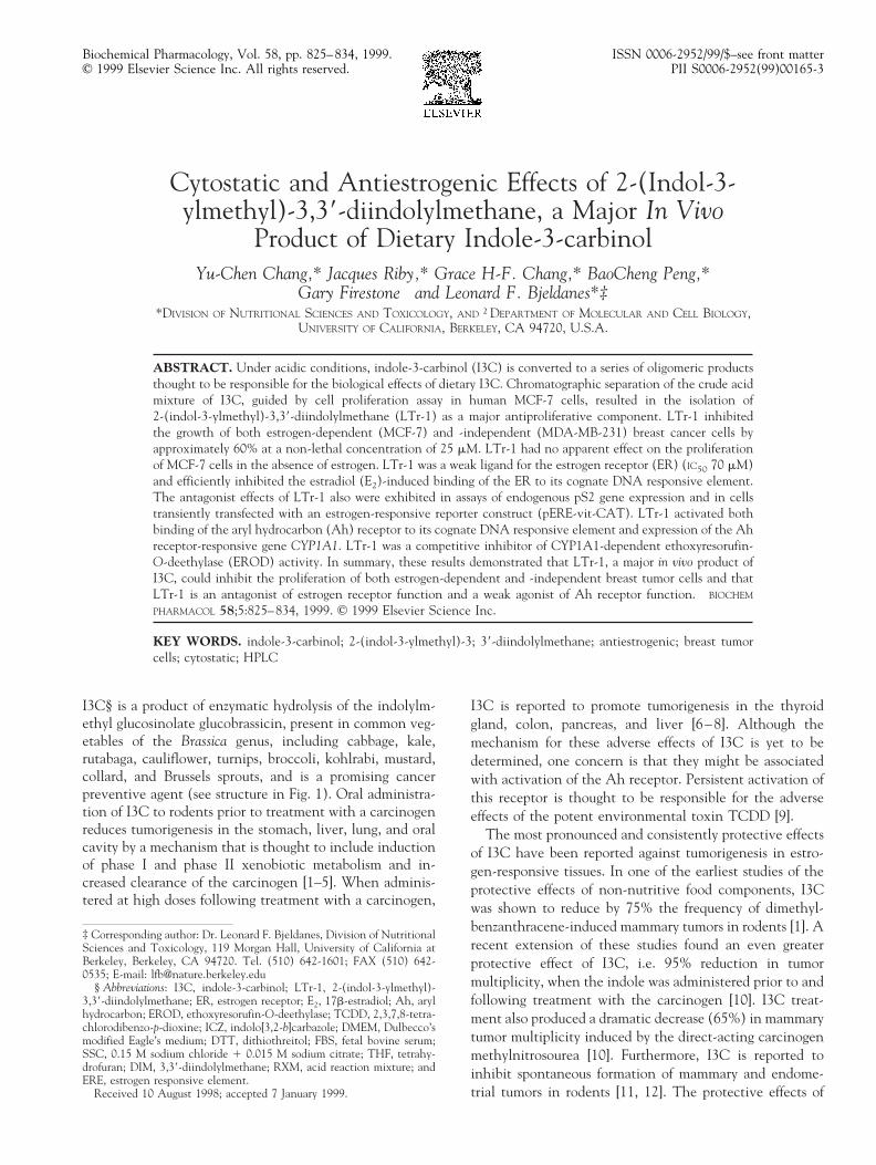
FIG. 6. Competitive binding to the ER. The binding of [3H]E2 (1 nM) to the ER from rat uterine cytosol was measured in the presenceof the unlabeled competitors, E2 (F), tamoxifen (E), and LTr-1 (l), at the concentrations indicated and reported as the percentageof binding in the absence of competitors. Results are presented as the averages of two independent determinations. Relative bindingaffinities were calculated using the concentration of competitor needed to reduce [3H]E2 binding by 50% as compared with theconcentration of unlabeled E2 needed to achieve the same result.
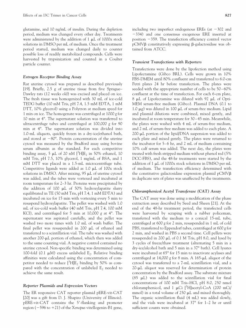
FIG. 7. Binding of nuclear proteins to the ERE. Gel mobilityshift analysis of nuclear extracts from estrogen-depleted MCF-7cells treated for 2 hr with DMSO (lanes 1, 4, 7, 10) or withLTr-1 (1.0 and 10.0 mM in lanes 2, 5, 8, 11 and 3, 6, 9, 12,respectively) and E2 (1 nM) (lanes 4–6 and 10–12). Amonoclonal antibody specific for the human ER also was addedto the incubation mixture for lanes 7–12. Arrows indicate thelocations of the free labeled probe (arrow #1), the ligand-responsive shifted band (arrow #2), and the antibody-super-shifted band (arrow #3).
830 Y-C. Chang et al.

creased cell counts apparently were not due to a generaltoxicity of the trimer, since we saw no evidence of cellkilling over the 5-day treatment period. Interestingly,LTr-1 exhibited no apparent effect on proliferation ofMCF-7 cells in the absence of E2.
Effects of LTr-1 on Estrogen Receptor Binding andFunction
Because LTr-1 inhibited the E2-induced proliferation ofMCF-7 cells, we examined the effects of this indolederivative on components of the ER signal transductionpathway. The relative binding affinity of LTr-1 for the ER,as measured by a competitive binding assay, indicated anIC50 of approximately 70 mM compared with 200 and 3.0nM measured for tamoxifen and E2, respectively (Fig. 6).Thus, LTr-1 exhibits a weak affinity for the ER.
We next examined by gel mobility shift assay the effectof LTr-1 on the binding activity of ER to its cognate DNAmotif. LTr-1 exhibited a strong concentration-dependentinhibitory effect on the E2-induced binding of ER to aconsensus ERE with nearly complete loss of the shiftedband at 10 mM LTr-1 (Fig. 7). In the absence of E2,however, LTr-1 exhibited weak agonist activity on EREbinding to DNA.
To determine whether LTr-1 can affect transcription ofestrogen-responsive genes, we examined its effects on ex-pression of the endogenous pS2 gene, often used as a markerof estrogen-responsive breast tumors, and on the pERE-vit-CAT reporter construct transiently transfected into MCF-7cells. The pERE-vit-CAT construct contains the promoterand 59-flanking region of the Xenopus vitellogenin geneupstream of the CAT structural gene. The results ofnorthern blot analysis indicated that E2-induced transcrip-tion of pS2 was inhibited in a concentration-dependentmanner (approximately 50% at 10 mM) by LTr-1 (Fig. 8).In the absence of E2, LTr-1 did not induce significanttranscription of pS2. A similar inhibitory effect of LTr-1was seen on E2-induced expression of the pERE-vit-CATreporter construct (Fig. 9). In this case, however, LTr-1exhibited a weak activation of this reporter in the absenceof E2. Thus, LTr-1 could suppress activation of E2-respon-sive genes at concentrations that inhibited breast tumorcell proliferation.
FIG. 8. Effect of LTr-1 on pS2 mRNA expression. Estrogen-depleted MCF-7 cells were treated for 48 hr with LTr-1 atconcentrations ranging from 0.1 to 10.0 mM, with (E) orwithout (F) E2 (1 nM). pS2 mRNA levels were measured bynorthern blot analysis (A) and normalized using GADPHmRNA as an internal standard (B). Results are presented as foldinduction over the DMSO control (averages of two independentdeterminations).
FIG. 9. Effect of LTr-1 on CAT expression from the ERE-vit-CAT reporter gene. MCF-7 cells were transiently transfectedwith the pERE-vit-CAT reporter plasmid and treated for 48 hrwith LTr-1 at the concentrations indicated, in the presence (F)or absence (E) of E2 (1 nM). CAT activity in cytosol prepara-tions from individual plates was normalized for protein concen-tration. Results are presented as fold induction over the DMSOcontrol (mean 6 range of two independent determinations).
Effects of an I3C Trimer in Cancer Cells 831
Effects of LTr-1 on Ah Receptor Signaling
We reported previously that LTr-1 has an appreciableaffinity for the Ah receptor [23]. Since persistent activationof the Ah receptor is thought to be responsible for the toxiceffects of certain ligands, including TCDD, we examinedfurther the effects of LTr-1 on this pathway.
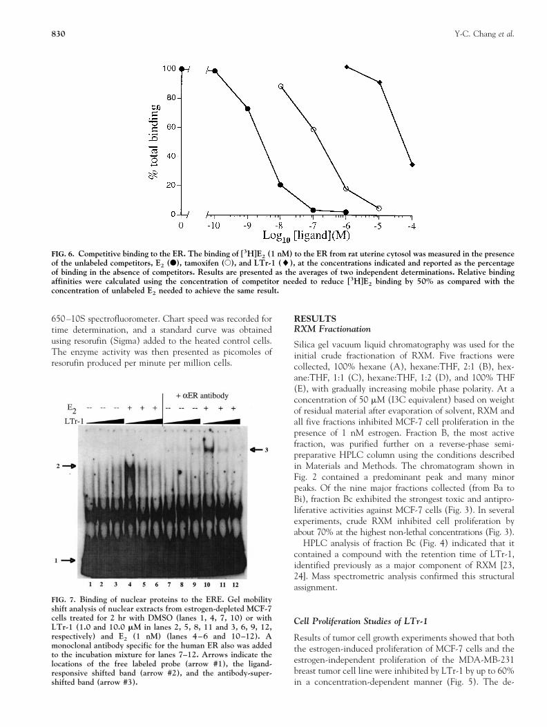
The effects of LTr-1 on binding of the Ah receptor to itscognate DNA motif, DRE, as determined by gel mobilityshift assay, are represented in Fig. 10. At a concentration of1 mM, LTr-1 promoted detectable binding of the Ahreceptor to the DRE. At a concentration of 10 mM, bindingwas as strong as the positive control, ICZ, indicating thatLTr-1 could efficiently transform the Ah receptor to aDNA binding form.
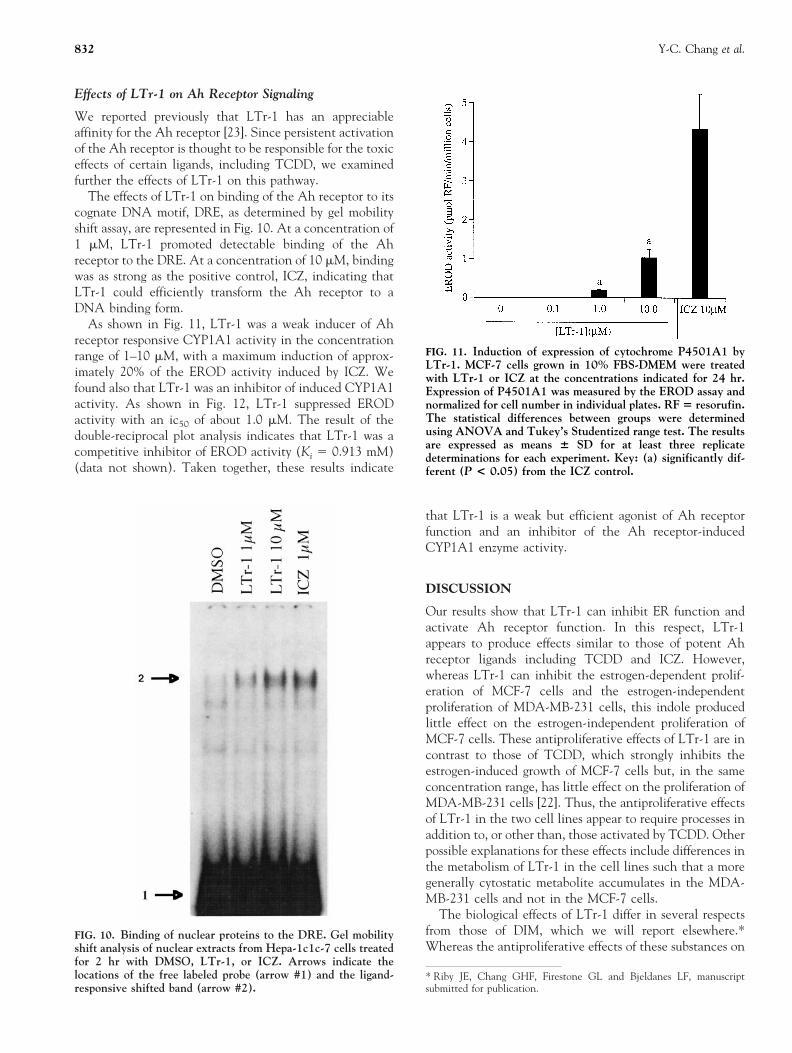
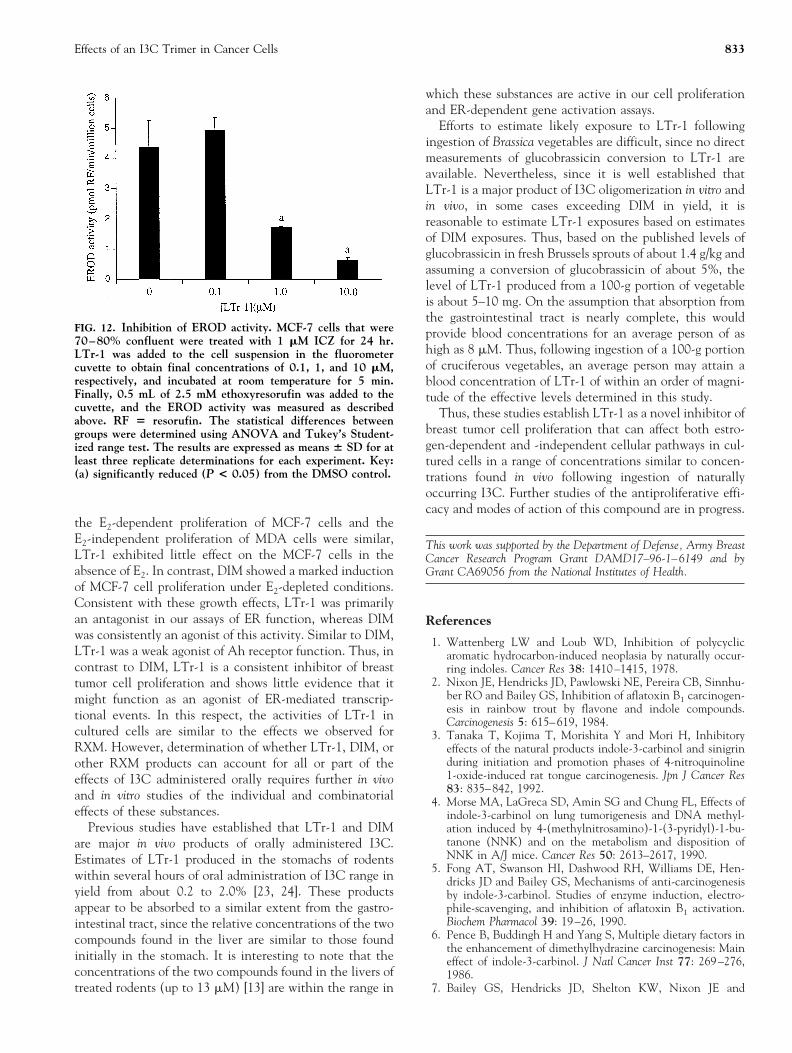
As shown in Fig. 11, LTr-1 was a weak inducer of Ahreceptor responsive CYP1A1 activity in the concentrationrange of 1–10 mM, with a maximum induction of approx-imately 20% of the EROD activity induced by ICZ. Wefound also that LTr-1 was an inhibitor of induced CYP1A1activity. As shown in Fig. 12, LTr-1 suppressed ERODactivity with an ic50 of about 1.0 mM. The result of thedouble-reciprocal plot analysis indicates that LTr-1 was acompetitive inhibitor of EROD activity (Ki 5 0.913 mM)(data not shown). Taken together, these results indicate
that LTr-1 is a weak but efficient agonist of Ah receptorfunction and an inhibitor of the Ah receptor-inducedCYP1A1 enzyme activity.
DISCUSSION
Our results show that LTr-1 can inhibit ER function andactivate Ah receptor function. In this respect, LTr-1appears to produce effects similar to those of potent Ahreceptor ligands including TCDD and ICZ. However,whereas LTr-1 can inhibit the estrogen-dependent prolif-eration of MCF-7 cells and the estrogen-independentproliferation of MDA-MB-231 cells, this indole producedlittle effect on the estrogen-independent proliferation ofMCF-7 cells. These antiproliferative effects of LTr-1 are incontrast to those of TCDD, which strongly inhibits theestrogen-induced growth of MCF-7 cells but, in the sameconcentration range, has little effect on the proliferation ofMDA-MB-231 cells [22]. Thus, the antiproliferative effectsof LTr-1 in the two cell lines appear to require processes inaddition to, or other than, those activated by TCDD. Otherpossible explanations for these effects include differences inthe metabolism of LTr-1 in the cell lines such that a moregenerally cytostatic metabolite accumulates in the MDA-MB-231 cells and not in the MCF-7 cells.
The biological effects of LTr-1 differ in several respectsfrom those of DIM, which we will report elsewhere.*Whereas the antiproliferative effects of these substances on
* Riby JE, Chang GHF, Firestone GL and Bjeldanes LF, manuscriptsubmitted for publication.
FIG. 10. Binding of nuclear proteins to the DRE. Gel mobilityshift analysis of nuclear extracts from Hepa-1c1c-7 cells treatedfor 2 hr with DMSO, LTr-1, or ICZ. Arrows indicate thelocations of the free labeled probe (arrow #1) and the ligand-responsive shifted band (arrow #2).
FIG. 11. Induction of expression of cytochrome P4501A1 byLTr-1. MCF-7 cells grown in 10% FBS-DMEM were treatedwith LTr-1 or ICZ at the concentrations indicated for 24 hr.Expression of P4501A1 was measured by the EROD assay andnormalized for cell number in individual plates. RF 5 resorufin.The statistical differences between groups were determinedusing ANOVA and Tukey’s Studentized range test. The resultsare expressed as means 6 SD for at least three replicatedeterminations for each experiment. Key: (a) significantly dif-ferent (P < 0.05) from the ICZ control.
832 Y-C. Chang et al.
the E2-dependent proliferation of MCF-7 cells and theE2-independent proliferation of MDA cells were similar,LTr-1 exhibited little effect on the MCF-7 cells in theabsence of E2. In contrast, DIM showed a marked inductionof MCF-7 cell proliferation under E2-depleted conditions.Consistent with these growth effects, LTr-1 was primarilyan antagonist in our assays of ER function, whereas DIMwas consistently an agonist of this activity. Similar to DIM,LTr-1 was a weak agonist of Ah receptor function. Thus, incontrast to DIM, LTr-1 is a consistent inhibitor of breasttumor cell proliferation and shows little evidence that itmight function as an agonist of ER-mediated transcrip-tional events. In this respect, the activities of LTr-1 incultured cells are similar to the effects we observed forRXM. However, determination of whether LTr-1, DIM, orother RXM products can account for all or part of theeffects of I3C administered orally requires further in vivoand in vitro studies of the individual and combinatorialeffects of these substances.
Previous studies have established that LTr-1 and DIMare major in vivo products of orally administered I3C.Estimates of LTr-1 produced in the stomachs of rodentswithin several hours of oral administration of I3C range inyield from about 0.2 to 2.0% [23, 24]. These productsappear to be absorbed to a similar extent from the gastro-intestinal tract, since the relative concentrations of the twocompounds found in the liver are similar to those foundinitially in the stomach. It is interesting to note that theconcentrations of the two compounds found in the livers oftreated rodents (up to 13 mM) [13] are within the range in
which these substances are active in our cell proliferationand ER-dependent gene activation assays.
Efforts to estimate likely exposure to LTr-1 followingingestion of Brassica vegetables are difficult, since no directmeasurements of glucobrassicin conversion to LTr-1 areavailable. Nevertheless, since it is well established thatLTr-1 is a major product of I3C oligomerization in vitro andin vivo, in some cases exceeding DIM in yield, it isreasonable to estimate LTr-1 exposures based on estimatesof DIM exposures. Thus, based on the published levels ofglucobrassicin in fresh Brussels sprouts of about 1.4 g/kg andassuming a conversion of glucobrassicin of about 5%, thelevel of LTr-1 produced from a 100-g portion of vegetableis about 5–10 mg. On the assumption that absorption fromthe gastrointestinal tract is nearly complete, this wouldprovide blood concentrations for an average person of ashigh as 8 mM. Thus, following ingestion of a 100-g portionof cruciferous vegetables, an average person may attain ablood concentration of LTr-1 of within an order of magni-tude of the effective levels determined in this study.
Thus, these studies establish LTr-1 as a novel inhibitor ofbreast tumor cell proliferation that can affect both estro-gen-dependent and -independent cellular pathways in cul-tured cells in a range of concentrations similar to concen-trations found in vivo following ingestion of naturallyoccurring I3C. Further studies of the antiproliferative effi-cacy and modes of action of this compound are in progress.
This work was supported by the Department of Defense, Army BreastCancer Research Program Grant DAMD17–96-1–6149 and byGrant CA69056 from the National Institutes of Health.
References
1. Wattenberg LW and Loub WD, Inhibition of polycyclicaromatic hydrocarbon-induced neoplasia by naturally occur-ring indoles. Cancer Res 38: 1410–1415, 1978.
2. Nixon JE, Hendricks JD, Pawlowski NE, Pereira CB, Sinnhu-ber RO and Bailey GS, Inhibition of aflatoxin B1 carcinogen-esis in rainbow trout by flavone and indole compounds.Carcinogenesis 5: 615–619, 1984.
3. Tanaka T, Kojima T, Morishita Y and Mori H, Inhibitoryeffects of the natural products indole-3-carbinol and sinigrinduring initiation and promotion phases of 4-nitroquinoline1-oxide-induced rat tongue carcinogenesis. Jpn J Cancer Res83: 835–842, 1992.
4. Morse MA, LaGreca SD, Amin SG and Chung FL, Effects ofindole-3-carbinol on lung tumorigenesis and DNA methyl-ation induced by 4-(methylnitrosamino)-1-(3-pyridyl)-1-bu-tanone (NNK) and on the metabolism and disposition ofNNK in A/J mice. Cancer Res 50: 2613–2617, 1990.
5. Fong AT, Swanson HI, Dashwood RH, Williams DE, Hen-dricks JD and Bailey GS, Mechanisms of anti-carcinogenesisby indole-3-carbinol. Studies of enzyme induction, electro-phile-scavenging, and inhibition of aflatoxin B1 activation.Biochem Pharmacol 39: 19–26, 1990.
6. Pence B, Buddingh H and Yang S, Multiple dietary factors inthe enhancement of dimethylhydrazine carcinogenesis: Maineffect of indole-3-carbinol. J Natl Cancer Inst 77: 269–276,1986.
7. Bailey GS, Hendricks JD, Shelton KW, Nixon JE and
FIG. 12. Inhibition of EROD activity. MCF-7 cells that were70–80% confluent were treated with 1 mM ICZ for 24 hr.LTr-1 was added to the cell suspension in the fluorometercuvette to obtain final concentrations of 0.1, 1, and 10 mM,respectively, and incubated at room temperature for 5 min.Finally, 0.5 mL of 2.5 mM ethoxyresorufin was added to thecuvette, and the EROD activity was measured as describedabove. RF 5 resorufin. The statistical differences betweengroups were determined using ANOVA and Tukey’s Student-ized range test. The results are expressed as means 6 SD for atleast three replicate determinations for each experiment. Key:(a) significantly reduced (P < 0.05) from the DMSO control.
Effects of an I3C Trimer in Cancer Cells 833
Pawlowski NE, Enhancement of carcinogenesis by the naturalanticarcinogen indole-3-carbinol. J Natl Cancer Inst 78:931–936, 1987.
8. Kim D, Han B, Ahn B, Hasegawa R, Shirai T, Ito N andTsuda H, Enhancement by indole-3-carbinol of liver andthyroid gland neoplastic development in a rat medium-termmultiorgan carcinogenesis model. Carcinogenesis 18: 377–381, 1997.
9. Schmidt JV and Bradfield CA, Ah receptor signaling path-ways. Annu Rev Cell Dev Biol 12: 55–89, 1996.
10. Grubbs CJ, Steele VE, Casebolt T, Juliana MM, Eto I,Whitaker LM, Dragnev KH, Kelloff GJ and Lubet RL,Chemoprevention of chemically-induced mammary carcino-genesis by indole-3-carbinol. Anticancer Res 15: 709–716,1995.
11. Bradlow HL, Michnovicz JJ, Telang NT and Osborne MP,Effects of dietary indole-3-carbinol on estradiol metabolismand spontaneous mammary tumors in mice. Carcinogenesis 12:1571–1574, 1991.
12. Kojima T, Tanaka T and Mori H, Chemoprevention ofspontaneous endometrial cancer in female Donryu rats bydietary indole-3-carbinol. Cancer Res. 54: 1446–1449, 1994.
13. Grose KR and Bjeldanes LF, Oligomerization of indole-3-carbinol in aqueous acid. Chem Res Toxicol 5: 188–193, 1992.
14. Stresser DM, Williams DE, Griffin DA and Bailey GS,Mechanisms of tumor modulation by indole-3-carbinol. Dis-position and excretion in male Fischer 344 rats. Drug MetabDispos 23: 965–975, 1995.
15. Chen Y-H, Riby J, Srivastava P, Bartholomew J, Denison Mand Bjeldanes L, Regulation of CYP1A1 by indolo[3,2-b]carbazole in murine hepatoma cells. J Biol Chem 270:22548–22555, 1995.
16. Chen I, Safe S and Bjeldanes L, Indole-3-carbinol and
diindolymethane as aryl hydrocarbon (Ah) receptor agonistsand antagonists in T47D human breast cancer cells. BiochemPharmacol 51: 1069–1076, 1996.
17. Stresser DM, Bjeldanes LF, Bailey GS and Williams DE, Theanticarcinogen 3,39-diindolylmethane is an inhibitor of cyto-chrome P-450. J Biochem Toxicol 10: 191–201, 1995.
18. Chen I, McDougal A, Wang F and Safe S, Aryl hydrocarbonreceptor-mediated antiestrogenic and antitumorigenic activ-ity of diindolylmethane. Carcinogenesis 19: 1631–1639, 1998.
19. Santell RC, Chang YC, Nair MG and Helferich WG, Dietarygenistein exerts estrogenic effects upon the uterus, mammarygland and the hypothalamic/pituitary axis in rats. J Nutr 127:263–269, 1997.
20. Chang T-C, Nardulli AM, Lew D and Shapiro DJ, The role ofestrogen response elements in expression of the Xenopus laevisvitellogenin B1 gene. Mol Endocrinol 6: 346–354, 1992.
21. Seed B and Sheen JY, A simple phase-extraction assay forchloramphenicol acyltransferase activity. Gene 67: 271–277,1988.
22. Dohr O, Vogel D and Abel J, Different response of 2,3,7,8-tetrachlorodibenzo-p-dioxin (TCDD)-sensitive genes in hu-man breast cancer MCF-7 and MDA-MB-231 cells. ArchBiochem Biophys. 321: 405–412, 1995.
23. Bjeldanes L, Kim JY, Grose KR, Bartholomew JC and Brad-field CA, Aromatic hydrocarbon responsiveness-receptor ago-nists generated from indole-3-carbinol in vitro and in vivo:Comparisons with TCDD. Proc Natl Acad Sci USA 88:9543–9547, 1991.
24. De Kruif CA, Marsman JW, Venekamp JC, Falke HE,Noordhoek J, Blaauboer BJ and Wortelboer HM, Structureelucidation of acid reaction products of indole-3-carbinol:Detection in vivo and enzyme induction in vitro. Chem BiolInteract 80: 303–315, 1991.
834 Y-C. Chang et al.