cytotoxic effects of klebsiella oxytoca strains isolated from patients
TRANSCRIPT

JOURNAL OF CLINICAL MICROBIOLOGY, Mar. 2010, p. 817–824 Vol. 48, No. 30095-1137/10/$12.00 doi:10.1128/JCM.01741-09Copyright © 2010, American Society for Microbiology. All Rights Reserved.
Cytotoxic Effects of Klebsiella oxytoca Strains Isolated from Patientswith Antibiotic-Associated Hemorrhagic Colitis or Other Diseases
Caused by Infections and from Healthy Subjects�†Martina M. Joainig,1,2 Gregor Gorkiewicz,1,3 Eva Leitner,4 Paul Weberhofer,2 Ines Zollner-Schwetz,2
Irmgard Lippe,5 Gebhard Feierl,4 Robert Krause,2 Thomas Hinterleitner,2Ellen L. Zechner,1* and Christoph Hogenauer2*
Institute of Molecular Biosciences, University of Graz, Graz, Austria1; Department of Internal Medicine, Medical University ofGraz, Graz, Austria2; Institute of Pathology, Medical University of Graz, Graz, Austria3; Institute of Hygiene, Microbiology and
Environmental Medicine, Medical University of Graz, Graz, Austria4; and Institute of Experimental andClinical Pharmacology, Medical University of Graz, Graz, Austria5
Received 4 September 2009/Returned for modification 30 October 2009/Accepted 23 December 2009
Antibiotic-associated hemorrhagic colitis (AAHC) is associated with Klebsiella oxytoca. This study analyzedwhether cytotoxic properties are linked to specific subtypes of K. oxytoca. Klebsiella isolates from stools of AAHCpatients, healthy carriers, and diarrhea patients as well as from infections of other organs were investigated.Cytotoxic effects on human epithelial cells were limited to the species K. oxytoca and were not detectable for anyother Klebsiella species. Isolates from AAHC patients and from stools showed the highest proportion ofcytotoxic strains. Urinary or respiratory tract isolates exhibited no cytotoxicity. Macrorestriction profiling ofstrains revealed no genetic relationships of AAHC isolates or the cytotoxic phenotype but identified thatdifferent K. oxytoca strains with different cytotoxic behaviors may be prevalent in the same AAHC patient.Under laboratory conditions, cytotoxicity was maximally effective after exponential bacterial growth and thendeclined despite the continued viability of K. oxytoca cells in culture. Given its capacity to induce AAHC andthat a high proportion of stool isolates tested cytotoxin positive, we argue that K. oxytoca should be consideredan opportunistic pathogen if detected in stools. The ability to induce disease after antibiotic treatment mostlikely represents an overgrowth of the toxin-producing bacterium due to an alteration of the normal colonicmicroflora.
Antibiotic-associated colitis (AAC) is a frequent adverseeffect observed when the normal bacterial flora is altered dueto antibiotic therapy. Most cases of AAC are caused by infec-tion by and extensive growth of Clostridium difficile, leading topseudomembranous colitis. A special form of AAC is antibi-otic-associated hemorrhagic colitis (AAHC), which was firstdescribed in 1978 (21) and has since been ascribed specificclinical, endoscopic, histopathological, and microbiologicalcharacteristics (9, 10). AAHC is not associated with C. difficileand was only recently shown to be caused by Klebsiella oxytoca(9, 10). AAHC is typically observed after a brief therapy withpenicillins, with a sudden onset of bloody diarrhea often incombination with severe abdominal cramps, which often re-quires hospitalization. The key features of AAHC upon endos-copy are mucosal hemorrhage and mucosal edema, usuallywith segmental distribution, commonly affecting the ascending
colon and the cecum (9). Histology typically resembles that ofcolitis induced by toxin-producing bacteria (10).
For the majority of patients with AAHC, stool testing re-veals K. oxytoca in significant amounts (�106 CFU/ml) (9, 23).This Gram-negative rod is ubiquitous in the environment (e.g.,soil and water) but can also be isolated from skin, mucousmembranes, and the intestines of humans and animals (19).Human infections with K. oxytoca resemble those with Kleb-siella pneumoniae; i.e., respiratory and urinary tracts are com-monly affected (e.g., nosocomial pneumonia), in addition tosoft tissue and hepatobiliary infections (6). Until recently, K.oxytoca was not considered to be an intestinal pathogen, and itspresence in stool has placed the organism as a constituent ofthe normal gut microflora. For the healthy population, coloni-zation of the intestine with K. oxytoca has been reported for1.6% to 9% of subjects (1, 4, 10, 23). It is important that K.oxytoca constitutively produces �-lactamases conferring resis-tance to amino- and carboxypenicillins (13), agents typicallygiven before the onset of AAHC. The association of K. oxytocawith AAHC was previously established by an animal modelusing a K. oxytoca strain isolated from a patient with AAHC incombination with an antibiotic to induce right-sided hemor-rhagic colitis (10).
In the 1990s, two independent groups reported that K. oxy-toca strains isolated from patients with AAHC produce a cy-totoxin, which caused cell death in cultured Hep2, Vero, CHO-K1, and HeLa cell lines as well as in an isolated intestinal-loop
* Corresponding author. Mailing address for Christoph Hogenauer:Department of Internal Medicine, Medical University of Graz, Auen-bruggerplatz 15, A8036 Graz, Austria. Phone: 43 316 385 4388. Fax: 43316 385 2648. E-mail: [email protected]. Mailingaddress for Ellen L. Zechner: Institute of Molecular Biosciences,University of Graz, Humboldtstraße 50, 8010 Graz, Austria. Phone: 43316 380 5624. Fax: 43 316 380 9019. E-mail: [email protected].
† Supplemental material for this article may be found at http://jcm.asm.org/.
� Published ahead of print on 6 January 2010.
817
Dow
nloa
ded
from
http
s://j
ourn
als.
asm
.org
/jour
nal/j
cm o
n 14
Feb
ruar
y 20
22 b
y 39
.124
.237
.5.
model (8, 15–17). In contrast, two laboratory strains of K.oxytoca exhibited no cytotoxicity, indicating that cytotoxin pro-duction might be strain specific (16, 17). The cytotoxin wasreported to be heat labile, insensitive to proteinase digestion,and of a low molecular mass (8, 16, 17). A detailed analysis ofthe chemical nature and molecular structure of the cytotoxin isnot yet available. Moreover, a causal link between toxin pro-duction in K. oxytoca and AAHC has not been established.
The aim of the current study was to assess whether cytotoxinproduction is specific to certain subtypes of K. oxytoca and totest the hypothesis that AAHC uniquely correlates with thosestrains. A total of 121 Klebsiella isolates were investigated,including K. oxytoca strains isolated from stool samples frompatients with AAHC, healthy carriers, and patients with colitis/diarrhea of other causes. K. oxytoca strains from infectionsinvolving other body sites and other Klebsiella spp. were ana-lyzed in comparison. The characterization of all isolates wasperformed by using genotypic and biochemical methods, andthe capacity of each isolate to induce cytotoxic effects on cul-tured eukaryotic cells was measured. A subset of strains wasgenotyped by macrorestriction profiling to assess their geneticrelatedness.
MATERIALS AND METHODS
Bacterial strains. The Klebsiella isolates used in this study are listed in TableS1 in the supplemental material. The isolates were obtained from patientstreated at the Medical University of Graz and from healthy volunteers. The studywas approved by the local institutional review board, and written informedconsent was obtained from all subjects. Two cytotoxin-negative reference iso-lates, K. oxytoca DSM 4798 (ATCC 8724) and DSM 5175 (ATCC 13182)(DSMZ, Braunschweig, Germany), and cytotoxin-producing strain MH 43-1(kindly provided by T. Chida, Department of Microbiology, Medical and DentalUniversity, Tokyo, Japan) (8) served as controls. The viewer was blinded to thesources of isolates. All strains were grown in tryptic soy broth (TSB) (Merck,Germany) and M9 minimal medium (Invitrogen, Lofer, Austria) at 37°C undergentle shaking overnight or on tryptic soy agar (Merck).
All Klebsiella isolates were subjected to biochemical analyses by using the API20E test (bioMerieux, Marcy l’Etoile, France). Indole production was assessedthree times independently with each isolate by using James (R2) reagent accord-ing to the manufacturer’s specifications (bioMerieux). The polygalacturonase(pehX) gene was previously shown to be specific for K. oxytoca among Klebsiellaspp.; thus, a 344-bp PCR amplification product of the pehX gene was employedfor the identification of all isolates according to a previously reported procedure(11). Taq polymerase (NEB, Bedford, MA) was applied according to the man-ufacturer’s specifications.
Isolates that were not unambiguously identified by means of API testing,indole production, and PCR were subsequently subjected to 16S rRNA genesequence analyses. The differentiation of species by 16S rRNA gene sequenceapplied sequence comparisons according to a method described previously byBoye and Hansen (3). This approach differentiates between Klebsiella speciesaccording to species-specific base changes (3, 20). PCR products were cyclesequenced with the BigDye termination cycle sequencing ready reaction kit (v.3.1;Applied Biosystems, Foster City, CA) and resolved by use of an ABI Prism 310genetic analyzer. Sequence data analysis was performed by using the NCBI BLASTserver (http://www.ncbi.nlm.nih.gov/BLAST/), and multiple sequence alignmentswere performed with SeqMan II (DNAStar Inc.). Reference sequences were Esch-erichia coli (GenBank accession no. J01695), K. oxytoca ATCC 13182T (GenBankaccession no. Y17655), K. pneumoniae ATCC 13883T (GenBank accession no.Y17656), K. planticola ATCC 33531T (GenBank accession no. Y17659), K. ornithi-nolytica 590681 (GenBank accession no. Y17662), and K. terrigena ATCC 33257T
(GenBank accession no. Y17658).Cytotoxin tissue culture assay. Cytotoxin production was monitored in a
modified cell culture assay as described previously (16). Hep2 cells were grownin minimal essential alpha medium with Earle’s balanced salt solution (Invitro-gen, Lofer, Austria) and with 10% fetal bovine serum, 100 �g/ml penicillin, and100 �g/ml streptomycin. Cultures were incubated at 37°C with 5% CO2 in 95%humidity. Cells were newly seeded every 48 h as recommended by the supplier
(European Collection of Cell Culture [ECACC], Wiltshire, United Kingdom).Thirty milliliters of TSB (Merck, Darmstadt, Germany) was inoculated with asingle bacterial colony and incubated for 14 to 16 h at 37°C with gentle agitation(180 rpm). Cytotoxic effects were measured for cultures grown to an opticaldensity at 600 nm (OD600) of 4 to 6. Cultures were then centrifuged at 5,000 rpmat 4°C for 20 min, and the supernatants were filtered through 0.2-�m celluloseacetate filters (Millipore, MA). Dilutions of the filtered supernatant were pre-pared in phosphate-buffered saline (PBS). A total of 1.5 � 104 Hep2 cells in 100�l culture medium were seeded per well in 96-well tissue culture plates (Greiner,Kremsmunster, Austria). Equivalent volumes (50 �l) of pure and diluted super-natants were added before incubation for 48 h. Eukaryotic cells were thenassessed microscopically for cytotoxic effects as described previously (8, 16). Theviability of Hep2 cells was assessed spectrophotometrically by using 3-(4,5-di-methyl-2-thiazolyl)-2,5-diphenyl-2H-tetrazolium bromide (MTT) (Sigma-Al-drich, St. Louis, MO). Cells were washed once with 200 �l PBS per well andincubated with 200 �l of PBS containing 5 mg/ml MTT for 2 h at 37°C. The MTTsolution was removed by aspiration, and cells were lysed with a 1:25 (vol/vol)solution of 96% acetic acid and 2-propanol (18). Absorbance units in the wellswere then determined at a 595-nm wavelength (microplate reader 550; Bio-Rad,Hercules, CA). Each dilution was measured in triplicate and compared to con-trols treated with PBS only. The 50% cytotoxic dose (CD50) was expressed as thefinal dilution of the bacterial supernatant wherein viability decreased to 50% ofthat of PBS-treated Hep2 cells. Statistical significance was determined by usingthe Fisher exact test to compare the AAHC isolates with Klebsiella isolates fromother test groups (SigmaPlot 11.0; Systat Software Inc., San Jose, CA).
Analysis of cytotoxin production during bacterial growth. Cells of clinicalisolate K. oxytoca 04/1O and the type strain K. oxytoca ATCC 13182 were grownin 2 ml TSB at 37°C for 48 h under gentle agitation. Bacterial cultures wereharvested at regular time points, and the cells were collected by centrifugation at5,000 rpm at 4°C for 20 min. Pellets were resuspended in 1 ml PBS and platedonto agar plates in serial dilutions. Bacterial supernatants were filtered andexamined by the MTT test as described above. Values are expressed as means �standard deviations (SD).
Strain typing of isolates by PFGE. Seventy Klebsiella strains were genotyped bymacrorestriction profiling with the restriction endonuclease XbaI and resolvedby pulsed-field gel electrophoresis (PFGE) analysis as described recently (7, 12).Briefly, bacterial cells grown overnight in TSB were pelleted, washed once with2 ml PBS, and diluted into PBS to an OD600 of 0.2. The suspension was mixedwith an equal amount of 2% low-melting agarose (FMC BioProducts, Rockland,ME) prepared in 0.5� Tris-borate-EDTA (TBE) buffer. Solidified blocks wereincubated in 1.3 ml of lysis buffer (0.25 mM EDTA [pH 8], 1% SDS) containing100 mg/ml proteinase K for 48 h at 50°C under gentle shaking. After 24 h, thebuffer was removed, and fresh buffer was added. Agarose blocks were washedthree times with 1.3 ml Tris-EDTA (TE) buffer containing 2 mM Pefabloc SC(Roche, Basel, Switzerland) at 37°C for 30 min. Subsequently, blocks wereincubated with 40 U XbaI (Fermentas, Ontario, Canada) and incubated at 37°Cunder gentle shaking for 20 h. DNA fragments were resolved in 1% peqGOLDuniversal agarose (Peqlab Biotechnologie GmbH, Erlangen, Germany) in 0.5�TBE buffer with the Pharmacia Biotech Gene Navigator apparatus (GE Health-care, Vienna, Austria). Electrophoresis was performed for 24 h at 6°C at aconstant voltage of 200 V (6 V/cm) with a linear ramp of pulse times from 1 to40 s. Gels were stained with ethidium bromide, and restriction patterns werevisualized under UV irradiation. Analysis of restriction fragment patterns wasperformed with ImageMaster 1D software (Amersham Pharmacia Biotech, Upp-sala, Sweden). A control strain, K. pneumoniae ATCC 4352, was run with eachgel to assess variation within gels. Matching and cluster analyses were done byusing the unweighted-pair group method with average linkages (UPGMA) withthe Dice coefficient.
RESULTS
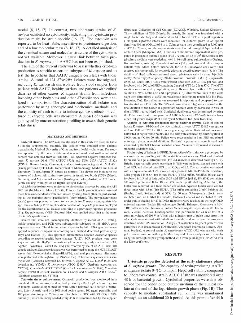
Cytotoxic properties detected at the early stationary phaseof K. oxytoca growth. The capacity of toxin-producing AAHCK. oxytoca isolate 04/1O to impair Hep2 cell viability comparedto laboratory control strain ATCC 13182 was monitored over48 h of bacterial growth. Cytolethal properties were first ob-served for the conditioned culture medium of the clinical iso-late at the end of the logarithmic growth phase (Fig. 1B). Thecapacity to mediate substantial cell killing was maintainedthroughout an additional 30-h period. At this point, after 44 h
818 JOAINIG ET AL. J. CLIN. MICROBIOL.
Dow
nloa
ded
from
http
s://j
ourn
als.
asm
.org
/jour
nal/j
cm o
n 14
Feb
ruar
y 20
22 b
y 39
.124
.237
.5.
of cultivation, no variation in the viability of isolate 04/1O wasobserved (Fig. 1A), but the culture medium no longer exhib-ited cytotoxic effects on Hep2 cells. Cytotoxin production byisolate 04/1O was not observed under similar conditions in M9minimal medium (not shown).
Clinical source of Klebsiella isolates. The clinical diagnosesfor the patients and the sources of isolation of Klebsiella strainsare shown in Table 1 and Table S1 in the supplemental mate-rial. Fifteen K. oxytoca isolates from 13 patients with AAHC(group I) were isolated during the active phase of colitis.AAHC was diagnosed based on typical clinical and endoscopicand/or radiological features (9, 10). All AAHC patients testednegative for C. difficile and other intestinal pathogenic bacte-ria. K. oxytoca strains of group II were obtained from patientswith C. difficile-negative AAC or from other forms of diarrheaor colitis with negative stool cultures for intestinal pathogens.
Thirteen K. oxytoca isolates (group III) were derived fromstools of healthy carriers without intestinal symptoms or pre-ceding antibiotic therapy. For comparison, K. oxytoca isolatesfrom other organ infection sites were also tested. These in-cluded isolates from the urinary tract (group IV; n � 10), therespiratory tract (group V; n � 16), bacteremia (group VI; n �13), and mucocutaneous infections (group VII; n � 16). K.pneumoniae isolates (group VIII; n � 19) and other Klebsiellaspecies (group IX; n � 5) originated from stool samples ofeither healthy volunteers or diarrhea patients.
Klebsiella identification. The results for the three differentmethods applied for strain identification are compared in Ta-ble S1 in the supplemental material. Results for API 20Ebiochemical testing, the indole reaction, and the K. oxytoca-specific pheX PCR analysis (11) were consistent for 105 of 124strains (121 clinical strains and 3 control strains). Discrepan-cies were observed for 21 isolates. 16S rRNA gene analysis wasused to clarify the taxonomy of uncertain strains (Table S1).Four of the 21 isolates were identified as being K. ornithino-lytica and one was identified as being K. planticola based on 16SrRNA gene analyses. Cytotoxin production by the 21 aberrantisolates was monitored with the cell culture assay. The fiveisolates found to reduce the viability of cultured Hep-2 cellswere all identified as being K. oxytoca isolates by 16S rRNAgene sequencing (see below).
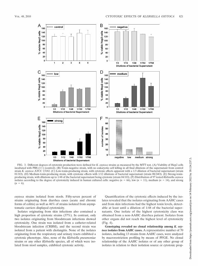
Cytotoxin production by K. oxytoca isolates. The capacity ofthese bacterial isolates to produce a cytotoxic substance wasassessed by the cultivation of Hep2 cells in standard mediumsupplemented with cell-free supernatant of the bacterial iso-late grown for 14 to 16 h. As illustrated in the optimized assayin Fig. 1, conditioned culture medium from a toxin-producingstrain grown to early stationary phase contains sufficient cyto-toxin for obvious detection even after extensive dilution. Arange of pure and serially diluted aliquots of filtered culturemedium from each isolate grown to this stage was tested in cellculture. Eukaryotic cell viability was evaluated microscopically(Fig. 2). The cytotoxic effect was evident by cell rounding anddetachment from the substratum, indicating cell death. Hep2cells also exhibited cell fragmentation typically observed forapoptosis. Comparative cell viability was determined quantita-tively based on MTT uptake and reduction with a colorimetricassay (18). The bacterial supernatant was defined as beingtoxin positive when the aliquot of undiluted supernatant addedto the tissue culture medium was sufficient to reduce the via-bility of the Hep2 cells by �50% compared to a supplement ofPBS alone. No discernible effects on Hep2 cells were observedupon microscopic evaluation or for the MTT test followingcultivation in the presence of PBS or conditioned growth me-dium from the toxin-negative control (Fig. 3A and B). Ninetytest strains (73%) showed no cytotoxin production. In contrast,cytotoxic effects were detectable for cultures containing themedium of 31 (27%) bacterial isolates. The 31 strains inducingpositive toxic effects were evaluated further and assigned var-ious levels of cytotoxicity, as defined in Fig. 3. Based on theseresults, 11 strains were assigned low toxin production (Fig. 3C).Fourteen strains induced medium cytotoxic effects (Fig. 3D),and 6 were identified as being highly cytotoxic (Fig. 3E). No-tably, all toxin-producing isolates were identified as being K.oxytoca isolates. From the total number of K. oxytoca isolatesinvestigated (97), 66 (68%) exhibited no evidence of toxin
FIG. 1. Cytotoxin production during growth of two K. oxytocastrains. (A) Growth curves of laboratory strain ATCC 13182 and clin-ical K. oxytoca isolate 04/1O, originating from an AAHC patient, weredetermined by colony counts on agar plates. (B) Effect of culturemedium from ATCC 13182 (cytotoxin negative) and AAHC isolate04/1O on Hep2 cell viability following 2 to 48 h of bacterial growth.The percent toxin effect represents the fraction of eukaryotic cellskilled after treatment with 48-fold dilutions of bacterial supernatantscompared to PBS-treated control cultures. The data represent themean values of 3 independent experiments � SD.
VOL. 48, 2010 CYTOTOXIC EFFECTS OF KLEBSIELLA OXYTOCA 819
Dow
nloa
ded
from
http
s://j
ourn
als.
asm
.org
/jour
nal/j
cm o
n 14
Feb
ruar
y 20
22 b
y 39
.124
.237
.5.

production and 31 (32%) reduced the viability of the culturedhuman cells (Fig. 3F).
Cytotoxicity of K. oxytoca from patients with AAHC andother infections. To examine whether cytotoxin production wascorrelated with other shared properties of Klebsiella strains,
isolates were classified into nine groups according to theirclinical diagnosis and the isolation source of bacterial strains(Table 1). K. oxytoca isolates from AAHC patients showed thehighest proportion of toxin-producing strains (69%) (Fig. 4).Cytotoxin production was also a common finding for other K.
TABLE 1. Klebsiella isolates used in this study
Groupa Descriptionb Diagnosis (no. of patients)c Isolation source No. ofPatients
No. of cytotoxicisolates/no. of
cytotoxin-negativeisolates
I AAHC AAHC (13) Stool culture 13/15d 9/6II Diarrhea AAC (7), diarrhea (4), IBD (2), hemorrhagic
colitis (1)Stool culture 14 8/6
III Healthy carrier Asymptomatic carriers (13) Stool culture 13 6/7IV Urinary tract UTI (7), complicated UTI (3) Urine culture 10 0/10V Respiratory tract VAP (7), pneumonia (5), exacerbated COPD
(2), bronchitis/bronchiectasis (2)Respiratory culture 16 0/16
VI Blood Bacteremia (10), CRBSI (3) Blood culture 13 2/11VII Skin DFS (6), CSSTI (3), other mucocutaneous
infections (7)Wound swab 16 6/10
VIII K. pneumoniae Asymptomatic carriers (9), AAC (7), IBD (2),hemorrhagic colitis (1)
Stool culture 19 0/19
IX Other Klebsiella species Hemorrhagic colitis (2), AAC (1), IBD (1),diarrhea (1)
Stool culture 5 0/5
a In total, 121 Klebsiella isolates were divided into 9 test groups according to the clinical diagnosis of patients and the source of isolation.b If not otherwise indicated, isolates were classified as being K. oxytoca strains.c Abbreviations: AAHC, antibiotic-associated hemorrhagic colitis; AAC, antibiotic-associated colitis; IBD, inflammatory bowel disease; UTI, urinary tract infection;
VAP, ventilator-associated pneumonia; COPD, chronic obstructive pulmonary disease; CRBSI, central venous catheter-related bloodstream infection; DFS, diabeticfoot syndrome; CSSTI, complicated skin and skin structure infection. The numbers of patients from each subclass are noted in parentheses.
d Fifteen isolates were derived from 13 patients.
FIG. 2. Cytotoxic effects of bacterial supernatants on cultured human cells. (A and B) Hep2 cells cultivated in medium supplemented withsupernatant (1/48 dilution) from toxin-negative K. oxytoca laboratory strain ATCC 13182 shown at �100 (A) and �400 (B) magnifications. (C andD) Hep2 cells incubated with bacterial supernatant obtained from K. oxytoca strain 04/1O isolated from a patient suffering from AAHC (1/48dilution) shown at �100 (C) and �400 (D) magnifications.
820 JOAINIG ET AL. J. CLIN. MICROBIOL.
Dow
nloa
ded
from
http
s://j
ourn
als.
asm
.org
/jour
nal/j
cm o
n 14
Feb
ruar
y 20
22 b
y 39
.124
.237
.5.
oxytoca strains isolated from stools. Fifty-seven percent ofstrains originating from diarrhea cases (acute and chronicforms of colitis) as well as 46% of strains isolated from asymp-tomatic carriers displayed cytotoxicity.
Isolates originating from skin infections also contained ahigh proportion of cytotoxic strains (37%). In contrast, onlytwo isolates originating from bloodstream infections showedcytotoxicity. One strain was isolated from a catheter-relatedbloodstream infection (CRBSI), and the second strain wasisolated from a patient with cholangitis. None of the isolatesoriginating from the respiratory and urinary tracts exhibited acytotoxic phenotype. Also, none of the Klebsiella pneumoniaestrains or any other Klebsiella species, all of which were iso-lated from stool samples, exhibited cytotoxic activity.
Quantification of the cytotoxic effects induced by the iso-lates revealed that the isolates originating from AAHC casesand from skin infections had the highest toxin levels, detect-able at least until a dilution of 1/48 of the bacterial super-natants. One isolate of the highest cytotoxicity class wasobtained from a non-AAHC diarrhea patient. Isolates fromother organs did not reach the highest level of cytotoxicity(Fig. 4).
Genotyping revealed no clonal relationship among K. oxy-toca isolates from AAHC cases. A representative number of 70isolates, including 13 strains from AAHC cases, were analyzedby macrorestriction profiling by means of PFGE. No clonalrelationship of the AAHC isolates or of any other group ofisolates in relation to their isolation source or cytotoxic prop-
FIG. 3. Different degrees of cytotoxin production were defined for K. oxytoca strains as measured by the MTT test. (A) Viability of Hep2 cellsincubated with PBS (1�) (control). (B) Toxin-negative strain, with no eukaryotic cell killing in all final dilutions of the supernatant from controlstrain K. oxytoca ATCC 13182. (C) Low-toxin-producing strain, with cytotoxic effects apparent with a 1/3 dilution of bacterial supernatant (strain01/1O). (D) Medium-toxin-producing strain, with cytotoxic effects with 1/12 dilutions of bacterial supernatant (strain 08/24O). (E) Strong-toxin-producing strain, with dilutions up to 1/48 of the bacterial supernatant being cytotoxic (strain 04/1O). (F) Distribution of 97 tested Klebsiella oxytocaisolates according to the degree of cytotoxicity induced in human cultured cells: negative (n � 66), low (n � 11), medium (n � 14), and strong(n � 6).
VOL. 48, 2010 CYTOTOXIC EFFECTS OF KLEBSIELLA OXYTOCA 821
Dow
nloa
ded
from
http
s://j
ourn
als.
asm
.org
/jour
nal/j
cm o
n 14
Feb
ruar
y 20
22 b
y 39
.124
.237
.5.
erties was evident by XbaI macrorestriction profiling (data notshown).
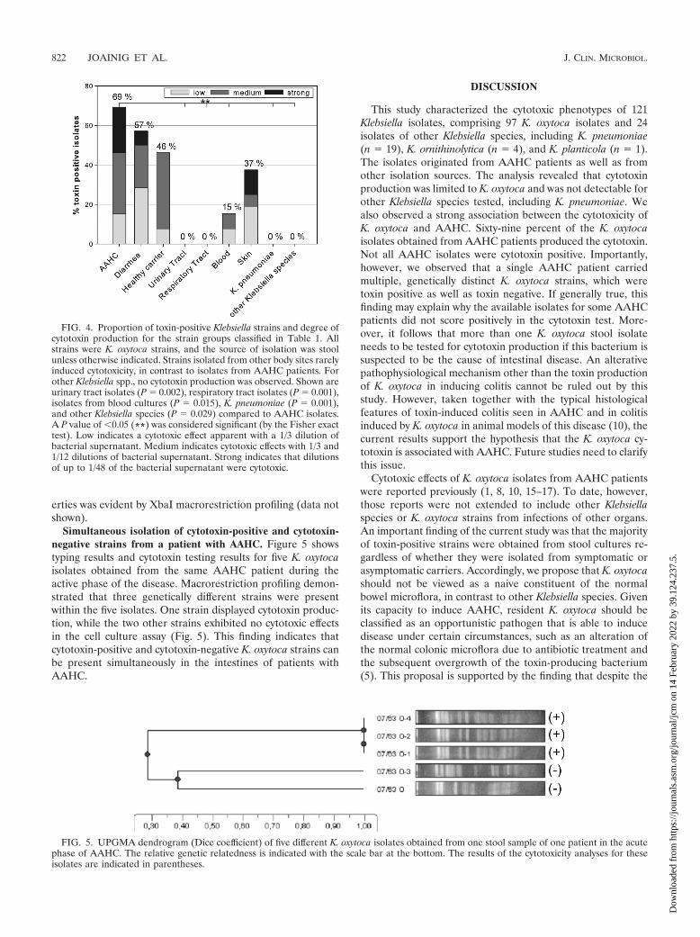
Simultaneous isolation of cytotoxin-positive and cytotoxin-negative strains from a patient with AAHC. Figure 5 showstyping results and cytotoxin testing results for five K. oxytocaisolates obtained from the same AAHC patient during theactive phase of the disease. Macrorestriction profiling demon-strated that three genetically different strains were presentwithin the five isolates. One strain displayed cytotoxin produc-tion, while the two other strains exhibited no cytotoxic effectsin the cell culture assay (Fig. 5). This finding indicates thatcytotoxin-positive and cytotoxin-negative K. oxytoca strains canbe present simultaneously in the intestines of patients withAAHC.
DISCUSSION
This study characterized the cytotoxic phenotypes of 121Klebsiella isolates, comprising 97 K. oxytoca isolates and 24isolates of other Klebsiella species, including K. pneumoniae(n � 19), K. ornithinolytica (n � 4), and K. planticola (n � 1).The isolates originated from AAHC patients as well as fromother isolation sources. The analysis revealed that cytotoxinproduction was limited to K. oxytoca and was not detectable forother Klebsiella species tested, including K. pneumoniae. Wealso observed a strong association between the cytotoxicity ofK. oxytoca and AAHC. Sixty-nine percent of the K. oxytocaisolates obtained from AAHC patients produced the cytotoxin.Not all AAHC isolates were cytotoxin positive. Importantly,however, we observed that a single AAHC patient carriedmultiple, genetically distinct K. oxytoca strains, which weretoxin positive as well as toxin negative. If generally true, thisfinding may explain why the available isolates for some AAHCpatients did not score positively in the cytotoxin test. More-over, it follows that more than one K. oxytoca stool isolateneeds to be tested for cytotoxin production if this bacterium issuspected to be the cause of intestinal disease. An alterativepathophysiological mechanism other than the toxin productionof K. oxytoca in inducing colitis cannot be ruled out by thisstudy. However, taken together with the typical histologicalfeatures of toxin-induced colitis seen in AAHC and in colitisinduced by K. oxytoca in animal models of this disease (10), thecurrent results support the hypothesis that the K. oxytoca cy-totoxin is associated with AAHC. Future studies need to clarifythis issue.
Cytotoxic effects of K. oxytoca isolates from AAHC patientswere reported previously (1, 8, 10, 15–17). To date, however,those reports were not extended to include other Klebsiellaspecies or K. oxytoca strains from infections of other organs.An important finding of the current study was that the majorityof toxin-positive strains were obtained from stool cultures re-gardless of whether they were isolated from symptomatic orasymptomatic carriers. Accordingly, we propose that K. oxytocashould not be viewed as a naive constituent of the normalbowel microflora, in contrast to other Klebsiella species. Givenits capacity to induce AAHC, resident K. oxytoca should beclassified as an opportunistic pathogen that is able to inducedisease under certain circumstances, such as an alteration ofthe normal colonic microflora due to antibiotic treatment andthe subsequent overgrowth of the toxin-producing bacterium(5). This proposal is supported by the finding that despite the
FIG. 4. Proportion of toxin-positive Klebsiella strains and degree ofcytotoxin production for the strain groups classified in Table 1. Allstrains were K. oxytoca strains, and the source of isolation was stoolunless otherwise indicated. Strains isolated from other body sites rarelyinduced cytotoxicity, in contrast to isolates from AAHC patients. Forother Klebsiella spp., no cytotoxin production was observed. Shown areurinary tract isolates (P � 0.002), respiratory tract isolates (P � 0.001),isolates from blood cultures (P � 0.015), K. pneumoniae (P � 0.001),and other Klebsiella species (P � 0.029) compared to AAHC isolates.A P value of �0.05 (**) was considered significant (by the Fisher exacttest). Low indicates a cytotoxic effect apparent with a 1/3 dilution ofbacterial supernatant. Medium indicates cytotoxic effects with 1/3 and1/12 dilutions of bacterial supernatant. Strong indicates that dilutionsof up to 1/48 of the bacterial supernatant were cytotoxic.
FIG. 5. UPGMA dendrogram (Dice coefficient) of five different K. oxytoca isolates obtained from one stool sample of one patient in the acutephase of AAHC. The relative genetic relatedness is indicated with the scale bar at the bottom. The results of the cytotoxicity analyses for theseisolates are indicated in parentheses.
822 JOAINIG ET AL. J. CLIN. MICROBIOL.
Dow
nloa
ded
from
http
s://j
ourn
als.
asm
.org
/jour
nal/j
cm o
n 14
Feb
ruar
y 20
22 b
y 39
.124
.237
.5.
fact that a high proportion of isolates originating from asymp-tomatic carriers showed cytotoxicity, their amounts in stoolswere significantly lower than amounts in stools from symptom-atic patients (�101 CFU/ml compared to 4 � 106 CFU/ml forAAHC patients) (23). The coresidence of toxin-positive andtoxin-negative isolates in patients and in healthy individuals isconsistent with the hypothesis that colitis may result from toxinproduction during the outgrowth of these strains followingantibiotic treatment. Moreover, for the patient group withacute or chronic diarrheal diseases, more than half of theisolates were cytotoxin positive. Thus, the role of cytotoxin-producing K. oxytoca in intestinal disease other than AAHCwill require further investigation (23).
Interestingly, cytotoxicity was also detected for a large pro-portion of skin isolates, which were derived primarily from footulcers. These wound infections are thought to be derived viafecal contamination (2). None of the isolates originating fromtypical Klebsiella infection sites, i.e., the urinary and respiratorytracts, showed a cytotoxic phenotype. Two isolates from blood-stream infections were cytotoxin positive: one was isolatedfrom a patient with CRBSI, and the second was obtained froma patient with cholangitis. It is reasonable to propose that theorigin of these infection-causing strains was the skin or intes-tine. In summary, we found that cytotoxin-positive K. oxytocaisolates most frequently originated from body sites with highrates of bacterial colonization, such as the intestine and skin,whereas isolates from the urinary and respiratory tracts werecytotoxin negative.
A rigorous identification of the isolates used here was per-formed with four different phenotypic and genotypic methods.These included API 20E testing, indole production, pehX PCR,and, ultimately, 16S rRNA gene analysis for ambiguously iden-tified isolates. The “gold standard” for differentiation betweenK. pneumoniae and K. oxytoca detects the tryptophanase-cata-lyzed conversion of tryptophan to indole in K. oxytoca. Impor-tantly, the phenotypic test can yield false-negative results dueto the loss of activity by some strains of K. oxytoca (14). Giventhat cytotoxin production was observed only for K. oxytocastrains, we tested another method for differentiation amongKlebsiella species using a PCR for the pehX gene (11). Al-though a previous report suggested a reliable differentiationbetween K. pneumoniae and K. oxytoca (11) by this method, weobtained inconsistent results for 10% of strains.
Macrorestriction analysis of DNA from 70 K. oxytoca iso-lates did not reveal evidence of clonal relationships, regardlessof the isolation source or cytotoxin phenotype. Thus, compar-ative genetics combined with a mutagenesis approach will benecessary to identify the genetic basis of cytotoxin productionin K. oxytoca. The expression of the cytotoxic phenotype islikely to be controlled by environmental cues. At present, noth-ing is known about this regulation. Under the laboratory con-ditions applied for this study, cytotoxic effects were first dis-cernible in the late logarithmic phase of K. oxytoca growth andreached a maximum at early stationary phase. The lack of adetectable cytotoxin effect at early time points of bacterialgrowth is likely to be due at least in part to the lower initialbacterial culture densities. Importantly, however, cytotoxicityfrom the medium of isolate 04/1O was rapidly lost after 42 h ofcultivation. At this time point, numbers of viable bacteria werepresent that were similar to those measured for cultures sup-
porting the maximal cytotoxin effect. This finding strongly sup-ports the conclusion that K. oxytoca releases specific substancesresponsible for the observed cytolethal effects on cultured hu-man cells. If the loss of Hep2 viability was due to bacterial celldebris or a growth-dependent lysis of the bacterial cells, thisgeneral toxicity would clearly not cease at later time points ofcultivation. Instead, the rapid decline in activity might reflectan acute loss of toxin production, its stability or activity, or acombination of these factors. K. oxytoca cytotoxicity has beenlinked to an inhibition of DNA synthesis (17), which agreeswell with the observed nuclear fragmentation in our eukaryoticcell culture system. This phenomenon is often accompanied byapoptosis of eukaryotic cells. The induction of the apoptoticpathway was observed previously for several bacterial toxins(22). At present, the molecular mechanisms underlying thecytolethal effects induced by K. oxytoca in this study remainunknown.
ACKNOWLEDGMENTS
We thank Susanne Hausler for her assistance in performing thePFGE and Christina Strempfl and Bernadette Neuhold for their tech-nical assistance.
This work was financed by the University of Graz, the MedicalUniversity of Graz (Hygiene Fonds), and Austrian Science Fund FWFproject P18607 (to E.L.Z.).
REFERENCES
1. Beaugerie, L., M. Metz, F. Barbut, G. Bellaiche, Y. Bouhnik, L. Raskine,J. C. Nicolas, F. P. Chatelet, N. Lehn, and J. C. Petit. 2003. Klebsiella oxytocaas an agent of antibiotic-associated hemorrhagic colitis. Clin. Gastroenterol.Hepatol. 1:370–376.
2. Bowler, P. G., and B. J. Davies. 1999. The microbiology of infected andnoninfected leg ulcers. Int. J. Dermatol. 38:573–578.
3. Boye, K., and D. S. Hansen. 2003. Sequencing of 16S rDNA of Klebsiella:taxonomic relations within the genus and to other Enterobacteriaceae. Int.J. Med. Microbiol. 292:495–503.
4. Degener, J. E., A. C. Smit, M. F. Michel, H. A. Valkenburg, and L. Muller.1983. Faecal carriage of aerobic gram-negative bacilli and drug resistance ofEscherichia coli in different age-groups in Dutch urban communities. J. Med.Microbiol. 16:139–145.
5. Dethlefsen, L., S. Huse, M. L. Sogin, and D. A. Relman. 2008. The pervasiveeffects of an antibiotic on the human gut microbiota, as revealed by deep 16SrRNA sequencing. PLoS Biol. 6:e280.
6. Donnenberg, M. S. 2005. Enterobacteriaceae, p. 2567–2586. In G. L.Mandell, J. E. Bennett, and R. Dolin (ed.), Bennett’s Principles andpractice of infectious diseases., 6th ed. Elsevier, Churchill Livingstone,Philadelphia, PA.
7. Gaillot, O., C. Maruejouls, E. Abachin, F. Lecuru, G. Arlet, M. Simonet, andP. Berche. 1998. Nosocomial outbreak of Klebsiella pneumoniae producingSHV-5 extended-spectrum beta-lactamase, originating from a contaminatedultrasonography coupling gel. J. Clin. Microbiol. 36:1357–1360.
8. Higaki, M., T. Chida, H. Takano, and R. Nakaya. 1990. Cytotoxic compo-nent(s) of Klebsiella oxytoca on HEp-2 cells. Microbiol. Immunol. 34:147–151.
9. Hogenauer, C., and T. Hinterleitner. 2008. Klebsiella oxytoca as a cause ofantibiotic-associated colitis, p. 293–311. In W. M. Scheld, S. C. Hammer, andJ. M. Hughes, Emerging Infections 8. ASM Press, Washington, DC.
10. Hogenauer, C., C. Langner, E. Beubler, I. T. Lippe, R. Schicho, G. Gork-iewicz, R. Krause, N. Gerstgasser, G. Krejs, and T. A. Hinterleitner. 2006.Klebsiella oxytoca as a causative organism of antibiotic-associated hemor-rhagic colitis. N. Engl. J. Med. 355:2418–2426.
11. Kovtunovych, G., T. Lytvynenko, V. Negrutska, O. Lar, S. Brisse, and N.Kozyrovska. 2003. Identification of Klebsiella oxytoca using a specific PCRassay targeting the polygalacturonase pehX gene. Res. Microbiol. 154:587–592.
12. Liu, P. Y., J. C. Tung, S. C. Ke, and S. L. Chen. 1998. Molecular epidemi-ology of extended-spectrum beta-lactamase-producing Klebsiella pneu-moniae isolates in a district hospital in Taiwan. J. Clin. Microbiol. 36:2759–2762.
13. Livermore, D. M. 1995. �-Lactamases in laboratory and clinical resistance.Clin. Microbiol. Rev. 8:557–584.
14. Maslow, J. N., S. M. Brecher, K. S. Adams, A. Durbin, S. Loring, and R. D.Arbeit. 1993. Relationship between indole production and differentiation of
VOL. 48, 2010 CYTOTOXIC EFFECTS OF KLEBSIELLA OXYTOCA 823
Dow
nloa
ded
from
http
s://j
ourn
als.
asm
.org
/jour
nal/j
cm o
n 14
Feb
ruar
y 20
22 b
y 39
.124
.237
.5.
Klebsiella species: indole-positive and -negative isolates of Klebsiella deter-mined to be clonal. J. Clin. Microbiol. 31:2000–2003.
15. Minami, J., S. Katayama, O. Matsushita, H. Sakamoto, and A. Okabe. 1994.Enterotoxic activity of Klebsiella oxytoca cytotoxin in rabbit intestinal loops.Infect. Immun. 62:172–177.
16. Minami, J., A. Okabe, J. Shiode, and H. Hayashi. 1989. Production of aunique cytotoxin by Klebsiella oxytoca. Microb. Pathog. 7:203–211.
17. Minami, J., S. Saito, T. Yoshida, T. Uemura, and A. Okabe. 1992. Biologicalactivities and chemical composition of a cytotoxin of Klebsiella oxytoca.J. Gen. Microbiol. 138:1921–1927.
18. Mosmann, T. 1983. Rapid colorimetric assay for cellular growth and survival:application to proliferation and cytotoxicity assays. J. Immunol. Methods65:55–63.
19. Podschun, R., and U. Ullmann. 1998. Klebsiella spp. as nosocomial patho-
gens: epidemiology, taxonomy, typing methods, and pathogenicity factors.Clin. Microbiol. Rev. 11:589–603.
20. Relman, D. A. 1993. Universal bacterial 16S rDNA amplification and se-quencing, p. 489–495. In D. H. Persing, T. F. Smith, F. C. Tenover, and T. J.White (ed.), Diagnostic Molecular Microbiology, principles and applications.American Society for Microbiology, Washington, DC.
21. Toffler, R. B., E. G. Pingoud, and M. I. Burrell. 1978. Acute colitis related topenicillin and penicillin derivatives. Lancet ii:707–709.
22. Weinrauch, Y., and A. Zychlinsky. 1999. The induction of apoptosis bybacterial pathogens. Annu. Rev. Microbiol. 53:155–187.
23. Zollner-Schwetz, I., C. Hogenauer, M. Joainig, P. Weberhofer, G. Gork-iewicz, T. Valentin, T. A. Hinterleitner, and R. Krause. 2008. Role ofKlebsiella oxytoca in antibiotic-associated diarrhea. Clin. Infect. Dis. 47:e74–e78.
824 JOAINIG ET AL. J. CLIN. MICROBIOL.
Dow
nloa
ded
from
http
s://j
ourn
als.
asm
.org
/jour
nal/j
cm o
n 14
Feb
ruar
y 20
22 b
y 39
.124
.237
.5.