dalia kamal eldien mohammed. respiratory tract infections (rtis) are any infection of the sinuses,...
TRANSCRIPT

Sputum CULTURELECTURE no 7
Dalia Kamal Eldien Mohammed

Respiratory tract infections (RTIs) are any infection of the sinuses, throat or lungs.
They're usually caused by viruses, but they can also be caused by bacteria.
Respiratory tract infections classified in to: Upper respiratory tract infections, which affect the
nose, sinuses and throat Lower respiratory tract infections, which affect the
airways and lungs
introduction
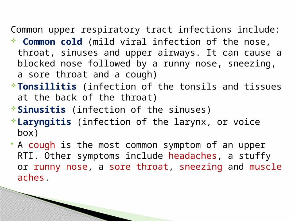
Common upper respiratory tract infections include: Common cold (mild viral infection of the nose, throat, sinuses
and upper airways. It can cause a blocked nose followed by a runny nose, sneezing, a sore throat and a cough)
Tonsillitis (infection of the tonsils and tissues at the back of the throat)
Sinusitis (infection of the sinuses) Laryngitis (infection of the larynx, or voice box) A cough is the most common symptom of an upper RTI. Other
symptoms include headaches, a stuffy or runny nose, a sore throat, sneezing and muscle aches.

Common lower RTIs include: Flu (this can affect either the upper or lower respiratory tract) Bronchitis (infection of the airways) Pneumonia (infection of the lungs) Bronchiolitis (an infection of the small airways that affects babies
and children younger than two years) Tuberculosis (persistent bacterial infection of the lungs) The main symptom of a lower RTI is also a cough, although it is
usually more severe and may bring up phlegm and mucus. Other possible symptoms are a tight feeling in the chest, increased rate of breathing,breathlessness and wheezing.

Lower respiratory tract infections are quite common in the general population, occurring with increased frequency in older individuals and those with chronic diseases or compromised immune function.
Etiologic diagnosis of the responsible pathogen is made by culture of respiratory tract secretions(sputum) or by isolation of a compatible organism from blood cultures.
Common infection of the lower respiratory tract is pneumonia
Lower Respiratory Tract Infection

Classification of pneumonia Pneumonia are classified to : A- Community acquired pneumonia common causes
Streptococcus pneumoniae& Mycoplasma pneumoniae
B- Hospital acquired pneumonia common causes Pseudomonas aeruginosa, Legionella pneumophila &Staphylococcus aureus

bacterial sputum culture is ordered when a health practitioner suspects that someone has a bacterial infection of the lungs or airways, such as bacterial pneumonia. This may show as changes in the lungs as seen on a chest x-ray.
Symptoms of pneumonia may include: Cough Fever, chills Muscle aches Fatigue Trouble breathing Chest pain Confusion Sometimes a sputum culture may be ordered after treatment of an
infection, to verify its efficacy.

Possible pathogens
Gram positive Gram negative Streptococcus pneumoniae Haemophilus influenzae Staphylococcus aureus Klebsiella pneumoniae Streptococcus pyogenes Pseudomonas aeruginosa Proteus species Yersina pestis Moraxella catarrhalis
Also Mycobacterium tuberculosis, Mycoplasmapneumoniae, and Legionella pneumophila.
Examination of sputum

S. pneumoniae and H. influenzae are the commonest causes of acute respiratory tract infections in tropical countries.
S. aureus, S. pyogenes, and H. influenzae are often secondary invaders in patients with influenza virus pneumonia.
M. pneumoniae causes primary atypical pneumonia. Fungi include Histoplasma capsulatum, Aspergillus species,
Candida albicans, Cryptococcus neoformans, and Nocardia

The presence of normal upper respiratory tract flora should be expected in sputum culture. Normal respiratory flora include:
Neisseria catarrhalis Candida albicans Diphtheroids alpha-hemolytic streptococci and some staphylococci.
Commensal bacteria of sputum

The specimen is sputum (is a thick fluid produced in the lungs and in the airways leading to the lungs). And blood for culture and serology
Sputum as it is being collected passes through the pharynx and the mouth. It therefore becomes contaminated with small numbers of commensal organisms
Give the patient a clean (need not be sterile), dry, wide-necked, leak-proof container, and request him or her to cough deeply to produce a sputum specimen.
If their is delaying use transport media – Amies or Stuart
specimen

Sputum container

How to Prepare the patient for the Test
Drinking a lot of water and other fluids at the night before the test, this may make it easier to cough up the sputum.
No food for one to two hours prior to expectorationRinsing the mouth by water prior to expectoration Do not use mouthwash before collecting a sputum sampleObtaining the specimen prior to antibiotic treatment

Macroscopical exam: Describe whether the sputum is: Purulent: Green-looking, mostly pus Mucopurulent: Green-looking with pus and mucus Mucoid: Mostly mucus Mucosalivary: Mucus with a small amount of saliva

Microscopical exam: Gram stain, examine the smear for pus cells and predominant
bacteria Giemsa stained preparation when histoplasmosis is suspected Giemsa or Wayson stained preparation when pneumonic plague
is suspected Antigen detection If the antisera are available

Culture the specimen To obtain as pure a culture as possible of a respiratory pathogen
it is necessary to reduce the number of commensals by Wash a purulent part of the sputum in about5 ml of sterile physiological saline. Inoculate in following media:-
Blood agar supports the growth of Gram positive cocci and most Gram negative rods, and is especially useful for evaluation of the colony morphology and hemolysis of streptococci.
Chocolate agar permits recovery of Haemophilus influenzae and other fastidious organisms that may grow less well on blood agar.
McConkey agar is selective for Gram negative bacteria and allows further classification into lactose ferment or non lactose ferment organisms.

Incubate the blood agar & McConkey agar aerobically and the chocolate agar plate in a carbon dioxide enriched atmosphere
In blood agar apply Optochin discs in the well (????).
Colonial morphology Gram stain Biochemical test Antimicrobial sensitivity test

A man 58 years of age, women 45 years old and her son, presenting to hospital with radiographic evidence of pneumonia and complaining of a 24-hour history of fever, chills, Muscle aches and cough, the doctor ask them to do sputum culture, proceed to identify the causative agents of each patient
Case study

Direct microscopical smear for the man
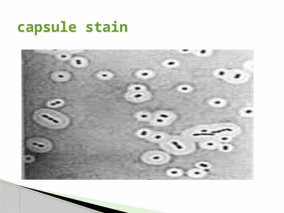
capsule stain

Blood agar with opichin disc

Chocolate blood agar


Indirect Gram stain

Catalase test

Bile solubility test

Direct microscopical smear for the women

Chocolate agar

Blood agar

Gram stain

What is the phenomenon seen here & what two bacteria are plated?

X& V factor requirement


Direct microscopical smear for the child

Blood agar

MacConkey agar

CLED

Indirect Gram stain

Gram stain (the capsule more visible)

capsule stain

Biochemical test (left one)

Indole & MR negative

VP, citrate& urease test positive

Motility(non motile)& kligler iron agar (e)

API (analytic profile index)test

Susceptibility tests should be performed only when the amount of cultural growth of a pathogen is significant.
Strains of S. pneumoniae should be tested on blood agar for susceptibility to penicillin, tetracycline, and erythromycin.
Penicillin-resistant isolates of Streptococcus penumoniae have been noted with increasing frequency, and have been clinically significant in patients with pneumonia .Because of this, disk diffusion susceptibility testing with a 1ug oxacillin is recommended as a screening test on all S. pneumoniae isolates from sputum a zone size less than 20 mm indicates reduced susceptibility.
H. influenzae strains should be tested for betalactamase production and susceptibility to ampicillin, tetracycline, and co-trimoxazole. Ampicillin and amoxicillin are broad-spectrum penicillin, active against Gram
positive bacteria (including enterococci) H. influenzae, and many coliforms. Flucloxacillin and cloxacillin are used to treat beta-lactamase (penicillinase)
producing
Antimicrobial sensitivity test

Sensitivity test
