darunavir/cobicistat module 2.6.1 … and motor activity there were no effects suggestive of...
TRANSCRIPT
1
DARUNAVIR/COBICISTAT
MODULE 2.6.1
NONCLINICAL INTRODUCTION
Issue Date: 13 SEPTEMBER 2013Department: Drug Safety SciencesDocument No.: EDMS-ERI-70120197
Confidentiality StatementThe information in this document contains trade secrets and commercial information that are privileged or confidential and may not be disclosed unless such disclosure is required by applicable law or regulations. In any event, persons to whom the information is disclosed must be informed that the information is privileged or confidential and may not be further disclosed by them. These restrictions on disclosure will apply equally to all future information supplied to you that is indicated as privileged or confidential.
2
MODULE 2.6.1
INTRODUCTION
DRV/COBI: 2.6.1 Introduction
3
TABLE OF CONTENTSIN-TEXT FIGURES.................................................................................................................... 4
IN-TEXT TABLES ..................................................................................................................... 5
ABBREVIATIONS..................................................................................................................... 6
1. INTRODUCTION.......................................................................................................... 7
2. PROPOSED CLINICAL USE....................................................................................... 9
DRV/COBI: 2.6.1 Introduction
4
IN-TEXT FIGURESFigure 1: Structural Formula of DRV............................................................................................ 8Figure 2: Structural Formula of COBI .......................................................................................... 8
DRV/COBI: 2.6.1 Introduction
5
IN-TEXT TABLES
No In-Text Tables
DRV/COBI: 2.6.1 Introduction
6
ABBREVIATIONSARV antiretroviralATZ atazanavirCOBI cobicistatCYP3A cytochrome P450 3ADRV darunavirEC50 50% effective concentrationFDC fixed dose combinationHIV human immunodeficiency virusIC50 median inhibitory concentrationPI protease inhibitor RTV ritonavir
DRV/COBI: 2.6.1 Introduction
7
1. INTRODUCTIONThe darunavir/cobicistat (DRV/COBI) fixed dose combination (FDC) tablet contains 800 mg DRV and 150 mg COBI and is developed by the Applicant in collaboration with Gilead Sciences Inc (Gilead).
Darunavir (PREZISTA®, formerly known as TMC114) is a human immunodeficiency virus (HIV) protease inhibitor (PI). Darunavir was developed in combination with low-dose ritonavir (RTV) as a pharmacoenhancer (booster) of DRV which inhibited the metabolism of DRV via cytochrome P450 (CYP) 3A. DRV, in combination with low dose RTV and with other antiretrovirals (ARVs), is indicated for the treatment of HIV-1 infection in adults and pediatric patients aged 3 years and above. In adults, the dosing regimen of DRV/RTV 800/100 mg once daily is recommended in ARV treatment-naive patients and ARV treatment-experienced patients without DRV resistance associated mutations.
Darunavir exhibits activity against wild-type HIV in acutely infected T-cell lines, human peripheral blood mononuclear cells and human monocytes/macrophages with median 50% effective concentration in cell-based assays (EC50) values ranging from 1.2 to 8.5 nM (0.7 to 5.0 ng/mL), which are well below the 50% cellular toxicity concentration range of 87 μM to > 100 μM. No significant increase in EC50 value was observed for the majority of recombinant clinical isolates resistant to at least 1 of the currently licensed PIs.
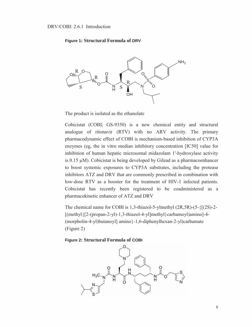
The chemical name of the active compound DRV is [(1S,2R)-3-[[(4-aminophenyl)sulfonyl](2-methylpropyl)amino]-2-hydroxy-1-(phenylmethyl)propyl]-carbamic acid (3R,3aS,6aR)-hexahydrofuro[2,3-b]furan-3-yl ester (Figure 1).
DRV/COBI: 2.6.1 Introduction
8
Figure 1: Structural Formula of DRV
NH
O
OH
NS
O
OO O
O
NH2
RR
S SR
The product is isolated as the ethanolate
Cobicistat (COBI; GS-9350) is a new chemical entity and structural analogue of ritonavir (RTV) with no ARV activity. The primary pharmacodynamic effect of COBI is mechanism-based inhibition of CYP3A enzymes (eg, the in vitro median inhibitory concentration [IC50] value for inhibition of human hepatic microsomal midazolam 1'-hydroxylase activity is 0.15 μM). Cobicistat is being developed by Gilead as a pharmacoenhancer to boost systemic exposures to CYP3A substrates, including the protease inhibitors ATZ and DRV that are commonly prescribed in combination with low-dose RTV as a booster for the treatment of HIV-1 infected patients. Cobicistat has recently been registered to be coadministered as a pharmacokinetic enhancer of ATZ and DRV
The chemical name for COBI is 1,3-thiazol-5-ylmethyl (2R,5R)-(5-{[(2S)-2-[(methyl{[2-(propan-2-yl)-1,3-thiazol-4-yl]methyl}carbamoyl)amino]-4-(morpholin-4-yl)butanoyl] amino}-1,6-diphenylhexan-2-yl)carbamate(Figure 2)
Figure 2: Structural Formula of COBI
DRV/COBI: 2.6.1 Introduction
9
The available nonclinical package is considered complete and in line with ICH, and CHMP guidelines and supports the proposed registration of DRV/COBI.
2. PROPOSED CLINICAL USEThe intended indication of the DRV/COBI 800/150 mg FDC is the same as the approved indication of the DRV/rtv 800/100 mg once daily regimen, ie, for the treatment of HIV-1 infection in HIV-1 infected treatment-naïve adult subjects and treatment-experienced adult subjects with no DRV resistance-associated mutations, and with plasma HIV-1 ribonucleic acid (RNA) <100,000 copies/mL and CD4+ cell count ≥100x106 cells/L. This is a so called ‘substitution indication’ of an already approved regimen, ie, the single agents have already been licensed for combined use at the same dose levels as in the FDC.
1
DARUNAVIR/COBICISTAT
MODULE 2.6.2
PHARMACOLOGY WRITTEN SUMMARY
DRV/COBI: 2.6.2 Pharmacology Written Summary
2
TABLE OF CONTENTS
IN-TEXT TABLES ..................................................................................................................... 3
ABBREVIATIONS..................................................................................................................... 4
1. BRIEF SUMMARY ....................................................................................................... 5
2. PRIMARY PHARMACODYNAMICS ........................................................................... 92.1. Darunavir ...................................................................................................................... 92.2. Cobicistat ...................................................................................................................... 9
2.2.1. Inhibition of Human CYP3A Activity by Cobicistat ....................................... 9
3. SECONDARY PHARMACODYNAMICS................................................................... 103.1. Darunavir .................................................................................................................... 103.2. Cobicistat .................................................................................................................... 10
3.2.1. Effect on Proteases and Proteasome Activity............................................ 103.2.2. Effects on Adipocytes................................................................................. 113.2.3. In Vitro Receptor Binding Potencies .......................................................... 113.2.4. In Vitro Cytotoxicity .................................................................................... 11
4. SAFETY PHARMACOLOGY..................................................................................... 124.1. Darunavir .................................................................................................................... 12
4.1.1. In Vitro Studies ........................................................................................... 124.1.1.1. Membrane Potassium (K+) Current (hERG Current).............................. 124.1.1.2. Cardiac Action Potential (Purkinje Fibers) .............................................. 124.1.2. In Vivo Studies ........................................................................................... 134.1.2.1. Cardiovascular Effects on Conscious, Telemetered Beagle Dogs......... 134.1.2.2. Gastro-Intestinal Transit Time in Rats .................................................... 134.1.2.3. Neurobehaviour and Motor Activity in Rats ............................................ 134.1.2.4. Pulmonary Safety in Rats ....................................................................... 14
4.2. Cobicistat .................................................................................................................... 144.2.1. In Vitro Studies ........................................................................................... 144.2.1.1. Effects on Ion Channels.......................................................................... 144.2.1.2. Effects on Action Potentials in Isolated Rabbit Cardiac Purkinje
Fibers ...................................................................................................... 154.2.1.3. Effects on Isolated Rabbit Hearts (Langendorff Method) ....................... 154.2.2. In Vivo Studies ........................................................................................... 194.2.2.1. Neurobehaviour and Motor Activity in Rats ............................................ 194.2.2.2. Cardiovascular Effects on Conscious, Telemetered Beagle Dogs ......... 204.2.2.3. Pulmonary Safety in Rats ....................................................................... 21
5. PHARMACODYNAMIC DRUG INTERACTIONS...................................................... 21
6. DISCUSSION AND CONCLUSIONS......................................................................... 22
7. TABLES AND FIGURES ........................................................................................... 25
8. REFERENCES ........................................................................................................... 25
DRV/COBI: 2.6.2 Pharmacology Written Summary
3
IN-TEXT TABLESTable 1: Effect of COBI and RTV on Various Activities Catalyzed by Human Hepatic Microsomal CYP3A Enzymes.................................................................................................... 9Table 2: Cytotoxicity of COBI in MT-2 and HepG2 Cells ........................................................12
DRV/COBI: 2.6.2 Pharmacology Written Summary
4
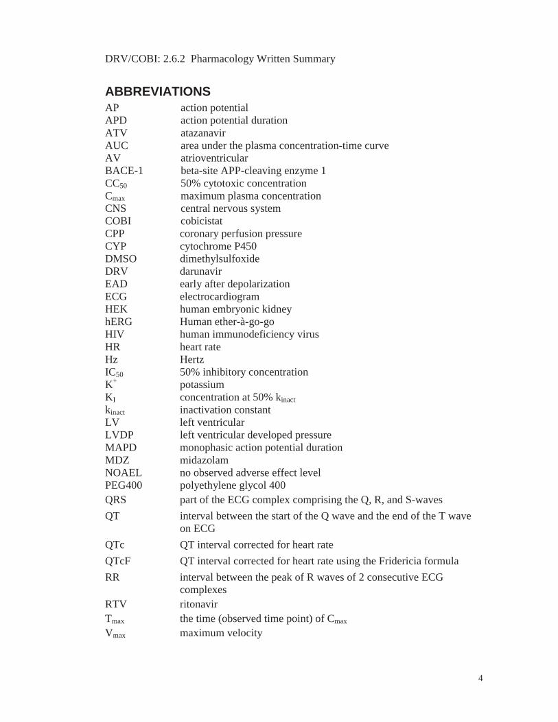
ABBREVIATIONSAP action potentialAPD action potential durationATV atazanavirAUC area under the plasma concentration-time curveAV atrioventricularBACE-1 beta-site APP-cleaving enzyme 1CC50 50% cytotoxic concentrationCmax maximum plasma concentrationCNS central nervous systemCOBI cobicistatCPP coronary perfusion pressureCYP cytochrome P450DMSO dimethylsulfoxideDRV darunavir EAD early after depolarizationECG electrocardiogramHEK human embryonic kidneyhERG Human ether-à-go-goHIV human immunodeficiency virusHR heart rateHz HertzIC50 50% inhibitory concentrationK+ potassiumKI concentration at 50% kinactkinact inactivation constantLV left ventricularLVDP left ventricular developed pressureMAPD monophasic action potential durationMDZ midazolamNOAEL no observed adverse effect levelPEG400 polyethylene glycol 400QRS part of the ECG complex comprising the Q, R, and S-wavesQT interval between the start of the Q wave and the end of the T wave
on ECGQTc QT interval corrected for heart rateQTcF QT interval corrected for heart rate using the Fridericia formulaRR interval between the peak of R waves of 2 consecutive ECG
complexesRTV ritonavirTmax the time (observed time point) of Cmax
Vmax maximum velocity
DRV/COBI: 2.6.2 Pharmacology Written Summary
5
1. BRIEF SUMMARYThe darunavir/cobicistat (DRV/COBI) fixed dose combination tablet contains 800 mg of the medicinal product DRV and 150 mg of the medicinal product COBI and is developed by the Applicant in collaboration with Gilead Sciences Inc (Gilead). Darunavir is a registered human immunodeficiency virus type 1 (HIV-1) protease inhibitor and COBI is a pharmacokinetic enhancer that has recently been approved as a pharmacokinetic enhancer of DRV and atazanavir (ATV). No nonclinical studies have been conducted with DRV in combination with COBI since the combined use of DRV and COBI is not expected to induce clinically relevant additive or synergistic effects. In this summary the pharmacology of DRV (also named TMC114 or Prezista®) and COBI (a structural analog of ritonavir [RTV]) is described. Both compounds have been investigated separately and therefore the individual DRV and COBI data are described in this document.
The primary and secondary pharmacodynamics, except the in vitro virology studies of DRV and COBI, are discussed, as well as a series of in vitro and in vivo safety pharmacology studies. The studies conducted are listed in Tabulated Summary 2.6.3.1 Overview. Details on the safety pharmacology studies can be found in Tabulated Summary 2.6.3.4.
Darunavir
The drug substance is a solvate (ethanolate). The dose levels used in all studies in this document are expressed as the nominal dose. Details concerning the study can be found in the Overview Table (Tabulated Summary 2.6.3.1).
Cardiovascular safety
In vitro, DRV at a concentration of 10 μM (5.9 μg/mL) in dimethylsulfoxide (DMSO) showed no significant effect on membrane potassium current inhuman Ether-à-go-go (hERG).T.human embryonic kidney (HEK) 293 cells and there were no effects on the electrophysiological cardiac action potential parameters in sheep isolated cardiac Purkinje fibers at the same concentration. In vivo cardio-hemodynamic and electrocardiogram (ECG) parameters did not change when dogs were dosed once up to 120 mg/kg DRV. Mean peak plasma concentration (Cmax) and area under the curve
DRV/COBI: 2.6.2 Pharmacology Written Summary
6
(AUC0-∞) values for male and female dogs were 16.6 and 15.0 μg/mL and 69.4 and 53.4 μg.h/mL, respectively, after a single administration at 120 mg/kg at the start of the 12-month dog study.
Gastrointestinal safety
There was no effect on gastrointestinal transit time of a charcoal solution after oral administration of 20, 200 and 2000 mg/kg DRV in rats.
Neurobehaviour and motor activity
There were no effects suggestive of neurological impairment or delayed neurotoxicity in rats dosed with DRV up to a single oral dose of 2000 mg/kg.
Pulmonary safety
Oral administration of DRV had no acute effects on respiration in rats at doses levels up to 2000 mg/kg when compared with vehicle.
Throughout this summary, clinical exposure data for DRV were derived from GS-US-210-0130 PK substudy in which HIV-1 infected subjects were treated with fixed dose DRV/COBI at 800/150 mg once daily for 24 weeks. Mean DRV exposure values amounted to 7.66 !g/mL (Cmax) and 81.6 !g.h/mL (AUC0-24h).
Cobicistat
Primary and Secondary Pharmacodynamics
No remarkable cytotoxicity was observed with COBI in vitro in human MT-2 and HepG2 cells, with 50% cytotoxic concentration (CC50) values of 89 and 44 !M, respectively (69.1 and 34.1 !g/mL). In vitro data indicate that COBI shows low potential for inhibition of host proteases (50% inhibitory concentration [IC50] > 30 !M) and a low potential for effects on adipocyte functions (lipid accumulation and glucose uptake).
Cardiovascular safety
Electrophysiology (in vitro patch clamp) studies indicated that COBI inhibited the hERG potassium current (IC50 = 1.8 !M [1.40 !g/mL]) and the hCav1.2 L-type calcium channel (IC50 = 6 !M [4.66 !g/mL]), but was a weak inhibitor of the hNav1.5 sodium channel (IC50 = 86.5 !M [67.1 !g/mL]). In
DRV/COBI: 2.6.2 Pharmacology Written Summary
7
rabbit Purkinje fibers (protein-free environment), COBI caused a shortening of the action potential duration (APD) at ∀ 1 !M (0.78 !g/mL); there was no evidence of triangulation, instability, or alternans predictive of prolongation of the QT interval. In Langendorff studies in isolated rabbit hearts (protein-free environment), COBI showed the potential to decrease left ventricular (LV) function and prolong the PR interval at ∀ 1 !M(0.78 !g/mL). No evidence of decreased left ventricular (LV) function was observed in clinical studies. When hearts were exposed to COBI in combination with ATV, effects on the PR interval and LV function were similar to the changes noted with COBI or ATV dosed alone.
In conscious telemetered dogs, there were no adverse effects on hemodynamic or ECG parameters up to 45 mg/kg (mean plasma COBI concentration 1 hour postdose was 7.7 !M [5.98 !g/mL]). Mild PR prolongation was noted primarily from 1 to 6 hours postdose, predominantly at 45 mg/kg and sporadically at 15 mg/kg, although mean PR intervals never exceeded the upper limits of normal for canines at any time point.
Based upon above data and ECG evaluations in the repeat-dose toxicity studies in dogs up to 39 weeks dosing (Module2.6.6/Section 3.2.2), COBIhas a low potential for QT prolongation, but may have a tendency to slightly prolong the PR interval. Data from the Langendorff studies also suggest that COBI may have the potential to decrease LV function at concentrations that also prolonged the PR interval. The shortening of the APD in rabbit Purkinje fibers, the PR prolongation, and the negative inotropic effects may be a consequence of interaction with cardiac calcium channels1,2.
In a thorough QT clinical study (Module2.7.4/Section 2.2.1), COBI demonstrated a lack of prolongation effects on the QTcF interval in healthy adult subjects at therapeutic and supratherapeutic exposures. A small but statistically significant negative association between COBI plasma concentration and QTc interval, and a modest, dose-related increase in PR interval, were observed in the QT/QTc study, which are not considered to be clinically significant. Further, echocardiograms performed in healthy subjects in Study GS-US-216-0116 (Module2.7.4/ Section 2.2.2) at baseline and after receiving 150 mg COBI for at least 15 days indicated no clinically significant change in LV function.
DRV/COBI: 2.6.2 Pharmacology Written Summary
8
Neurobehaviour and motor activity
A single oral dose of 50 mg/kg caused no effects on the central nervous system (CNS) in rats. At higher doses (∀ 150 mg/kg), decreased arousal and locomotor activity, salivation, and a decrease in body temperature and motor activity were noted 2 and 6 hours postdose. These effects at ∀ 150 mg/kg may represent a general toxicity response and are not considered a direct effect on the CNS.
Pulmonary safety
Cobicistat had no effects on respiratory parameters in rats after single oral doses up to 500 mg/kg.
DRV/COBI: 2.6.2 Pharmacology Written Summary
9
2. PRIMARY PHARMACODYNAMICS2.1. Darunavir
Darunavir is an inhibitor of the dimerization and of the catalytic activity of HIV-1 protease. It selectively inhibits the cleavage of HIV encoded Gag-Pol polyproteins in virus infected cells, thereby preventing the formation of mature infectious disease particles. Darunavir has potent in vitro activity against both wild type and multi-drug resistant HIV-1 strains.
For more detailed primary pharmacodynamics of DRV, please refer to Module2.7.2/Summary of Virology.
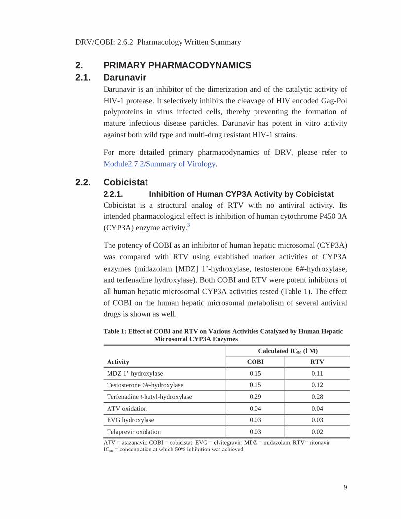
2.2. Cobicistat2.2.1. Inhibition of Human CYP3A Activity by CobicistatCobicistat is a structural analog of RTV with no antiviral activity. Itsintended pharmacological effect is inhibition of human cytochrome P450 3A (CYP3A) enzyme activity.3
The potency of COBI as an inhibitor of human hepatic microsomal (CYP3A)was compared with RTV using established marker activities of CYP3A enzymes (midazolam [MDZ] 1’-hydroxylase, testosterone 6#-hydroxylase, and terfenadine hydroxylase). Both COBI and RTV were potent inhibitors of all human hepatic microsomal CYP3A activities tested (Table 1). The effect of COBI on the human hepatic microsomal metabolism of several antiviral drugs is shown as well.
Table 1: Effect of COBI and RTV on Various Activities Catalyzed by Human Hepatic Microsomal CYP3A Enzymes
ActivityCalculated IC50 (!!M)
COBI RTV
MDZ 1’-hydroxylase 0.15 0.11
Testosterone 6#-hydroxylase 0.15 0.12
Terfenadine t-butyl-hydroxylase 0.29 0.28
ATV oxidation 0.04 0.04
EVG hydroxylase 0.03 0.03
Telaprevir oxidation 0.03 0.02ATV = atazanavir; COBI = cobicistat; EVG = elvitegravir; MDZ = midazolam; RTV= ritonavirIC50 = concentration at which 50% inhibition was achieved
DRV/COBI: 2.6.2 Pharmacology Written Summary
10
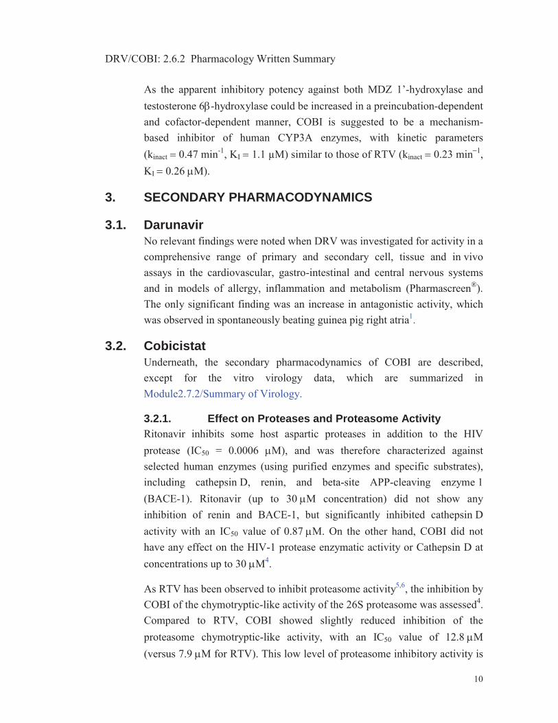
As the apparent inhibitory potency against both MDZ 1’-hydroxylase and testosterone 6#-hydroxylase could be increased in a preincubation-dependent and cofactor-dependent manner, COBI is suggested to be a mechanism-based inhibitor of human CYP3A enzymes, with kinetic parameters (kinact ∃ 0.47 min-1, KI ∃ 1.1 μM) similar to those of RTV (kinact ∃ 0.23 min%1, KI ∃ 0.26 !M).
3. SECONDARY PHARMACODYNAMICS
3.1. DarunavirNo relevant findings were noted when DRV was investigated for activity in a comprehensive range of primary and secondary cell, tissue and in vivo assays in the cardiovascular, gastro-intestinal and central nervous systems and in models of allergy, inflammation and metabolism (Pharmascreen®).The only significant finding was an increase in antagonistic activity, which was observed in spontaneously beating guinea pig right atria1.
3.2. CobicistatUnderneath, the secondary pharmacodynamics of COBI are described, except for the vitro virology data, which are summarized in Module2.7.2/Summary of Virology.
3.2.1. Effect on Proteases and Proteasome ActivityRitonavir inhibits some host aspartic proteases in addition to the HIV protease (IC50 = 0.0006 !M), and was therefore characterized against selected human enzymes (using purified enzymes and specific substrates), including cathepsin D, renin, and beta-site APP-cleaving enzyme 1 (BACE-1). Ritonavir (up to 30 !M concentration) did not show any inhibition of renin and BACE-1, but significantly inhibited cathepsin D activity with an IC50 value of 0.87 !M. On the other hand, COBI did not have any effect on the HIV-1 protease enzymatic activity or Cathepsin D at concentrations up to 30 !M4.
As RTV has been observed to inhibit proteasome activity5,6, the inhibition by COBI of the chymotryptic-like activity of the 26S proteasome was assessed4. Compared to RTV, COBI showed slightly reduced inhibition of the proteasome chymotryptic-like activity, with an IC50 value of 12.8 !M (versus 7.9 !M for RTV). This low level of proteasome inhibitory activity is
DRV/COBI: 2.6.2 Pharmacology Written Summary
11
unlikely to be significant at maximal unbound clinical exposure level of 0.095 !M COBI.
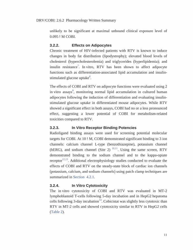
3.2.2. Effects on AdipocytesChronic treatment of HIV-infected patients with RTV is known to induce changes in body fat distribution (lipodystrophy); elevated blood levels of cholesterol (hypercholesterolemia) and triglycerides (hyperlipidemia); and insulin resistance7. In vitro, RTV has been shown to affect adipocyte functions such as differentiation-associated lipid accumulation and insulin-stimulated glucose uptake8.
The effects of COBI and RTV on adipocyte functions were evaluated using 2 in vitro assays9, monitoring normal lipid accumulation in cultured human adipocytes following the induction of differentiation and evaluating insulin-stimulated glucose uptake in differentiated mouse adipocytes. While RTV showed a significant effect in both assays, COBI had no or a less pronounced effect, suggesting a lower potential of COBI for metabolism-related toxicities compared to RTV.
3.2.3. In Vitro Receptor Binding PotenciesRadioligand binding assays were used for screening potential molecular targets for COBI. At 10 !M, COBI demonstrated significant binding to 3 ion channels: calcium channel L-type (benzothiazepine), potassium channel (hERG), and sodium channel (Site 2) 10,11. Using the same screen, RTV demonstrated binding to the sodium channel and to the kappa-opiate receptor12,13. Additional electrophysiology studies conducted to evaluate the effects of COBI and RTV on the steady-state block of cardiac ion channels (potassium, calcium, and sodium channels) using patch clamp techniques are summarized in Section 4.2.1.
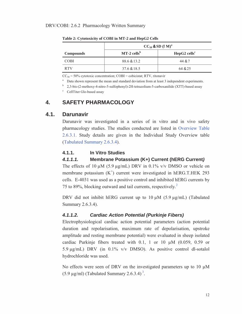
3.2.4. In Vitro CytotoxicityThe in vitro cytotoxicity of COBI and RTV was evaluated in MT-2 lymphoblastoid T-cells following 5-day incubation and in HepG2 hepatoma cells following 3-day incubation14. Cobicistat was slightly less cytotoxic than RTV in MT-2 cells and showed cytotoxicity similar to RTV in HepG2 cells (Table 2).
DRV/COBI: 2.6.2 Pharmacology Written Summary
12
Table 2: Cytotoxicity of COBI in MT-2 and HepG2 Cells
Compounds
CC50 && SD (!M)a
MT-2 cellsb HepG2 cellsc
COBI 88.6 & 13.2 44 & 7
RTV 37.6 & 18.5 64 & 25
CC50 = 50% cytotoxic concentration; COBI = cobicistat; RTV, ritonavira Data shown represent the mean and standard deviation from at least 3 independent experiments.b 2,3-bis-(2-methoxy-4-nitro-5-sulfophenyl)-2H-tetrazolium-5-carboxanilide (XTT)-based assayc CellTiter Glo-based assay
4. SAFETY PHARMACOLOGY
4.1. DarunavirDarunavir was investigated in a series of in vitro and in vivo safety pharmacology studies. The studies conducted are listed in Overview Table 2.6.3.1. Study details are given in the Individual Study Overview table (Tabulated Summary 2.6.3.4).
4.1.1. In Vitro Studies4.1.1.1. Membrane Potassium (K+) Current (hERG Current)The effects of 10 μM (5.9 μg/mL) DRV in 0.1% v/v DMSO or vehicle on membrane potassium (K+) current were investigated in hERG.T.HEK 293 cells. E-4031 was used as a positive control and inhibited hERG currents by 75 to 89%, blocking outward and tail currents, respectively.2
DRV did not inhibit hERG current up to 10 μM (5.9 μg/mL) (Tabulated Summary 2.6.3.4).
4.1.1.2. Cardiac Action Potential (Purkinje Fibers)Electrophysiological cardiac action potential parameters (action potential duration and repolarisation, maximum rate of depolarisation, upstroke amplitude and resting membrane potential) were evaluated in sheep isolated cardiac Purkinje fibers treated with 0.1, 1 or 10 μM (0.059, 0.59 or 5.9 μg/mL) DRV (in 0.1% v/v DMSO). As positive control dl-sotalol hydrochloride was used.
No effects were seen of DRV on the investigated parameters up to 10 μM (5.9 μg/ml) (Tabulated Summary 2.6.3.4) 3.
DRV/COBI: 2.6.2 Pharmacology Written Summary
13
4.1.2. In Vivo Studies4.1.2.1. Cardiovascular Effects on Conscious, Telemetered
Beagle DogsFour conscious, telemetered male beagle dogs were dosed orally via gavage at single escalating doses of 0 (vehicle), 30, 60 or 120 mg/kg of DRV dissolved in polyethylene glycol 400 (PEG400) (1mL/kg) 4. The interval between administrations was 3 to 5 days. Arterial blood pressure, heart rate and lead II ECG (PR, RR, QRS and QT intervals and QTc) were measured continuously at least 30 minutes before dosing up to 6 hours post-dose.
No effect on cardio-hemodynamic and ECG parameters was measured up to 120 mg/kg (Tabulated Summary 2.6.3.4). Toxicokinetic data were not measured in this study, but mean Cmax and AUC0-∞ values for male and female dogs were 16.6 and 15.0 μg/mL and 69.4 and 53.4 μg.h/mL, respectively, after a single administration at a similar dose level (120 mg/kg) at the start of the 12-month dog study (see Module2.6.6/Section3.1.2.1).
4.1.2.2. Gastro-Intestinal Transit Time in RatsDarunavir was examined for its ability to induce a delay of the gastro-intestinal transit time in male Wistar rats (5/group)5. This was determined by measuring the maximal travel in a time period of 30 minutes of a charcoal suspension applied 30 minutes after a single oral (gavage) administration of vehicle (40%v/v PEG400), 20, 200 and 2000 mg/kg DRV in PEG400. Atropine served as positive control (= delay in gastric emptying).
No effect of DRV on gastrointestinal transit time was detected up to 2000 mg/kg (Tabulated Summary 2.6.3.4).
4.1.2.3. Neurobehaviour and Motor Activity in RatsA single oral (gavage) dose of DRV at 0 (vehicle; 40 % v/v PEG400), 20, 200 or 2000 mg/kg was given to male Wistar rats (10/group) to assess behavioral, neurologic and autonomic parameters before treatment and 1, 6 and 24 hours post-dose. Chlorpromazine hydrochloride was also administered as a positive control.
DRV did not affect the neurofunctional integrity up to 2000 mg/kg(Tabulated Summary 2.6.3.4) 6.
DRV/COBI: 2.6.2 Pharmacology Written Summary
14
4.1.2.4. Pulmonary Safety in RatsMale Wistar rats (5/group) received a single oral (gavage) dose of DRV in PEG400 at 0 (vehicle), 20, 200 or 2000 mg/kg to assess pulmonary safety.7
Breathing frequency and tidal volume were measured during 100 minutes after dosing. Methacholine chloride was used as positive control.
No acute effects of DRV on respiration were measured up to 2000 mg/kg(Tabulated Summary 2.6.3.4).
4.2. Cobicistat4.2.1. In Vitro Studies4.2.1.1. Effects on Ion ChannelsThe in vitro effects of COBI at 0.3, 1, 3, and 10 !M (0.23, 0.78, 2.33 and 7.76 !g/mL; in a vehicle of 0.3% DMSO in HEPES-buffered physiological saline) on membrane K+ current were determined. (Tabulated Summary 2.6.3.4)15. Cobicistat inhibited hERG K+ current by3.2% at 0.3 !M (0.23 !g/mL), 37.1% at 1 !M (0.78 !g/mL), 64.2% at 3 μM(2.33 !g/mL), and 89.5% at 10 !M (7.76 !g/mL) (versus 0.8% in the vehicle control). The IC50 for the inhibitory effect of COBI on hERG K+ current was 1.8 !M (1.40 !g/mL; Hill coefficient ∃ 1.3). Under identical conditions, the positive control (60 nM terfenadine) inhibited hERG potassium current by (mean; n ∃ 2) 86.3% confirming the sensitivity of the test system to hERG inhibition.
In a second study, the potential effects of COBI on the steady-state block of cardiac ion channels were determined using patch clamp techniques. Cobicistat inhibited the hERG potassium current (IC50 = 1.9 !M[1.47 !g/mL]) and the hCav1.2 L-type calcium channel (IC50 = 6 !M[4.66 !g/mL]), but was a weak inhibitor of the hNav1.5 sodium channel (IC50 = 86.5 !M [67.1!g/mL]). (Tabulated Summary 2.6.3.4)16. Forcomparison, effects of RTV on the steady-state block of cardiac ion channels were also determined. The IC50 for the hERG channel was 8.75 μM, with a Hill coefficient of 1.11. However, the presence of precipitates were noted in all physiological saline solutions at concentrations > 7.7 μM RTV. Owing to the low solubility limit of RTV in the sodium and calcium channel physiological solutions, the IC50 and Hill coefficients could not be determined for RTV inhibition of the cardiac sodium and calcium channels.
DRV/COBI: 2.6.2 Pharmacology Written Summary
15
4.2.1.2. Effects on Action Potentials in Isolated Rabbit Cardiac Purkinje Fibers
Since COBI was inhibitory at the IKr (hERG), Cav1.2, and Nav1.5 ion channels, further testing was conducted with rabbit Purkinje fibers. Effects of COBI and RTV on cardiac action potential (APs) using Purkinje fibers excised from adult female rabbit ventricles were assessed at 4 concentrations (0.03, 0.1, 1, and 10 !M [0.0023, 0.078, 0.78 and 7.76 !g/mL]), with increasing concentrations added sequentially to 4 fiber preparations at 2 stimulus frequencies (1 and 2 Hz); AP parameters were compared to time-matched vehicle controls (Tabulated Summary 2.6.3.4)17. As positive control dl-sotalol hydrochloride was used
Cobicistat at 1 and 10 !M (0.78 and 7.76 !g/mL) caused a shortening of the action potential duration (APD60 and APD90) that was statistically significant over the effect produced by the vehicle at a stimulus frequency of 1 Hz. At a stimulus frequency of 2 Hz, only the shortening of APD60 and APD90
observed at 10 !M (7.76 !g/mL) was statistically significant. Changes in other AP parameters were not statistically different compared to those produced by the vehicle (rabbit Purkinje fiber Tyrode’s solution + 0.3 !MDMSO) suggesting a low potential for QT prolongation for COBI. Ritonavir at 9.6 !M produced a decrease in the rate of conduction, Vmax (dv/dtmax), which was statistically significant over that produced by the vehicle at stimulus frequencies of 1 and 2 Hz. The other AP parameters were not affected in a statistically significant manner over those observed with the vehicle.
4.2.1.3. Effects on Isolated Rabbit Hearts (Langendorff Method)
Two studies18, 19 were conducted to evaluate the effects of COBI (first study) and COBI, ATV, as well as a combination of ATV and escalating doses of COBI (second study) on cardiac hemodynamic and electrophysiologic parameters in isolated female rabbit hearts.
In the first study, COBI concentrations of 0.3, 1, 3, and 10 !M (0.23, 0.78, 2.33 and 7.76 !g/mL) were applied (Tabulated Summary 2.6.3.4)18. In one group of hearts (n = 4 per vehicle and test group), the atrioventricular (AV) node was ablated and the heart paced to circumvent QT effects secondary to drug-induced changes in heart rate (HR) and to measure rate dependent
DRV/COBI: 2.6.2 Pharmacology Written Summary
16
effects of COBI. Changes in the QT interval; QRS duration of the ECG together with the monophasic action potential duration at 30, 60, and 90 percent repolarization (MAPD30, MAPD60, and MAPD90); triangulation (MAPD90-MAPD30); stability of the MAP; and LV contractility (LVDP, dP/dtmin and dP/dtmax) were evaluated. In another group of hearts (n = 4 per vehicle and test group), the ECG was measured from free running hearts and the effects of COBI on the RR and PR intervals were determined.
Exposure to 0.3, 1, 3, and 10 !M COBI (0.23, 0.78, 2.33 and 7.76 !g/mL)shortened the QT interval by 8.8% to 20%. The average QT interval was significantly different from baseline at 3 and 10 !M (2.33 and 7.76 !g/mL)COBI only. The QRS interval was unaffected at concentrations up to 10 !M(7.76 !g/mL) COBI.
Exposure to 0.3, 1, 3, and 10 !M COBI (0.23, 0.78, 2.33 and 7.76 !g/mL)caused a dose-dependent shortening of the MAPD30, 60, 90 by 3.8% to 43.9%. Compared to baseline, statistically significant decreases were noted at ≥3 !M for MAPD30, ≥1 !M for MAPD60, and at ≥3 !M for MAPD90. Triangulation was slightly reduced after exposure to 0.3 and 1 !M COBI(0.23 and 0.78 !g/mL) and slightly increased after exposure to 3 and 10 !M(2.33 and 7.76 !g/mL), although none of the triangulation values were statistically different to baseline values. In addition, stability of the MAP (beat-to-beat variability) and reverse use-dependency of MAPD60 were not significantly affected by COBI.
Cobicistat reduced left ventricular contractility of the isolated heart in a dose-dependent manner. LVDP was reduced by 7% to 61%; dP/dtmin by 7.5% to 91%; and dP/dtmax by 10.7% to 88.6%. Decreases in LVDP, dP/dtmin, and dP/dtmax were statistically different from baseline at 1, 3, and 10 !M COBI (0.78, 2.33 and 7.76 !g/mL).
In free running hearts, COBI increased the PR interval by 4.3%, 16.6%, 41.3%, and 95.5% at concentrations of 0.3, 1, 3, and 10 !M, respectively(0.23, 0.78, 2.33 and 7.76 !g/mL). The PR interval was statistically different from baseline after exposure to 3 and 10 !M (2.33 and 7.76 !g/mL). Also, in 3 of the 4 hearts tested, second degree AV block developed after exposure to 10 !M COBI. Cobicistat significantly increased the RR interval by 25.8% and 63.5% at 3 and 10 !M (2.33 and 7.76 !g/mL), respectively.
DRV/COBI: 2.6.2 Pharmacology Written Summary
17
In conclusion, COBI was associated with negative inotropic effects and shortening of the APD on the isolated rabbit heart at concentrations ≥ 1 !M(0.78 !g/mL). At ≥ 3 !M (2.33 !g/mL), decreases in the QT interval and increases in the PR and RR intervals were noted. There were no notable effects on the QRS interval, triangulation, and stability. There were no remarkable effects noted on hemodynamic, electrophysiologic, and electrocardiographic parameters at a concentration of 0.3 !M COBI.
In a second study, the effects of COBI alone (0.15, 0.45, 1.5, and 4.5 μM[0.12, 0.35, 1.16 and 3.49 !g/mL]), ATV alone (1.5, 4.5, 15, and 45 μM), as well as a combination of ATV (fixed, 1.5 μM) and escalating concentrations of COBI (0.045, 0.15, 0.45, and 1.5 μM [0.035, 0.12, 0.35 and 1.16 !g/mL]) on cardiac hemodynamic and electrophysiologic parameters of isolated rabbit hearts were evaluated (Tabulated Summary 2.6.3.4)19. Hearts (4 per test group) from female New Zealand White rabbits were perfused in Langendorff constant-flow mode with Krebs-Henseleit buffer and exposed to increasing concentrations of test articles for 15 minutes at each concentration. Left ventricular (LV) function, including reduced LVDP, contractility (LV dP/dtmax), relaxation (LV dP/dtmin), and coronary perfusion pressure (CPP) were determined to assess the effects of the test articles on hemodynamic function. Heart rate; QT, QRS, and PR intervals; MAPD30, MAPD50 and MAPD90; and action potential triangulation (MAPD90%MAPD30) were determined to assess the effects of the test articles on cardiovascular electrophysiology. At each concentration of test article, mean values were compared to baseline values. In addition, the appearance of early after depolarizations (EADs) on the monophasic action potential waveform was also quantified.
The concentrations of COBI and ATV were measured in DMSO stock solutions and post-perfusion medium. As initial analyses showed low concentrations of COBI in post-perfusion samples compared to target values (35%-48% of target); additional research was conducted to investigate whether these low values were reflective of low stability of COBI in perfusion buffer, or were due to problems encountered in extraction of COBI from perfusion buffer prior to analysis. These results suggested that inadequate extraction of COBI from perfusion buffer was responsible for the low measured COBI concentrations in post perfusion samples. As a result,
DRV/COBI: 2.6.2 Pharmacology Written Summary
18
measured concentrations of COBI and ATV in perfusion samples are not considered reflective of probable exposures achieved in the Langendorff study, and nominal concentrations are noted below.
Exposure to COBI at concentrations ≥ 1.5 μM (1.16 !g/mL) was associated with significant decreases in LV function, including LVDP (%50% decrease at 1.5 μM), contractility (%50% decrease at 1.5 μM), and impairment of relaxation (%54% decrease at 1.5 μM [1.16 !g/mL]). Significant increases in CPP were noted at ≥ 1.5 μM (37% increase at 1.5 μM [1.16 !g/mL]), suggesting a possible vasoconstrictive effect of the compound.
Exposure to ATV was associated with slight, but not statistically significant, decreases in LV function (LVDP, LV dP/dtmax, LV dP/dtmin) at concentrations ≥ 15 μM. There were no notable effects of ATV on CPP at concentrations up to 15 μM.
When hearts were exposed to escalating concentrations of COBI (≤ 1.5 μM[1.16 !g/mL]) in combination with 1.5 μM ATV, the negative inotropic effects of 1.5 μM COBI appeared slightly reduced, with LV developed pressure decreasing approximately 30% compared to baseline (versus 50% with 1.5 μM COBI alone). In addition, there were no notable effects of the combination on CPP, suggesting a reversal of the putative COBI-induced vasoconstriction.
The highest concentration of the combination (1.5 μM COBI/1.5 μM ATV) significantly decreased heart rate by 26% compared to baseline, as compared to an 8% decrease with 1.5 μM COBI alone, and to a 12% decrease with 1.5 μM ATV alone.
Exposure to 4.5 μM COBI (3.49 !g/mL) or 15 μM ATV significantly increased the PR interval by 62% and 45% (versus baseline), respectively. Trends toward increased PR interval were noted with both compounds alone at concentrations of 1.5 μM COBI (23% increase) and 1.5 μM ATV (10% increase). When tested in combination, a significant increase in the PR interval was noted at the highest concentration of 1.5 μM COBI/1.5 μM ATV (37% increase versus baseline) only.
There were no notable changes in QRS duration, QT interval, MAPD, or triangulation at concentrations up to 4.5 μM COBI (3.49 !g/mL).
DRV/COBI: 2.6.2 Pharmacology Written Summary
19
Atazanavir, when tested alone, was associated with significant increases in MADP90 at 45 μM, and a trend towards increases in triangulation at 4.5 μM that reached statistical significance at 45 μM. In addition, 1 of 4 hearts developed ventricular tachycardia, and 2 hearts developed AV dissociation at 45 μM ATV. There were no notable changes in QRS duration, QT interval, MAPD, or triangulation with combinations of COBI and ATV. Further, COBI, ATV, and combinations of COBI and ATV, were not associated with the development of EADs.
In summary, all 3 regimens (COBI, ATV, and COBI/ATV in combination) were associated with negative inotropic effects on the isolated rabbit heart. When 1.5 μM COBI (1.16 !g/mL) was coadministered with 1.5 μM ATV, effects on LV function were similar to the decreases noted at 1.5 μM COBI alone. Decreases in HR and increases in the PR interval were noted with both compounds alone, and with the highest concentration of the combination (1.5 μM COBI/1.5 μM ATV). There were no notable effects of the combination on QRS, QT interval, MAPD, and triangulation, and there was no association with EADs. There were no remarkable effects noted on hemodynamic, electrophysiologic, or electrocardiographic parameters at concentrations of 0.45 μM COBI (0.35 !g/mL), 4.5 μM ATV, or with the combination at 0.45 μM COBI plus 1.5 μM ATV.
4.2.2. In Vivo Studies4.2.2.1. Neurobehaviour and Motor Activity in RatsFemale Sprague-Dawley rats (8/group) were given a single oral gavage dose of vehicle (95% propylene glycol [PG], 5% ethanol [EtOH] with 0.005M HCl) or 50, 150, or 500 mg/kg of COBI to assess CNS effects (functional observation battery and motor activity evaluation).(Tabulated Summary 2.6.3.4)20.
No CNS effects were observed in rats dosed at 50 mg/kg. Decreases in arousal incidence in the observation arena were noted in rats treated with 150 and 500 mg/kg at 2 hours postdose and were still present at the 6-hour postdose assessment. Furthermore, rats administered COBI at doses of 150 and 500 mg/kg displayed slight decreases in locomotor activity level (qualitative) in the arena during the 2-hour postdose assessment. The decreases in locomotor activity in the arena were still evident during the 6-hour postdose assessment. A 4% and 8% decrease in the body temperature
DRV/COBI: 2.6.2 Pharmacology Written Summary
20
was noted following doses of 150 and 500 mg/kg, respectively. The decreased temperature reached a maximum at 2 hours postdose for animals dosed with 150 mg/kg COBI and returned to predose and control values by 6 hours postdose. For animals dosed with 500 mg/kg COBI, the maximum effect was seen at 6 hours postdose. Slight to moderate salivation was seen at the 30 minutes postdose assessment following doses of 150 and 500 mg/kg. Doses of 150 and 500 mg/kg COBI caused statistically significant decreases in motor activity (quantitative) at 2 hours postdose. Decreases in motor activity were still evident at the 6-hour postdose assessment at 500 mg/kg.
Based on these findings, the no-observed-adverse-effect-level (NOAEL) for this study was 50 mg/kg.
4.2.2.2. Cardiovascular Effects on Conscious, Telemetered Beagle Dogs
In a dose escalation design, each of 4 male dogs received vehicle (95% PG, 5% EtOH [with 0.005M HCl]), 5, 15, and 45 mg/kg COBI as a single oral gavage dose with a minimum of 2 days between each dose (Tabulated Summary 2.6.3.4)21. Arterial blood pressures (mean arterial pressure, systolic blood pressure, diastolic blood pressure, and pulse pressure), HR , and quantitative ECG intervals were evaluated. Plasma samples were taken from all animals at approximately Tmax (1 hour postdose) and analyzed for levels of COBI.
There were no compound-related effects on any hemodynamic parameter, no qualitative waveform abnormalities, and no effects on HR or QRS interval parameters. After oral administration of COBI, an increase in the mean PR interval was observed, predominantly following the high dose (45 mg/kg) and sporadically at the mid dose (15 mg/kg). The magnitude of the mean PR interval prolongation was mild with PR interval increases up to 12.2 msec compared to predose values. However the range of absolute PR interval values was 91.8 to 99.6 msec, which did not exceed the upper limits of normal for canines (130 msec) at any time point. Administration of COBI caused a mild increase in the mean QTc interval only following the high (45 mg/kg) dose. The magnitude of the QTc interval prolongation was mild (∋ 4%), unlikely to be biologically significant, and not considered adverse. There was no effect of the low dose (5 mg/kg) on any quantitative ECG parameter. Plasma samples taken 1 hour postdose to assess exposure showed
DRV/COBI: 2.6.2 Pharmacology Written Summary
21
mean concentrations of 560, 3770, and 5960 ng/mL (0.7, 4.9, and 7.7 !M, respectively) following doses of 5, 15, and 45 mg/kg, respectively.
Based on these findings, the NOAEL for COBI in this study was at least 45 mg/kg.
4.2.2.3. Pulmonary Safety in RatsGroups of 6 female Sprague-Dawley rats received a single oral gavage administration of vehicle (95% PG, 5% EtOH with 0.005 M HCl), 50, 150, or 500 mg/kg COBI to evaluate its pharmacological effects on the respiratory system (Tabulated Summary 2.6.3.4)22.
No treatment-related effects were measured on respiratory rate, tidal volume, or derived minute volume. Based on these results, the NOAEL for respiratory effects in the rat was considered to be 500 mg/kg COBI, the highest dose administered.
5. PHARMACODYNAMIC DRUG INTERACTIONSFor pharmacodynamic drug interactions, please refer to Module2.7.2Summary of Virology.
DRV/COBI: 2.6.2 Pharmacology Written Summary
22
6. DISCUSSION AND CONCLUSIONSDarunavir
Darunavir has been tested in 2 in vitro safety pharmacology studies at concentrations significantly exceeding the free plasma concentration determined in patients treated with a dose of 800/150 mg DRV/COBI (human free Cmax for DRV is 0.46 μg/mL calculated based on a human total Cmax of 7.66 μg/mL and an estimated plasma protein binding = 94%). In vitro, DRV at a concentration of 10 μM (5.9 μg/mL) in DMSO showed no significant effect on membrane potassium current in hERG.T.HEK 293 cells and there were no effects on the electrophysiological cardiac action potential parameters in sheep isolated cardiac Purkinje fibers at the same concentration corresponding to 13-fold the clinical unbound concentration at recommended doses.
In vivo, DRV (in PEG400) administered to 4 conscious telemetered dogs had no effect on cardio-hemodynamic and ECG parameters following single oral (gavage) doses of up to 120 mg/kg. Although DRV systemic exposure was not determined in the telemetry study, mean Cmax and AUC0-∞ values for male and female dogs were 16.6 and 15.0 μg/mL and 69.4 and 53.4 μg.h/mL, respectively, after a single administration at a similar dose level (120 mg/kg) at the start of the 12-month dog study (see ToxicologyTabulated Summary 2.6.7.7.F). DRV peak plasma levels in dogs are higher (2.0- to 2.2-fold) than those attained in humans (mean Cmax= 7.66 μg/mL) at therapeutic doses, AUC values are slightly below the clinical exposure (mean AUC0-24h =81.6 μg.h/mL). In addition, no treatment-related effects on heart rate or ECG morphology were noted in vivo after repeated administration in dogs (Module2.6.6/Section3.1.2).
In rats, after single oral administration of up to 2000 mg/kg DRV, there was no effect on gastrointestinal transit time of a charcoal solution, no relevant effects on neurobehaviour and motor activity and no acute effects on respiration.
Overall, DRV safety pharmacology studies did not detect any significant nonclinical safety signals. Therefore, DRV is considered to have no potential for cardiovascular, pulmonary or nervous system effects that could be of any concern for the use of DRV in combination therapy (DRV/COBI) for the treatment of HIV 1 infection at fixed dose of 800/150 mg.
DRV/COBI: 2.6.2 Pharmacology Written Summary
23
Cobicistat
Cobicistat is a structural analog of RTV, which retains its potent mechanism-based inhibition of human CYP3A, but lacks anti-HIV activity. Under physiological conditions, no inhibition of HIV-1 replication was detected at concentrations of COBI as high as 90 !M. These results indicate that COBI is devoid of antiretroviral activity at concentrations exceeding the clinical exposures by over 300-fold (see Module2.7.2/Section 4.2.3). The nonclinicalin vitro virology data of COBI, including demonstration of a specific lack of anti-HIV-1 activity, are described in the nonclinical virology summary contained in Module2.7.2 Summary of Virology.
Unlike RTV, COBI did not show any effects on the host protease cathepsin D and was less inhibitory against the host proteasome activity. Compared with RTV, COBI also showed similar or lower cytotoxicity in human lymphoid and hepatic cell lines. In vitro data from studies with differentiated adipocytes suggest that COBI may have reduced effects on lipid metabolism and adipocyte functions compared to RTV.
Safety pharmacology studies were conducted to determine the potential effects of COBI on the central nervous, cardiovascular and respiratory systems. In the rat CNS study, there were no significant neurotoxic effects; changes were limited to salivation, decreases in arousal, locomotor and motor activities, and decreases in body temperature at doses of 150 mg/kg and above. The NOAEL was 50 mg/kg. Decreases in body temperature are commonly observed in rodents after xenobiotic exposure, and most likely represent an adaptive thermoregulatory response unique to rodents, rather than a direct effect on the CNS23,24,25. Similarly, decreases in arousal and motor activity may represent a general toxicity response rather than a direct CNS response. No adverse effects were observed in the rat respiratory study (NOAEL 500 mg/kg).
Patch clamp studies indicated that COBI inhibited the hERG potassium current (IC50 1.8 !M) and the hCav1.2 L-type calcium channel (IC50 6 !M), but was a weak inhibitor of the hNav1.5 sodium channel (IC50 86.5 !M). In rabbit Purkinje fibers (protein-free environment), which are considered more sensitive to drug-induced APD prolongation and EADs than fibers isolated from dog and several other species26, COBI caused a shortening of the APD
DRV/COBI: 2.6.2 Pharmacology Written Summary
24
at ∀ 1 μM; there was no evidence of triangulation, instability, or alternans predictive of prolongation of the QT interval.
In a Langendorff study in rabbit hearts (protein-free environment) conducted with COBI alone, negative inotropic effects and shortening of the APD was noted at ∀ 1 !M. In a second Langendorff study in rabbit hearts, COBI produced similar effects (PR interval prolongation and decreases in LV function) at concentrations ≥ 1.5 !M. When hearts were exposed to COBI in combination with ATV, effects on PR interval and LV function were similar to the decreases noted with COBI alone. Cobicistat had no notable effects alone, or in combination with ATV, on QRS and QT intervals, MAPD, or triangulation; and there were no EADs.
In conscious telemetered dogs, there were no adverse effects on hemodynamic and ECG parameters up to 45 mg/kg, the highest dose administered. Cobicistat plasma levels 1 hour after dose administration at 45 mg/kg were between 2530 and 8950 ng/mL (3.3 to 11.5 μM; mean of 7.7 μM). Compared to vehicle control values, mild prolongation in PR intervals were noted primarily from 1 to 6 hours postdose, although mean PR intervals never exceeded the upper limits of normal for canines at any time point27,28. Further, based on the results of the Japanese QT PRODACT studies and others, the mild increases in QTc (∋ 4%) noted from 13 to 24 hours postdose at 45 mg/kg are unlikely to be biologically significant29,30.
Although COBI inhibits the L-type calcium ion channel and K+
hERG-current at low micromolar concentrations, data from the Purkinje fiber assay, the cardiovascular dog study, and ECG evaluations in the repeat-dose toxicity studies in dogs up to 39 weeks duration (Module2.6.6/Section 3.2.2) suggest that COBI has a low potential for QT prolongation, but may have a tendency to slightly prolong the PR interval. Of note, in the 39-week dog toxicity study, there were no notable effects on the QT and PR intervals at dose levels up to 20 mg/kg/day. Mean COBI Cmax values during Week 39 at 20 mg/kg were between 7090 to 8405 ng/mL (9.1 to 10.8 μM). The shortening of the APD in rabbit Purkinje fibers and the mild delay in the PR interval in dogs may be a consequence of interaction with cardiac calcium channels31,32.
DRV/COBI: 2.6.2 Pharmacology Written Summary
25
COBI has shown the potential to decrease LV function and prolong the PR interval in the isolated rabbit heart at ∀ 1 !M, which is approximately 10-fold above the anticipated unbound clinical exposure at the 150 mg COBI dose. However, as the fraction of unbound COBI is lower in plasma samples obtained in clinical studies (2.49% to 3.23%) compared to the in vitro studies, including clinical studies in subjects with moderate hepatic impairment or severe renal impairment, the potential of COBI to decreaseLV function and prolong PR is expected to be low in patients. In a thorough QT clinical study (Module2.7.4/Section 2.2.1), COBI demonstrated a lack of prolongation effects on the QTcF interval in healthy adult subjects at therapeutic and supratherapeutic exposures. A small but statistically significant negative association between COBI plasma concentration and QTc interval, and a modest, dose-related increase in PR interval, were observed in the QT/QTc study, which are not considered to be clinically significant. Further, echocardiograms performed in healthy subjects in Study GS-US-216-0116 at baseline and after receiving 150 mg COBI for at least 15 days indicated no clinically significant change in LV function (Module 2.7.4/Section2.2.2).
In summary, safety pharmacology studies with COBI did not reveal any significant safety findings, with the exception of the Langendorff studies. However, no clinically significant cardiovascular changes have been observed at clinical exposures up to 4-fold higher than those achieved at the clinical dose of 150 mg COBI.
Overall, the pharmacodynamic and pharmacological assessment of COBI supports the effective and safe use of this agent in combination therapy for the treatment of HIV-1 infection.
7. TABLES AND FIGURESTabulated Summaries are located in Module 2.6.3.
8. REFERENCESReports in bold are submitted. Reports and literature in black are available upon request.
DRV/COBI: 2.6.2 Pharmacology Written Summary
26
DARUNAVIR REFERENCES
Report Title1. Nonclinical Pharmacology Report TMC114-NC118. Evaluation of
the activity of TMC114 in tissue, animal and anti-infective in vitro assays. (Nov ).
2. Nonclinical Pharmacology Report TMC114-NC103 ( DPHN1001). Effect of TMC114 on hERG currents recorded from stably transfected HEK293 cells. . (Sep
).
3. Nonclinical Pharmacology Report TMC114-NC105 ( DPHN1005). Effect of TMC114 on action potential parameters in sheep isolated cardiac purkinje fibers. . (Oct ).
4. Nonclinical Toxicology Report TMC114-NC108 (Quintiles DPHN1003). Cardiovascular effects of TMC114 in conscious, telemetered Beagle dogs. . (Nov ).
5. Nonclinical Toxicology Report TMC114-NC120 ( V3120). General pharmacology studies: gastro-intestinal transit time study after oral administration of TMC114 in albino rats.
. (Mar ).
6. Nonclinical Toxicology Report TMC114-NC116 ( V3176). Safety pharmacology study: neurobehavioral observations and automated motor activity assessment after single dose oral administration of TMC114 in the rat. . (Feb ).
7. Nonclinical Toxicology Report TMC114-NC117 ( V3228). Safety pharmacology studies: respiratory assessment after single dose oral administration of TMC114 in the rat. . (Mar ).
COBICISTAT REFERENCES
1. Guth BD. Preclinical cardiovascular risk assessment in modern drug development. Toxicol Sci 2007;97 (1):4-20
2. Dai DZ. Two patterns of ion channelopathy in the myocardium: perspectives for development of anti-arrhythmic agents. Curr Opin Investig Drugs 2005;6 (3):289-97.
DRV/COBI: 2.6.2 Pharmacology Written Summary
27
3. Nonclinical pharmacokinetics report AD-216-2028. Inhibition of human CYP3A activity by GS-9350 in vitro.
4. Nonclinical pharmacology report PC-216-2001. Activity of GS-9350 against HIV-1 and host proteases.
5. Andre P, Groettrup M, Klenerman P, de Giuli R, Booth BL, Jr., Cerundolo V, et al. An inhibitor of HIV-1 protease modulates proteasome activity, antigen presentation, and T cell responses. Proc Natl Acad Sci USA 1998;95 (22):13120-4.
6. Nguyen AT, Gagnon A, Angel JB, Sorisky A. Ritonavir increases the level of active ADD-1/SREBP-1 protein during adipogenesis. AIDS 2000;14 (16):2467-73.
7. Carr A, Samaras K, Burton S, Law M, Freund J, Chisholm DJ, et al. A syndrome of peripheral lipodystrophy, hyperlipidaemia and insulin resistance in patients receiving HIV protease inhibitors. AIDS 1998;12 (7):F51-F8.
8. Murata H, Kruz PW, Mueckler M. The mechanism of insulin resistance caused by HIV protease inhibitor therapy. J Biol Chem 2000;275 (27):20251-4.
9. Nonclinical pharmacology report PC-216-2004. In vitro effects of GS-9350 on adipocytes.
10. Nonclinical toxicology report TX-168-2007. LeadProfilingScreen Data Report for Gilead Sciences, Inc.; GS-340649
11. Nonclinical toxicology report TX-168-2011. Individual Tests Data Report for Gilead Sciences, Inc.; GS-340649
12. Nonclinical pharmacology report PC-137-2004. LeadProfilingScreen Data Report for Gilead Sciences, Inc.; GS-017415
13. Nonclinical pharmacology report PC-168-2005. Individual Tests Data Report for Gilead Sciences, Inc.; GS-017415
14. Nonclinical pharmacology report PC-216-2003. Cytotoxicity profile of GS-9350.
15. Nonclinical toxicology report TX-216-2009. Effects of COBI on Isolated Hearts (non-GLP).
16. Nonclinical toxicology report TX-216-2015. Effects of GS-9350 and GS-017415 on Cardiac Ion Channels Expressed in Human Embryonic Kidney Cells.
DRV/COBI: 2.6.2 Pharmacology Written Summary
28
17. Nonclinical toxicology report TX-168-2012. Effect of GS-340649 and GS-017415 on Action Potentials in Isolated Rabbit Cardiac Purkinje Fibers (non-GLP).
18. Nonclinical pharmacology report PC-216-2007. A Pharmacological Assessment of the Effect of COBI on the Respiratory System of the Albino Rat (GLP).
19. Nonclinical pharmacology report PC-216-2009. An Examination of the Cardiovascular Effects of COBI, ATV and COBI + ATV on the Isolated Heart of the Female Rabbit (Langendorff Method) (non-GLP).
20. Nonclinical toxicology report TX-216-2006. A Pharmacological Assessment of the Effect of COBI on the Central Nervous System of the Albino Rat (GLP)
21. Nonclinical toxicology report TX-216-2008. A Pharmacological Assessment of the Effect of COBI on the Cardiovascular System of the Beagle Dog Using Telemetry (GLP)
22. Nonclinical toxicology report TX-216-2007. A Pharmacological Assessment of the Effect of COBI on the Respiratory System of the Albino Rat (GLP).
23. Watkinson WP, Gordon CJ. Caveats regarding the use of the laboratory rat as a model for acute toxicological studies: modulation of the toxic response via physiological and behavioral mechanisms. Toxicology 1993;81 (1):15-31.
24. Watkinson WP, Campen MJ, Wichers LB, Nolan JP, Costa DL. Cardiac and thermoregulatory responses to inhaled pollutants in healthy and compromised rodents: modulation via interaction with environmental factors. Environ Res 2003;92 (1):35-47.
25. Redfern WS, Strang I, Storey S, Heys C, Barnard C, Lawton K, et al. Spectrum of effects detected in the rat functional observational battery following oral administration of non-CNS targeted compounds. J Pharmacol Toxicol Methods 2005;52 (1):77-82.
26. Lu HR, Marien R, Saels A, De Clerck F. Species plays an important role in drug-induced prolongation of action potential duration and early afterdepolarizations in isolated Purkinje fibers. J Cardiovasc Electrophysiol 2001;12 (1):93-102.
DRV/COBI: 2.6.2 Pharmacology Written Summary
29
27. Sasaki H, Shimizu N, Suganami H, Yamamoto K. QT PRODACT: inter-facility variability in electrocardiographic and hemodynamic parameters in conscious dogs and monkeys. J Pharmacol Sci 2005;99 (5):513-22.
28. Soloviev MV, Hamlin RL, Shellhammer LJ, Barrett RM, Wally RA, Birchmeier PA, et al. Variations in hemodynamic parameters and ECG in healthy, conscious, freely moving telemetrized beagle dogs. Cardiovasc Toxicol 2006;6 (1):51-62.
29. Redfern WS, Carlsson L, Davis AS, Lynch WG, MacKenzie I, Palethorpe S, et al. Relationships between preclinical cardiac electrophysiology, clinical QT interval prolongation and torsade de pointes for a broad range of drugs: evidence for a provisional safety margin in drug development. Cardiovasc Res 2003;58 (1):32-45.
30. Toyoshima S, Kanno A, Kitayama T, Sekiya K, Nakai K, Haruna M, et al. QT PRODACT: in vivo QT assay in the conscious dog for assessing the potential for QT interval prolongation by human pharmaceuticals. J Pharmacol Sci 2005;99 (5):459-71.
31. Dai DZ. Two patterns of ion channelopathy in the myocardium: perspectives for development of anti-arrhythmic agents. Curr Opin Investig Drugs 2005;6 (3):289-97.
32. Guth BD. Preclinical cardiovascular risk assessment in modern drug development. Toxicol Sci 2007;97 (1):4-20.
MODULE 2.6.3
PHARMACOLOGY TABULATED SUMMARY
1
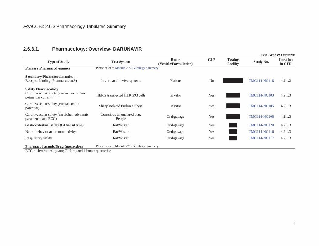
2.6.3.1. Pharmacology: Overview- DARUNAVIRTest Article: Darunivir
Type of Study Test System Route(Vehicle/Formulation)
GLP TestingFacility Study No. Location
in CTDPrimary Pharmacodynamics Please refer to Module 2.7.2 Virology Summary
Secondary PharmacodynamicsReceptor binding (Pharmascreen®) In vitro and in vivo systems Various No TMC114-NC118 4.2.1.2
Safety PharmacologyCardiovascular safety (cardiac membranepotassium current) HERG transfected HEK 293 cells In vitro Yes TMC114-NC103 4.2.1.3
Cardiovascular safety (cardiac actionpotential) Sheep isolated Purkinje fibers In vitro Yes TMC114-NC105 4.2.1.3
Cardiovascular safety (cardiohemodynamicparameters and ECG)
Conscious telemetered dog, Beagle Oral/gavage Yes TMC114-NC108 4.2.1.3
Gastro-intestinal safety (GI transit time) Rat/Wistar Oral/gavage Yes TMC114-NC120 4.2.1.3
Neuro-behavior and motor activity Rat/Wistar Oral/gavage Yes TMC114-NC116 4.2.1.3
Respiratory safety Rat/Wistar Oral/gavage Yes TMC114-NC117 4.2.1.3
Pharmacodynamic Drug Interactions Please refer to Module 2.7.2 Virology SummaryECG = electrocardiogram; GLP = good laboratory practice
DRV/COBI: 2.6.3 Pharmacology Tabulated Summary
2
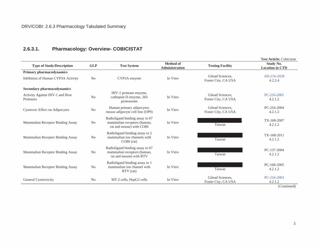
2.6.3.1. Pharmacology: Overview- COBICISTAT
Test Article: Cobicistat
Type of Study/Description GLP Test System Method of Administration Testing Facility Study No.
Location in CTDPrimary pharmacodynamics
Inhibition of Human CYP3A Activity No CYP3A enzyme In Vitro Gilead Sciences,Foster City, CA USA
AD-216-20284.2.2.4
Secondary pharmacodynamics
Activity Against HIV-1 and Host Proteases No
HIV-1 protease enzyme, cathepsin D enzyme, 26S
proteasomeIn Vitro Gilead Sciences,
Foster City, CA USAPC-216-2001
4.2.1.2
Cytotoxic Effect on Adipocytes No Human primary adipocytes; mouse adipocyte cell line (OP9) In Vitro Gilead Sciences,
Foster City, CA USAPC-216-2004
4.2.1.2
Mammalian Receptor Binding Assay NoRadioligand binding assay to 67 mammalian receptors (human,
rat and mouse) with COBIIn Vitro ,
TaiwanTX-168-2007
4.2.1.2
Mammalian Receptor Binding Assay NoRadioligand binding assay to 2 mammalian ion channels with
COBI (rat)In Vitro
TaiwanTX-168-2011
4.2.1.2
Mammalian Receptor Binding Assay NoRadioligand binding assay to 67 mammalian receptors (human,
rat and mouse) with RTVIn Vitro
TaiwanPC-137-2004
4.2.1.2
Mammalian Receptor Binding Assay NoRadioligand binding assay to 1 mammalian ion channel with
RTV (rat)In Vitro
TaiwanPC-168-2005
4.2.1.2
General Cytotoxicity No MT-2 cells, HepG2 cells In Vitro Gilead Sciences,Foster City, CA USA
PC-216-20034.2.1.2
(Continued)
DRV/COBI: 2.6.3 Pharmacology Tabulated Summary
3
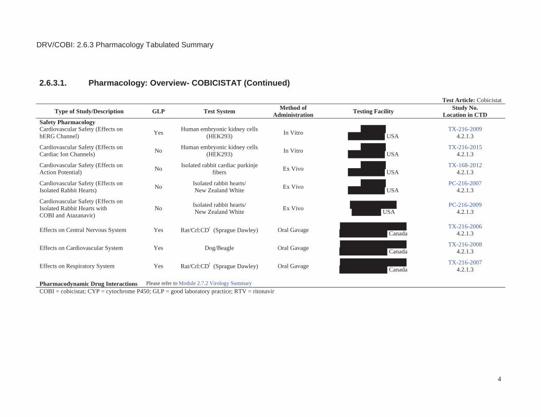
2.6.3.1. Pharmacology: Overview- COBICISTAT (Continued)
Test Article: Cobicistat
Type of Study/Description GLP Test System Method of Administration Testing Facility Study No.
Location in CTDSafety PharmacologyCardiovascular Safety (Effects onhERG Channel) Yes Human embryonic kidney cells
(HEK293) In Vitro USA
TX-216-20094.2.1.3
Cardiovascular Safety (Effects on Cardiac Ion Channels) No Human embryonic kidney cells
(HEK293) In Vitro USA
TX-216-20154.2.1.3
Cardiovascular Safety (Effects on Action Potential) No Isolated rabbit cardiac purkinje
fibers Ex Vivo USA
TX-168-20124.2.1.3
Cardiovascular Safety (Effects on Isolated Rabbit Hearts) No Isolated rabbit hearts/
New Zealand White Ex Vivo USA
PC-216-20074.2.1.3
Cardiovascular Safety (Effects on Isolated Rabbit Hearts with COBI and Atazanavir)
No Isolated rabbit hearts/ New Zealand White Ex Vivo USA
PC-216-20094.2.1.3
Effects on Central Nervous System Yes Rat/Crl:CD! (Sprague Dawley) Oral Gavage CanadaTX-216-2006
4.2.1.3
Effects on Cardiovascular System Yes Dog/Beagle Oral Gavage CanadaTX-216-2008
4.2.1.3
Effects on Respiratory System Yes Rat/Crl:CD! (Sprague Dawley) Oral Gavage CanadaTX-216-2007
4.2.1.3
Pharmacodynamic Drug Interactions Please refer to Module 2.7.2 Virology SummaryCOBI = cobicistat; CYP = cytochrome P450; GLP = good laboratory practice; RTV = ritonavir
DRV/COBI: 2.6.3 Pharmacology Tabulated Summary
4
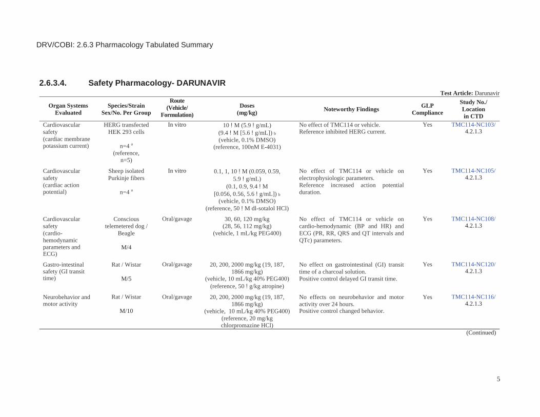
2.6.3.4. Safety Pharmacology- DARUNAVIRTest Article: Darunavir
Organ SystemsEvaluated
Species/StrainSex/No. Per Group
Route(Vehicle/
Formulation)Doses
(mg/kg) Noteworthy Findings GLPCompliance
Study No./Locationin CTD
Cardiovascular safety(cardiac membranepotassium current)
HERG transfected HEK 293 cells
n=4 a(reference,
n=5)
In vitro 10 !M (5.9 !g/mL) (9.4 !M [5.6 !g/mL]) b
(vehicle, 0.1% DMSO) (reference, 100nM E-4031)
No effect of TMC114 or vehicle.Reference inhibited HERG current.
Yes TMC114-NC103/ 4.2.1.3
Cardiovascular safety(cardiac actionpotential)
Sheep isolated Purkinje fibers
n=4 a
In vitro 0.1, 1, 10 !M (0.059, 0.59,5.9 !g/mL)
(0.1, 0.9, 9.4 !M[0.056, 0.56, 5.6 !g/mL]) b
(vehicle, 0.1% DMSO)(reference, 50 !M dl-sotalol HCl)
No effect of TMC114 or vehicle on electrophysiologic parameters.Reference increased action potential duration.
Yes TMC114-NC105/4.2.1.3
Cardiovascular safety(cardio-hemodynamicparameters and ECG)
Conscious telemetered dog /
Beagle
M/4
Oral/gavage 30, 60, 120 mg/kg(28, 56, 112 mg/kg)
(vehicle, 1 mL/kg PEG400)
No effect of TMC114 or vehicle on cardio-hemodynamic (BP and HR) and ECG (PR, RR, QRS and QT intervals and QTc) parameters.
Yes TMC114-NC108/4.2.1.3
Gastro-intestinalsafety (GI transit time)
Rat / Wistar
M/5
Oral/gavage 20, 200, 2000 mg/kg (19, 187,1866 mg/kg)
(vehicle, 10 mL/kg 40% PEG400)(reference, 50 !g/kg atropine)
No effect on gastrointestinal (GI) transit time of a charcoal solution.Positive control delayed GI transit time.
Yes TMC114-NC120/4.2.1.3
Neurobehavior andmotor activity
Rat / Wistar
M/10
Oral/gavage 20, 200, 2000 mg/kg (19, 187,1866 mg/kg)
(vehicle, 10 mL/kg 40% PEG400) (reference, 20 mg/kg chlorpromazine HCl)
No effects on neurobehavior and motor activity over 24 hours.Positive control changed behavior.
Yes TMC114-NC116/4.2.1.3
(Continued)
DRV/COBI: 2.6.3 Pharmacology Tabulated Summary
5
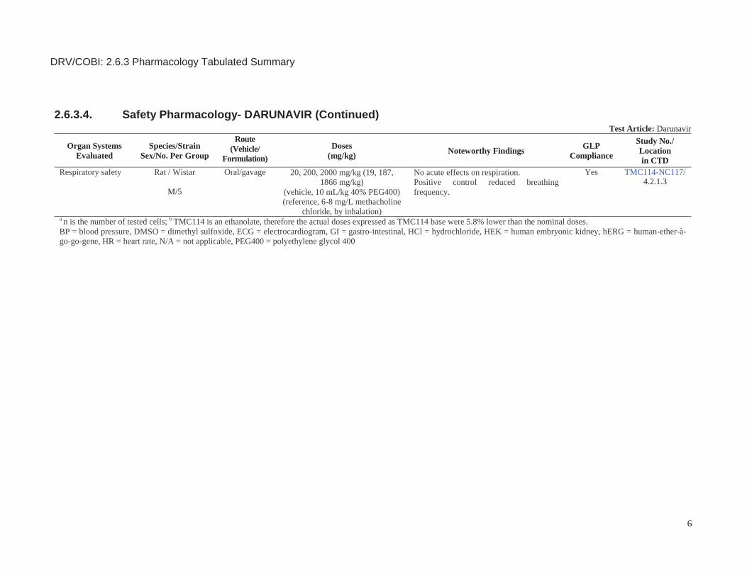
2.6.3.4. Safety Pharmacology- DARUNAVIR (Continued)Test Article: Darunavir
Organ SystemsEvaluated
Species/StrainSex/No. Per Group
Route(Vehicle/
Formulation)Doses
(mg/kg) Noteworthy Findings GLPCompliance
Study No./Locationin CTD
Respiratory safety Rat / Wistar
M/5
Oral/gavage 20, 200, 2000 mg/kg (19, 187,1866 mg/kg)
(vehicle, 10 mL/kg 40% PEG400) (reference, 6-8 mg/L methacholine
chloride, by inhalation)
No acute effects on respiration.Positive control reduced breathing frequency.
Yes TMC114-NC117/ 4.2.1.3
a n is the number of tested cells; b TMC114 is an ethanolate, therefore the actual doses expressed as TMC114 base were 5.8% lower than the nominal doses.BP = blood pressure, DMSO = dimethyl sulfoxide, ECG = electrocardiogram, GI = gastro-intestinal, HCl = hydrochloride, HEK = human embryonic kidney, hERG = human-ether-à-go-go-gene, HR = heart rate, N/A = not applicable, PEG400 = polyethylene glycol 400
DRV/COBI: 2.6.3 Pharmacology Tabulated Summary
6
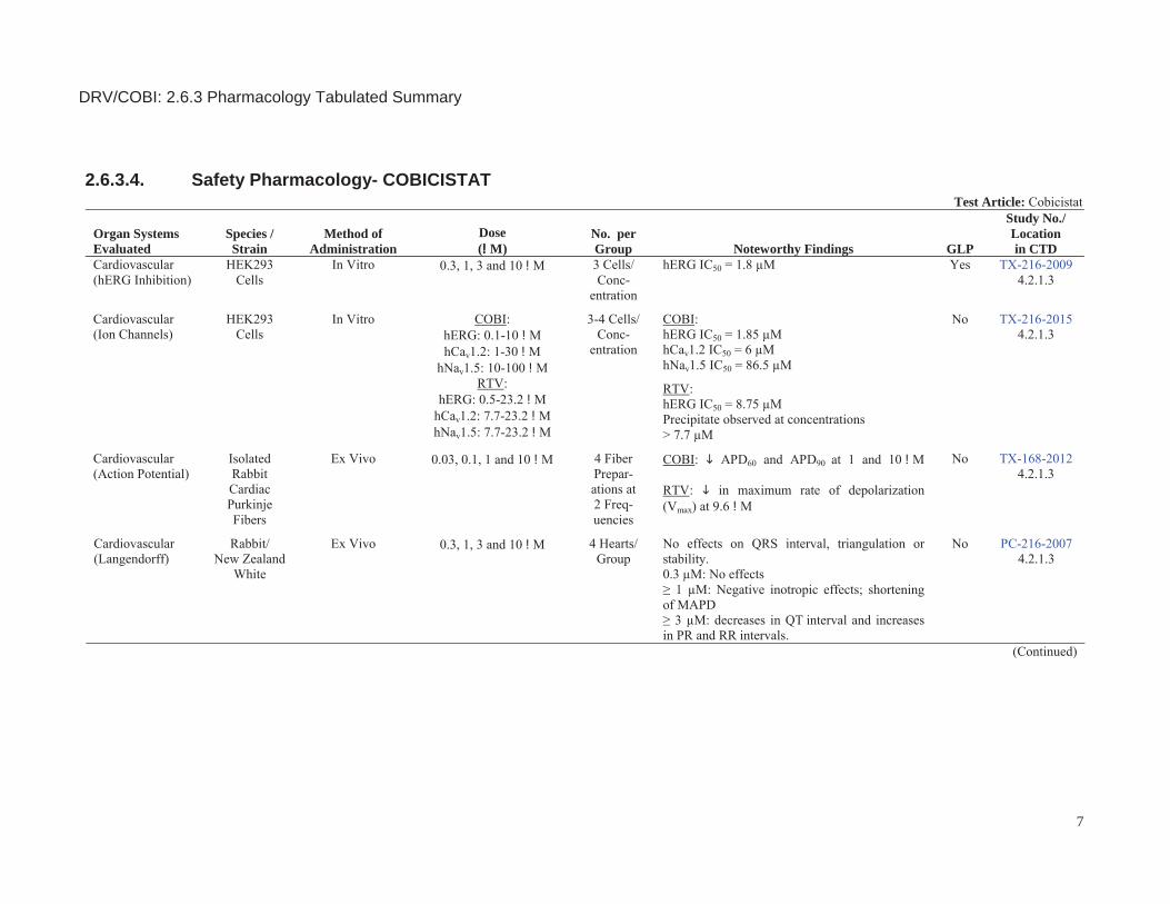
2.6.3.4. Safety Pharmacology- COBICISTATTest Article: Cobicistat
Organ Systems Evaluated
Species / Strain
Method of Administration
Dose(!!M)
No. per Group Noteworthy Findings GLP
Study No./Locationin CTD
Cardiovascular (hERG Inhibition)
HEK293 Cells
In Vitro 0.3, 1, 3 and 10 !M 3 Cells/ Conc-
entration
hERG IC50 = 1.8 μM Yes TX-216-20094.2.1.3
Cardiovascular (Ion Channels)
HEK293 Cells
In Vitro COBI:hERG: 0.1-10 !MhCav1.2: 1-30 !M
hNav1.5: 10-100 !MRTV:
hERG: 0.5-23.2 !MhCav1.2: 7.7-23.2 !MhNav1.5: 7.7-23.2 !M
3-4 Cells/ Conc-
entration
COBI:hERG IC50 = 1.85 μMhCav1.2 IC50 = 6 μMhNav1.5 IC50 = 86.5 μM
RTV:hERG IC50 = 8.75 μMPrecipitate observed at concentrations > 7.7 μM
No TX-216-20154.2.1.3
Cardiovascular(Action Potential)
Isolated Rabbit Cardiac Purkinje Fibers
Ex Vivo 0.03, 0.1, 1 and 10 !M 4 Fiber Prepar-ations at 2 Freq-uencies
COBI: APD60 and APD90 at 1 and 10 !M
RTV: in maximum rate of depolarization (Vmax) at 9.6 !M
No TX-168-20124.2.1.3
Cardiovascular(Langendorff)
Rabbit/New Zealand
White
Ex Vivo 0.3, 1, 3 and 10 !M 4 Hearts/Group
No effects on QRS interval, triangulation or stability.0.3 μM: No effects≥ 1 μM: Negative inotropic effects; shortening of MAPD≥ 3 μM: decreases in QT interval and increases in PR and RR intervals.
No PC-216-20074.2.1.3
(Continued)
DRV/COBI: 2.6.3 Pharmacology Tabulated Summary
7
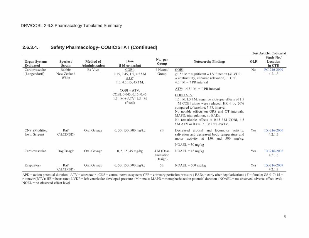
2.6.3.4. Safety Pharmacology- COBICISTAT (Continued)Test Article: Cobicistat
Organ Systems Evaluated
Species / Strain
Method of Administration
Dose(!!M or mg/kg)
No. per Group Noteworthy Findings GLP
Study No./Locationin CTD
Cardiovascular(Langendorff)
Rabbit/New Zealand
White
Ex Vivo COBI:0.15, 0.45, 1.5, 4.5 !M
ATV:1.5, 4.5, 15, 45 !M,
COBI + ATV:COBI: 0.045, 0.15, 0.45, 1.5 !M + ATV: 1.5 !M
(fixed)
4 Hearts/Group
COBI: ≥1.5 !M = significant LV function ( LVDP,
contractility, impaired relaxation), CPP 4.5 !M = PR interval
ATV: ≥15 !M = PR interval
COBI+ATV: 1.5 !M/1.5 !M: negative inotropic effects of 1.5 �M COBI alone were reduced; HR by 26% compared to baseline; PR interval.No notable effects on QRS and QT intervals, MAPD, triangulation; no EADs.No remarkable effects at 0.45 !M COBI, 4.5 !M ATV or 0.45/1.5 !M COBI/ATV.
No PC-216-20094.2.1.3
CNS (Modified Irwin Screen)
Rat/Crl:CD(SD)
Oral Gavage 0, 50, 150, 500 mg/kg 8 F Decreased arousal and locomotor activity, salivation and decreased body temperature and motor activity at 150 and 500 mg/kg.
NOAEL = 50 mg/kg
Yes TX-216-20064.2.1.3
Cardiovascular Dog/Beagle Oral Gavage 0, 5, 15, 45 mg/kg 4 M (Dose Escalation
Design)
NOAEL = 45 mg/kg Yes TX-216-20084.2.1.3
Respiratory Rat/Crl:CD(SD)
Oral Gavage 0, 50, 150, 500 mg/kg 6 F NOAEL = 500 mg/kg Yes TX-216-20074.2.1.3
APD = action potential duration ; ATV = atazanavir ; CNS = central nervous system; CPP = coronary perfusion pressure ; EADs = early after depolarizations ; F = female; GS-017415 = ritonavir (RTV); HR = heart rate ; LVDP = left ventricular developed pressure ; M = male; MAPD = monophasic action potential duration ; NOAEL = no-observed-adverse-effect level; NOEL = no-observed-effect level
DRV/COBI: 2.6.3 Pharmacology Tabulated Summary
8