data sheet - amsbio
TRANSCRIPT

Data Sheet
Hedgehog Signaling Pathway Gli Reporter – NIH3T3 Cell Line
Catalog #: 60409 Product Description The Gli Reporter – NIH3T3 Cell Line is designed for monitoring the activity of the hedgehog signaling pathway. The hedgehog pathway controls stem cell growth in embryonic and adult tissues and promotes tumor growth in a number of human cancers. The mammalian hedgehog proteins, Sonic Hedgehog (Shh), Indian Hedgehog (Ihh), and Desert Hedgehog (Dhh) activate hedgehog signaling by binding to their membrane receptor “Patched” (PTCH). This binding releases PTCH inhibition of Smoothened (Smo) and allows Smo to activate the Gli family of transcription factors, leading to transcription and expression of hedgehog signal target genes. The Gli Reporter – NIH3T3 Cell Line contains the firefly luciferase gene under the control of Gli responsive elements stably integrated into NIH3T3 cells. Luciferase expression correlates with activation of the hedgehog signaling pathway. This cell line is validated for its response to stimulation with murine Sonic Hedgehog and to treatment with inhibitors of the hedgehog signaling pathway. Application
• Monitor hedgehog signaling pathway activity.
• Screen for activators or inhibitors of the hedgehog signaling pathway. Format Each vial contains ~1.5 X 10⁶ cells in 1 ml of 10% DMSO. Storage Immediately upon receipt, store in liquid nitrogen. Functional Validation and Assay Performance The following assays are designed for 96-well format. To perform the assay in different tissue culture formats, the cell number and reagent volume should be scaled appropriately. Materials Required but Not Supplied
• Recombinant Mouse Sonic Hedgehog (mShh) (R&D Systems # 461-SH-025)
•Cyclopamine (AMSBIO # 27013: inhibitor of hedgehog pathway (Smo inhibitor)
•Vismodegib (GDC-0449) (AMSBIO # 27010): inhibitor of hedgehog pathway (Smo inhibitor)
• Assay medium: Opti-MEM Reduced Serum Medium (Invitrogen #31985-062) with 0.5% calf serum (Hyclone # SH30072.03), 1% non-essential amino acids (Hyclone

#SH30238.01), 1 mM Na-pyruvate (Hyclone # SH30239.01), 10 mM HEPES (Hyclone # SH30237.01), and 1% Pen/Strep (hyclone # SV30010).
• 96-well tissue culture plate or 96-well tissue culture-treated white clear-bottom assay plate
•One-Step Luciferase Assay System (AMSBIO # 60690)
• Luminometer Mycoplasma testing The cell line has been screened using the PCR-based VenorGeM Mycoplasma Detection kit (Sigma-Aldrich) to confirm the absence of Mycoplasma species. Culture conditions Cells should be grown at 37° with 5% CO2 using DMEM medium (Hyclone #SH30243.01) supplemented with 10% Bovine Serum (Hyclone # SH30072.03), not Fetal Bovine Serum (FBS), 1% Penicillin/Streptomycin (Hyclone # SV30010), and 500 μg/ml of Geneticin (LifeTechnologies #11811031). It may be necessary to adjust the percentage of CO2 in the incubator depending on the NaHCO3 level in the basal medium.
It is recommended to quickly thaw the frozen cells from liquid nitrogen in a 37°C water-bath, transfer to a tube containing 10 ml of growth medium without Geneticin, spin down cells, resuspend cells in pre-warmed growth medium without Geneticin, transfer resuspended cells to
a T25 flask and culture in a CO2 incubator at 37°C. At first passage, switch to growth medium containing Geneticin. Cells should be split before they reach complete confluence. To passage the cells, rinse cells with phosphate buffered saline (PBS), and detach cells from the culture vessel with 0.05% Trypsin/EDTA. Add complete growth medium and transfer to a tube, spin down the cells, then resuspend cells and seed appropriate aliquots of cell suspension into new culture vessels. Subcultivation ration: 1:10 to 1:20 weekly or twice a week. To freeze cells, rinse cells with phosphate buffered saline (PBS), and detach cells from the culture vessel with 0.05% Trypsin/EDTA. Add complete growth medium and transfer to a tube, spin down the cells, then resuspend cells in chilled calf serum plus 10% DMSO. Transfer cells
to -80°C overnight before placing in liquid nitrogen for long term storage.
A. Dose response of Gli Reporter – NIH3T3 cells to mouse Sonic Hedgehog (mShh)
1. Harvest Gli Reporter – NIH3T3 cells from culture in growth medium and seed cells at a density of 25,000 cells per well into white clear-bottom 96-well microplate in 100 µl of growth medium without Geneticin.
2. Incubate cells at 37°C in a CO2 incubator for 16-20 hours.
3. The next day, the cells should reach confluency. It is critical for the cells to reach confluency in the wells before treatment.
Carefully remove the medium from the wells and avoid disrupting the cell monolayer. The cells are prone to detach at this stage. We recommend using a pipettor, not an aspirator, to remove the medium. Add 50 µl of threefold serial dilution of mShh in assay medium to stimulated wells. Add 50 µl of assay medium to the unstimulated control wells. Add 50 µl of assay medium to cell-free control wells (for determining background luminescence). Set up each treatment in at least triplicate.
4. Incubate the plate at 37°C in a CO2 incubator for 24 to 30 hours.
5. Perform luciferase assay using the One-Step Luciferase Assay System according to the protocol provided: Add 100 µl of One-Step Luciferase reagent per well and rock at room temperature for ~20 minutes. Measure luminescence using a luminometer.
If using luciferase reagents from other vendors, follow the manufacturer’s assay protocol.
6. Data Analysis: Subtract the average background luminescence (cell-free control wells)
from the luminescence reading of all wells. The fold induction of Gli luciferase reporter expression = background-subtracted luminescence of mShh-stimulated well / average background-subtracted luminescence of unstimulated control wells.
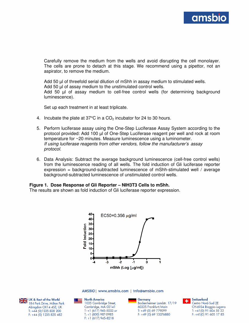
Figure 1. Dose Response of Gli Reporter – NIH3T3 Cells to mShh. The results are shown as fold induction of Gli luciferase reporter expression.

B. Inhibition of mShh-induced reporter activity by inhibitors of hedgehog signaling pathway in Gli Reporter - NIH3T3 cells
1. Harvest Gli Reporter – NIH3T3 cells from culture in growth medium and seed cells at a density of 25,000 cells per well into white clear-bottom 96-well microplate in 100 µl of growth medium without Geneticin.
2. Incubate cells at 37°C in a CO2 incubator for 16-20 hours.
3. The next day, the cells should reach confluency. It is critical for the cells to reach confluency in the wells before treatment.
Carefully remove the medium from wells and avoid disrupting the cell monolayer. The cells are prone to detach at this stage. We recommend using a pipettor, not an aspirator, to remove the medium. Prepare stock solution of hedgehog pathway inhibitor (Cyclopamine or GDC-0449) in DMSO. Dilute the inhibitor stock in assay medium and add 45 µl of diluted inhibitor in assay medium to the wells. The final concentration of DMSO in assay medium can be up to 0.5%. Add 45 µl of assay medium with same concentration of DMSO without inhibitor to inhibitor control wells. Add 45 µl of assay medium with DMSO to cell-free control wells (for determining background luminescence). (Table 1)
4. Incubate the plate at 37°C in a CO2 incubator for 1-2 hours.
5. Add 5 µl of diluted mShh in assay medium to stimulated wells (final [mShh] = 1 µg/ml). Add 5 µl of assay medium to the unstimulated control wells (cells without inhibitor and mShh treatment for determining the basal activity). Add 5 µl of assay medium to cell-free control wells. Set up each treatment in at least triplicate. (Table 1)
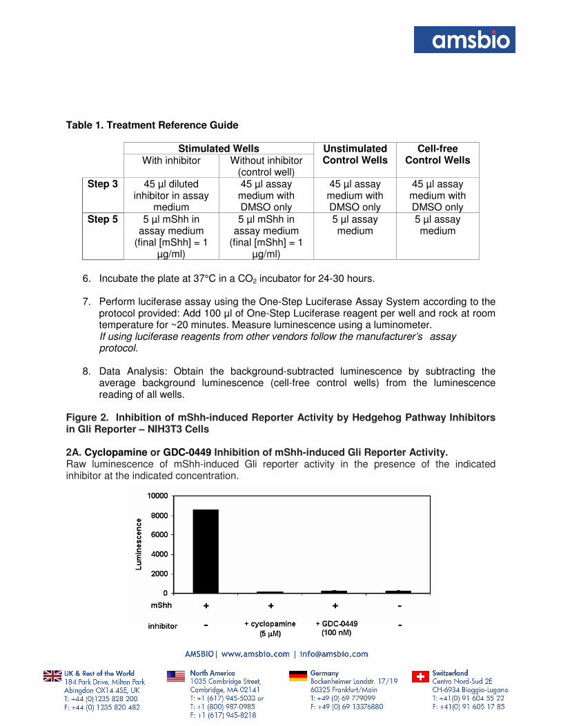
Table 1. Treatment Reference Guide
Stimulated Wells Unstimulated Control Wells
Cell-free Control Wells With inhibitor Without inhibitor
(control well) Step 3 45 µl diluted
inhibitor in assay medium
45 µl assay medium with DMSO only
45 µl assay medium with DMSO only
45 µl assay medium with DMSO only
Step 5 5 µl mShh in assay medium
(final [mShh] = 1 µg/ml)
5 µl mShh in assay medium
(final [mShh] = 1 µg/ml)
5 µl assay medium
5 µl assay medium
6. Incubate the plate at 37°C in a CO2 incubator for 24-30 hours.
7. Perform luciferase assay using the One-Step Luciferase Assay System according to the protocol provided: Add 100 µl of One-Step Luciferase reagent per well and rock at room temperature for ~20 minutes. Measure luminescence using a luminometer.
If using luciferase reagents from other vendors follow the manufacturer’s assay protocol.
8. Data Analysis: Obtain the background-subtracted luminescence by subtracting the
average background luminescence (cell-free control wells) from the luminescence reading of all wells.
Figure 2. Inhibition of mShh-induced Reporter Activity by Hedgehog Pathway Inhibitors in Gli Reporter – NIH3T3 Cells 2A. Cyclopamine or GDC-0449 Inhibition of mShh-induced Gli Reporter Activity. Raw luminescence of mShh-induced Gli reporter activity in the presence of the indicated inhibitor at the indicated concentration.

2B. GDC-0449 Inhibition Dose Response Curve The results are shown as percentage of luminescence. The background-subtracted luminescence of cells stimulated with mShh in the absence of GDC-0449 was set at 100%.
References
1. Kinzler KW et al. (1990) The GLI gene encodes a nuclear protein which binds specific sequences in the human genome. Mol Cell Biol. 10(2):634-642.
2. Mullor JL et al. (2002) Pathways and consequences: Hedgehog signaling in human disease. Trends Cell Biol. 12(12):562-569.
3. Peukert S et al. (2010) Small-molecule inhibitors of the hedgehog signaling pathway as cancer therapeutics. ChemMedChem. 5(4):500-512.
Related Products Product Name Catalog # Size Cyclopamine 27013 5 mg Vismodegib (GDC-0449) 27010 10 mg Wnt Signaling Pathway TCF/LEF Reporter – HEK293 Cell Line
60501 1 Vial
TCF/LEF Reporter Kit (Wnt/β-catenin Signaling Pathway) 60500 500 Reactions One-Step Luciferase Assay System 60690-1 10 ml One-Step Luciferase Assay System 60690-2 100 ml