deborah michel oct 2014 - research-groups.usask.ca of flow cytometry... · what is flow cytometry...
TRANSCRIPT

D e b o r a h M i c h e l O C T 2 0 1 4
Basics of a Flow Cytometer
http://www.usask.ca/pharmacy-nutrition/research/groups/FlowCytometryUserGroup/Home.php

What is Flow Cytometry
flow cytometry is the measurement of
cells/particles in a flow system, which delivers the cells/particles (0.2 to 150 µm) singly past a point of measurement.
Points to consider Flow Light Detection
detector
www.bdbiosciences.com

What can a Flow Cytometer Tell Us About a Cell/Particle?
Relative size by Forward Scatter
Relative granularity or internal complexity by
Side Scatter
Research question by Relative fluorescence intensity (ie marker)
www.2010.igem.org
Right angle light detector
Incident

Examples of application of forward and side scattering
high internal complexity and large size
www.labome.com

Examples of application of forward and side scattering
Nanodiamond and titanium oxide particles in cellular uptake studies using HeLa cells increases the side scatter with little effect on forward scattering
S. Alwani and Dr. Badea research

What is Fluorescent Light?
The fluorochrome
absorbs energy from the laser
The fluorochrome releases the absorbed energy by emission of photons of a longer wavelength
www.bdbiosciences.com
www.uni-leipzig.de
excitation emission

Fluorescence
www.bdbiosciences.com

FACSCalibur
Fluidics Introduces and focuses the cells for interrogation
Optics Generates and collects the light signals
Electronics Converts the optical signal to digital signal, processes the
signal and communicate with the computer

Fluidics
www.bdbiosciences.com

Hydrodynamic focusing
Slower moving sample stream is injected into a faster moving sheath stream
Surface tension and laminar flow causes the sample to be “wicked off” into a narrower faster moving stream within the sheath stream (stream within a stream)
Alignment of cells within this stream are controlled by velocity of the two streams
www.bdbiosciences.com
Flow cell
Higher resolution
Lower resolution
(Quantitative)
(Qualitative)

Optics
Excitation/Emission 2 lasers (488 and
635nm) 4 filters BP and LP
(FL1 to 4)
www.cnbc.pt
Levels: E-1 E00 E01 E02 E03
Photomultiplier tubes
Pre-Amplifier
488 laser
FL1 530/30nm
www.uni-leipzig.de

Electronics
Pulse height (H), area
(A) and width (W)
Converts analog signals to proportional digital signals
Practical Flow Cytometry Haematology Diagnostics
H
W
A

Electronics
Volt Histogram (one parameter)
www.bdbiosciences.com

Plotting Data
www.bdbiosciences.com
Channel values

Plot Stats (Cell Quest Pro)
100 to 1000
Peak Channel 286
Peak 108
300
𝑴𝑴𝑴𝑴 =𝒙𝒊𝑴 𝑮𝑴𝑮 𝑴𝑴𝑴𝑴 = 𝟏𝟏
∑ 𝒍𝑮𝒍 𝒙𝒊𝑴
M. Poorghorban and Dr. Badea research

Types of Plots
Histogram Dot plot Density plot
Contour plot 3D plot
Dr. Badea research

Flow Cytometer Setup
Set the forward and side scatter detectors for your
untreated, unstained cell population of interest
Set fluorescence detectors sensitivities
Using more than one fluorescence marker? Do you need to correct spectral overlap with compensation?
Collect data from your samples

Setting FSC and SSC
Set forward and side scatter detectors to untreated cells.
HeLa cells
Beads

Setting Fluorescence Detectors
Set FL1 and FL2 detectors so that the auto fluorescence from unstained cells are set within the first log decade (100 to 101)
PE (FL2)
FITC (FL1)

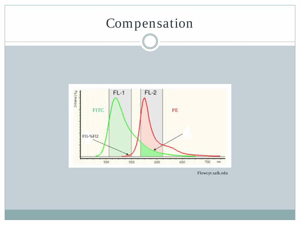
Compensation
When analysing
more than one color be careful of spectral overlap
Digital FACS have software for this.
FITC PE
Flowcyt.salk.edu
Fl2-%Fl1

Compensation
Cells + FITC
FL2 – % FL1
Q1 Q2
Q3 Q4 Y mean
Quadrant Statistics
Quad Events X Mean Y mean
Q1 0 *** ***
Q2 0 *** ***
Q3 7198 3.87 3.27
Q4 2786 336.3 3.32
FITC (FL1)
PE (FL2)
PE (FL2)
FITC (FL1)
Fl2-%FL1
Compensation
Fl1-%Fl2
Flowcyt.salk.edu
FITC PE

Compensation
Cells + FITC
Cells + PE
FL2 – % FL1
Q1 Q2
Q3 Q4 Y mean
Quadrant Statistics
Quad Events X Mean Y mean
Q1 0 *** ***
Q2 0 *** ***
Q3 7198 3.87 3.27
Q4 2786 336.3 3.32
FITC (FL1)
PE (FL2)
FL1 – % FL2
Q1 Q2
Q3 Q4
Quadrant Statistics
Quad Events X Mean Y mean
Q1 318 3.51 600.71
Q2 0 *** ***
Q3 1035 3.86 4.03
Q4 0 *** ***
X mean
PE (FL2)
PE (FL2) PE
(FL2)
FITC (FL1)
FITC (FL1) FITC (FL1)
Fl2-%FL1
Fl1-%FL2

Quiz
Over compensated Under compensated Compensated
A. Doig et al. Practical Flow Cytometry in Haematology Diagnosis

Gating
Isolate populations of interest
Gating an area will make your analysis more specific
Can remove dead cells and debris
Cannot discriminate between cells with the same
scattering properties
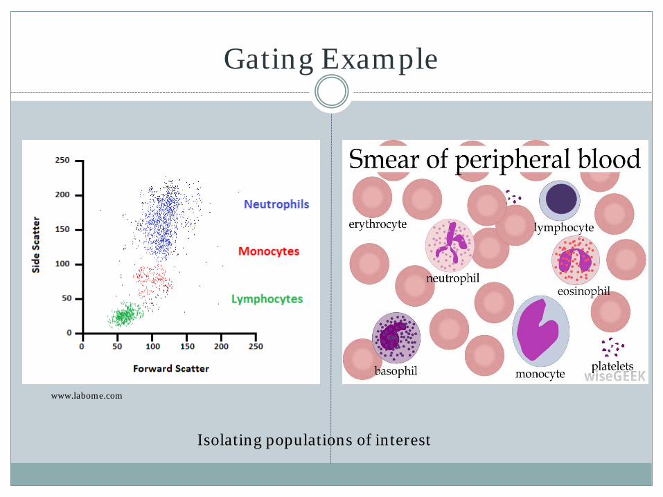
Gating Example
www.labome.com
Isolating populations of interest
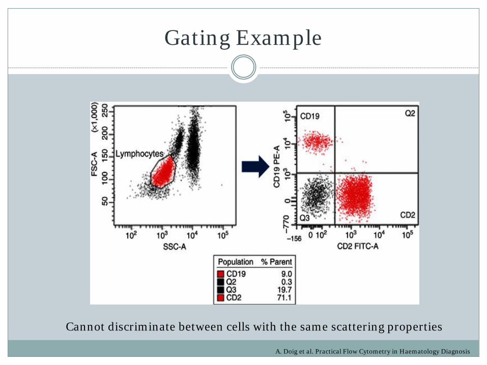
Gating Example
A. Doig et al. Practical Flow Cytometry in Haematology Diagnosis
Cannot discriminate between cells with the same scattering properties

Back Gating Example
A. Doig et al. Practical Flow Cytometry in Haematology Diagnosis
Leucocytes CD45+
Monocytes CD14+
Lymphocytes CD14-, CD45+Bright
Back gating of P1
Fluorescence of P2
Which population is Lymphocytes?

Applications
Immunophenotyping / Intracellular antigens
measurement DNA/RNA: cell cycle, aneuploidy,
endoreduplication, kinetics DNA base ratios Chromatin structure Apoptosis (DNA degradation, mitochondrial
membrane potential, permeability changes, caspase activity)
Membrane potential Membrane fluidity Membrane fusion/runover Intracellular calcium (ions) flux Intracellular pH Sulfhydryl groups/glutathione Cell viability Cell tracking and proliferation Intracellular reactive oxygen species (Oxidative
burst) Cell proliferation Cell enumeration Cell volume and morphological complexity Cell pigments (f.ex. chlorophyll or phycoerythrin)
Drug delivery Multidrug resistance (MDR) Phagocytosis Pathogen-host cell adherence Differentiation Identification of “stem cells” Reticulocyte, platelet etc analysis Microparticles analysis Assessing infection/transfection levels Monitoring of the electropermeabilization of cells Cytotoxicity assay Enzymatic activity Cell activation Protein-protein interactions (FRET, split-GFP) Protein modifications, phospho-proteins Activation of signalling pathways Cytokine Secretion Sorting (f.ex sperm sorting for sex preselection) Karyotyping Telomere length

Build Your Own Flow Cytometer
36th Annual Course in Flow Cytometry, Brunswick Maine, 2012