debre markos universty school of medicine …
TRANSCRIPT

1
DEBRE MARKOS UNIVERSTY SCHOOL OF
MEDICINE DEPARTMENT OF MEDICAL
PHYSIOLOGY
INTRODUCTION TO MEDICAL PHYSIOLOGY
BY :Melkamu Tilahun (Medical physiologist)

2
What is Human Physiology
❖ Branch of biomedical sciences that deals with normal body
function.
❖ It is sometimes called the science of regulation of physiologic
parameters of the body.
❖ Fields of Physiology range from simple viral physiology, bacterial
physiology, cellular physiology to the most complex human
physiology
Why do we learn Physiology?
❖ To explain the physical and chemical factors that are responsible for
the origin, development, and progression of life.
❖ To diagnose and treat abnormalities (pathologies).
❖ Once we have known the normal physiologic phenomena, it is
not difficult to detect abnormalities.
3

Historical background
➢ Physiology is an experimental science to which a number of
scientists contributed a lot.
➢ William Harvey in 1628:- Described the direction of blood
circulation and other aspects of circulatory system.
➢ Claude Bernard:- 100 years ago French physiologist described
that every cell in the body is bathed with the fluid (Extra cellular
fluid). ECF contains all the needed substances for cells.
➢ Cells are capable of living ,growing, and performing their special
functions as long as the proper concentrations of oxygen, glucose,
different ions, amino acids, fatty substances, and other constituents
are available in this internal environment. 4
5
Historical background…
➢ Walter Cannon, another great physiologist of the 1st half of 19th
century, termed the maintenance of constant conditions in the ECF
as homeostasis
✓ Essentially all organs and tissues of the body perform functions
that help maintain these constant conditions.
➢ Physiology as a quantitative science which all physiological
parameters are expressed in numbers and units
➢ Physiology has a strong link with disciplines like:
▪ Anatomy
▪ Biochemistry
▪ Pathology
▪ Pharmacology
▪ Physics etc

Homeostasis
➢ The term homeostasis mean that maintenance of static or constant
conditions in the internal environment (ECF) of the body.
➢ To maintain homeostatic environment all organs and tissues use
Feedback control system.
➢For example
✓ Lungs maintain the normal concentration of respiratory gases in
blood.
✓ The CVS transports required substances and removes waste products,
✓ The kidneys maintain constant ionic concentration
✓ GIT maintains internal environment by providing nutrients ,water
and electrolytes to the body. 6
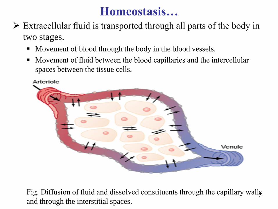
Homeostasis…➢ Extracellular fluid is transported through all parts of the body in
two stages.
▪ Movement of blood through the body in the blood vessels.
▪ Movement of fluid between the blood capillaries and the intercellular
spaces between the tissue cells.
Fig. Diffusion of fluid and dissolved constituents through the capillary walls
and through the interstitial spaces.7
Regulatory systems of homeostasis
➢ The two systems in the body designed for controlling homeostasis:
1. Nervous system
2. Endocrine system
1. The nervous regulatory mechanism
➢ The nervous system regulates body functions through generation of
action potential and release of neurotransmitters.
➢ To bring about complete communication among various structures
of the body NS act through reflex arc.
8

The nervous regulatory mechanism…
➢ Reflex arc is a pathway of neural reflex. It composed of five structures:
1. Receptors (change detectors and transducers)
2. Sensory system (carries nerve signal to the integrating center )
3. Integrating center (evaluates the information and gives feedback)
4. Motor system ( carries information away from the integrating center)
5. Effectors (receive motor feedback and accordingly act which result
desirable biological responses)
9

10
Stimulus (Mechanical, Chemical, thermal, etc)
Receptor (Neural transduction)
Sensory System
Integrating center (Brain and spinal cord)
Motor System (FB)
Effector (Muscle,gland) =Desirable biological responses
Fig. Reflex Arc

Regulatory systems of homeostasis cont’d…..
2. The hormonal regulatory mechanism
Hormones are chemical messengers secreted by endocrine glands, and
transported via blood to the target organs including other glands.
Examples:
Parathyroid glands secrete parathyroid hormone to the kidneys, bone and
small intestine = [Ca2+]
Aldosterone from adrenal cortex to the kidneys, intestine [Na+]
Anti-diuretic hormone (ADH) causes water retention from the kidneys and
intestine.
An organism is said to be in a state homeostasis when its internal environment
contains an optimum amount of nutrients, gases, electrolytes, water, hormones,
enzymes and temperature. 11

Ca2+homeostasis
12
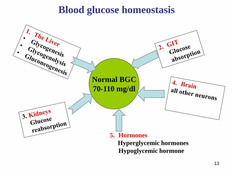
Blood glucose homeostasis
13
Normal BGC
70-110 mg/dl
5. Hormones
Hyperglycemic hormones
Hypoglycemic hormone

Regulatory systems of homeostasis cont’d…..
Common Properties Of Hormones And Neurotransmitters
- Both are released in small amount
- Both have receptors on the target organs
- Both act by altering their target organs
- Both work towards common goal →Homeostasis
But there are differences
- Nervous regulation is faster but hormones is slower
- Nervous effects are diffused but hormones is mostly localized
14

Some important Homeostatic Values
Body fluid volume (40 L) ECF = 15L
ICF = 25L
Osmolality 300 mosm/L (285 – 300 mosm/L)
PH 7.35 – 7.45
Blood Gases PCO2 = 40 – 46 mm Hg
PO2 = 40 – 104 mm Hg
Body Temperature 36.3 – 37.1OC
Electrolytes (ECF) Ca2+=10 mg/dL or 5 meq/L or
K+ = 4 meq/L
Na+= 142 meq/L
Cl-= 103 meq/L
HCO3- =27 meq/L 15
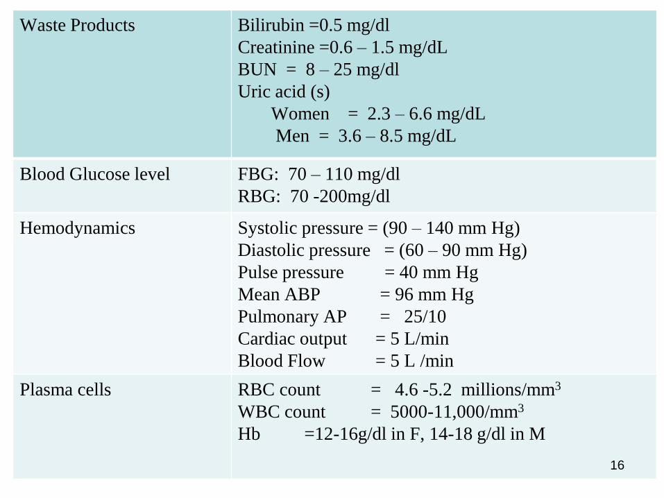
Waste Products Bilirubin =0.5 mg/dl
Creatinine =0.6 – 1.5 mg/dL
BUN = 8 – 25 mg/dl
Uric acid (s)
Women = 2.3 – 6.6 mg/dL
Men = 3.6 – 8.5 mg/dL
Blood Glucose level FBG: 70 – 110 mg/dl
RBG: 70 -200mg/dl
Hemodynamics Systolic pressure = (90 – 140 mm Hg)
Diastolic pressure = (60 – 90 mm Hg)
Pulse pressure = 40 mm Hg
Mean ABP = 96 mm Hg
Pulmonary AP = 25/10
Cardiac output = 5 L/min
Blood Flow = 5 L /min
Plasma cells RBC count = 4.6 -5.2 millions/mm3
WBC count = 5000-11,000/mm3
Hb =12-16g/dl in F, 14-18 g/dl in M
16

Feedback control mechanisms of the homeostasis
➢ Feedback control mechanism is a means by which our body tries to
maintain its homeostatic environment
➢ There are two types of feed back mechanisms:
A. Negative Feedback Mechanism (NFM)
B. Positive Feedback Mechanism (PFM)
17
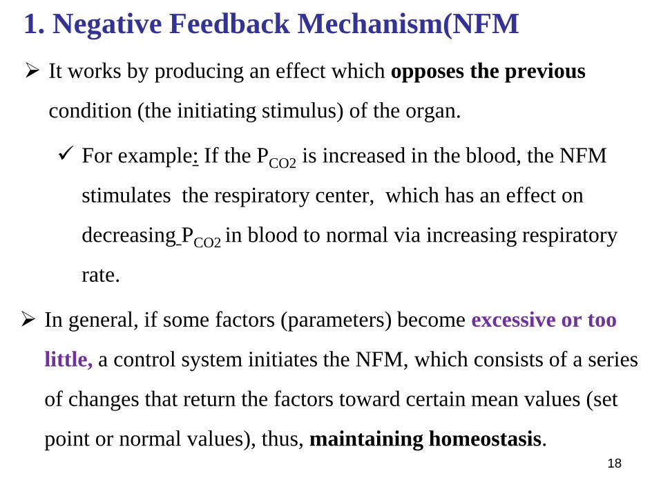
1. Negative Feedback Mechanism(NFM
➢ It works by producing an effect which opposes the previous
condition (the initiating stimulus) of the organ.
✓ For example: If the PCO2 is increased in the blood, the NFM
stimulates the respiratory center, which has an effect on
decreasing PCO2 in blood to normal via increasing respiratory
rate.
➢ In general, if some factors (parameters) become excessive or too
little, a control system initiates the NFM, which consists of a series
of changes that return the factors toward certain mean values (set
point or normal values), thus, maintaining homeostasis. 18

19
20

The Positive Feedback Mechanism (PFM)
➢ It works by producing an effect which enhances or repeats the
same action like that of the starting stimulus. It also called vicious
circle and disturbs the internal environment and cause disease and
death.
For example, if a person suffers from a heart attack that damages
the heart function, then the heart pumps less amount of blood to the
tissues including the heart muscle and brain. Because the heart
muscle does not get sufficient nutrients and O2, the activity of the
heart becomes weaker and weaker and the weaker the heart the
lesser blood is pumped and then death may occur. 21

Examples of the PFM
1. Blood clotting is an example of a very valuable use of PF
2. Generation and propagation of the action potential.
✓ Stimulated nerve fiber opening of Na+ channels entry of few Na+
stimulates the opening of more and more Na+ channels.
3. Labor during child birth
Uterine contraction is enhanced as the head of the baby stretches the cervix
generation of action potentials AP reaches H another AP posterior
pituitary release of oxytocin into the blood contraction of uterine muscle
more and more stretching and more and more contraction. The only way to
stop this kind of phenomenon is by removing the stimulus
4. LH-surge: immediately before ovulation.22

LH surge: the positive feedback mechanism
23
HT
Pituitary
Ovary
GnRH
LH
Estrogen
>200 µg/ml
activates
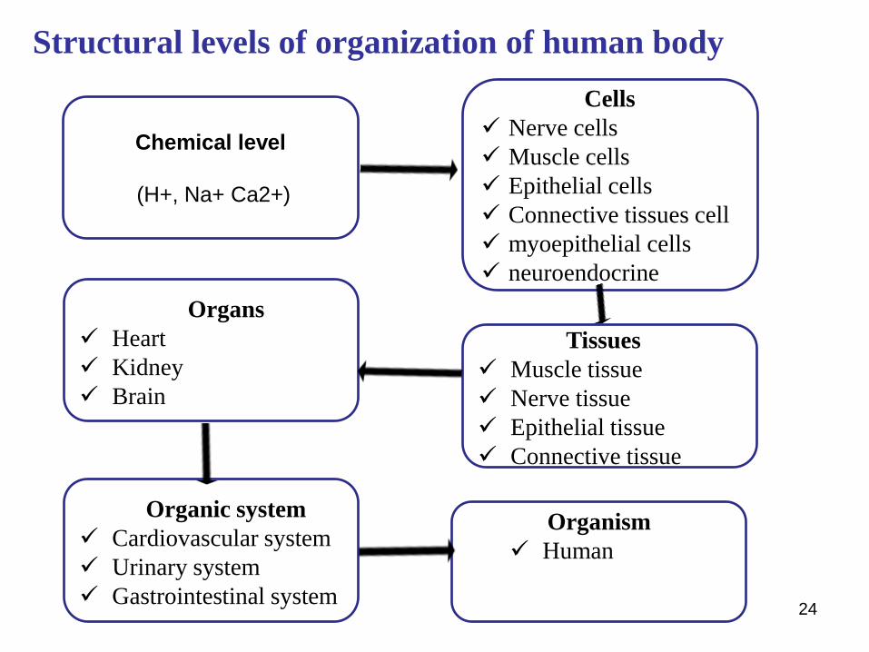
Structural levels of organization of human body
24
Chemical level
(H+, Na+ Ca2+)
Cells
✓ Nerve cells
✓ Muscle cells
✓ Epithelial cells
✓ Connective tissues cell
✓ myoepithelial cells
✓ neuroendocrine
Tissues
✓ Muscle tissue
✓ Nerve tissue
✓ Epithelial tissue
✓ Connective tissue
Organs
✓ Heart
✓ Kidney
✓ Brain
Organic system
✓ Cardiovascular system
✓ Urinary system
✓ Gastrointestinal system
Organism
✓ Human

25
Structural levels of organization
Cell physiology
➢ Cells are functional & structural units of the body
There are tow types of cells:
A. Cells without typical nucleus = prokaryotes
B. Cells with nucleus = eukaryotes
26

Cell physiology….o Prokaryotes: (Eg. bacteria)
Smaller (1-10 m)
No cytoskeleton
Generally no membrane-bound organelles
RNA and protein synthesis in same compartment
Small circular chromosome
Generally very small & unicellular
o Eukaryotes:
Genetic material mostly in nucleus
Larger (10-100 m)
Cytoskeleton present
Membrane-bound organelles present
RNA synthesis in nucleus, protein synthesis in cytoplasm 27
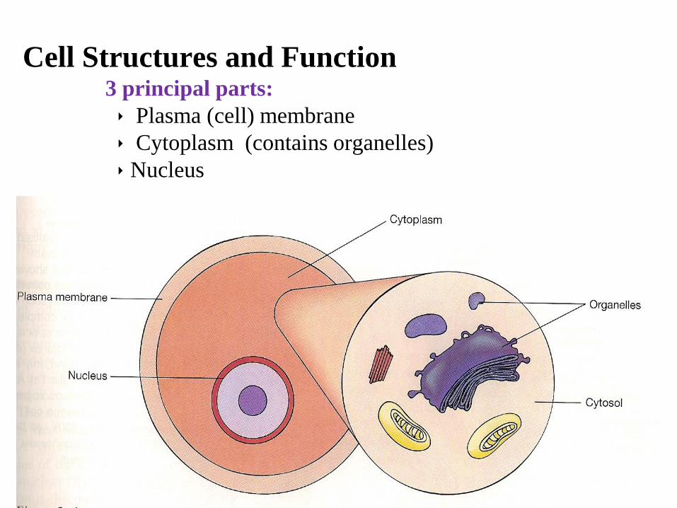
Cell Structures and Function3 principal parts:
Plasma (cell) membrane
Cytoplasm (contains organelles)
Nucleus
28

29

30
Generalized cellComponents of cells
1. A typical cell has two parts: nucleus and cytoplasm.
2. Nucleus is separated from the cytoplasm by a nuclear
membrane
3. The cytoplasm is separated from the surrounding fluid (ECF)
by the plasma membrane
The different substances that make up the cell are collectively called
protoplasm. Protoplasm is composed of five basic substances:
1. Water
2. Electrolytes
3. Proteins
4. Lipids
5. Carbohydrates.

31
The plasma membrane
➢ It is a sheet-like structure that surrounds (encloses) the cell,
separating the cellular contents from the ECF.
➢ It is entirely composed of proteins and lipids in a ratio of 55:43
respectively, and only 3% of carbohydrates.
Percent proportion:
1. Proteins: 55 %
Phospholipids 25 %
2. Lipids: 42 % Cholesterol 13 %
Neutral tats 4 %
3. Carbohydrate: 3 %
The level of cholesterol determines rigidity of the membrane.

32
Function of the plasma membrane
1. Separates cellular contents from the ECF
2. Regulates the passage of substances in and out.
▪ It is semi-permeable allowing some substances to pass through
it excluding others. This creates unequal distribution of ions on
both sides of the membrane.
3. It provides receptors for NTs, hormones and drugs.
4. It is a means of cell to cell contact.
5. Plays an important role in the generation and transmission of
electrical impulse in nerves and muscle.
6. Involved in the regulation of cell growth and proliferation.
33
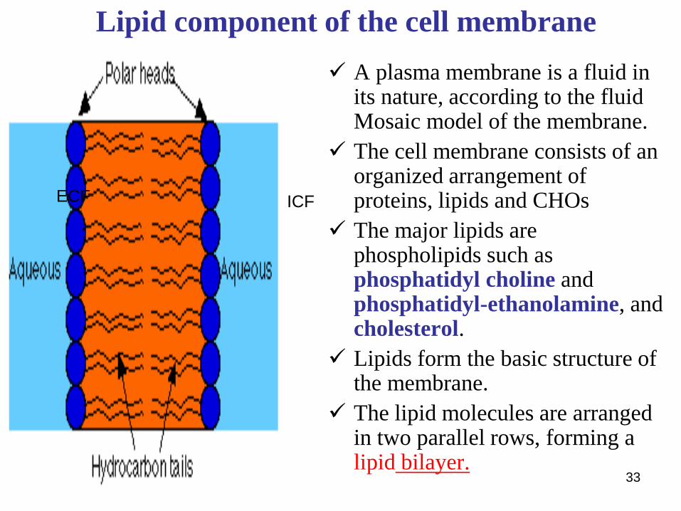
Lipid component of the cell membrane
✓ A plasma membrane is a fluid in its nature, according to the fluid Mosaic model of the membrane.
✓ The cell membrane consists of an organized arrangement of proteins, lipids and CHOs
✓ The major lipids are phospholipids such as phosphatidyl choline and phosphatidyl-ethanolamine, and cholesterol.
✓ Lipids form the basic structure of the membrane.
✓ The lipid molecules are arranged in two parallel rows, forming a lipid bilayer.
ECF ICF

34
The plasma membrane…
➢ It is believed that globular proteins are embedded in the lipid
bilayers and that these proteins participate in the transport of lipid-
insoluble particles through the plasma membrane, some integral
proteins act as carriers and channels.
➢ The cell membrane is surrounded by a cell coat or glycocalyx, which
is made up of glycolipids and glycoproteins.
➢ It uses the site of hormonal receptors and antigenic activity in
blood groups.
➢ The phospholipids component is organized into a double layer with
their hydrophobic (tail) and polar (hydrophilic) heads.

35
The plasma membrane…

36
The plasma membrane…
➢ The physical orientation of the lipid bilayer structures is that the
hydrophilic ends of the lipid molecules line up facing the ICF and
ECF.
➢ The hydrophobic tails of the molecules face each other in the
interior of the bilayer.
➢ The lipid bilayer portion of the cell membrane is impermeable to
water and water soluble substances such as ions, glucose, urea
and others.
➢ Fat soluble substances such as O2, CO2, N2, alcohol and drugs can
diffuse through the membrane.

37
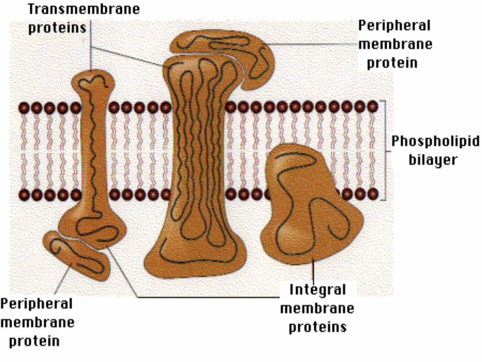
Membrane proteinsIntegral and Peripheral proteins
A. Integral or intrinsic proteins: interdigitated in the hydrophobic
center of the lipid bilayer.
➢ Transmembrane proteins are integral proteins that span the entire
bilayer.
➢ Transmembrane proteins serve as:
1. Channels through which ions pass
2. Carriers which actively transport materials across the bilayer
e.g. glucose
3. Pumps which actively transport ions
4. Receptors for neurotransmitters and hormones
➢ Integral proteins that are present only on one side of the membrane,
serve primarily as enzymes.
38

39
Membrane proteins: channels

40
Membrane proteins (cont’d)
B. Peripheral or extrinsic proteins: bind to the hydrophilic polar
heads of the lipid or on integral proteins.
➢ Peripheral proteins that bind to the intracellular surface contribute to
the cytoskeleton.
➢ Peripheral proteins that bind to the external surface contribute to the
glycocalyx (a cell coat that is composed of glycolipids and
glycoproteins to cover the cell membrane)
➢ Extrinsic protein not use for Channel ,Carrier but use as receptor &
enzymes
41
Membrane carbohydrates
➢ Attached invariably on the outside surface of the membrane,
binding with protruded integral proteins and lipid, they form
glyco-proteins and glyco-lipids (glycocalyx).
➢ They play a role in
1. Immune reaction (antigenical importance)
2. Cell to cell attachment
3. Act as receptors for NTs, hormones and drugs

Cytoplasm
➢ is the portion of the cell found between nucleus and cell membrane
➢ contains organelles and cytosol
➢ Cytosol is the fluid portion of cytoplasm
➢ Organelles are specialized compartments or subunit within a
cell that has a specific function
➢ An organelle is usually separately enclosed within its own plasma
membrane.
42

43
The nucleus
➢ The nucleus is the control center for the cells.
➢ It contains the genes, which are units of heredity.
➢ Chemically each gene consists of highly compressed DNA in the
form of chromosomes
➢ Genes control cellular activity by determining the type of
proteins, enzymes, and other substances that are made by the
cell.
➢ The nucleus is also the site of RNA synthesis.

The nucleus (cont’d)
➢ There are three kinds of RNA
❖ Messenger RNA (mRNA), which carries the instruction from
DNA for protein synthesis to the cytoplasm
❖ Ribosomal RNA (rRNA), which moves to the cytoplasm where
it becomes the site of protein synthesis.
❖ Transfer RNA (tRNA), serves as an amino acid transporter
system within the cell for protein synthesis.
➢ Nucleotides are composed of nitrogen containing bases purine (A,
G) and pyrimidin (C, T) as well as deoxyribose sugar conjugated
by phosphate.44

45
The nucleus (cont’d)
➢ In RNA, the pyrimidin base T is replaced by U and the 5-carbon
sugar is ribose.
➢ In addition to the chromatin, the nucleus contains one or two round
bodies called nucleoli for rRNA synthesis.
➢ The nuclear contents are surrounded by a double walled nuclear
membrane.
➢ The pores present in this membrane allow fluids, electrolytes,
RNA, and other materials to move between the nuclear and
cytoplasmic comportments.

46
Function of nucleotides
1. Building units of nucleic acid DNA, RNA
2. High energy molecules (ATP, GTP)
3. Biosynthetic mediators (UDP-glycogen)
4. Regulator of chemical reaction in the cell e., g,. cAMP
5. Act as coenzyme (NAD, FAD)

47
48
Cellular organelles
➢ inner organs of the cell. These include the ribosomes, endoplasmic
reticulum (ER), Golgi apparatus, mitochondria, lysosomes,
peroxisomes and the cytoskeletal system (microtubules and
microfilaments).
Ribosomes:
▪ Are the sites of protein synthesis in the cell
▪ Small particles composed of rRNA and proteins
▪ Found in two forms: attached to the wall of ER or as free
ribosomes.
➢ Free ribosomes are found in two forms
▪ Scattered in the cytoplasm
▪ Clustered (aggregated) to form functional units called
polyribosomes

49
Endoplasmic Reticulum (ER)
➢ It is an extensive membranous structure that connects various parts
of the inner cell.
➢ ER is also connected with the nuclear membrane.
➢ There are two types of ER: rough ER and smooth ER.
➢ The rER is associated with ribosomes.
➢ The function of rER is to segregate proteins that are being exported
from the cell.
➢ rER is the site of protein synthesis
50
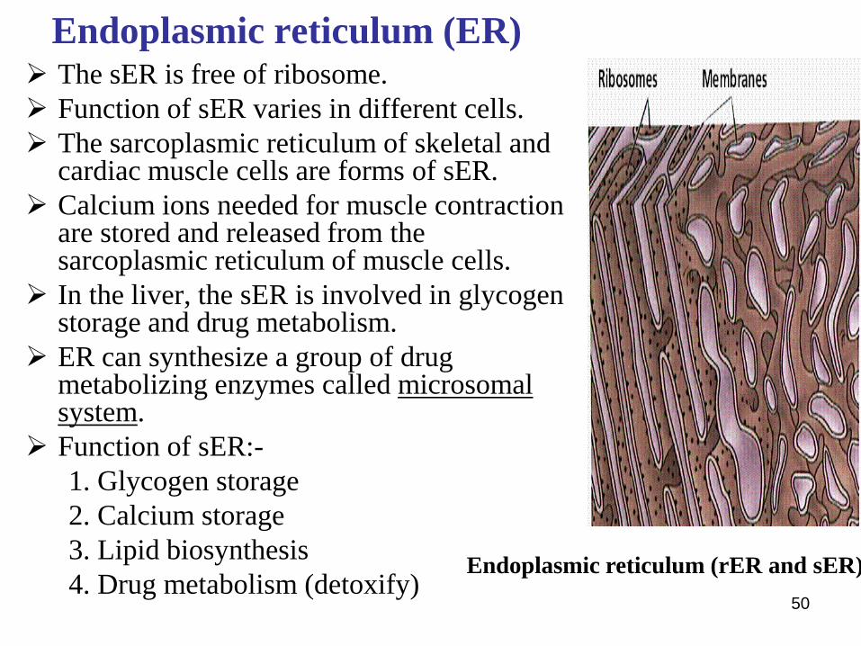
Endoplasmic reticulum (ER)➢ The sER is free of ribosome.
➢ Function of sER varies in different cells.
➢ The sarcoplasmic reticulum of skeletal and cardiac muscle cells are forms of sER.
➢ Calcium ions needed for muscle contraction are stored and released from the sarcoplasmic reticulum of muscle cells.
➢ In the liver, the sER is involved in glycogen storage and drug metabolism.
➢ ER can synthesize a group of drug metabolizing enzymes called microsomal system.
➢ Function of sER:-
1. Glycogen storage
2. Calcium storage
3. Lipid biosynthesis
4. Drug metabolism (detoxify) Endoplasmic reticulum (rER and sER)

51
Golgi Complex
➢ The Golgi complex consists of
flattened membranous saccules
and cisterns that communication
with the ER and acts as a
receptacle for hormones and other
substances that the ER produces.
➢ It then modifies and packages
these substances into secretory
granules.

52
Rough ER and Golgi complex
53
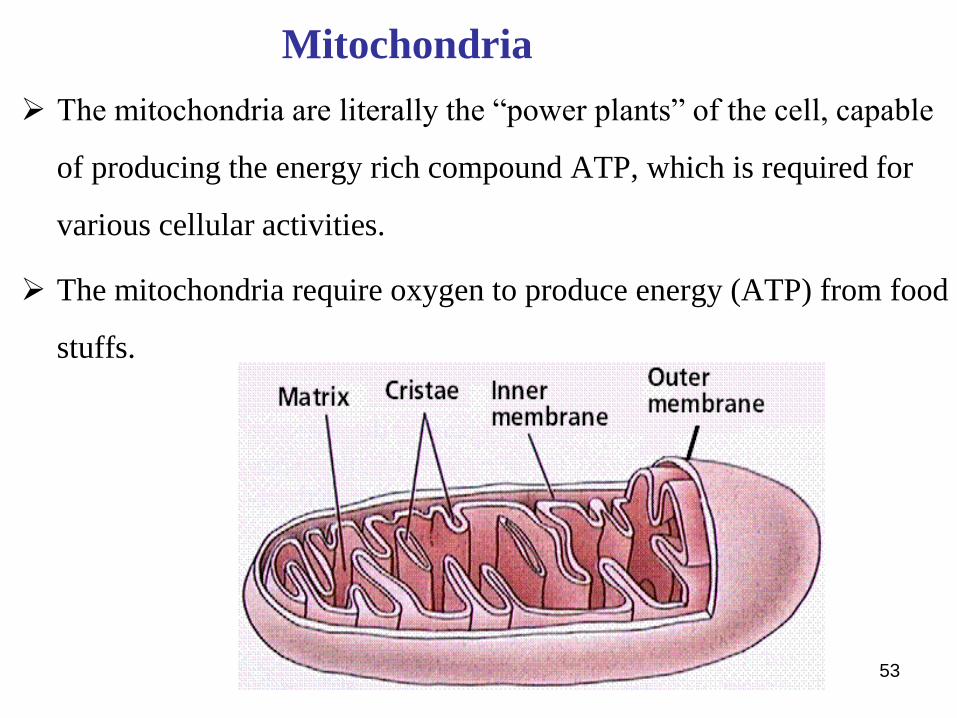
Mitochondria
➢ The mitochondria are literally the “power plants” of the cell, capable
of producing the energy rich compound ATP, which is required for
various cellular activities.
➢ The mitochondria require oxygen to produce energy (ATP) from food
stuffs.

Lysosomes➢ Lysosomes are vesicular organelles that form by breaking off from
the Golgi apparatus and then dispersing throughout the cytoplasm.
➢ The lysosomes provide an intracellular digestive system that allows
the cell to digest
▪ Damaged cellular structures,
▪ Food particles that have been ingested by the cell, and
▪ Unwanted matter such as bacteria.
➢ It is surrounded by a typical lipid bilayer membrane and is filled with
large numbers of small granules 5 to 8 nanometers in diameter, which
are protein aggregates of as many as 40 different hydrolase
(digestive) enzymes.
➢ The membrane surrounding the lysosome prevents the enclosed
hydrolytic enzymes from coming in contact with other substances in
the cell and, therefore, prevents their digestive actions.
54

Peroxisomes
➢ Also called small bodies
➢ Spherical in shape
➢ Surrounded by single membrane
➢ Have protective role in that they secrete chemical that converts
harmful substances into harmless
➢ e.g., Catalase produced by peroxisomes change H2O2 to H2O and
O2 H2O2 O2 +H2O
Catalase
55

56
Cytoskeletal system of the cell
➢ They are microfilaments and
microtubules, rigid threadlike structures
dispersed through out the cytoplasm.
Function of cytoskeletal system:
1. Maintain shape of the cells. eg.Neurofibrils in axon
2. Serve as a transport system for the movement of compounds and organelles within the cell. egaxoplasmic transport
3. Construct the mitotic spindle eg. Centroils
4. Provide for the support and movement of cilia and flagella
5. Cell to cell contact: to fasten cell membranes together
Microtules organized as 9+2 doublets

57
Transport through the cell membrane
➢ Substances are transported through the cell membrane by:
1. Simple diffusion
2. Osmosis
3. facilitated diffusion
4. active transport (1° and 2°) and
5. vesicular transport mechanisms.
✓ Endocytosis
✓ Exocytosis
ECF
ICF
58
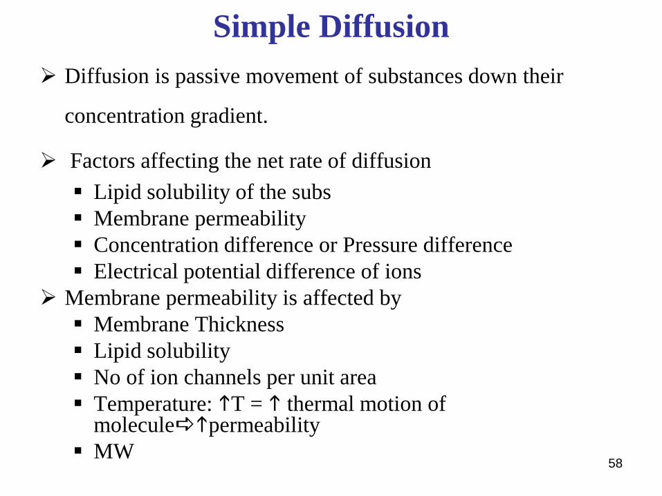
Simple Diffusion
➢ Diffusion is passive movement of substances down their
concentration gradient.
➢ Factors affecting the net rate of diffusion
▪ Lipid solubility of the subs
▪ Membrane permeability
▪ Concentration difference or Pressure difference
▪ Electrical potential difference of ions
➢ Membrane permeability is affected by
▪ Membrane Thickness
▪ Lipid solubility
▪ No of ion channels per unit area
▪ Temperature: T = thermal motion of moleculepermeability
▪ MW

59
Simple Diffusion
➢ Rate of diffusion is determined by the following factors
summarized in the formula shown below.
S. A. T. C
➢ Rate of diffusion = D MW
Where, C = Change of concentration
S = Solubility in lipid
A = Surface area of the membrane
T = Temperature
D = Distance or membrane thickness
MW = Molecular wt of substances
➢ Examples: Substances that are transported by simple diffusion are
CO2, O2, alcohol, lipid soluble drugs and ions through specific
channels.

60
Osmosis➢ It is the power of movement of H2O from an area of higher amount of
water to an area of lower amount of water through the semipermeable membrane.
➢ The direction of movement of water is governed by the amount of osmoticaly active particles (solutes).
➢ The pressure that opposes osmosis of water is called osmotic pressure
➢ H2O molecules have very small (0.3 nm) in diameter, so that they can not traverse the lipid bilayer simply. Instead they pass through specific water channels called aquaporins: Five aquaporins (AQ1….AQ5) have been identified in the body.
61
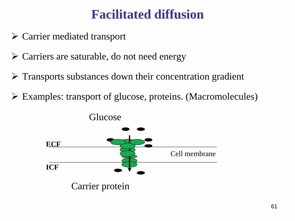
Facilitated diffusion
➢ Carrier mediated transport
➢ Carriers are saturable, do not need energy
➢ Transports substances down their concentration gradient
➢ Examples: transport of glucose, proteins. (Macromolecules)
Glucose
Carrier protein
Cell membrane
ECF
ICF
62

63
Active transport
➢ Substances are transported
against concentration,
electrochemical gradient, up hill
direction.
➢ Used for the transport of Na+,
K+, Ca2+, Fe2+, H+, Cl-
➢ Consumes energy in the form of
ATP
Primary active transport
➢ Carrier protein is involved
➢ Consumes energy from ATP
➢ Carrier protein is anti-porter
Common examples
1. Na+ - K+ ATPase
2. H+ - K+ ATPase
3. Ca2+ ATPase

64
Active transport: Na+ - K+ ATPase
Na-K-Pump
❖ It pumps 3Na+ outward and 2K+ inward
❖ It maintains Na outside and ↓K+ inside
❖ It maintains electropositive outside and electronegative, inside.
❖ Na+ - K+ pump is a carrier protein that is made up of two subunits. It has 3 binding sites for Na+ inside
❖ It has 2 binding sites for K+ on the outside
❖ It has ATPase activity inside. ATP = ADP + ---P + energy.
❖ Energy brings conformational change of the pump so that Na+ pumped outward and K+ inward.

65
Secondary active transport
▪ Carrier protein is involved
▪ Consumes energy, but not ATP
▪ Carrier protein is symporter
▪ Uniport carriers: Carry single substance to one direction
▪ Antiport carriers: Carry two substances in opposite directions
▪ Symport carriers: Carry two substances into the same direction

66
Vesicular transport
There are two types:
1. Endocytosis:
Pinocytosis
phagocytosis
2. Exocytosis

Cell Junctions (Intercellular Connections)
➢ Multicellular organisms (eg. human) exist because cells are able to
bind to each other
➢ Cells may adhere either directly to each other in cell-cell adhesion
or to extracellular components that provide a structural framework
for cell binding
➢ The binding of cells to the ECM is termed cell matrix or cell-
substratum adhesion
➢ Cell adhesion is not just a structural element that binds things
together, it is also a highly dynamic process
67

Cell Junctions (Intercellular Connections)…
➢ Molecules that mediate cell adhesion are called cell adhesion
molecules (CAMs)
➢ The majority, but not all, of CAMs belong to one of four protein
families:
Cadherins
Immunoglobulin (Ig) family,
Selectins (Selectins are Calcium-dependent, Cell to Cell
Surface Carbohydrate Binding Proteins.
Integrins (are receptors that mediate attachment between a cell
and the tissues surrounding it,68
Cell Junctions (Intercellular Connections)…
➢ CAMs are transmembrane proteins and have:
1. An extracellular domain that participates in adhesion,
2. A transmembrane domain that anchors the protein in the cell membrane,
3. A cytoplasmic domain that mediates attachment to the cytoskeleton
➢ Adhesive binding by adhesion molecules may be either homophilic,
meaning that the molecule binds to another of the same type, or
heterophilic, meaning that binding is to a molecule of different type
➢ Cell-cell and cell-matrix junctions are diverse in structure and they
do more than physical binding
69
Cell Junctions (Intercellular Connections)…
Four main functions of cell-cell and cell-matrix junctions are:
i . Anchoring junctions (adheres, desmosomes, hemidesmosomes)
ii. Occluding junctions (tight junctions)
iii.Channel forming junctions (gap junctions)
iv. Signal relaying junctions (chemical synapses and immunological
synapses)
70
1. Tight Junctions➢ Regulate paracellular permeability and cell polarity (spatial
differences in the shape, structure, and function of cells).
➢ Are made up of ridges—half from one cell and half from the
other—which adhere so strongly at cell junctions that they almost
obliterate the space between the cells
➢ Surround the apical margins of the cells in epithelia such as
the intestinal mucosa and the walls of the renal tubules
➢ Permit the passage of some ions and solute in between adjacent
cells (paracellular pathway)
➢ Spaces between cells or, more precisely regulate the permeability
of these spaces selectively71
1. Tight Junctions…
➢ Prevent the movement of proteins in the plane of the membrane,
helping to maintain the different distribution of transporters and
channels in the apical and basolateral cell membranes that make
transport across epithelia possible
➢ act as “fences” to separate the molecules in the apical and
basolateral membranes, thus helping to maintain cell polarity
➢ The transmembrane components of tight junctions are the proteins
occludin and claudin
➢ In the cytoplasm occludin and claudin interact with the actin
cytoskeleton through attaching cell proteins72

73
Tight junction….

2. Desmosomes…
➢ Are spotlike or punctate junction
➢ Maintain tissue integrity by providing strong intercellular
adhesion and acting as a link between the cytoskeletons of adjacent
cells
➢ are linked to keratin intermediate filaments inside the cell, then
extend across the plasma membrane to associate with identical
cadherins of an adjacent cell
➢ Abundant in skin, heart, neck of uterus where they are needed to
withstand mechanical stress
74

3. Adherens junctions
Provide strong mechanical attachments between
adjacent cells
The primary function of the adherens junction is cell-
cell adhesion
Cadherins linked to actin microfilaments in the
cytoplasm extend out of the cell and bind to cadherins
of an adjoining cell
Found in the heart and epithelial tissue
75
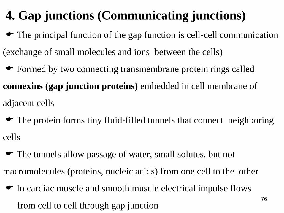
4. Gap junctions (Communicating junctions)
The principal function of the gap function is cell-cell communication
(exchange of small molecules and ions between the cells)
Formed by two connecting transmembrane protein rings called
connexins (gap junction proteins) embedded in cell membrane of
adjacent cells
The protein forms tiny fluid-filled tunnels that connect neighboring
cells
The tunnels allow passage of water, small solutes, but not
macromolecules (proteins, nucleic acids) from one cell to the other
In cardiac muscle and smooth muscle electrical impulse flows
from cell to cell through gap junction 76

77

INTERCELLULAR SIGNALING
Cells need to be able to communicate to other cells and respond to
environmental changes
For multi-cellular organisms, cell-cell communication is important
For unicellular organisms, they need to be able to respond to
physical and chemical changes in their environment
78

79Ultimate effects of cell signaling

80
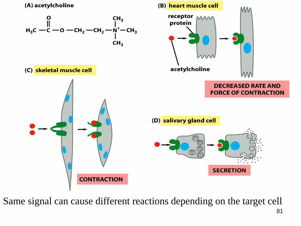
Five forms of intercellular signaling (Cell-cell communication)
(E)
ing on the target cell s depending on the target cell
81
Same signal can cause different reactions depending on the target cell
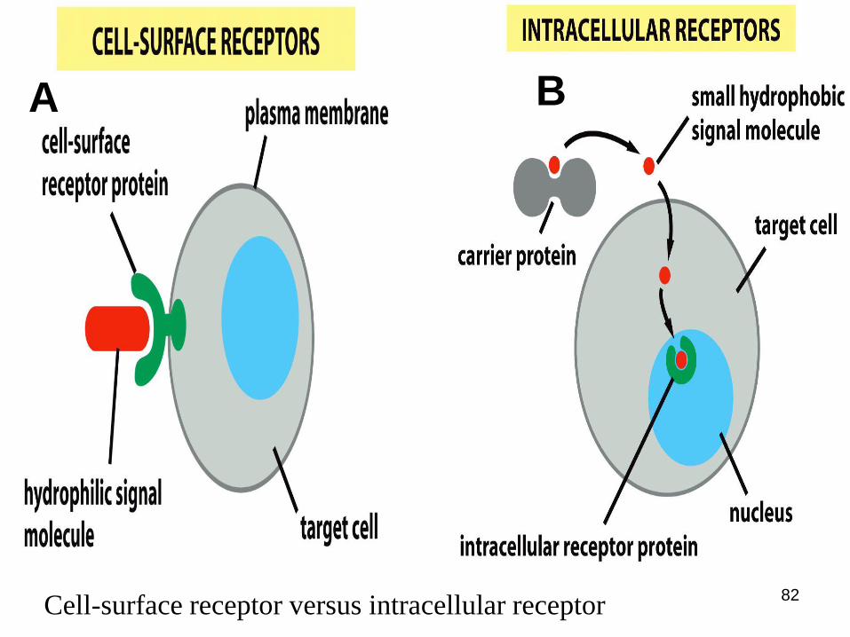
Cell-surface receptor versus intracellular receptor82
BA
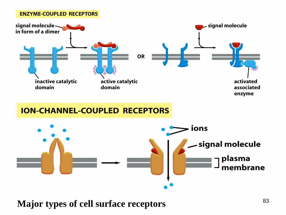
Major types of cell surface receptors 83

84
ICF•Water
•High K+, Po43-,
Mg2+
•Nutrients, gases
•Hormones
ECF• Water
• High Na+, Cl- , Ca2+ and HCO3-
• Nutrients: glucose, aa, lipids
• Gases: O2, CO2
• Hormones, Enzymes
Fluid environment of the body
➢ 60% of human body is made up of fluid▪ Sex; adult men 60% of total body weight
adult women 55% of total body weight
▪ Age; neonate 70% of total body weight
> 60 years men 52%, women 46% of total body weight
➢ Body fluid is distributed in 2 compartments
1. Intracellular fluid compartment (ICF) : two thirds of the body water
2.Extracellular fluid compartment (ECF): one third of the body water

Body fluid compartment cont’d………
➢ These two fluid compartments differ strikingly in terms of their
electrolyte composition
➢ But the fluid compartments solute concentrations (osmolarity) are
normally equal (no an osmotic difference between cells cytoplasm
and ECF)
➢ The ECF further subdivided into two major sub compartments:
✓ The interstitial fluid and lymph, comprises three fourths of the ECF
✓ The blood plasma, comprises about one fourth of the ECF
➢ The blood plasma, interstitial fluid, and lymph are nearly identical
in composition, except for the higher protein concentration in the
plasma 85
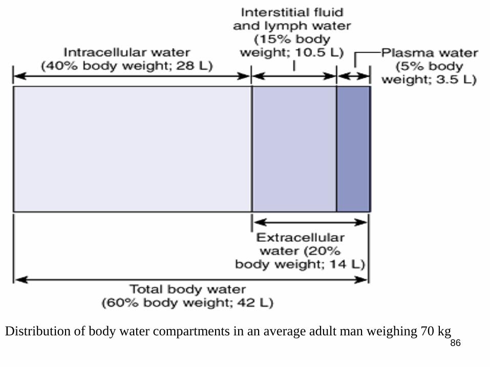
Distribution of body water compartments in an average adult man weighing 70 kg86

Body fluid compartment cont’d……
➢ In addition to above mentioned, ECF compartment includes
transcellular fluid that amounts to about 1% to 3% of body weight
➢ Transcellular fluids include cerebrospinal fluid, aqueous humor of
the eye, secretions of the digestive tract and associated organs
(saliva, bile, pancreatic juice), renal tubular fluid and bladder urine,
synovial fluid, and sweat
➢ Transcellular fluids are not plasma ultrafiltrates (as are interstitial
fluid and lymph); so they have a distinct ionic composition
87

Constituents of Extracellular and Intracellular Fluids
• Ionic composition of plasma and interstitial fluid is similar (plasma
and interstitial fluid are separated by highly permeable capillary
membranes)
• Higher concentration of protein in the plasma than in interstitial fluid
✓ Intracellular fluid is separated from the extracellular fluid by a cell membrane
(highly permeable to water but not to most of the electrolytes in the body)
✓ Intracellular fluid contains large amounts of potassium and phosphate ions
plus moderate quantities of magnesium and sulfate ions
✓ The intracellular fluid contains only small quantities of sodium and chloride
ions and almost no calcium ions
88

Fig. Major cations and anions of the ICF and ECF89

Water Balance
➢ People normally stay in a stable water balance; that is, water input
and output are equal
➢ Body fluid volume is kept remarkably constant
➢ Daily intake of water:
• Ingested in the form of liquids and water in the food, 2100 ml/d
• Synthesized in the body due to oxidation of nutrients, 200 ml/day
➢ Daily loss of body water:
• Insensible water loss 700 ml/day
• Fluid loss in sweat, 100 ml/day
• Water loss in feces, 100 ml/day
• Water loss by the kidneys (urine), 1400 ml/day
➢ The most important means by which the body maintains a balance
between water intake and output is by controlling the rates at which
the kidneys excrete water (0.5 L/day to 20 L/day)90
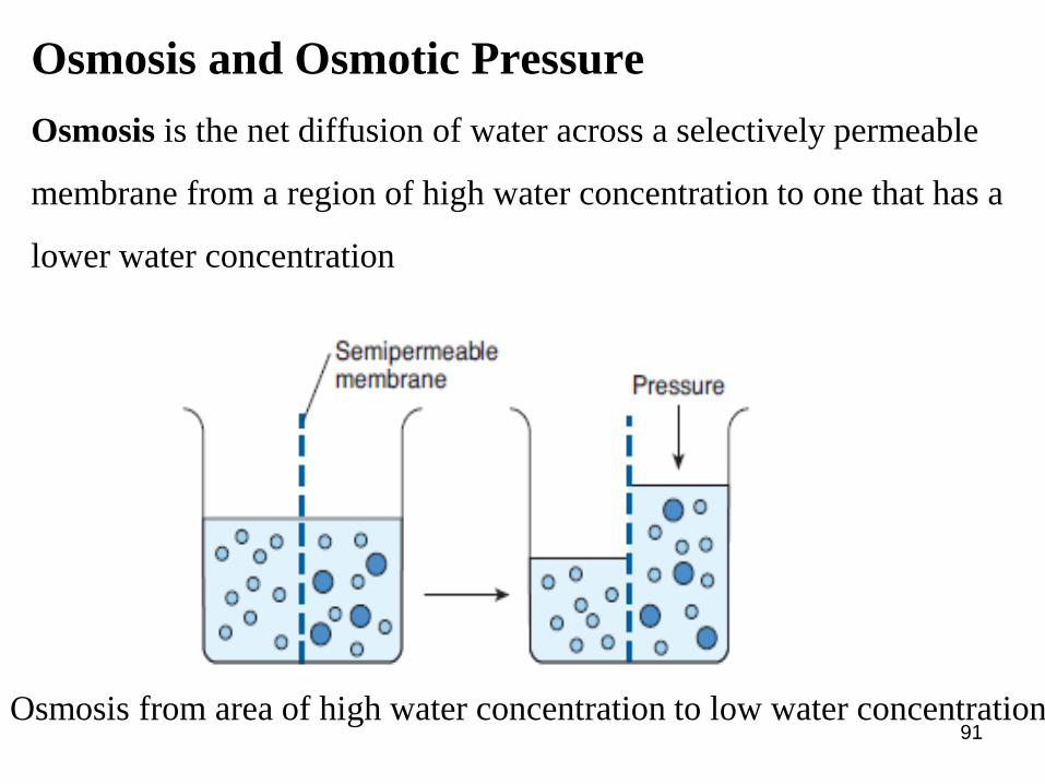
Osmosis and Osmotic Pressure
Osmosis is the net diffusion of water across a selectively permeable
membrane from a region of high water concentration to one that has a
lower water concentration
91Osmosis from area of high water concentration to low water concentration
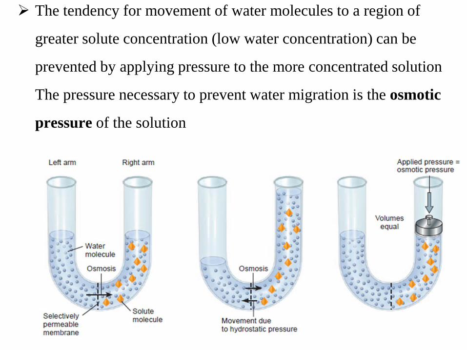
➢ The tendency for movement of water molecules to a region of
greater solute concentration (low water concentration) can be
prevented by applying pressure to the more concentrated solution
The pressure necessary to prevent water migration is the osmotic
pressure of the solution
92

Isotonic, Hypotonic, and Hypertonic Fluids
➢ Cell is placed in Isotonic solution (280-300mOsm/L) the cells will not
shrink or swell because the water concentration in the intracellular and
extracellular fluids is equal.eg. 0.9 % sodium chloride.
➢ Cell is placed into hypotonic solution (<282 mOsm/L) → diffusion of
water into the cell → swelling of the cell.
➢ Cell is placed in a solution (>282 mOsm/L) water will flow out of the
cell into the extracellular fluid, while the cell shrinks.
93

94