decay fungi of oaks and associated hardwoods for western
TRANSCRIPT

32
ArboristWESTERN
Winterl 2010
largely of lignin that becomes part of soil humus which resists further degradation. This brown-rot residue is an important component of the carbon sequestered in for-est soil. White-rot fungi frequently decay hardwoods, and brown-rot fungi usually colonize conifers, but many exceptions occur. The decayed wood within the tree can take different forms, including “stringy rot,” “spongy rot,” “pocket rot,” “cubical rot,” and “laminated rot.” Each of these decay types has different physical properties that affect the amount of strength remaining in the wood. In brown-rot decay, large amounts of strength loss occur early in the decay process due to the rapid depolymerization of cellulose (Cowling, 1961).
Decay fungi of oaks and associated hardwoods for western arboristsJessie A. Glaeser and Kevin T. Smith
E XAMINATION OF TREES FOR THE PRESENCE and extent of decay should be part of any hazard tree assessment. Identification of the fungi respon-
sible for the decay improves prediction of tree performance and the quality of management decisions, including tree pruning or removal. Scouting for Sudden Oak Death (SOD) in the West has drawn attention to hardwood tree species, particularly in the urban forest where native or introduced hardwoods may predominate. Consequently, the tree risk assessment specialist needs a working knowledge of the fungi associated with hardwood decay. We present here some of the common fungi responsible for decay of hardwoods, particularly of oak (Quercus spp.), tanoak (Lithocarpus densiflorus), and chinquapin (Castanopsis spp.) in Western North America.
Experts group wood decay fungi by various criteria. Academic mycologists use evolutionary kinship, often revealed by analysis of microscopic structures and genetic material. Direct observations can group fungi based on habitat, spatial position, and the appearance of the de-cayed wood (Tainter and Baker, 1996). An eco-nutritional approach groups some wood decay fungi as saprotrophs that attack wood in service or as felled logs, slash, or snags (Toupin et al., 2008) or as pathogens that decay wood in living trees. Pathogenic wood decay fungi can be further subdivided based on the type of wood degraded and the position of the fungi in the living tree. Heartrot fungi can decay heartwood in living trees despite the tree’s ability to produce protective chemicals and low oxygen conditions in the central cylinder (Highley and Kirk, 1979). When sapwood is exposed by mechanical injury, many saprot fungi can act as primary pathogens and directly kill living cells in advance of infection. These include canker rot and many root rot fungi (Shortle et al., 1996). Fruiting bodies of saprot fungi around the outer circumference of the stem can indicate structural weakness, particularly to arborists, tree climbers and land managers.
Wood decay fungi are also categorized by the appear-ance of decayed wood. White-rot fungi degrade the lignin, cellulose and hemicellulose of wood, leaving behind a white or off-white residue. Some white-rot fungi produce many small pockets of decay throughout the infected vol-ume of wood. Brown-rot fungi degrade the cellulose and hemicellulose in the wood cell wall but do not significantly degrade the lignin. Brown-rotted wood in advanced decay is often seen as more or less cubical fragments. Eventually, brown-rooted wood becomes a brown residue, composed
Definitions of mycological terms(Gilberston and Ryvarden, 1987)
Annulus – a ring found on the stipe of certain
mushrooms.
Applanate – thin, flattened horizontally. Usually
used to describe sessile fruiting bodies or the pile-
ate portion of effused-reflexed fruiting bodies.
Effused –reflexed – a fruiting body that is partially
resupinate and partially shelving into the pileus.
Mycelium – the vegetative stage of the fun-
gus, usually observable as a mass of individual
threads, termed “hyphae.”
Ochraceous – a yellowish buff color.
Pileus – the portion of a fruiting body with a sterile
upper surface and a fertile lower surface.
Resupinate – flat.
Rhizomorph – a macroscopic strand, often re-
sistant to drying, that spreads throughout the soil.
Black in Armillaria species.
Sessile – without a stipe.
Stipe – stalk-like or stem-like structure that sup-
ports the pileus.
Stipitate – with a stipe. The stipe can be central
or attached laterally (eccentric).
Ungulate – hoof-shaped.

Arborist
33
WESTERN
Winter 2010
Many decay fungi can be categorized using the above static criteria. However, the great advance in understand-ing the biology of wood decay in living trees involves the compartmentalization process (Shigo, 1984), a foundation concept in forest pathology (Manion, 2003). Prior to the de-scription of the compartmentalization process, heartrot was thought to form by the direct infection of dead heartwood exposed by injury. In the compartmentalization concept, heartrot generally begins with the infection of sapwood by a succession of fungi. The spread of those fungi throughout the tree is resisted through the boundaries and barriers of compartmentalization (Smith 2006). As the vascular cambium continues to produce new xylem, and healthy sapwood continues to be converted into heartwood, the infection appears to become more-or-less centered in the middle of the tree.
More important than questions of what constitutes a true “heartrot,” or whether trees that do not produce heart-wood can have heartrot, or whether decay of heartwood in a living tree is the product of a saprophyte or a pathogen, is to recognize that trees actively respond to injury and colonization by decay fungi. Infections in a young tree can produce a cascade of processes that result in long-standing decay and cavities in large, mature individuals. The amount of decayed wood and the size of the wood cavities are important criteria to consider during hazard tree assessments.
The types of decay, morphological features of the fruiting body, and a short discussion about the impact on hazard tree analysis are presented for some of the major decay fungi associated with oaks and other hardwoods in Western North America. Common synonyms are listed following an “=” sign. Many of the polypore fungi listed were considered in the genus Polyporus in older texts.
I. Pathogenic fungiAbortiporus biennis – fruiting bodies are annual, sessile
or sometimes with a lateral or central stalk, sometimes distorted with pores covering the entire surface. Upper surface, when it is present, is white to pale brown, azonate to slightly zonate, hairy or nearly smooth. Spore-bearing surface poroid, pores round to angular, 2 – 4 per mm, white bruising red. A white-rot of dead hardwoods and a white trunk-rot of living hardwoods, sometimes fruiting on the ground from subterranean roots (Binion et al., 2006).
Annulohypoxylon (= Hypoxylon) thouarsianum – “Cramp balls”. Fruiting body a dark brown to black, carbonaceous and hemispherically-shapped disk 1.0 – 5.0 cm in diameter, usually with small bumps or pimples that are discharge points of the spore-bearing sacs. Especially common on Quercus agrifolia (coast live oak), and Lithocarpus densifora (tanbark oak) (Wood, 2010) in the Sudden Oak Death zone and can be an opportunistic pathogen (Dr. D. Rizzo, personal communication). When the disc is cut in half, the
Abortiporus biennis Annulohypoxylon thouarsianum
Abortiporus biennis
34
ArboristWESTERN
Winterl 2010
internal tissue is fibrous to wooly, with radial to faintly concentric lines, grey and brown, sometimes appearing silky and lustrous. Fruiting bodies are scattered to clustered on bark, often developing after fall rains and persisting into the next season (Woods, 2010). May be confused with saprotrophic species of Daldinia, but the warted, bumpy surface distinguishes the two genera. Biscogniauxia mediter-ranea may appear quite similar macroscopically and can also be an opportunistic pathogen (Dr. T. Siewicki, personal communication).
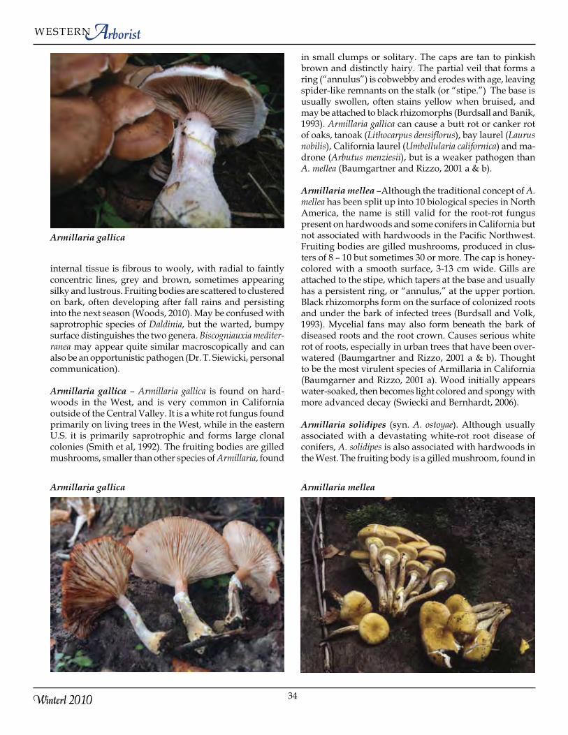
Armillaria gallica – Armillaria gallica is found on hard-woods in the West, and is very common in California outside of the Central Valley. It is a white rot fungus found primarily on living trees in the West, while in the eastern U.S. it is primarily saprotrophic and forms large clonal colonies (Smith et al, 1992). The fruiting bodies are gilled mushrooms, smaller than other species of Armillaria, found
in small clumps or solitary. The caps are tan to pinkish brown and distinctly hairy. The partial veil that forms a ring (“annulus”) is cobwebby and erodes with age, leaving spider-like remnants on the stalk (or “stipe.”) The base is usually swollen, often stains yellow when bruised, and may be attached to black rhizomorphs (Burdsall and Banik, 1993). Armillaria gallica can cause a butt rot or canker rot of oaks, tanoak (Lithocarpus densiflorus), bay laurel (Laurus nobilis), California laurel (Umbellularia californica) and ma-drone (Arbutus menziesii), but is a weaker pathogen than A. mellea (Baumgartner and Rizzo, 2001 a & b).
Armillaria mellea –Although the traditional concept of A. mellea has been split up into 10 biological species in North America, the name is still valid for the root-rot fungus present on hardwoods and some conifers in California but not associated with hardwoods in the Pacific Northwest. Fruiting bodies are gilled mushrooms, produced in clus-ters of 8 – 10 but sometimes 30 or more. The cap is honey-colored with a smooth surface, 3-13 cm wide. Gills are attached to the stipe, which tapers at the base and usually has a persistent ring, or “annulus,” at the upper portion. Black rhizomorphs form on the surface of colonized roots and under the bark of infected trees (Burdsall and Volk, 1993). Mycelial fans may also form beneath the bark of diseased roots and the root crown. Causes serious white rot of roots, especially in urban trees that have been over-watered (Baumgartner and Rizzo, 2001 a & b). Thought to be the most virulent species of Armillaria in California (Baumgarner and Rizzo, 2001 a). Wood initially appears water-soaked, then becomes light colored and spongy with more advanced decay (Swiecki and Bernhardt, 2006).
Armillaria solidipes (syn. A. ostoyae). Although usually associated with a devastating white-rot root disease of conifers, A. solidipes is also associated with hardwoods in the West. The fruiting body is a gilled mushroom, found in
Armillaria gallica
Armillaria gallica Armillaria mellea

Arborist
35
WESTERN
Winter 2010
large clusters, with a brownish ring (“annulus”). The base of the stipe is frequently pointed. The cap is brown, cov-ered with dark scales, and can be very large – up to 1 foot in diameter (Burdsall and Volk, 1993, Burdsall and Volk, 2008, Volk 2010). It has not been observed in California (Baumgartner and Rizzo, 2001 a & b.)
Fomes fomentarius – “Tinder conk”. Perennial, hard, woody, horse hoof-shaped conk with prominent zones and furrows, broadly attached to substrate. Upper surface usually pale to dark grey or brown. Spore-bearing surface poroid, concave, tan to cream-colored when young becom-ing ochraceous to pale brown with age. Pores round, 2 – 4 per mm. Solitary or in groups on living trees, stumps and logs. Causes a white spongy mottled heartrot of living trees; continues to fruit on dead stumps and logs (Binion et al., 2008; Gilbertson and Ryvarden, 1986). Decay first appears as a light brown discoloration with the wood
remaining quite firm. Wood with advanced decay is yel-low-white, soft and spongy, and frequently contains brown to black zone lines. Small radial cracks filled with yellow fungal mycelium develop, giving the decay a mottled ap-pearance (Allen et al., 1996). Most common on birch but has been reported on oak.
Ganoderma applanatum – “Artist’s Conk”. Fruiting body perennial, 5 – 52 cm wide or even larger, convex, hoof-shaped to fan-shaped, stalkless. Upper surface hard, concentrically zonate and furrowed, gray to brown to gray-ish brown. Spore-bearing surface poroid, white at first, becoming off white to dingy yellow with age, staining brown upon bruising (which allows it to be used by artists as a canvas). Pores very small, 4 – 6 per mm. Solitary or in overlapping clusters on stumps, logs, or wounds of living trees. Common. (Binion et al., 2008). Forms a mottled white-rot of roots, root crown and trunks. In the East, the pres-ence of conks can be cause for immediate removal of the affected tree as conks are often associated with advanced decay and potential failure (Luley, 2005). In the West, conks are not as common on oak but more frequently observed on big leaf maple (Acer macrophyllum) and bay laurel (Lau-rus nobilis) where they are not associated with advanced decay (Shaw, 2010). Oaks with fruiting bodies are often extensively decayed and may have an elevated failure rate. The fungus often enters tree through wounds in exposed roots and base of tree. Decay commonly extends 1 – 2 m above and below the fruiting body. Decline and mortality more pronounced during periods of environmental stress (Swiecki and Bernhardt, 2006). Ganoderma brownii, which is more hoof shaped and which has an orange pore surface, is a closely related species that occupies a similar niche in California but is not common.
Ganoderma lucidum – “Reishi” or “Varnish Conk”. Found in California but not the Rocky Mountains or Pacific North-
Armillaria solidipes (syn. A. ostoyae)
Fomes fomentarius
Ganoderma applanatum

36
ArboristWESTERN
Winterl 2010
west (Swiecki and Bernhardt, 2006). Fruiting body annual, 2.5 – 3.5 cm wide, semi-circular to fan-shaped or kidney-shaped, surface with concentric zones and furrows, shiny, dark red, reddish-brown to orange-brown becoming ochre or yellow toward the margin. Stalk lateral, 2.5 – 10 cm long and 0.5 – 4 cm thick. Spore-bearing surface poroid, off-white to yellow initially but becoming brown with age or upon bruising. Pores small, 4 – 7 per mm. (Binion et al., 2008). Causes white root-rot and butt-rot of living native hard-woods and exotic ornamental hardwood trees and shrubs. Fruiting bodies develop at or near the ground line (Gilb-ertson and Ryvarden, 1986). In California, fruiting bodies generally do not form until there is extensive decay with an elevated risk of failure (Swiecki and Bernhardt, 2006). This is in contrast to the eastern and midwestern U.S. where the presence of fruiting bodies alone is usually not reason for tree removal (Luley, 2005). The closely related varnish conks, Ganoderma tsugae and G. oregonense, are found on conifers.
Grifola frondosa – “Maitake”, “Hen of the Woods”, or “Sheep’s Head”. Fruiting body composed of compound clusters up to 30 cm across made up of individual fan-shaped to club-shaped lobes developing from a lateral, thick, branched stalk. Upper surface ochre-brown to gray-ish-brown or blackish-brown. Spore-bearing surface poroid, white to off-white, pores round to angular, 1 – 3 per mm. Occurs in small to massive clusters at the base of living oak trees or stumps. Usually considered a weak pathogen and may occur at the base of the same tree for many years (Binion et al., 2008). It causes a white-rot and butt-rot, predominantly of oak but also on other hard-woods and conifers. Rare in the Pacific Northwest (Gilbert-son and Ryvarden, 1986) but may escape from cultivation; not reported from California (Dr. T. Swiecki, personnal communication). Often found on oaks that have no other symptoms of decay. Not usually a cause for immediate removal (Luley, 2005).
Ganoderma lucidum Grifola frondosa
Grifola frondosa Hericium erinaceus

Arborist
37
WESTERN
Winter 2010
Hericium erinaceus – “Hedgehog Fungus”, “Bear’s Head Tooth”, “Lion’s Mane”, or ‘Pom Pom Mushroom”. Fruit-ing body annual, solitary, 10 – 20 cm wide. Spore-bearing surface consists of many closely-packed, slender, icicle-like teeth 2 – 5 cm long. Teeth are white when young becoming yellow, brownish, or sometimes reddish with age. Young fruiting bodies are edible although may cause allergic reactions in some individuals. Causes a white pocket rot of living trees, and is associated with wounds. Decayed tissue initially spongy and eventually degrades to leave a large cavity (Swiecki and Bernhardt, 2006).
Inonotus andersonii (= Poria andersonii)– A resupinate (flat) polypore that forms sheet-like fruiting bodies on dead wood underneath the bark and sometimes between outer layers of the sapwood. Fruiting bodies initially cinnamon brown but become black with age, usually 0.3 – 1.0 m long. Pores cir-cular to angular, 1 – 6 per mm. Spore deposits on inner bark
initially bright sulphur yellow, becoming brown with time. Very common pathogen on living oaks, causing white rot of heartwood and strips of decay in the sapwood. Advanced decay appears bleached, is very light in weight, and crumbles easily. Also forms cankers and kills the cambium resulting in dieback, failure and mortality (Swiecki and Bernhardt, 2006). There are dozens of other resupinate polypores that are not easily distinguished without a microscope. Some of these species are pathogenic, but most are saprotrophic.
Inonotus hispidus – “Shaggy Polypore”. Fruiting body broadly attached, usually solitary, up to 10 X 15 cm wide by 8 cm deep. Top, including the edge, is reddish orange, becoming reddish brown to nearly black with age, no zonations, and many coarse hairs when young (“hispid”). Pore surface yellow-brown becoming dark brown with age. Pores angular, 1 – 3 per mm, becoming eroded and uneven. Causes a white heartrot of living oaks (Binion et al., 2008).
Inonotus andersonii Inonotus andersonii
Inonotus andersonii Inonotus hispidus

38
ArboristWESTERN
Winterl 2010
Capable of killing sapwood in living trees and is commonly associated with trunk cankers on oaks. In Arizona, it is a major decay fungus of Arizona black walnut (Juglans major) (Gilbertson and Ryvarden, 1986).
Inonotus dryadeus (= Pseudoinonotus dryadeus) – “Weeping Conk”. Fruiting bodies annual but persistent, developing at the trunk base or on roots below the soil surface, variable in size but can be very large (up to 75 cm wide), initially soft but becomes dried and cracked with age. Top surface of fresh fruiting bodies are yellowish to brown, blackening with age, and may have many droplets of amber-colored exudates. Lower surface buff with fine circular to angular pores, 4 – 6 per mm. Causes a slowly developing white-rot root disease and butt-rot with most of decay concentrated in larger roots. Affected trees may have significant amounts of root decay and an elevated risk of windthrow (Luley, 2005; Swiecki and Bernhardt, 2006).
Inonotus dryophilus – Very similar to Inonotus hispidus in appearance except lacking the hispid (hairy) upper surface (Binion et al., 2008). Pore surface initially buff becoming dark reddish brown, rough, pores angular, 1 – 3 per mm with thin border tissue that breaks up so pores become eroded and irregular with age. Causes a white-rot of heartwood in the trunk of living oaks and also decays strips or sections of sapwood, forming elongate cankers and killing the cambium. Brown mycelium accumulates in the decayed wood in advanced decay (Gilbertson and Ryvarden, 1986). Fruiting occurs well above the ground line (Luley, 2005). One of the most serious pathogens of living oaks in California associated with decline, failure, and mortality (Swiecki and Bernhardt, 2006).
Laetiporus gilbertsonii – Fruiting bodies fleshy, shelving (overlapping), up to 20 cm wide, with a lateral narrow or wide stipe or sessile. Upper surface pale salmon-orange or
Inonotus dryadeus Inonotus dryophilus
Inonotus dryophilus Inonotus dryophilus

Arborist
39
WESTERN
Winter 2010
pale pinkish-orange to tan or light brown in age, sometimes nearly white. Pore surface lemon-yellow to pale lemon-yel-low. Pores initially circular becoming more angular with age, 2 – 4 per mm, present along stipe to attachment point. Causes a brown-rot of Quercus and Eucalyptus species in living trees or dead trunks and logs (Burdsall and Banik, 2001). The decay is a cubical brown heartrot that may lead to failure in the main stem or butt. Decay may progress into major roots (Luley, 2005). The presence of Laetiporus fruiting bodies is often an indicator of extensive decay and should be taken seriously. Laetiporus gilbertsonii var. pallidus is similar but has a pale orange to pale brown pileus surface and a white pore surface. Laetiporus conifericola is similar but grows only on conifers (Burdsall and Banik, 2001).
Omphalotus olivascens – “Jack O’Lantern Fungus”. This fungus is associated with oaks and eucalyptus and is often found at the base of hardwood stumps and on buried roots,
fruiting in fall through winter. The upper surface is dull brown to orange, sometimes with olive tones. The cap is convex, 5 – 15 cm in diameter; the cap margin is initially rolled over but expands and becomes wavy with age. Gills may be slightly lighter in color than the cap, continue onto the upper stalk, and can bioluminesce in fresh specimens although this is often difficult to observe. The stalk is 5-15 cm long, 1-4 cm thick, central to off-central, tapers, smooth, yellowish-olive, with brown stains at the base (Wood, 2010). This fungus can be an opportunistic pathogen (Dr. D. Rizzo, personal communication) and causes a white rot. It is poisonous and has been confused with orange chantrelles.
Phellinus everhartii – Fruiting body is perennial, stalkless, and hoof-shaped, generally less than 6 [high] x 13 [wide] x 8 [deep] cm. Upper surface of conk is dark brown to black, velvety when young but becoming smooth and eroded with
Laetiporus gilbertsonii Omphalotus olivascens
Laetiporus gilbertsonii var. pallidus Phellinus everhartii

40
ArboristWESTERN
Winterl 2010
age. The spore bearing surface is poroid, with a velvety ap-pearance, dark chocolate brown, pores circular to angular, 5 -6 per mm. Causes a white-heart rot of living oak. (Binion et al, 2006; Gilbertson and Ryvarden, 1987). There are sev-eral other similar species that are difficult to distinguish without a microscope. Common in the Southwest.
Phellinus gilvus– “Mustard Yellow Polypore”. Fruiting body annual to perennial, sessile or slightly effused-re-flexed, solitary or shelving (overlapping) in large numbers. Very variable in appearance. Upper surface is dark yellow-brown to rusty brown, velvety when young, becoming smooth with age, tapering to a sharp margin. Spore-bearing surface poroid, reddish brown to dark purple brown, pores circular to angular, 1 – 5 per/mm (Binion et al, 2006). Can cause a white-rot of heartwood of living oaks and a uniform white rot of dead wood (Gilbertson and Ryvarden, 1987). Most common conk on oaks (Quercus) and tanbark oak
(Lithocarpus densiforus) in California (Wood, 2010).
Phellinus robustus – Fruiting body very hard, perennial, sessile or effused-reflexed, may ungulate or be rather flat (applanate), up to 12 [high] x 20[wide] x 11 [deep] cm. Upper surface brown to blackish with a yellowish or grey-brown pore surface. Pores circular, 7 – 9 per mm. Tube lay-ers form in rows, visible if conk is cross-sectioned. Forms a white rot of living hardwoods (Gilberson and Ryvarden, 1987). Most common species of Phellinus on oak in northern California and often associated with tree failure. In north-ern CA, usually reported high on the bole and associated with a heartrot (Dr. T. Swiecki, personal communication). In southern California and the Southwest, found at the base of affected trees and thought to be a root and butt rot (Gilbertson and Ryvarden, 1987).
II. SaprotrophsArmillaria nabsnona – Armillaria nabsnona is found on many hardwoods in western North America, including oak, but most commonly on Alnus species. Macroscopic characters that distinguish A. nabsnona from other North American species of Armillaria include a more orange col-oration when fresh and a narrower stipe in comparison to the size of the cap. The stipe is darker than other Armillaria species, especially when dried. There are no scales, but small black hairs may be present on the surface of the pi-leus, a similar situation to that found in A. mellea. It is often associated with dead wood in riparian zones and causes a white rot (Volk et al., 1996). In California, A. nabsnona is restricted to the northwestern redwood forest area and is primarily associated with living and dead alder (Alnus rubra), tanoak (Lithocarpus densiflorus) and California laurel (Umbellularia californica) (Baumgartner and Rizzo, 2001b).
Bjerkandera adusta – “Smoky Polypore”. Fruiting body effused-reflexed to sessile, shelf-like, frequently in large
Phellinus gilvus
Phellinus robustus Armillaria nabsnona

Arborist
41
WESTERN
Winter 2010
numbers, frequently coalescing to form larger fruiting bodies or uniting to form large sheets on the underside of logs. Upper surface is pale yellow-white to pale creamy-buff, becoming grayish-white with age. Smooth to finely fuzzy. Pore surface pale gray to dark gray, sometimes with a brown tint. Pores 5 – 7 per mm, circular, becoming an-gular with age, gray. White-rot of fallen wood or standing snags (Binion et al, 2006). Most common on aspen,
Cerrena unicolor – “Mossy Maze Polypore”. Fruiting body annual, sessile or effused-reflexed, semicircular, of-ten shelving and coalescing laterally. Upper surface gray to brownish gray, very hairy-fuzzy, usually green from growth of associated algae, zonate to somewhat zonate. Spore-bearing surface with maze-like pores, pale ivory to gray, 3 – 4 pores per mm. (Binion et al., 2006) It is a symbiont of the wood wasp Tremex columba (Gilbertson and Ryvarden, 1986). An aggressive white saprot than can decay a large portion of the trunk and can also kill the cambium as a canker rot. Infected stems are subject to breakage. A common invader after damage from ice storms and other wounds (Luley, 2005).
Daedalea quercina – “Thick Maze Polypore”. Fruiting bod-ies annual or perennial, broadly attached, bracket-like, 5 – 15 cm wide, upper surface convex to flat, uneven, white to pale brown or grayish brown. Pore surface irregular, pores 1 mm or wider in diameter, walls between pores thick, pores usually maze-like to nearly gill-like, white to light brown, at times with pinkish tones. Solitary or in small groups. Rare west of Mississippi River (Binion et al, 2006). Slowly pro-gressing brown-rot in butt and trunk that progresses to form hollow cavities. Usually not a reason for immediate removal but affected trees should be monitored (Luley, 2005).
Daedaelopsis confragosa – “Thin-walled Maze Flat Polypo-re”. Fruiting bodies annual but persistent, bracket-like, 2.5 Bjerkandera adusta
Cerrena unicolor Daedalea quercina

42
ArboristWESTERN
Winterl 2010
– 15 cm wide, upper surface convex to flat with a thin, sharp margin, usually zonate. May have matted hair on surface or be smooth. Gray to brown or reddish brown. Pore surface poroid to maze-like or gill-like, white to brownish, bruising pinkish. Extremely variable in appearance. Causes a white-rot on dead wood. On oak and many other hardwoods. (Binion et al., 2006) Infrequent in the West but more com-mon in the Northwest (Gilbertson and Ryvarden, 1986).
Irpex lacteus – “Milk-white Toothed Polypore”. Fruiting bodies annual, broadly effused to effused-reflexed and sessile. Often fruiting bodies fuse laterally to make large sheets that totally cover broken or dead branches. Upper surface white to pale cream-color, hairy, sometimes zonate. Pore surface appears spiny because of erosion and split-ting of pores, white to pale cream-colored. Pores initially angular, 2-3 per mm. White-rot on fallen branches and dead branches still attached to the tree. (Binion et al, 2008).
Daedaelopsis confragosa
Irpex lacteus Ischnoderma resinosum
Irpex lacteus

Arborist
43
WESTERN
Winter 2010
Ischnoderma resinosum – “Resinous Polypore”. Fruiting body annual, fan-shaped to semicircular, up to 25 cm wide. Margin rounded. Upper surface velvety or hairy becoming smooth with age, often radially wrinkled, zonate with dark brown to rusty brown wrinkles. Pores creamy white to ochraceous, bruising brown, circular to angular, 4 – 6 per mm. Young fruiting bodies exude amber-colored droplets. Fruiting bodies solitary or in groups on logs, stumps, and standing trees (Binion, et al, 2008). The decay is a white-rot that is yellowish and stringy to spongy with a strong odor of anise (Gilbertson and Ryvarden, 1986).
Lenzites betulina – “Multicolor Gill Polypore” or “Gilled Polypore”. Fruiting body annual but persists over winter, kidney-, fan-shaped or semicircular, 3 – 10 cm wide. Up-per surface velvety to hairy with distinct multi-colored zones or grooves in shades of pink, tan, gray, yellow,
orange or brown; older specimens may be green from an algal associate. Pores are gill-like, radiating from point of attachment. Often found in overlapping groups or shelving on dead branches, logs and stumps (Binion etal., 2008). Absent or rare in the Rocky Mountains but present in the Pacific Northwest. Often on Betula but also found on many other hardwoods and conifers (Gilbertson and Ryvarden, 1986).
Pleurotus ostreatus – “Oyster Mushroom”. A large, fleshy, white fruiting body that is a select edible. Tends to be gray to white and relatively thin-fleshed on oaks to thick fleshed, grey-brown shelves on cottonwood and willow. The cap can be 5 – 25 cm in diameter, convex to nearly flat at matu-rity with a margin that is lobed to wavy, especially when young. Surface of the cap is smooth. The gills are white although may become yellowish with age and continue onto the upper portion of the stalk, when present. The stalk
Pleurotus ostreatus
Lenzites betulina

44
ArboristWESTERN
Winterl 2010
is often absent but when present is short and thick, 0.5-3.0 cm long, 0.5-2.0 cm thick, eccentric or lateral with dense white hairs at the base. The fruiting body may have the odor of anise when fresh. It is usually found in a cluster of overlapping shelves on logs and boles of hardwoods. Fruiting begins in early fall and may continue through winter in the mild climates of California (Wood, 2010). The fungus is a white-rotter and can be an opportunistic pathogen. (Dr. D. Rizzo, personal communication).
Polyporus species: – The genus Polyporus is a large group of poroid fungi that can be recognized by their central to lateral stipe. Most species have a light to deep brown upper surface and are tough when fresh and woody when dried. Some of the common species are: P. arcularius, a smallish polypore (up to 4 cm wide) with a central stipe and radially arranged hexagonal pores; P. badius (= Royoporus badius), a fairly large but thin polypore (up to 15 cm broad) with a
central or lateral stipe that is black and minutely hairy at its base; and P. brumalis, which is of medium size (up to 8 cm wide) with a central or lateral stipe and angular pores. All cause a white-rot of woody debris and dead trees (Binion, 2008; Gilbertson and Ryvarden, 1987).
Schizophyllum commune – “Split-gill Fungus”. Fruit-ing body a leathery, fan-shaped bracket, 1-3.5 cm broad, frequently lobed or fused at the base with other brackets; upper surface densely hairy, light greyish-brown when moist, ashy grey to white when dry; lower surface light grey consisting of well spaced, longitudinally split gills; stipe usually absent; flesh thin, light grey to brown, tough. Can be found year-round, usually in clusters, on dead boles and branches. (Wood, 2010). Causes a white rot decay and can be an opportunistic pathogen on stressed trees (Dr. D. Rizzo, personal communication).
Polyporus sp.
Stereum hirsutum
Schizophyllum commune

Arborist
45
WESTERN
Winter 2010
Stereum species – Species of Stereum are easily recognized by their relatively thin fruiting bodies that are often fan-shaped to paddle-shaped and their smooth spore-bearing undersurface that lacks pores, gills or other supporting structures. Many resemble other decay fungi and are only recognized as belonging to the genus Stereum when the smooth lower surface is observed. Stereum hirsutum forms shelves that are sessile to effused reflexed, has an upper surface that is initially wooly, becoming smooth with age, with concentric zones of orange brown to grayish tan colors, and a smooth brownish spore-bearing surface that bruises yellow when fresh, usually found on logs. Stereum ochraceoflavum forms typical shell-shaped fruiting bodies but also saucer-shaped fruiting bodies that may partially or completely surround the sticks on which they grow. The upper surface is buff to tan-brown, indistinctly zoned, and uniformly hairy (Wood, 2010). All Stereum species cause a white saprot and are associated with dead trees and woody
debris. Several other Stereum species grow on conifers, especially S. sanguinolentum.
Trametes species – The genus Trametes contains many com-mon saprot fungi with broad host and geographical ranges. The group is characterized primarily by microscopic char-acteristics – the poroid fruiting bodies are formed from three different types of fungal hyphae which give them a characteristically hard, leathery texture. Trametes versicolor, the “Turkey Tail Fungus” has a zonate, multicolored upper surface varying from hairy to smooth or velvety in narrow concentric zones. It often forms large clusters of shelves. Trametes hirsuta is also zonate but with much more subdued colors, predominantly shades of white, gray and tan with concentric zones of thick and thin woolliness, becoming smooth with age, cream-colored to grayish yellow and only faintly zonate to azonate. All of these fungi are strong white-rotters. (Binion et al., 2008) Trametes versicolor can
Trametes versicolor
Stereum hirsutum
Trametes hirsutum
Trametes hirsuta (lower surface)

46
ArboristWESTERN
Winterl 2010
attack and colonize cambium adjacent to dead wood and form cankers. The presence of Trametes and other saprot fungi is an indication that the branch or section of trunk is dead and decayed. Sanitation pruning to remove infected branches is recommended since some of them can infect and colonize healthy tissue (Luley, 2005).
Trichaptum biforme – “Violet-toothed Polypore”. Fruiting bodies annual, sessile or effused-reflexed, solitary or shelv-ing, often coalescing to form large sheets. Upper surface is gray to tan, hairy to smooth, with concentric zones of thick and thin woolliness. Pore surface is purple to pink especially at the margins but becoming brown with age. Pores initially angular, 3 – 5 per mm but eventually eroding and splitting to form teeth. Common on fallen oak debris, logs and stumps (Binion et al., 2008). Forms a white pocket rot of sapwood on dead hardwoods. The wood becomes lacy and fragile with small empty pockets (Gilbertson and Ryvarden, 1987). Trichaptum abietinus is similar, but grows on conifers.
Jessie A. Glaeser, USFS, Northern Research Station, Madison, WI 53726 ([email protected]) Kevin T. Smith, USFS, Northern Research Station, Durham, NH 03824 ([email protected]) Photos used with permission from Drs. Harold H. Burd-
sall, Jr., Michael Emberger http://www.messiah.edu/Oakes/fungi_on_wood/, Jessie A. Glaeser, Larry Grand, Michael Kuo www.mushroomexpert.com, Daniel L. Lind-
ner, Bruce Lundy, Thomas J.Volk http://TomVolkFungi.net, Ted Swiecki http://phytosphere.com/and Michael Wood www.mykoweb.com “Fungi of California”.
Trichaptum biforme
United We Plant! Thanks to our 30+ Volunteer Certified Arborists,
we properly planted over 4,000 trees on Make a Difference Day, October 23, 2010.
You’re the Best!
Volunteer ArboristsAli Summers Amber Krebbers Bob Keeley Brian Koch Cortney Poole Dan Howell Dave Ephron Gonzalo Magana Greg Saenz Jeff Wooten Jeremy Rappoport Joe Garcia Joseph Eves Joshua Chavez Julie Broughton
Julie Luna Ken Menzer Kurt Stegen Mark Frizzell Mary Jane Marlow Mike Campbell Monica Seyfried
Patrick Denney Paul Rider Peter Lance Robert Ritzman Robert Wagoner Roger Boddaert Roger Snell
Sam Oludunfe Scott Jones Steve Hunt Susan Stiltz Tim O’Shay
Sponsoring OrganizationsCalifornia ReLeaf American Recovery & Reinvestment Act USDA Forest Service Valley Crest
Photo: Sam Oludunfe