deep vein thrombosis care pathway – the siapav...
TRANSCRIPT

DVT CARE PATHWAY
1.0 Revision; 01/01/2017 Page1of25
SOCIETA’ ITALIANA DI ANGIOLOGIA E PATOLOGIA VASCOLARE
DEEP VEIN THROMBOSIS CARE PATHWAY – THE SIAPAV MODEL
tHE
Writing and Editorial Committee:
SIAPAV Executive Board (Annalisa Di Folca, delegate Team Leader)
SIAPAV Study and Research Centre (Raffaele Pesavento, delegate Team Leader)
This document was prepared in collaboration with the following National Scientific
Societies: ALT, FIMMG, SICVE, SIDV-GIUV, SIMG, SISET

DVT CARE PATHWAY
1.0 Revision; 01/01/2017 Page2of25
Rationale and aim
Managed care and clinical governance are among the most adopted strategies in complex health systems to put under control or influence health care quality, accessibility, costs and prices, use and outcomes. The development of clinical pathways (CPs) is a way to achieve the goals of offering good, appropriate medical care at a lower cost. The CPs are tools through which evidence-based guidelines (GLs) on a specific disease or clinical topic can be implemented in a clinical process, dealing with the local resources, availability and costs. The most important characteristic of CPs is that of developing a health care process shared among all the actors involved, achieving high standards of appropriate care and efficient integration. The developing of CPs is a step-by-step process through the details of how to plan and implement a pilot test, and how to use the results to refine the pathway. It spells out the concepts involved and issues to consider, then details in six steps how to create a clinical pathway: - Form a multidisciplinary team and choose a team leader - Select the patient population and a top priority clinical problem - Gather needed information on current practices - Research best practices - Identify differences between current and best practices - Draft the pathway
After peer reviewing and refinement, the pathway is ready for pilot implementation. Why a vascular scientific society should deal with CPs, as the proper development must be tailored to the needs of a single reality or operating territory? The caring role of the modern Angiologists will be significantly affected by the overall process of reorganization of health systems and it is important that they will take a leading role in order to exploit their own specialist role and avoid marginalization due to the inherently multi-specialized nature of certain diseases. Besides an educational role, we believe that a scientific society can contribute to improve the competence of its members in this field, and provide care pathway models useful for a well-balanced local development, among business management, good clinical practice and the protection of patients' best interest.

DVT CARE PATHWAY
1.0 Revision; 01/01/2017 Page3of25
Developed by: [insert the local multidisciplinary working group]
Reference person: [insert]
Approved by: [insert hospital officer]
Date: ……. Revision:………….

DVT CARE PATHWAY
1.0 Revision; 01/01/2017 Page4of25
INDEX
1. Introduction Page 5 2. Targets Page 6 3. References to Regulations Page 6 4. Glossary and Abbreviations Page 7 5. Literature search and evaluation strategy Page 8 6. Processes and duty evaluation Page 9 6.1 Triage Page 9 6.2 Initial assessment Page 10 6.3 Diagnostic procedures Page 11 6.3.1 Pre-test clinical probability: Wells Score Page 12 6.3.2 D-Dimer measuring Page 12 6.3.3 Angiologic Consultancy /Clinical and Instrumental examination Page 14 6.3.4 Ultrasound exams and diagnostic strategies Page 14 6.3.5 Complementary diagnostic imaging Page 15 6.3.6 Management of suspected DVT over the week-end/nocturnal hours Page 16 6.4 Management of the patients with confirmed DVT in the ED Page 17 6.4.1 Reperfusion therapy Page 17 6.4.2 Caval filter insertion Page 20 6.4.3 Home treatment Page 20 6.4.4 Hospitalization Page 22 6.5 Management of the patients with DVT excluded Page 22 6.6 Follow-up Page 22 6.6.1 Management of the patients under home treatment Page 22 6.6.2 Management of the patients discharged after hospitalization Page 23 7. Implementation deadlines Page 24 8. Outcome/Process Indicators Page 24 9. References Page 24 10. Attached documents index Page 25

DVT CARE PATHWAY
1.0 Revision; 01/01/2017
Page5of25
1. INTRODUCTION Venous thromboembolism (VTE) is a clinical condition characterized by a pathologic activation of the coagulation cascade in the venous vessels leading to the formation of intraluminal thrombosis, which often can fragment and dislodge, thus embolizing in the pulmonary artery system. Usually, venous thromboemboli originate from the lower limb veins but also from other venous districts, in the right heart chambers or in the pulmonary arteries. Deep vein thrombosis (DVT) is defined as the partial or complete occlusion of deep veins in the lower limbs and/or in the abdomen or pelvis. DVT is a vascular disease potentially severe and lethal because of the conferred risk of early or late sudden death. If immediate treatment is not given, a fatal event could occur in up to 50% of cases in the first three months. Moreover, DVT can lead to the postthrombotic syndrome (PTS), a late VTE complication that can be highly invalidating and with relevant economic and social costs. The DVT incidence is about 1/1000 subjects in the general population, which varies with age, with minimal values during infancy (0.005%) and exponentially increases up to 5-25/1000 subjects aged over 70 years. VTE is still the third leading cardiovascular disease, after myocardial infarction and stroke, is the third cause of death in the general population and the first in hospitalized patients. Moreover, it is constantly increasing for several reasons, as the life span lengthens, traumas, cancer and surgical interventions increase. The diagnosis of DVT is still a challenging issue for physicians and the health system, as the clinical onset is frequently subtle and vague leading to a difficult early diagnosis, which is critical to prevent the embolic damage. Up to 70% of DVT is asymptomatic, up to 30-50% of DVT patients has a concomitant pulmonary embolism (PE), and up to 20% of distal DVTs extend to the proximal veins. The diagnosis of DVT is currently based on validated and well-standardized algorithms including clinical probability models, lab test as the D-dimer (DD) and non-invasive imaging tests. Despite this, the peculiar nature of DVT still leads to a number of diagnostic delays and false negative or false positive diagnoses, with obvious clinical implications and risk of under-treatment or overtreatment. An appropriate assessment of the individual risk profile and disease presentation (e.g. unprovoked vs provoked, cancer-associated etc.) should be already performed during the acute phase of a DVT as it influences the therapeutic indication, the therapeutic management and follow up. Finally, an appropriate therapeutic management of DVT should bring down the risk of early death, of early and late recurrences and of SPT, without excessive costs.
DVT CARE PATHWAY
1.0 Revision; 01/01/2017
Page6of25
2. TARGETS This CP is aimed at:
• Improving the quality and consistency of the DVT care, based on the guidelines
adopted by the working group (WG), leading to appropriate clinical decisions. • Improving the planning of diagnostic and therapeutic pathways, setting Hospital
standards. • Improving information and communication aspects with the patient. • Improving the monitoring of the actual quality of care administered, through the
definition of the appropriate indicators of process and outcome and the creation of an efficient system for collecting and analysing data.
This document describes the journey of patients admitted to the Emergency Department (ED) with clinical suspicion of DVT, it defines the subsequent procedures and evaluations aimed at confirming or excluding the diagnosis, the outset of the therapy and the criteria for home or hospital care. A set of operative instructions have been included in this pathway, regarding how to use the DVT diagnostic algorithm and how to initially treat the patients with confirmed diagnosis. The following patients have been excluded from this document: - those with thoracic pain, dyspnoea of recent onset or any other clinical finding indicating
a suspected PE ( for whom a specific CP is reserved) - those with asymptomatic DVT - those with superficial vein thrombosis ( for whom a specific care pathway is reserved) N.B.: as this is a care pathway model, it is aimed at offering a set of suggestions to create a local care pathway: all parts of the document could be thus modified according to the criteria of developing a “real-life” care pathway, i.e. the “four W’s rule” ( Who, Where, When, hoW) and the “F.A.I.A.U” setting ( Finding, Appraising, Integrating, Adapting, Updating), bearing in mind the local obstacles to the implementation of the pathway and the means to overcome them.
3. REFERENCES TO REGULATIONS
[Here the working group should insert all the appropriate references pertinent to care pathways established by local or national government health care institutions]

DVT CARE PATHWAY
1.0 Revision; 01/01/2017
Page7of25
4. GLOSSARY AND ABBREVIATIONS
Acronym Definition APSAC Anisoylated Plasminogen Streptokinase Activator Complex
aPTT Activated Partial Thromboplastin Time
CFM Doppler colour flow imaging
CP Clinical Pathway
CT Computed tomography ( scan)
CUS/CCUS Compressive Ultrasound/Complete Compressive Ultrasound
DD D-dimer
DOAC Direct Oral Anticoagulant
DVT Deep Vein Thrombosis
ED Emergency Department
EKG Electrocardiogram
ESC European Society of Cardiology
GL Guideline
GP General Practioner
ICD9CM The International Classification of Diseases, Ninth Revision, Clinical Modification
IVC Inferior Vena Cava
IVCF Inferior Vena Cava Filter
LMWH Low Molecular Weight Heparin
MR Magnetic Resonance
UFH Unfractioned heparin

DVT CARE PATHWAY
1.0 Revision; 01/01/2017
Page8of25
PE Pulmonary Embolism
NSAID Nonsteroidal Anti-Inflammatory Drugs
OAT Oral Anticoagulant Therapy
PLTs Platelets
PT/INR Prothrombin Time/ International Normalized Ratio
PTP Pre-Test Probability
PTS Post-Thrombotic Syndrome
rtPA Recombinant tissue Plasminogen Activator
SK Streptokinase
TT Thrombin Time
U Units
UK Urokinase
US Ultrasound
VKA Vitamin K Antagonist
VTE Venous ThromboEmbolism
WG Working Group
5. LITERATURE SEARCH AND EVALUATION STRATEGY
As recommended by the most recent and accredited national and European guidelines on the creation of clinical pathways, the WG has firstly carried out both the revision of the current evidence and the search of the National and International Guidelines through the following keywords: “ Venous thrombosis”, “Vein thrombosis”, “Venous thromboembolism”, and limiting the search through the following keywords :”English, Italian, published in the last 3 years, Humans, Practice Guideline” (1-3). The following Databases have been examined: NCG (National Guideline Clearinghouse), CMA (Canadian Medical Association), NeLH GF (National Electronic Library for Health

DVT CARE PATHWAY
1.0 Revision; 01/01/2017
Page9of25
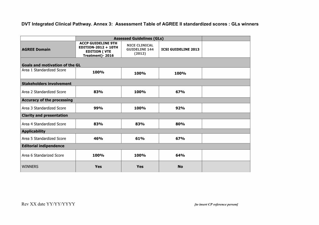
Guidelines Finder), SIGN (Scottish Intercollegiate Guidelines Network), PNLG (Piano Nazionale Linee Guida), Medline, NICE (National Institute of Clinical Excellence). The Guidelines of interest have been firstly identified through the literature search of the sources cited and then evaluated according to the criteria proposed by Grilli et al. (4) (see Annex 1). Notwithstanding the exclusion of at least a guideline after this evaluation, the WG decided to still put it in the second stage of evaluation, in which the AGREE II criteria were applied (Appraisal of Guidelines for Research and Evaluation) (5) (see Annex 2). Finally, the WG has selected the guideline "NICE Clinical Guideline 144" of 2012 (6-8), which got the best score and was considered more applicable to the Italian Health System (see Annex 3). For some specific aspects of the CP, the WG has decided to take into consideration the recommendations of the guidelines "Antithrombotic Therapy and Prevention of Thrombosis, 9th ed: American College of Chest Physicians Evidence-Based Clinical Practice Guidelines, 2012 (9, 10) and SIDV-Giuv, 2012 (11).
6. PROCESSES AND DUTY EVALUATION Care pathway model with admission to the Emergency Department In the following lines, the various stages of the care paths included in the flow chart are described in detail (see Annex 4).
6.1 Triage
All patients presenting/arriving at the ED undergo a “triage” following a standard pattern with colour codes. This procedure is aimed at early detection of the main clinical problem and its urgency level by assigning a colour code. The legislation stipulates that the trained nurse is the professional deputy to do the Triage (Official Gazette No. 114 of 17 May 1996 General Series "Act of agreement between the State and regions for the approval of the guidelines on the health emergency system under Decree of the President of the Republic March 27, 1992" and "Guidelines on training, retraining and permanent training of personnel working in emergency-urgency system "State-Regions Conference of 22 May 2003). To achieve this objective, the nurse uses a specific data collection that makes use of inspection and anamnestic elements and is divided into the following three steps: 1st Step: consists in recognizing, through the inspection, patients with ongoing alterations of one or more vital functions (Circulation, Airway, Breathing - CAB). The inspection is performed directly in the course of collection of personal and logistic data; if it is indicative of alteration of vital functions, the nurse assigns a red code without proceeding to subsequent steps. 2nd Step: consists in recognizing, through a “standardized interrogation", a situation at risk of developing a sudden alteration of vital functions. The nurse with a yellow code identifies patients who fall into this category. Specifically, the patient who presents/arrives at the emergency room reporting a symptom or sign consistent with

DVT CARE PATHWAY
1.0 Revision; 01/01/2017
Page10of25
DVT, provided that vital alterations (red code) have been excluded, receives a yellow code in case of: - Coexistence of dyspnea, episode of loss of consciousness or palpitations (cardinal symptoms) with or without associated chest pain. In all other cases, the state of distress is then evaluated. 3rd Step: aimed at distinguishing the remaining patients (i.e. not previously identified with red or yellow code), in 2 other groups according to their "state of distress": - The patients who should avoid prolonged waiting because, although not at risk, show a relevant "state of distress" (identified by green code) - Patients with no risk priorities, or severe distress (identified by white code). The encoding of the 3rd step is carried out through information already collected in the previous steps, together with the clinical observation of the patient. The triage is performed by the nursing staff able to attribute the emergency code that addresses the patient directly to the proper ED area and attending physician. 6.2 INITIAL ASSESSMENT
Red Code No procedure is expected according to a red code. Indeed, in case of patients presenting/arriving to the ED with suspected PE/ chest pain who are assigned a red code the staff must refer to the specific CP. Yellow Code Patients with suspected DVT who are assigned a yellow code are subjected to post-triage examination, within 10 minutes of arrival in the ED, by the attending physician (usually the one already operating in the red area). The physician will decide, based on the clinical evaluation, possible arterial blood gas analysis, 12-lead EKG, which patients will continue their diagnostic workup in the red area (see also PE/chest pain CPs) and who will be followed by the attending physician of the green area. Green code Patients with suspected DVT who are assigned a green code and those with initial yellow code in whom symptomatic PE is excluded, undergo the diagnostic evaluation (see section 6.3) by the attending physician in the green area, according to the waiting list. White Code In general, not applicable (suspected DVT does not qualify for the assignment of a white code. [If the local ED protocols provide for it the related instructions should be inserted here]

DVT CARE PATHWAY
1.0 Revision; 01/01/2017
Page11of25
6.3 DIAGNOSTIC PROCEDURES
The ED nurse: - takes a blood sample for basic laboratory tests (blood count, PT / INR, aPTT, S-Nitrogen, S-creatinine, S- sodium, S-potassium, S-chloride) and DD assay. The ED physician performs/evaluates: • Accurate medical history and possible comorbidities. • General physical examination (respiratory, cardiovascular, nervous system, bones and joints, neurological etc.). • The presence of edema in one or more lower/upper limbs under the muscle fascia (reduced calf ballottement), the presence of possible collateral venous network (sign of Pratt), the presence of cyanotic or pale limb colour. • The presence of pain, either spontaneous or exacerbated by limb movements.
• The pre-test clinical probability (PTP) using the simplified Wells’ score. If this first-choice tool is not used, the physician may state a reasoned clinical judgment defining in any case the DVT PTP is likely or unlikely (6.3.1) (Table 1). • Whether to prescribe chest X-ray, to be performed within 30-60 minutes. • The results of the DD assay (paragraph 6.3.2). • If able, a simplified CUS (paragraph 6.3.4). If the diagnostic suspicion is not excluded or CUS reveals not compressible veins, the ED physician prescribes the angiologic clinical-instrumental examination (paragraph 6.3.3). If the suspected diagnosis is excluded the ED physician should consider the differential diagnosis with other diseases presenting with similar clinical picture. In patients admitted for suspected DVT, the clinical picture is generally limited to the lower or upper limbs. On the other hand, in some patients , as those with a history of malignancy, hereditary thrombophilia, use of hormonal therapy or other comorbidities, the nurse and the ED physician should suspect DVT in other locations (eg. those with headache in thrombosis of the cerebral sinuses, individuals with acute anuric renal failure in renal vein thrombosis etc.). In these cases, the ED physician will prescribe either directly or supported by a specialist (neurologist, nephrologist, angiologist etc.) an ECD, if appropriate or a CT angiography, if not contraindicated. [If specific pathways are provided for these forms of DVT, the link should be inserted here] Note: the clinical use of standardized algorithms including PTP, DD and ultrasound examination are validated only for a lower limb DVT.

DVT CARE PATHWAY
1.0 Revision; 01/01/2017
Page12of25
Table 1. The WELLS’Score
Items Points Active cancer (Treatment or palliation within 6 months) 1 Previously documented DVT 1 Bedridden recently >3 days or major surgery within four weeks 1 Paralysis, paresis, or recent plaster immobilization of the lower extremity 1 Localized tenderness along the deep venous system 1 Entire leg swollen 1 Calf swelling >3 cm compared to the other leg (Measured 10 cm below tibial tuberosity)
1 Pitting edema, confined to symptomatic leg 1 Collateral (nonvaricose) superficial veins present 1 Alternative diagnosis to DVT as likely or more likely -2
Interpretation Overall score:
≥2 points
DVT Likely
<2
points
DVT Unlikely
6.3.1 Pre-test clinical probability assessment: Wells Score
A validated scoring system to estimate the DVT PTP is generally characterized by a number of items based on clinical criteria (medical history, symptoms, clinical signs). The different scores are able to define a number of classes of clinical probability, useful for increasing the accuracy of an instrumental diagnostic test and allow the construction of diagnostic algorithms aimed at minimizing false either negative or false positive diagnoses and to optimize the required resources. The estimate of the PTP should be applied before performing instrumental diagnostic tests that are always needed to confirm or exclude a DVT. Indeed, the clinical diagnosis alone of DVT is unreliable. Among the available validated scoring systems the most widely adopted is the Wells Score (Table 1); in its most recent simplified version it includes 10 items and two probability categories (DVT likely or unlikely).
6.3.2 D-Dimer measuring
• The DD is a degradation product of fibrin stabilized by covalent crosslinks. Its presence in the circulation is linked to the activation of coagulation with fibrin formation, stabilization of fibrin by Factor XIII (activated by thrombin) and subsequent proteolysis by the fibrinolytic system. DD is detectable at low concentrations in the blood of healthy individuals indicating an equilibrium between the formation and lysis of fibrin under physiological conditions. The DD plasmatic concentration increases in all specific or non-specific clinical situations associated with fibrin-formation and fibrinolysis. Regarding the DVT diagnosis, the plasmatic concentration threshold used

DVT CARE PATHWAY
1.0 Revision; 01/01/2017
Page13of25
in the clinical practice was chosen to obtain a high negative predictive value. Thus, a normal value of DD excludes DVT with a very high probability, while a high value does neither exclude nor confirm it. The test should be integrated in a diagnostic algorithm that involves the assessment of the pre-test clinical probability and the ultrasound examination. It is important to remember that: - The DD plasma concentration often increases with age: currently has not been recommended a set of values of DD adjusted for age authorized for clinical routine use. - Anticoagulant therapy, either with heparin or with oral anticoagulants, reduces the DD levels. - The fibrin deposits, both intra- and extra-vascular, can be a source of fibrin degradation products. - The DD plasma concentration may be normal even in the presence of a thrombotic process for: a) Hypo-fibrinolysis, b) acute thrombotic event dating back over 7-14 days; c) use of insensitive assay method; d) cut-off incorrect values. Below are some physiological and pathological conditions associated with increased levels of DD and that may need to be considered in the differential diagnosis: • Old age • Neonatal Period • Physiological and pathological Pregnancy • Patients hospitalized • Patients with functional disabilities • Aortic dissection • Malignancies • Infections (especially Gram-negative) • Trauma • Burns • Disseminated intravascular coagulation • Stroke • Ischemic heart disease • Aneurysms • Peripheral Arterial Disease • Heart failure • Sub arachnoid haemorrhage • More bleeding • Liver disease • Kidney disease • Inflammatory bowel disease • Systemic lupus erythematosus, rheumatoid arthritis etc. • Thrombolytic therapy

DVT CARE PATHWAY
1.0 Revision; 01/01/2017
Page14of25
6.3.3 Angiologic Consultancy / Clinical and Instrumental examination The Angiologist: • Assesses the ED files. • Performs the clinical exam. • Chooses the most appropriate diagnostic strategy among those that are formally validated for clinical use (paragraph 6.3.4). • Performs ultrasonography [in case of US performed by a sonographer a specific instructions set should be created], usually choosing the procedure (simplified CUS, C-CUS,CFM) indicated in the adopted diagnostic strategy (paragraph 6.3.4). • When indicated, prescribes additional diagnostic tests (e.g. CT scanning, MR venography,) (paragraph 6.3.5) [at this point the exact modalities of patient sending to the X-ray room should be specified] • Gives the final diagnosis 6.3.4 Ultrasound exams and diagnostic strategies
The Angiologist chooses, as a rule, one of the formally validated diagnostic strategies, according to the following factors: clinical judgment, patient’s characteristics (availability for serial exams and follow-up, special anatomical conditions), availability of the ultrasound equipment and dedicated facilities/services. The ultrasound exam can be performed as follows: - Simplified compression ultrasonography (CUS): it is simple, fast, procedure with a quick learning curve; it is based on the B-mode insonation of the common femoral vein and the popliteal vein until the trifurcation, before and after compression of the vessel with the ultrasound probe. If the exam is negative, it cannot be considered conclusive and should be repeated after one week unless it is included in a validated diagnostic algorithm. The CUS does not give any morphologic information on the the iliac-caval, visceral veins and those along distal lower limbs. The technique has only been formally validated for the study of the lower limbs, but is also commonly used for the study of the superficial venous vessels. - Complete CUS (CCUS): it is a compressive ultrasound exam extended to the deep distal veins of the lower limbs, according to standardized patterns. It is a simple, fast enough technique only when relevant anatomic obstacles are absent and requires a more complex learning curve. If the examination is negative, it excludes DVT. Nevertheless, several clinicians still prefer to include it in a clinical-instrumental algorithm. - Doppler colour flow imaging (CFM): it is a more complex technique, quite fast only if performed as a limited exam; it takes longer time when performed as a full exam and requires specific expertise. The examination displays and studies the veins using the B-mode, Doppler spectral analysis, colour flow mapping. All the deep and superficial veins of the lower limbs, including the iliac ones and the inferior cava vein should be studied. A negative result

DVT CARE PATHWAY
1.0 Revision; 01/01/2017
Page15of25
excludes DVT. The exam does not require preceding complementary tests and could be used alone. Nevertheless, several clinicians still prefer to include it in a clinical-instrumental algorithm. The examination can also explore the main visceral venous vessels (renal veins, mesenteric, portal vein) and other districts (subclavian veins and upper limbs, jugular veins). However, no validated multi-step algorithms are available for these districts. - All ultrasound exams may incidentally discover pathological images of the surrounding tissues, which should be reported. In this case, the Angiologist and the attending physician should prescribe additional imaging test, if appropriate. - Diagnostic Strategy (lower limbs): DVT is excluded when: • PTP unlikely + negative DD • CCUS/CFM is normal DVT is confirmed when: • PTP unlikely + positive DD + positive CUS • PTP likely + negative DD + positive CUS • CCU/CFM is positive A further diagnostic workup or serial US tests (after 48 hours and/or 7 days) is recommended when: • PTP likely + positive DD + negative CUS • PTP unlikely + negative DD + positive CUS • CCUS/CFM difficult to perform -- The patients with excluded VTE and no other indications to hospitalization could be discharged -- The patients with inconclusive VTE diagnosis and no other indications to hospitalization could be discharged with the prescription of CUS/CCUS/CFM within 48 hours and/or 7 days; if the further test is negative the diagnosis is definitively excluded; if positive the diagnosis is confirmed -- All patients for whom a serial US examination cannot be guaranteed should undergo a CFM test. -- Pregnant women, patients with massive edema of the lower limbs and those with suspected DVT in other districts than lower limbs must undergo a CFM test - In case of suspected ipsilateral recurrent lower limb DVT the test of choice is the CUS with application of the appropriate diagnostic criteria for thrombotic recurrence (i.e. an increase of the residual diameter of the vein ≥ 4 mm compared with the last available value). In case of negative ultrasound examination associated with a negative DD the diagnosis is ruled out. In the presence of positive DD, negative CUS and no other indications for the hospitalization the patient can be discharged with prescription for a further CUS test within 7 days; if the latter test is still negative, the diagnosis is ruled out
6.3.5 Complementary diagnostic imaging
The Angiologist and/or the ED physician will prescribe radiological examinations with contrast medium (CT or MR angiography) in the case of persisting strong diagnostic doubts with inconclusive diagnostic tests and in the following circumstances:

DVT CARE PATHWAY
1.0 Revision; 01/01/2017
Page16of25
- Diagnostic ultrasound examination not executable - Suspected visceral deep vein thrombosis (inferior vena cava, pelvic veins, renal, mesenteric, portal vein, hepatic veins, the superior vena cava, intracranial veins) In pregnant women and in children performing CT or MR with contrast medium must be weighed together with the radiologist after a careful balancing between the advantage of getting a clear diagnosis and the risks for the mother and the fetus and the possibility of minimizing the exposure to ionizing radiation ( eg. Lung scan) The indication to venography is limited to carefully selected cases or when the other diagnostic techniques cannot be executed or were non-diagnostic. [The Local WG should model and describe this part of the pathway according to the actual availability of imaging techniques and to the related logistics, timing, human resources and materials]. 6.3.6 Management of suspected DVT over the week-end/nocturnal hours [To possibly change the path according to the service hours of unit/ angiology clinic] During the hours in which the Angiologist is not on duty (nights, public holidays), the ED physician: - Prescribes the angiologic clinical- instrumental examination, which will be carried out within 24 hours on weekdays, within 72 hours on public holidays, and decides whether to discharge the patient until the angiologic test is performed or to admit him/her in the observation room or in a medical ward, as a rule in the presence of other specific indications for hospitalization and in any case according to clinical judgment; - Evaluates if immediately starting anticoagulant therapy while awaiting the specialist diagnosis, according to PTP and DD values and/or the simplified CUS as follows: - PTP unlikely, DD Negative: the administration of weight-adjusted doses of LMWH is not indicated until the angiologic test is performed. - PTP unlikely and positive DD or PTP likely with any DD: the administration of weight-adjusted doses LMWH is indicated while waiting the angiologic test.
If the ED physician performs a simplified CUS, the scenarios are the following: - Positive CUS and presence of exclusion criteria for a home therapy (see also section 6.4.3):
he/she admits the patient in an area of hospitalization. - Positive CUS and no exclusion criteria for home therapy: (see also section 6.4.3): he/she
prescribes a treatment with weight-adjusted LMWH or a single-drug approach DOAC (Annex 6), discharges the patient, prescribes and plans the angiologic clinical-instrumental test within 24-72 hours.
- Negative CUS: the administration of weight-adjusted doses of LMWH is not indicated until the angiologic test is performed. Note: It may still be useful to reassess PTP and DD and in case of a significant discrepancy with CUS the ED physician may still consider starting an anticoagulant therapy and/or hospitalizing the patient.

DVT CARE PATHWAY
1.0 Revision; 01/01/2017
Page17of25
6.4 Management of the patients with confirmed DVT in the ED The Angiologist: • Considers whether there are indications for urgent reperfusion therapy of venous occluded vessels (Paragraph 6.4.1). • Considers whether there are contraindications to anticoagulation and the possible indication to the placement of inferior vena cava filter (IVCF), preferably removable (in agreement with the ED physician and the radiologist) (paragraph 6.4.2). • Assesses the patient’s eligibility to home treatment (paragraph 6.4.3) and proposes the next path (home treatment or hospitalization) (paragraph 6.4.4). • Evaluates and prescribes the pharmacologic (and mechanical) treatment (see Annex 6) • If the patient is discharged he/she suggests the appropriate "follow-up" (section 6.6), the advice for both the General Practioner (GP) and the patient/caregiver, the release of the information "brochures" for the patient and the caregiver. [To insert here any possible other procedures/processes delegated to nursing staff or support as training of caregivers etc.]
6.4.1 Reperfusion Therapy Thrombolysis or surgical thrombectomy
The angiologist evaluates the indication of an urgent reperfusion treatment of the occluded venous vessels and decides whether to prescribe it. [At this point, the local WG, according to the locally available technologies and procedures, should insert the proper operating instructions (e.g. Interventional Radiology or Angiology or Vascular Surgery Unit with sufficient skill and volume of procedures). In case of lack of locally available facilities, an operating instruction on how to send the patient to a tertiary or hub vascular centre should be included] The indications are as follows: - Phlegmasia cerulea dolens with impending venous gangrene present by less than two weeks (Provided that the contraindications described in Table 2 have been assessed) - Iliac- femoral DVT if:
o Symptoms present by less than two weeks o No relevant comorbidities ( i.e. young patients) o Expected survival of at least one year o Provided that the contraindications described in Table 2 have been assessed
In case of confirmed indication to thrombolysis/ surgical thrombectomy: ……. [at this point an appropriate operating instruction should be inserted by the local WG, according to several questions and items: who is the person in charge? Radiologist? Vascular Surgeon? Cardiologist? Angiologist? Collegial consulting instructions; Admission Unit etc.]
Notes: The reperfusion therapy may be medical or surgical (i.e. systemic or cath or pharmacomechanical thrombolysis, surgical thrombectomy). It has several relevant limitations (haemorrhagic risk, surgical risk, complexity and high costs) and is supported by studies with important methodological limits. The anticoagulant treatment should be guaranteed to all patients undergoing thrombolysis or thrombectomy, with the same methods and principles used in patients treated with anticoagulation alone. The fibrinolytic to be used are listed in Table 3. [Insert only those in the local hospital therapeutic Handbook]

DVT CARE PATHWAY
1.0 Revision; 01/01/2017
Page18of25
Table 2. Contraindications to pharmacological thrombolysis Absolute: - Prior haemorrhagic stroke or recent cerebrovascular non-haemorrhagic accident - Active internal bleeding - Intracranial neoplasm or recent head injury - Suspected aortic dissection - Recent surgery (in the last two weeks) - Pregnancy - Blood Pressure > 200/120 mmHg - Allergy to streptokinase or APSAC (using rtPA) Relative: - Severe high blood pressure> 180/100 mmHg - Bleeding diathesis or ongoing use of anticoagulants - History of previous cerebrovascular accident haemorrhagic not with complete recovery - Trauma or surgery for more than two weeks - Active peptic ulcer disease - Haemorrhagic retinopathy - Previous treatment with SK or APSAC

DVT CARE PATHWAY
1.0 Revision; 01/01/2017
Page19of25
Table 3. Fibrinolytic drugs [Insert only those actually available in the local hospital
Streptokinase (SK)
- - Risk of immediate type allergic reactions (for the pre-existence of antibodies from previous streptococcal infection) or after sensitization (not be repeated within 6 months from the first administration)
- -Possible premedication methylprednisolone 40 mg i.v.
- - Patient to be monitored during the first half hour of administration
• Dose: 250,000 U in 20 minutes, followed by 100,000 U / hour for 24-48 hours (for 2- 3 days, if DVT massive, but with greater risk of bleeding)
Urokinase (UK) - - High cost of production - - Dose: 4,400 U / kg in 20 minutes followed
by 4,400 U / kg / hour, or at doses halved in association with heparin, or at even lower doses in the treatment locoregional
Recombinant plasminogen tissue activator (rt-PA)
- - Main activator of fibrinolysis in the blood - -
- - elective lytic action on thrombus size, but at therapeutic doses results in alterations of hemostasis and haemorrhagic complications like other thrombolytic
- - In Italy the use of rt-PA is authorized for the treatment of acute myocardial infarction and DVT with clinically suspected pulmonary embolism, no DVT
Complex acylated streptokinase activator plasminogen- (APSAC)
A prolonged fibrinolytic treatment requires monitoring of blood coagulation factors, as a precaution. Although the prolongation of PTT and the thrombin time (TT) may be used to assess the biological effect of thrombolytic treatment, there is not an appropriate method of monitoring therapy. The parameter that best reflects the effectiveness is the TT, but there is no clear correlation with the clinic. On suspension, the treatment should be continued with intravenous heparin at therapeutic doses, if the aPTT ratio is already higher than 2 . [the roles and functions of health personnel responsible for monitoring / withdrawals etc. should be described]

DVT CARE PATHWAY
1.0 Revision; 01/01/2017
Page20of25
6.4.2 Caval filter insertion
In patients with lower limbs or iliac-caval, proximal DVT the Angiologist assesses if there are absolute transient or permanent contraindications to anticoagulation (Table 4). If case of such contraindications, if obvious contraindications or circumstances that advise against the indication (e.g. very elderly, terminal patients) are absent (Table 5.6), the Angiologist proposes the placement of a vena cava filter. [to describe the paths / following instructions, depending on the availability or otherwise of an interventional radiology/cardiology unit and local practices; to define the professionals involved, the need for a preliminary assessment of the radiologist and the ED physician, the possible admission in a medical unit that will activate the radiologist consultation] Table 4. Absolute contraindications to anticoagulant therapy • Active clinically significant bleeding during or just before starting anticoagulant therapy • Progression of DVT/onset or recurrent PE despite a well conducted anticoagulation (must be clearly assessed the adequacy of the dose of the used anticoagulant drugs and/or the inability to increase INR between 3 and 4 or to replace oral anticoagulation with LMWH) • Very high risk of bleeding Table 5. Absolute contraindications to the placement of vena cava filter • Lack of vascular access to the ICV • Implantation site in the ICV inadequate due to extensive thrombosis • Dilation of the ICV exceeding 40 mm in diameter
Table 6. Relative contraindications to the placement of vena cava filter • Severe coagulopathy • Septic emboli • Detection of positive blood cultures Note: The type of filter, removable or permanent, the choice of the venous access, right- side trans-jugular or trans-femoral and the vena cava filter position, above or below the renal veins, are decided by the Radiologist / Cardiologist in agreement with the Angiologist.
6.4.3 Home treatment
The Angiologist evaluates the indication for the home treatment of the diagnosed DVT, according to the inclusion and exclusion criteria included in the check-list (Table 7). Provided that no contraindications exist, he/she subsequently offers the patient and family the home treatment (see Annex 6 for the choice of the appropriate drug type and regimen )[ At this point the local WG should describe in detail the available paths and persons in charge as the GP, primary care nurses, lab, point of care tests, etc.) Provided that the patient has signed the informed consent, the Angiologist: - Gives verbal instructions and provides written instructions for the home management of

DVT CARE PATHWAY
1.0 Revision; 01/01/2017
Page21of25
anticoagulation and compression therapy. [If qualified nursing staff directly provide the instructions, the text should be amended accordingly] - Entrusts the patient and family to the nursing staff to be trained on the injection procedure and the handling of the vials, syringes, needles etc. (only if DOAC single-drug approach is not prescribed) - Activates the link with the GP, if provided, to communicate the start of the home therapy [any alternatives should be described at this point or deleted] - Prescribes the planned lab tests and follow-up specialist consultancies (and/or thrombosis clinic) [or describe alternative procedures/paths] - Plans the first follow-up medical and instrumental exam ( CUS/CFM; usually after 3 months) - Encourages early ambulation. The ED attending physician (if appropriate): - Re-evaluates the clinical case, the specialist's decisions and the criteria for drug therapy and home discharge - Orders the administration of the first dose of the anticoagulant drug chosen (LMWH or fondaparinux or DOAC (see also Annex 6) - Maintains the patient under observation for further 30-60 minutes prior to discharge - Re-evaluates the patient, provides the attached clinical documentation and proceeds to discharge. Table 7. Exclusion criteria of a home treatment of DVT - Refusal of a home treatment - Symptomatic PE (according with the ESC risk class) - Massive DVT - Swelling of the entire limb - Acrocyanosis - Phlegmasia cerulea dolens - Iliac or caval DVT - Need for oxygen therapy - High risk of bleeding with anticoagulants - Active bleeding - Bleeding episode from less than 4 weeks - Recent surgery or trauma (<7 days) - Thrombocytopenia (PLTs <100 000 x 106 / L) - Coagulopathy (INR> 1.4, aPTT> 40 sec) - Intracerebral or intrahepatic metastasis - Severe renal insufficiency - Severe hepatic impairment - Uncontrolled high systemic blood pressure - Relevant comorbidities or special needs - Need of hospitalization for the underlying disease - Severe pain requiring opiate analgesia - Dementia ( only if home caregiving is suboptimal) - Communication problems with the specialist, GP or hospital - Forced Immobilization (only if home caregiving is suboptimal)
DVT CARE PATHWAY
1.0 Revision; 01/01/2017
Page22of25
6.4.4 Hospitalization The Angiologist and the ED physician admit the patient in a medical ward (Angiology, Medicine) according to the following, general, rules:
- When at least one criterion of exclusion from home therapy is met - When a closer monitoring of the anticoagulant therapy (e.g. very low/very high body weight etc.) is deemed preferable - When the hospitalization is preferred for other reasons (e.g. the need for complex differential diagnosis, some suspicious forms of malignancy that need prompt assessment, acute and / or active comorbidities) [To describe whether the subsequent path - hospitalization- will be managed by the angiologist, the ED physician or both. To define the methods of patient transportation, especially if particularly complex e.g. need for ambulance for intra-hospital transport etc.] The ED physician orders the administration of the first dose of LMWH, UFH or DOAC especially if the transfer to the ward is delayed.
6.5 Management of the patient with DVT excluded The Angiologist: • Considers the differential diagnosis and suggest any possible further investigations/ consultations, in the ED or office-based. • Describes the possible presence of injuries / other diseases responsible for the clinical picture, which are found during the ultrasound vascular examination. • Addresses the patient to the ED physician who shall:
- Discharge the patient with the prescription of CUS / Control CCUS after 48 hours or 7 days, according to the Angiologist’s prescription/proposal (paragraph 6.3.4).
- Hold the patient in the ED, in an observation area or hospitalize him/her based on other indications.
6.6 Follow- up 6.6.1 Management of the patient under home treatment The Angiologist describes, in his/her final clinical report, the method of managing the follow-up of the subjects treated at home and prescribes the next exams - Gives the patient the information brochure containing contact details with the health facilities in case of adverse events during the early phase of home treatment.
DVT CARE PATHWAY
1.0 Revision; 01/01/2017
Page23of25
- Prescribes blood count / platelet count after 5 days, and possibly after 10 and 15 days if the patient is treated with heparin (first check after 48 hours if the subject has been exposed to heparin in the previous 6 months)
- Prescribes the follow-up clinical exam and creatinine dosage after 3 months if the patient is treated with DOAC - Prescribes the next INR if the patient is treated with VKA - Optional: - Screening for thrombophilia (at the end of anticoagulant treatment or limited to genetic and immunological tests) - Screening for occult malignancy (usually blood count, erythrocyte sedimentation rate, total and fractionated proteins, the standard urine test, looking for faecal occult blood x 2, complete abdomen ultrasound, clinical and instrumental breasts, uterus, prostate examination. - Any other targeted surveys, specialist consultations, etc. - Angiologic clinical and instrumental exam after 3 months of anticoagulation. - In the presence of an isolated distal DVT for which was prescribed the only ultrasound surveillance or a no more than 4-6 weeks treatment follow-up exams should be prescribed (CUS / CCUS after 7-14- 30 days in the first case; angiologic clinical-instrumental examination after 4 to 6 weeks in the latter case) The ED physician describes, in his/her final report, the prescriptions of the Angiologist [If needed, to replace with "attach a copy of the specialist clinical report," according to the local procedures]
6.6.2 Management of the patient discharged after hospitalization
The attending physician describes, in his/her final clinical report, the method of managing the follow-up of the subjects discharged from a hospitalization and prescribes the next exams; - Gives the patient the information brochure containing contact details with the health facilities in case of adverse events during the early phase of home treatment. - Prescribes blood count / platelet count after 5 days, and possibly after 10 and 15 days if the patient is treated with heparin (first check after 48 hours if the subject has been exposed to heparin in the previous 6 months)
- Prescribes the follow-up clinical exam and creatinine dosage after 3 months if the patient is treated with DOAC - Prescribes the next INR if the patient is treated with VKA - Optional: - Screening for thrombophilia (at the end of anticoagulant treatment or limited to genetic and immunological tests) - Screening for occult malignancy (usually blood count, erythrocyte sedimentation rate, total and fractionated proteins, the standard urine test, looking for faecal occult blood x 2, complete abdomen ultrasound, clinical and instrumental breasts, uterus, prostate examination. - Any other targeted surveys, specialist consultations, etc. - Angiologic clinical and instrumental exam after 3 months of anticoagulation.

DVT CARE PATHWAY
1.0 Revision; 01/01/2017
Page24of25
- In the presence of an isolated distal DVT for which was prescribed the only ultrasound surveillance or a no more than 4-6 weeks treatment follow-up exams should be prescribed (CUS / CCUS after 7-14- 30 days in the first case; angiologic clinical-instrumental examination after 4 to 6 weeks in the latter case)
The attending physician describes, in his/her final report, the prescriptions of the Angiologist. [If needed, to replace with "attach a copy of the specialist clinical report," according to the local procedures] 7. IMPLEMENTATION DEADLINES
This document will come into force from the date of publication. The referent of the CP will be responsible for updating this document at the end of the third year and /or in case of significant new evidence on the subject He/she should also organize update WG meetings if deemed necessary for the update of the document itself.
8. OUTCOME/PROCESS INDICATORS Indicator: - Number of patients accessing the Emergency Department (ED): n(1) - Number of patients accessing the ED with suspected DVT: n(2) - Percentage of patients accessing the ED with suspected DVT of total accesses in ED: [n(1) / n(2)*100 - Percentage of patients with suspected DVT stratified for DVT pre-test probability - Percentage of patients with DVT stratified by the ward of hospitalization - Percentage of shorter hospitalizations among total hospitalizations for DVT - Number of prescribed DVT home treatments compared to the total number of patients admitted to ED for suspected DVT -Percentage of patients with DVT excluded re-admitted to the ED, among those discharged within a year from the ED and stratified by clinical probability 9. REFERENCES
1) Bero LA, Grilli R, Grimshaw JM, Harvey E, Oxman AD,Thomson MA. Closing the gap between research and practice: an overview of systematic reviews of interventions to promote the implementation of research findings. BMJ 1998;317:465-468.
2) Grol R, Grimshaw J. From best evidence to best practice. Effective implementation of change in patients’ care. Lancet 2003;362:1225-30.
3) Guyatt GH, Oxman AD et al; GRADE Working Group. GRADE: an emerging consensus on rating quality of evidence and strength of recommendations. BMJ2008; 336:924-6.

DVT CARE PATHWAY
1.0 Revision; 01/01/2017
Page25of25
4) Grilli R, Magrini N, Penna A, Mura G, Liberati A. Practice guidelines developed by specialty societies: the need for a critical appraisal. Lancet, 2000 Jan 8;355(9198):103-106.
5) AGREE Next Steps Consortium (2009). The AGREE II Instrument [Electronic version] http://www.agreetrust.org
6) National Institute for Health and Care Excellence; Venous thromboembolic diseases: the management of venous thromboembolic diseases and the role of thrombophilia testing. CG144. 2012; London: National Institute for Health and Care Excellence.
7) National Institute for Health and Care Excellence. Venous thromboembolic diseases: the management of venous thromboembolic diseases and the role of thrombophilia testing. CG144; Appendix A-M. 2012; London: National Institute for Health and Care Excellence.
8) Dupras D, Bluhm J, Felty C, Hansen C, Johnson T, Lim K, Maddali S, Marshall P, Messner P, Skeik N. Institute for Clinical Systems Improvement. Venous Thromboembolism Diagnosis and Treatment.http://bit.ly/VTE0113. Updated January 2013.
9) Wells PS, et al. Evaluation of D-dimer in the diagnosis of suspected deep-vein thrombosis. New Engl J Med 2003;349:1227
10) Bates SM, Jaeschke R, Stevens SM, Goodacre S, Wells PS, Stevenson MD, Kearon C, Schunemann HJ, Crowther M, Pauker SG, Makdissi R, Guyatt GH; American College of Chest Physicians. Diagnosis of DVT: Antithrombotic Therapy and Prevention of Thrombosis, 9th ed: American College of Chest Physicians Evidence-Based Clinical Practice Guidelines. Chest. 2012 Feb;141(2 Suppl):e351S- 418S. doi:10.1378/chest.11-2299. PubMed PMID: 22315267; PubMed Central PMCID: PMC3278048.
11) Kearon C, Akl EA, Comerota AJ, Prandoni P, Bounameaux H, Goldhaber SZ, Nelson ME, Wells PS, Gould MK, Dentali F, Crowther M, Kahn SR; American College of Chest Physicians. Antithrombotic therapy for VTE disease: Antithrombotic Therapy and Prevention of Thrombosis, 9th ed: American College of Chest Physicians Evidence-Based Clinical Practice Guidelines. Chest. 2012 Feb;141(2 Suppl):e419S- 94S. doi: 10.1378/chest.11-2301. Erratum in: Chest. 2012 Dec;142(6):1698-1704. PubMed PMID: 22315268; PubMed Central PMCID: PMC3278049.
12) Antignani PL, Benedetti-Valentini F, Aluigi L, Baroncelli TA, Camporese G,Failla G, Martinelli O, Palasciano GC, Pulli R, Rispoli P, Amato A, Amitrano M,Dorigo W, Gossetti B, Irace L, Laurito A, Magnoni F, Minucci S, Pedrini L, Righi D, Verlato F. Diagnosis of vascular diseases. Ultrasound investigations--guidelines. Int Angiol. 2012 Oct;31(5 Suppl 1):1-77. PubMed PMID:23470846.
10. ATTACHED DOCUMENTS INDEX Annex 1: Assessment Table of "Grilli Criteria" Annex 2: Table of standardized assessment scores "AGREE II" Annex 3: Summary table of standardized scores "AGREE II": Annex 4: Patient with suspected DVT flowchart Annex 5: List of codes ICD9CM Annex 6: Pharmacological and compressive Therapy
Rev XX ,date YY/YY/YYYY [to insert here CP reference person ]
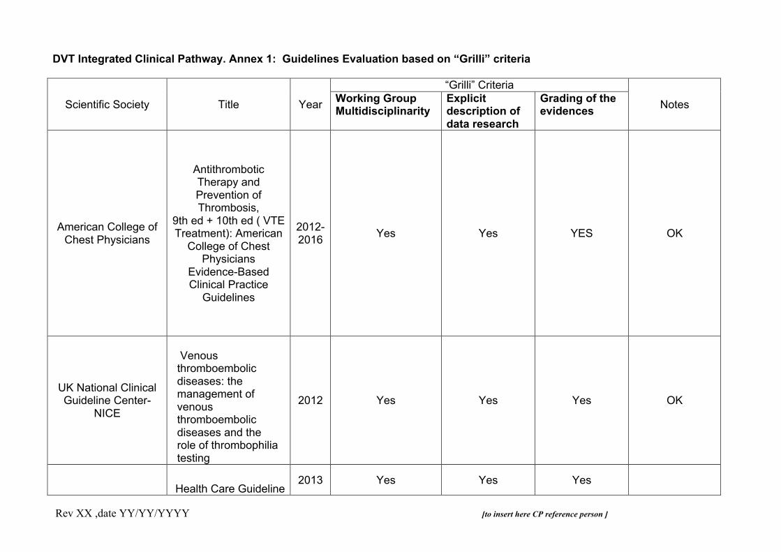
DVT Integrated Clinical Pathway. Annex 1: Guidelines Evaluation based on “Grilli” criteria
Scientific Society Title Year
“Grilli” Criteria
Notes Working Group Multidisciplinarity
Explicit description of data research
Grading of the evidences
American College of
Chest Physicians
Antithrombotic Therapy and Prevention of Thrombosis,
9th ed + 10th ed ( VTE Treatment): American
College of Chest Physicians
Evidence-Based Clinical Practice
Guidelines
2012-2016 Yes Yes YES OK
UK National Clinical Guideline Center-
NICE
Venous thromboembolic diseases: the management of venous thromboembolic diseases and the role of thrombophilia testing
2012 Yes Yes Yes OK
Health Care Guideline
2013 Yes Yes Yes
Rev XX ,date YY/YY/YYYY [to insert here CP reference person ]
ICSI- Institute for Clinical Systems
Improvement
Venous Thromboembolism
Diagnosis and Treatment
Rev XX date YY/YY/YYYY [to insert CP reference person]
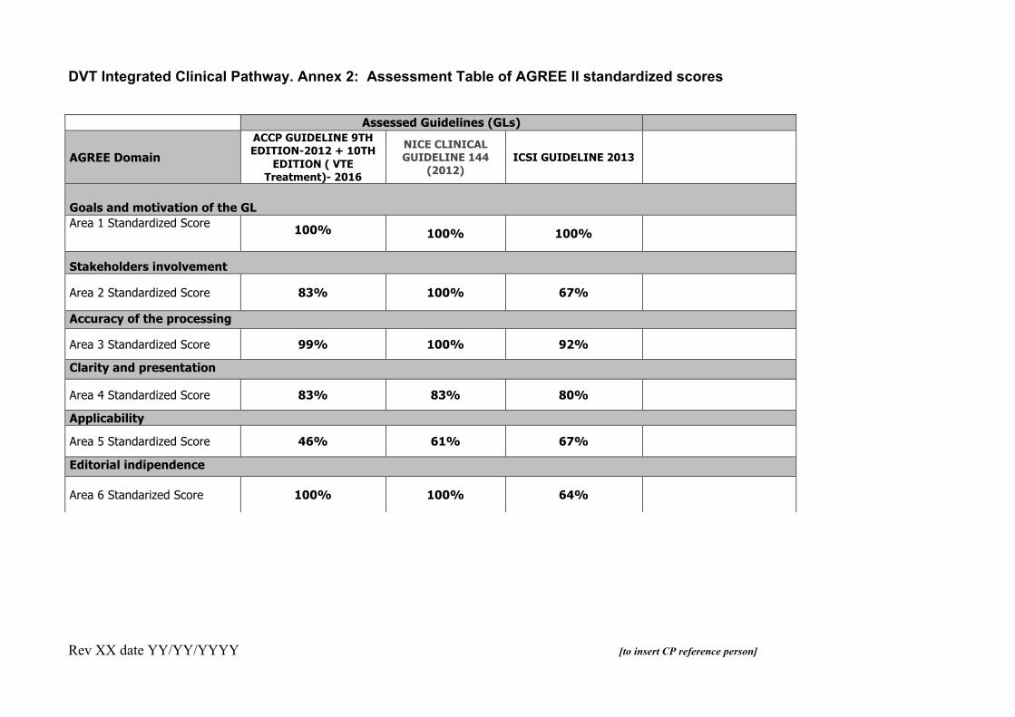
DVT Integrated Clinical Pathway. Annex 2: Assessment Table of AGREE II standardized scores
Assessed Guidelines (GLs)
AGREE Domain
ACCP GUIDELINE 9TH EDITION-2012 + 10TH
EDITION ( VTE Treatment)- 2016
NICE CLINICAL GUIDELINE 144
(2012) ICSI GUIDELINE 2013
Goals and motivation of the GL Area 1 Standardized Score 100% 100% 100%
Stakeholders involvement
Area 2 Standardized Score 83% 100% 67%
Accuracy of the processing
Area 3 Standardized Score 99% 100% 92%
Clarity and presentation
Area 4 Standardized Score 83% 83% 80%
Applicability
Area 5 Standardized Score 46% 61% 67%
Editorial indipendence
Area 6 Standarized Score 100% 100% 64%
Rev XX date YY/YY/YYYY [to insert CP reference person]
DVT Integrated Clinical Pathway. Annex 3: Assessment Table of AGREE II standardized scores : GLs winners
Assessed Guidelines (GLs)
AGREE Domain
ACCP GUIDELINE 9TH EDITION-2012 + 10TH
EDITION ( VTE Treatment)- 2016
NICE CLINICAL GUIDELINE 144
(2012) ICSI GUIDELINE 2013
Goals and motivation of the GL Area 1 Standardized Score 100% 100% 100%
Stakeholders involvement
Area 2 Standardized Score 83% 100% 67%
Accuracy of the processing
Area 3 Standardized Score 99% 100% 92%
Clarity and presentation
Area 4 Standardized Score 83% 83% 80%
Applicability
Area 5 Standardized Score 46% 61% 67%
Editorial indipendence
Area 6 Standarized Score 100% 100% 64%
WINNERS Yes Yes No

ER Nursing Staff
ER Physician/ Laboratory staff
ER Physician/Radiologist
Vascular Physician/Angiologist
Vasc Phys- Angiologist/ ER Physician
Vasc Phys- Angiologist/ ER Physician/Radiologist
Vasc Phys- Angiologist/ ER Physician/ Internista/ Primary
care Nursing Staff/General Practioner
Note 1: Use ER protocols forthe colour coding and the assessment of chest pain
Note 2: History, Wells Score for DVT, D-Dimer, Consider simplified CUS if available (not compulsory), general and cardiopulmonary physical exam, consider insertion of a venous line
Secondary pathway 1: Apply the PE/chest pain pathway/flowchart
Noea 3: Consider differential diagnosis with ultrasound or other imaging procedures
Note 4: CUS or Colour-Coded duplex scanning according to the chosen diagnostic startegy. In selected cases perform CT/MR scanning
Secondary pathway 2: Evaluate the inclusion/exclusion criteria of cath/pharmacomechanic thrombolysis or thrombectomy. Evaluate the inclusion/exclusion criteria of the insertion of a caval filter, either removable or definitive
Secondary pathway 3:- Evaluate the inclusion/exclusion criteria of home treatment;- Evaluate the treatment straetgies:-- LMWHs or fondaprinux, wheter followed by VKA/DOAC or not;--DOAC ( single-drug approach)-- If sever clinical instability/severe renal insufficiency: Hospitalize and evaluate intravenous UFH--if HIT(T): use the available and authorized druge ( lepirudin,bivalirudin, argatroban, danaparoid)- Administer the dose of the chosen anticoagulant drug
Note 4: - Home treatment: entrust the patient to the GP-Hospitalization: standard or early discharge according to the attending physician's decisions
ACTIVITIES RECORDS/NOTES
Triage: lower limbs oedema/suspected deep vein thrombosis (see note 1)
Initial Assessment (see note 2)
Suspected PE?
Yes Secondarypathway n. 1
Suspected DVT?
NoO
STOP (see note 3)
NoO
Yes
Angiologic consultancy/clinical and instrumental examination (see note 4)
Is DVT confirmed?
NoO
STOP see Note 3
Yes
Secondary pathway n.3
Indication forthrombolysis/caval filter?
Yes
NoO
Secondarypathway n. 2
Early dischargeor
Home treatment
(see Note 4)
Hospitalization
(see note 4)
Flow-chart 1: Patient with suspected DVT admitted in ER
Rev. XX ; date of revision YY/YY/YYYY
PERSON IN CHARGE

PA_TVP_SIAPAV
Rev. XX date YY/YY/YYYY [to insert CP reference person]
DVT Integrated Clinical Pathway - Annex 5
ICD9CM Codes
(“The International classification of Diseases – 9 th revision – Clinical Modification” 2007 Italian version) Code Diagnosis
453.2 vena cava thrombosis 451.81 Iliac/femural vein thrombosis
451.19 Popliteal vein thrombosis
451.19 Tibial veins thrombosis

Annex 6. Pharmacologic and compressive therapy (thrombolysis not included) Anticoagulant Therapy [ to eliminate the anticoagulant drugs locally not available/authorized] The mainstay of pharmacological treatment of DVT is anticoagulation. It has the following objectives: • Prevent the local extension of the thrombus and the embolization; • Promote or accelerate the spontaneous fibrinolysis; • Prevente the recurrence of venous thromboembolic episodes; • Prevent the long-term complications (e.g. post-thrombotic syndrome). It should be as timely and appropriate as possible. There are several categories of anticoagulant drugs, each characterized by a specific safety and efficacy profile: in Europe unfractionated heparin (sodium or calcium) (UFH), low molecular weight heparins (LMWHs), fondaparinux, two vitamin K antagonists (warfarin and acenocoumarol), four oral direct anticoagulants (DOAC) ( dabigatran, rivaroxaban,apixaban, edoxaban) are currently approved for the treatment of VTE (DVT and/or PE). Before starting an anticoagulant therapy the Angiologist should carefully consider the contraindications to the treatment (see Table 2). It is important to bear in mind that, after an acute episode of VTE, the risk of complications varies in time and an appropriate anticoagulant therapy should be administered accordingly. Three phases can be identified in detail, as follows: an acute or early phase lasting for the first 7 days, in which the risk of death and/or early recurrent VTE is at the maxium; a subsequent phase, lasting three months, in which there is a consistent risk of early recurrencies and inegligible risk of death; a later phase, of indefinite duration, in which there is a variable risk of late recurrencies and other complications as the post-thrombotic syndrome and/or a chronic thromboembolic pulmonary hypertension. For each of these phases a specific anticoagulant treatment (initial, long-term and extended) should be proposed. Initial anticoagulation The initial treatment of DVT consists in the administration of a rapid onset injective drug as UFH i.v. LMWHs s.c., fondaparinux s.c. or a rapid onset single-drug approach DOAC. The Angiologist and/or the Emergency Department (ED) physician choose the appropriate drug to administrate based on Hospital Pharmacy availability, and on clinical considerations (e.g. a proximal extension of the thrombosis, need of thrombolysis, body weight, renal failure, pregnancy, allergies, history of heparin-induced thrombocytopenia (HIT), cancer, home treatment etc.). Notably, in the current major guidelines DOAC are considered either equivalent to the standard anticoagulant therapy or the first choice drugs

in non-cancer patients. Before starting anticoagulant treatment the following lab test should be performed: PT/INR, aPTT, platelets count, hemoglobin concentration, serum creatinine and creatinine clearance ( calculated with the Cockroft-Gault formula). - LMWHs/fondaparinux/ UFH:
o LMWHs have a lower risk of bleeding and HIT compared to UFH. In addition, they - as well as fondaparinux - have more favourable pharmacological profile than UFH due to the fixed-dose or adjusted body weight dose administration without need of routine monitoring.
o On the other hand, LMWHs and fondaparinux are characterized by predominantly renal excretion, leading to a potential accumulation and increased bleeding risk in patients with severe renal impairment (creatinine clearance <30 ml/min). In these patients, fondaparinux is contraindicated, for LMWHs the dosage is usually halved or adjusted by means of measuring the plasmatic anti-Xa activity and UFH remains the preferred therapy. In patients with creatinine clearance between 30 and 50 ml/min caution and clinical monitoring are recommended.
o The LMWHs have a different dosage depending on the specific molecule (see Table 1).
o The dose of fondaparinux is 5 mg s.c. O.D. for patients with body weight < 50 kg,
7.5 mg s.c. O.D. for patients with body weight between 50 kg and 100 kg, 10 mg s.c. O.D. for those weighing > 100 kg.
o The dose of UFH i.v. is equal to a bolus of 5000 IU, followed by a continuos
infusion at a dose able to keep an aPTT ratio between 1.5 to 2.5.
o If UFH or LMWHs are being used, the Angiologist should prescribe a platelet count between the 5th and the 7th day of treatment or within the 2nd day in patients recently exposed to heparin and at least one other count after 7-10 days if the treatment is prolonged beyond the 7th day. After the first 30 days of heparin treatment no additional controls are needed. The routine platelet count monitoring is not needed when fondaparinux or DOAC are used.
o The DOAC have a different dosage depending on the specific molecule (see
Table 2.
o If anticoagulation with VKA is chosen, the early initiation of the drug (eg, same day as parenteral therapy is started) and continuation of parenteral anticoagulation for a minimum of 5 days and until the INR is 2.0 or above for at least 24 h is recommended.
o In patients with active cancer a LMWH s.c, administered at least for the first six months is the current recommended anticoagulant treatment, at full doses for the first four weeks and subsequently at a dose equal to the 75% of the therapeutic dose, given once a day.
o In acute DVT during pregnancy the use of vitamin K antagonists or DOAC are
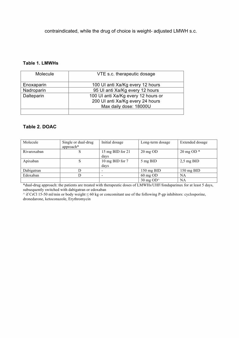
contraindicated, while the drug of choice is weight- adjusted LMWH s.c.
Table 1. LMWHs
Molecule VTE s.c. therapeutic dosage
Enoxaparin 100 UI anti Xa/Kg every 12 hours Nadroparin 95 UI anti Xa/Kg every 12 hours Dalteparin 100 UI anti Xa/Kg every 12 hours or
200 UI anti Xa/Kg every 24 hours Max daily dose: 18000U
Table 2. DOAC Molecule Single or dual-drug
approach* Initial dosage Long-term dosage Extended dosage
Rivaroxaban S 15 mg BID for 21 days
20 mg OD 20 mg OD *
Apixaban S 10 mg BID for 7 days
5 mg BID 2,5 mg BID
Dabigatran D - 150 mg BID 150 mg BID Edoxaban D - 60 mg OD NA 30 mg OD^ NA *dual-drug approach: the patients are treated with therapeutic doses of LMWHs/UHF/fondaparinux for at least 5 days, subsequently switched with dabigatran or edoxaban ^ if CrCl 15-50 ml/min or body weight ≤ 60 kg or concomitant use of the following P-gp inhibitors: cyclosporine, dronedarone, ketoconazole, Erythromycin