defects in the genes coding for cartilage extracellular
TRANSCRIPT

DEFECTS IN THE GENES CODING FOR CARTILAGE EXTRACELLULAR MATRIX PROTEINS AS A CAUSE OF OSTEOARTHRITIS AND MULTIPLE EPIPHYSEAL DYSPLASIA
EVELIINAJAKKULA
Faculty of Medicine,Department of Medical Biochemistry
and Molecular Biology,Biocenter Oulu,
University of Oulu
OULU 2005


EVELIINA JAKKULA
DEFECTS IN THE GENES CODING FOR CARTILAGE EXTRACELLULAR MATRIX PROTEINS AS A CAUSE OF OSTEOARTHRITIS AND MULTIPLE EPIPHYSEAL DYSPLASIA
Academic Dissertation to be presented with the assent ofthe Faculty of Medicine, University of Oulu, for publicdiscussion in Auditorium of the Medipolis (Kiviharjuntie 11), on May 27th, 2005, at 10 a.m.
OULUN YLIOPISTO, OULU 2005

Copyright © 2005University of Oulu, 2005
Supervised byProfessor Leena Ala-Kokko
Reviewed byDocent Kati ElimaDocent Marjo Kestilä
ISBN 951-42-7732-5 (nid.)ISBN 951-42-7733-3 (PDF) http://herkules.oulu.fi/isbn9514277333/
ISSN 0355-3221 http://herkules.oulu.fi/issn03553221/
OULU UNIVERSITY PRESSOULU 2005

Jakkula, Eveliina, Defects in the genes coding for cartilage extracellular matrixproteins as a cause of osteoarthritis and multiple epiphyseal dysplasia Faculty of Medicine, Department of Medical Biochemistry and Molecular Biology, Biocenter Oulu,University of Oulu, P.O.Box 5000, FIN-90014 University of Oulu, Finland 2005Oulu, Finland
AbstractThe role of sequence variations in genes encoding cartilage extracellular matrix (ECM) proteins werestudied in osteoarthritis (OA) and multiple epiphyseal dysplasia (MED). The cartilage collagen genesCOL2A1, COL9A1, COL9A2, COL9A3, COL11A1, and COL11A2 were screened for sequencevariations in 72 Finnish probands and one US family with primary early-onset hip and/or knee OA.Altogether 239 sequence variations were found, of which 16 were not present in the controls. Sevenof the unique variations — four in COL11A1, two in COL11A2, and one in COL2A1 — were studiedfurther, because they resulted in the substitution of conserved amino acids or were predicted to affectmRNA splicing. Association analysis was performed by genotyping 6–12 common polymorphismsfrom each gene in 72 OA patients and 103 controls; no common predisposing alleles were identified.The results, however, suggest that mutations in the minor cartilage collagen genes can be the causeof OA in a subgroup of OA patients.
Two MED families with clinical and radiographic features suggestive of a collagen IX mutationwere studied. Mutation screening of COL9A1, COL9A2, and COL9A3 yielded negative results.Instead, an R718W mutation in COMP was identified in both families. Clinical and radiographicoverlap between patients with collagen IX mutations and patients with COMP mutations points to acommon supramolecular complex pathogenesis.
Clinical, radiological and molecular analyses of known MED genes were performed on a cohortof 29 consecutive MED patients. The DTDST mutation was identified in four patients (14%), theCOMP mutation in three (10%), and the MATN3 mutation in three (10%). Two new distinctphenotypic entities were identified in patients in whom no mutation was found. The findings suggestthat mutations in the above mentioned known MED genes are not the major cause of MED and areresponsible for less than half of the cases. The existence of additional MED loci is supported by theexclusion of known loci and finding of the specific subgroups among these patients.
The results suggest that genetic defects in ECM genes can predispose to OA and cause MED, eventhough the major genes involved in both disorders remain to be found.
Keywords: chondrodysplasia, multiple epiphyseal dysplasia, osteoarthritis


Acknowledgements
This work was carried out at the Collagen Research Unit of the Department of Medical Biochemistry and Molecular Biology, University of Oulu, and Biocenter Oulu, and at the Center for Gene Therapy, Tulane University Health Sciences Center, New Orleans, USA, during the years 1999-2005.
I wish to express my deepest gratitude to my supervisor, Professor Leena Ala-Kokko, for her continuous encouragement and never-ending optimism throughout these years and for arranging me the opportunity for me to work at the Center for Gene Therapy in New Orleans. I would like to express my sincere thanks to Professors Peppi Karppinen, Kari Kivirikko, and Taina Pihlajaniemi for providing excellent research facilities and maintaining a high-standard scientific environment. In addition, I wish to express my respect for Professor Ilmo Hassinen for his outstanding scientific work in the field of energy metabolism. My sincere thanks are to Professor Darwin J Prockop for giving me the honour of working in his laboratory in New Orleans.
Docents Kati Elima and Marjo Kestilä are acknowledged for their thorough and valuable comments on this thesis. I wish to thank Sandra Hänninen, M.Sc., for her careful revision of the language of the manuscript.
All the collaborators who participated in the projects presented in this thesis deserve my sincere thanks. I am grateful to Miia Melkoniemi for teaching me the basics of lab work at start of this research project and for fruitful collaboration with the osteoarthritis project throughout these years. I had the opportunity to work alongside a team of expert clinicians, including Professor Heikki Kröger, Docent. Ilkka Kiviranta, Professor Andrea Superti-Furga, and Docent Outi Mäkitie, and their clinical and scientific expertise has been essential in completing the research projects presented in this thesis. Harald H.H. Göring is acknowledged for his collaboration with the statistical aspects of the osteoarthritis project.
The colleagues at the Department of Medical Biochemistry and Molecular Biology, University of Oulu, and at the Center for Gene Therapy, New Orleans deserve my warmest thanks. I want to especially thank all the members of group Ala-Kokko, for the cheerful moments and for the scientific and non-scientific discussions in the offices and outside the lab. I wish to thank Minna Männikkö, our always friendly post-doc, for her help with research problems and for her support and friendship over the years. The time

spent in Philadelphia and New Orleans was made much more enjoyable by sharing the work and leisure time with Noora Noponen-Hietala, Minta Lumme, Jaana Peters, and Ahmad Sahraravand; thank you for all the cheerful memories.
I wish to thank Aira Harju, Minta Lumme, Justing Manges, Marjorie McGants, Helena Satulehto, Marika Oikarinen, and Christina Troxell for their excellent technical assistance.
I warmly thank our group members Heini Hartikka, Noora Noponen-Hietala and Miia Melkoniemi for their contributions to the practical arrangement of the dissertation. I thank them for sharing the sometimes hectic moments during the preparation of the doctoral dissertation and during the organization of the party.
My sincere thanks go to Auli Kinnunen and Marja-Leena Kivelä for their always efficient and friendly secretial assistance, to Pertti Vuokila, Marja-Leena Karjalainen, and Seppo Lähdesmäki for keeping the everyday lab-life running smoothly, and to our computer crew, especially to Risto Helminen, for their help with computers. The assistance of the staff of the Center for Gene Therapy, New Orleans, is also greatly acknowledged.
I wish to express my warmest gratitude to my parents, Eero and Maaret, for all their loving care and support over the years, and for giving me an excellent basis for my life. My sister Elina and brother Eerikki both deserve my sincere thanks for many joyful moments , which brightened the days filled with research work and medical studies, and for their friendship. I want to warmly thank my colleagues and dear friends, especially Outi Jauhola, Virpi Koskela, and Elise Saarela, for sharing the many aspects of being a medical student and the first steps of our clinical carrier, and for being such good friends, even if I sometimes seemed to be constantly in the lab.
This work was financially supported by the Finnish Medical Foundation and the Oulu University Scholarship Foundation.
Oulu, March 2005 Eveliina Jakkula

Abbreviations
ACG1B achondrogenesis type 1B AO2 atelosteogenesis type 2 BMI body mass index BMP bone morphogenetic protein bp base pair(s) C- carboxy- cDNA complementary DNA CDMP1 cartilage-derived morphogenetic protein 1 CI confidence interval COL collagenous domain COLyAx human gene for the α(x) chain of collagen y COMP cartilage oligomeric matrix protein CSGE conformation-sensitive gel electrophoresis DTD diastrophic dysplasia DTDST diastrophic dysplasia sulphate transporter EBV Ebstein-Barr virus ECM extracellular matrix EGF epidermal growth factor FGF fibroblast growth factor FRZB frizzled motif associated with bone development Gly glycine IGF-1 insulin like growth factor-1 Ihh Indian hedgehog protein IL-1 interleukin-1 IL4R interleukin 4 receptor α JSN joint space narrowing kb kilobasepair(s) MATN3 matrilin-3 MED multiple epiphyseal dysplasia MLS multipoint lod score MMP matrix metalloproteinase

MRI magnetic resonance imaging mRNA messanger RNA N- amino- NC non-collagenous OA osteoarthritis OPLL ossification of the posterior longitudinal ligament of the spine OR odds ratio OSMED otospondylomegaepiphyseal dysplasia PCR polymerase chain reaction PSACH pseudoachondroplasia PTHrP parathyroid hormone related protein rER rough endoplasmic reticulum rMED recessive multiple epiphyseal dysplasia ROA radiological OA RT-PCR reverse transcriptase PCR SD standard deviation SED spondyloepiphyseal dysplasia SNP single nucleotide polymorphism T3- type III repeat TGF-β transforming growth factor- β THR total hip replacement TKR total knee replacement TNF-α tumor necrosis factor- α VDR vitamin D receptor VEGF vascular endothelial growth factor vWFA vonWillebrand factor A-like domain X any amino acid Y any amino acid

List of original articles
This thesis is based on the following articles, which are referred to in the text by their Roman numerals: I Jakkula E, Melkoniemi M, Kiviranta I, Lohiniva J, Räinä SS, Warman ML, Ahonen
K, Kröger H, Göring HHH, Ala-Kokko L (2005). The role of sequence variations within the genes encoding collagen II, IX and XI in Non-syndromic, early-onset osteoarthritis. Osteoarthritis Cartilage, in press
II Jakkula E, Lohiniva J, Capone A, Bonafe L, Marti M, Schuster V, Giedion A, Eich G, Boltshauser E, Ala-Kokko L, Superti-Furga A (2003). A recurrent R718W mutation in COMP results in multiple epiphyseal dysplasia with mild myopathy: clinical and pathogenetic overlap with collagen IX mutations. J Med Genet 40:942-948
III Jakkula E, Mäkitie O, Czarny-Ratajzak M, Jackson GC, Damignani R, Susic M, Briggs MD, Cole WC, Ala-Kokko L (2005). Mutations in the known genes are not the major cause for MED; distinctive phenotypic entities among patients with no identified mutations. Eur J Hum Genet 13:292-301
In addition, some unpublished data are presented.


Contents
Abstract Acknowledgements Abbreviations List of original articles Contents 1 Introduction ...................................................................................................................13 2 Review of the literature .................................................................................................15
2.1 Cartilage .................................................................................................................15 2.2 Development of the skeleton and the formation of synovial joints ........................15 2.3 Structure and function of the growth plate and epiphyseal cartilage ......................18 2.4 Structure and function of articular cartilage ...........................................................20
2.4.1 Collagenous components of cartilage ..............................................................22 2.4.1.1 Collagen II ................................................................................................22 2.4.1.2 Collagen IX...............................................................................................24 2.4.1.3 Collagen XI...............................................................................................25
2.4.2 Noncollagenous components of cartilage ........................................................28 2.4.2.1 Cartilage oligomeric matrix protein..........................................................28 2.4.2.2 Matrilin-3..................................................................................................29 2.4.2.3 Diastrophic dysplasia sulphate transporter ...............................................30
2.5 Osteoarthritis (OA).................................................................................................31 2.5.1 Clinical features, diagnosis and classification of OA ......................................31 2.5.2 Prevalence of OA.............................................................................................32 2.5.3 Etiopathogenesis of OA...................................................................................32 2.5.4 Inheritance of OA ............................................................................................34
2.5.4.1 Association analysis of candidate genes in OA ........................................35 2.5.4.2 Genome-wide linkage studies of OA........................................................36
2.6 Multiple epiphyseal dysplasia (MED) ....................................................................40 2.6.1 Clinical and radiological features of MED ......................................................40 2.6.2 Molecular genetics of MED.............................................................................41
2.6.2.1 Mutations in COMP..................................................................................41 2.6.2.2 Mutations in COL9A1, COL9A2 and COL9A3.......................................42

2.6.2.3 Mutations in MATN3................................................................................42 2.6.2.4 Mutations in DTDST ................................................................................43
3 Outlines of the present study .........................................................................................46 4 Materials and methods...................................................................................................48
4.1 Patients and controls (I-III).....................................................................................48 4.2 Conformation sensitive gel electrophoresis (CSGE) analysis (I-III) ......................49
4.2.1 Mutation screening of the COL2A1, COL9A1, COL9A2, COL9A3, COL11A1, and COL11A2 genes in OA patients (I) .......................49 4.2.2 Mutation screening of the COL9A1, COL9A2, COL9A3, COMP, MATN3 and DTDST genes in MED patients (II, III) .........................49
4.3 Sequencing of PCR products (I-III)........................................................................50 4.4 RT-PCR (I, III)........................................................................................................50 4.5 Genotyping of polymorphisms for OA association analysis (I)..............................51 4.6 Linkage analysis of candidate genes in MED families 7 and 9 (III).......................51 4.7 Statistical analysis (I)..............................................................................................51
5 Results ...........................................................................................................................53 5.1 Mutation and association analysis of cartilage collagen genes in patients with primary early-onset hip and/or knee OA (I) ..................................53
5.1.1 Identification of mutations in cartilage collagen genes in OA patients with early-onset hip and/or knee OA...............................................................53 5.1.2 No evidence for the association of polymorphisms in cartilage collagens with primary early-onset hip and/or knee OA .................................................54
5.2 Identification of mutations in MED patients and the clinical and radiographical analysis of MED patients (II, III)....................................................55
5.2.1 Identification of a recurrent R718W mutation in COMP and the description of the associated MED phenotype (II) ....................................55 5.2.2 Results of the mutation screening of COL9A1, COL9A2, COL9A3, COMP, DTDST, and MATN3 in the Canadian MED patient cohort (III)........55 5.2.3 Linkage analysis of candidate genes in the families of Canadian MED patients 7 and 9 and the results of RT-PCR analysis of COMP in these patients (III)........................................................................................56 5.2.4 Identification of two new phenotypic entities among patients with no mutations in the known MED genes (III) ...........................................57
6 Discussion .....................................................................................................................58 6.1 The role of genetic factors in OA ...........................................................................58 6.2 Allelic and non-allelic heterogeneity in MED in relation to genotype-phenotype correlations........................................................................62
References Appendix

1 Introduction
Hyaline cartilage is the most common, prototypic form of cartilage and it is found in the articular surfaces of joints, cartilaginous anlagen of developing bone, epiphyseal growth plates, costochondral cartilages, and the cartilaginous parts of the trachea, bronchi, larynx, and nose. Hyaline cartilage has an important role throughout the human’s lifetime from the development of the cartilaginous anlagen of the skeleton during the embryonic stage until adulthood, when its main role is to enable smooth joint articulation that resists the tensile and compressive forces applied to the joints. The unique composition and structure of the hyaline cartilage makes these functions possible.
The mature hyaline cartilage contains only a few cells embedded in an abundant extracellular matrix (ECM). The major components of this ECM are collagens and proteoglycans. Collagen II is the major cartilage collagen interacting with the minor cartilage collagens IX and XI to form heterotypic collagen fibrils. Collagens III, VI, X, XII, XIII, XIV, and XVI also contribute to the mature cartilage matrix. The main proteoglycan of cartilage is aggrecan, which is mainly responsible for the compressive stiffness of the hyaline cartilage, whereas collagen fibrils are responsible for providing the tensile strength of the cartilage tissue. Noncollagenous proteins present in cartilage, such as cartilage oligomeric matrix protein (COMP) and matrilins, have an important function in cartilage structure by providing interactions between different components of the ECM as well as in cell-matrix interactions, and play a role in cartilage metabolism and regulating chondrocyte proliferation.
Defects in the genes encoding these components can lead to disturbed formation and maintenance of hyaline cartilage. This may cause a wide spectrum of disorders ranging from perinatally lethal, severe chondrodysplasias to mild chondrodysplasias, where precocious osteoarthritis as a result of the degeneration of articular cartilage is often part of the phenotype. One example of the mild chondrodysplasias is multiple epiphyseal dysplasia (MED), in which the normal endochondral ossification process is disturbed. Abnormalities in epiphyseal and growth plate cartilage result, leading to delayed and irregular ossification. Patients usually present symptoms such as joint pain, stiffness, or waddling gait during childhood or later in life, as early-onset osteoarthritis.
Osteoarthrosis or osteoarthritis (OA) is the most common joint disorder affecting millions of people worldwide. The hallmark finding of OA is the gradual degradation of

14
articular cartilage. The etiology of OA is still not well understood, but it has long been known to have a strong genetic component. Despite the numerous studies performed and advances in genetics, the major disease-causing or predisposing genes in OA have not yet been identified.
The characterization of genetic defects will not only give us more information about the function of the proteins, but will also lead to a better understanding of the pathogenesis of the diseases. With that knowledge, it might be possible in the future to develop treatments for e.g. OA patients which would not only treat the symptoms but also address the cause of the disease.
The goal of this thesis was to study the role of sequence variations in the cartilage collagen genes in primary hip and knee OA, as well as to study the role of mutations in known MED genes and the phenotype-genotype correlation in MED. Mutation and association analyses of COL2A1, COL9A1, COL9A2, COL9A3, COL11A1, and COL11A2 of patients with primary, early-onset hip and/or knee OA were performed. The genotype-phenotype correlation in MED was further improved by a thorough mutation and clinical analysis of MED patients, resulting in the identification of two new phenotypic entities among patients in whom the mutation screening of known MED genes (COMP, COL9A1, COL9A2, COL9A3, MATN3, and DTDST) had yielded negative results.

2 Review of the literature
2.1 Cartilage
Cartilage is a specialized form of connective tissue, and there are three different types of cartilage in the human body. The most common type of cartilage is hyaline cartilage, which originally forms the cartilaginous model of the developing skeleton and is later replaced by bone during the process of endochondral ossification of the long bones and vertebrae. It can also be found in the growth plates until puberty. In adults, hyaline cartilage can be found in the articular surfaces of joints, in costochondral cartilages, and in the cartilaginous parts of the trachea, bronchi, larynx, and nose. The other types of cartilage are fibrocartilage, found in the annulus fibrosus of the intervertebral disk, tendinous and ligamentous insertions, menisci, the symphysis pubis, and insertions of joint capsules, and elastin cartilage, found in the pinna of the ears, in the epiglottis, and in the arytenoid cartilages of the larynx. (Schiller 1994.)
The properties of these different kinds of cartilage are due to their different structures. Two features common to all these cartilage types are the relatively low content of cells, chondrocytes, and the abundance of ECM secreted by the chondrocytes. The cartilage tissue is highly hydrated, and the main components of the ECM are collagens and proteoglycans, where collagens provide the fibril framework for the amorphous material consisting of mainly of proteoglycans and other proteins. The fibril network of hyaline cartilage consists mainly of collagen II fibrils, whereas in fibrocartilage the main fibril-forming collagen is collagen I. In elastin cartilage the main structural component is elastin. Therefore, the different functional properties of the cartilage types arise from their structural components. (Schiller 1994.)
2.2 Development of the skeleton and the formation of synovial joints
Skeletal development has fascinated quite a large number of researchers, and currently the major steps of skeletal development are relatively well known for the mouse; the

16
molecular background and regulation of these processes are still being studied in more detail. The main phases of skeletal development are summarized below. (For reviews see DeLise et al. 2000, Olsen et al. 2000 and Kronenberg 2003.)
The formation of the skeleton is a highly organized, complex process that begins with the migration of undifferentiated mesenchymal cells to areas destined to become bone and joints. The next step in the process is mesenchymal cell condensation. Cells from the cranial neural crest condense to form craniofacial skeleton, those from the somites give rise to the axial skeleton, and cells from the lateral mesodermal plate form condensations that give rise to the limb bones. From this point on, the development continues differently in the craniofacial versus other parts of the skeleton. The majority of the bones of the face and skull form via intramembranous ossification, in which undifferentiated mesenchymal cells convert directly into bone-forming osteoblasts. In contrast, the skeletal components of the axis, pelvis, and limbs form by endochondral ossification. Prior to condensation, mesenchymal cells secrete an ECM rich in hyaluronan and collagen I. It is known that the transcription factor Sox-9 (SRY-type high mobility group box-containing transcriptor factor) is essential for converting cells of condensations into chondrocytes, and it acts further at every stage of chondrocyte differentiation. Bone morgphogenetic proteins (BMPs), members of the transforming growth factor-β (TGF-β) family, also have an important role in the formation of mesenchymal condensations as shown by the finding that various inactivating mutations in the Bmp5 gene lead to either several abnormal or absent condensations in short ear mice (Kingsley et al., 1992). As the differentiation of chondrocytes begins as a result of the interactions of multiple signalling pathways, these developing chondrocytes begin to produce cartilage-specific proteins, such as collagens II, IX and XI, and aggrecan, while the expression of collagen I is turned off. The cartilaginous anlagen elongate and expand in width as a result of chondrocyte proliferation and the deposition of ECM. Chondrocytes in the central region undergo maturation to hypertrophic chondrocytes shortly after their formation. These cells start producing a differently composed ECM characterized by the expression of collagen X and angiogenic factors such as vascular endothelial growth factor (VEGF), which induce sprouting angiogenesis from the perichondrium surrounding the cartilaginous anlage. Osteoblasts, osteoclasts, and hematopoietic cells arrive along with the blood vessels. In these primary ossification centers, the hypertrophic cartilage is degraded, hypertrophic chondrocytes undergo apoptosis, and in the end of this process, cartilage is replaced by bone. Simultaneously, a collar of compact bone is formed by osteoblasts in the perichondrium encapsulating the primary ossification center, which is then located inside a bone tube and the diaphysis of bone is formed. (For more detailed review see DeLise et al. 2000, Olsen et al. 2000, Kronenberg 2003.)
The processes of joint specification or patterning (those which determine where a joint will form) and joint cavity formation (how a joint will form) both require precise regulation. The idea that joint cavity formation occurs within an apparently uninterrupted extracellular matrix was first presented as early as in the late 19th century (Bernays 1878). The synovial joints form between opposing discrete regions of mesenchymal expansion, whose location and length are predetermined by previous limb patterning events. The mechanisms regulating joint cavitation must lead to the formation of a non-adherent plane of cleavage, which involves both a local, precisely defined loss of tensile strength within the fragile interzone, where opposing skeletal elements meet, and the separation of

17
the cartilaginous surfaces by a matrix that facilitates their almost frictionless motion against one another. This interzone consists of three morphologically different layers: an intermediate looser cell layer consisting of mesenchymal cells, which becomes trapped between the ends of two outer chondrogenic layers consisting of densely packed mesenchymal cells at the interface of the interzone and the cartilaginous epiphyses. As the development progresses and the cartilaginous skeletal elements continue to expand, these interzonal regions become increasingly flattened.
It is now clear that the cavitation process does not appear to involve merely a liquification of the ground substance or cells, but rather a series of continuous, progressive changes in cell differentiation status leading to the distinct range of connective tissue of the joint (Lamb et al. 2003). There are two opinions of how the cavitation occurs. The first and still widely accepted theory suggests the formation of a partial cavity within the interzone as a result of local changes in the ECM and its enlargement by mechanical factors (Whillis 1940, Andersen & Bro-Rasmussen 1961, Murray & Drachman 1969, Doskocil 1985). The second one is based on selective cell degeneration and increased apoptosis within the interzone (Mitrovic 1977, 1978; Nalin et al. 1995, Abu-Hijleh et al. 1997). Some more recent studies (Ito & Kida 2000, Kavanagh et al. 2002) have concluded that apoptosis does not contribute to knee joint cavitation, but rather changes in the organization, shape, and contents of the interzone are essential. After the joint cavity has been formed, the cells in the outer chondrogenic layers of the interzone differentiate into chondrocytes that form the articular cartilage of the joint, while cells from the perichondrium differentiate and form the adjacent tendons and ligaments. (For a review, see Lamb et al. 2003.)
In recent studies the function of signalling pathways involved in the joint formation has been highlighted. Wnt-14, a secreted growth factor of the Wnt-gene family, is known to be highly expressed in joint-forming regions in the interzone and the neighbouring nonchondrogenic cells (Hartmann & Tabin 2001). This Wnt-14 expression seems to promote the interzone phenotype in prechondrogenic mesenchymal cells, and thus it has been suggested that Wnt-14 expression dictates the position of future joints. Another signalling pathway involved is the CDMP1/Gdf5 pathway. Normally Gdf5 (growth/differentiation factor 5) is expressed in developing joints throughout the cavitation process, and mutations in the genes coding for cartilage-derived morphogenetic protein 1 (CDMP1) and its mouse homolog, Gdf5, have been shown to cause defects in joint formation resulting in the shortening or missing of phalanges (for reviews, see Shum & Nuckolls 2002 and Lamb et al. 2003.) The molecular mechanisms of joint formation and articular chondrocyte differentiation are not currently fully understood, but the results of ongoing research will hopefully better describe the normal developmental processes in the future. (For reviews, see Shum & Nuckolls 2002 and Lamb et al. 2003.)
As the development of the skeleton continues after the formation of joints and the elongation of the cartilaginous anlagen, secondary ossification centers are formed within the chondroepiphysis at the ends of long bones distal to growth plates (Figure 1). This process is less well known when compared to the formation of primary ossification centers, but it is thought that the appearance of cartilage canals might participate in the initiation of secondary ossification centers. These canals contain blood vessels and penetrate from the perichondrium through the noncalcified cartilage matrix into the

18
avascular epihyseal cartilage, but their exact role is still unclear. Roach et al. (1998) suggested, based on their studies and the previous literature, that prior to the initiation of the ossification center, several cartilage canals converge around the future ossification center, and hypertrophy of the chondrocytes at this site is induced by factors diffusing from the canals. This is then followed by the same sequential steps, leading to the mineralization of cartilage as in the growth plate, which is described in more detail in the next chapter. As the secondary ossification centers are formed at the ends of the epiphyseal cartilage, a cartilaginous growth plate is left between them. The process of chondrocyte proliferation and differentiation in this growth plate then controls the longitudinal growth of the skeleton. (Ballock & O’Keefe 2003, Olsen et al. 2000.)
Fig. 1. Schematic presentation of a developing bone with different zones of grown plate shown.
2.3 Structure and function of the growth plate and epiphyseal cartilage
The growth plate can be divided into a series of anatomic zones (Figure 1) that represent unique morphological and biochemical stages during chondrocyte differentiation. The resting zone facing the epiphysis consists of chondrocytes which are in a relatively quiescent state, and it contains the highest proportion of ECM when compared to other zones of the growth plate. The next zone beneath the resting zone is the proliferating zone, where chondrocytes begin to divide, assuming a flattened appearance and becoming organized into columns. In the maturation zone, the rate of synthesis of ECM components is increased, allowing the separation of recently divided cells from each other. The hypertrophic zone consists of terminally differentiated chondrocytes, which

19
have increased cellular volume and increased alkaline phosphatase activity and collagen X expression. (Ballock & O’Keefe 2003.)
The general composition of this ECM is fairly similar to that of the ECM in the articular cartilage, consisting of collagens, proteoglycans, other noncollagenous proteins and water. The composition varies somewhat between different zones. The major collagen in the growth plate is collagen II (von der Mark & von der Mark 1977, Mizoguchi et al. 1990), and collagens IX and XI are also highly expressed (Mendler et al. 1989, Ballock & O’Keefe 2003). In addition, collagens VI, XII, and XIII have been found in the growth plate cartilage (Keene et al. 1988, Sandberg et al. 1989, Gregory et al. 2001). The principal proteoglycan in the growth plate is aggrecan (Mundlos et al. 1991), and other smaller proteoglycans such as decorin and biglycan are present (Heinegård & Oldberg 1989). COMP is found in human (Hecht et al. 2004), rat (Hedbom et al. 1992, Shen et al. 1995), bovine (Hedbom et al. 1992) and porcine (Ekman et al. 1997) epiphyseal and growth plate cartilage, and its strongest expression seems to be in the territorial matrix of the proliferative zone. Matrilin-3 is also expressed in all regions of the growth plate and epiphyseal cartilage both in humans (Kleemann-Fischer et al. 2001) and in mice (Klatt et al. 2000, Segat et al. 2000, Klatt et al. 2002, Ko et al. 2004), with the strongest expression being found in the territorial and interterritorial regions. The characteristic protein expressed by hypetrophic chondrocytes is collagen X (Schmid & Linsenmayer 1985).
Mineralization of cartilage ECM occurs in a somewhat directional pattern. Matrix vesicles, which are formed by the budding of the chondrocyte plasma membrane and deposited into the surrounding ECM of the hypertrophic zone, serve as a nidus for mineralization. Capillary loops from the metaphysis invade the mineralized cartilage, entering the lacuna of hypertrophic chondrocytes and bringing the osteoblasts, chondroclasts, and stromal cells needed for the completion of endochondral ossification. The growth plate is dependent on the diffusion of nutrients and oxygen from these vascular structures since the growth plate itself is avascular. The process of chondrocyte proliferation and differentiation in the growth plate controls the longitudinal growth of the skeleton. The rate of longitudinal bone growth diminishes as the developing skeleton approaches maturity, and the proliferation of growth plate chondrocytes decreases. Estrogen-mediated physeal closure occurs after puberty in humans and some other mammals; the growth plate is completely resorbed and the fusion of the epiphysis to the metaphysis occurs. (For reviews, see Ballock & O’Keefe 2003 and Olsen et al. 2000.) The molecular mechanisms of this closure are still being studied, but it has been suggested that estrogen might promote the programmed replicative senescence of growth plate chondrocytes (Weise et al. 2001).
Both in vivo and in vitro studies using mouse models have shown that Indian hedgehog protein (Ihh), secreted by prehypertrophic and early hypertrophic chondrocytes in the developing bone, stimulates chondrocyte proliferation directly. Through the stimulation of parathyroid hormone-related protein (PTHrP) synthesis, Ihh determines the distance from the end of the bone at which chondrocytes stop proliferating and undergo hypertrophic differentiation. At this site, Ihh also has an effect on perichondrial cells, converting them into osteoblasts that form the bone collar. Many of the twenty-two fibroblast growth factor (FGF) genes and four FGF receptor genes are expressed during endochondral bone formation. To summarize the FGF studies, FGF signalling seems to

20
decrease chondrocyte proliferation both directly and by suppressing Ihh expression. BMP signalling antagonizes the effects of FGF signalling at several levels and promotes chondrocyte proliferation by increasing Ihh expression in prehypertrophic chondrocytes. In summary, chondrocyte proliferation during chondrogenesis and endochondral ossification is regulated by FGFs, BMPs, PTHrP, Ihh, cell-cell and cell-matrix adhesion and interactions, ECM components, and biomechanical signals. (For reviews, see Baitner et al. 2000, DeLise et al. 2000, Olsen et al. 2000, Shum & Nuckolls 2002, Horton 2003, and Kronenberg 2003). Various mutations have been characterized in the genes involved in these processes both in humans (reviewed by Superti-Furga et al., 2001, Shum & Nuckolls 2002, Kornak & Mundlos 2003, Zelzer & Olsen 2003) and in mice (reviewed by McLean et al. 2001), which lead to disturbances in cartilage formation and endochondral ossification and result in various chondrodysplasia phenotypes.
2.4 Structure and function of articular cartilage
Articular cartilage is a hypocellular, aneural, alymphatic, and avascular tissue covering the ends of the bones in diarthrodial joints. The specific structural organization of the extracellular matrix produced by sparsely scattered chondrocytes is responsible for providing resistance to compressive forces, distributing load and, together with the synovial fluid, enabling frictionless movement of joints. The cell volume averages only approximately 1-2 % of the total cartilage volume in human adults and with aging, the number of chondrocytes and the production of the ECM decreases progressively. (Huber et al. 2000, Poole et al. 2001, Aigner & Stöve 2003.)
The organization of the ECM, chondrocytes and the proportion of different ECM components varies between the different zones of articular cartilage. There are four different horizontal layers: the superficial, transitional (intermediate), deep, and calcified zones (Figure 2). The superficial zone at the joint surface consists of flattened chondrocytes aligned parallel to the surface and surrounded by densely packed layers of thin collagen fibrils running parallel to each other and the surface. This zone, due to its collagen composition, provides the highest tensile properties found in articular cartilage. In the deeper zones, cell density decreases, while the diameter of collagen fibrils increases and their orientation becomes more random. Aggrecan content reaches its the maximum concentration in the deep zone. The chondrocytes in the calcified zone usually have a hypertrophic phenotype expressing collagen X, but this calcified matrix is not fully resorbed and provides structural integration with the subchondral bone. (For reviews, see Huber et al. 2000, Poole et al. 2001, and Aigner & Stöve 2003.)

21
Fig. 2. Schematic presentation of the structure and zones of articular cartilage.
The ECM of articular cartilage is composed of tissue fluid, which is composed of water, dissolved ions, gases, small proteins, and metabolites, and macromolecules, which include collagens, proteoglycans, and noncollagenous proteins. Approximately 80 % of the weight of articular cartilage comes from water bound to proteoglycans, whereas collagens account for about 2/3 of the dry weight of cartilage. Collagen II is the major cartilage collagen forming heterotypic collagen fibrils with the minor cartilage collagens IX and XI (Huber et al. 2000, Poole et al. 2001, Aigner & Stöve 2003, Eyre 2004).
In addition, collagen X is expressed by chondrocytes in the calcified zone of normal articular cartilage. Collagen VI, ubiquitously found in most tissues, forms a branched filamentous network in the pericellular region of chondrocytes that can bind other ECM components of the pericellular matrix, such as decorin, fibromodulin, hyaluronan and fibronectin (Huber et al. 2000, Poole et al. 2001, Eyre 2002, Aigner & Stöve 2003). In addition, collagens III (Wotton & Duance 1994, Young et al. 2000), XII (Gregory et al. 2001), and XVI (Lai & Chu 1996) have been detected in articular cartilage, but they are not considered to be cartilage-specific as their main expression is in noncartilaginous tissues.
The major proteoglycan of cartilage is aggrecan, which consists of a protein core as well as keratan sulphate and chondroitin sulphate glycosaminoglycan chains. The hydration of these side chains provides the compressive stiffness of cartilage, and the subsequent swelling of the molecules is restricted by the fibrillar collagen network. The aggrecan monomers bind to hyaluronan and link protein to form large aggregates (Knudson & Knudson 2001). In addition, numerous non-collagenous proteins are expressed in articular cartilage, such as small proteoglycans containing leucine-rich

22
repeats (e.g decorin, biglycan, fibromodulin, and lumican), which have been shown to interact with the fibrillar collagens, perlecan, COMP, fibrilin-1, lubricin, chondroadherin, and proline-arginine-rich end leucine rich protein (PRELP) (see Roughley 2002, Poole et al. 2001, and Svensson et al. 2001.) Mutations in the genes encoding these proteins usually lead to disturbances in the developing cartilage or cartilage integrity as shown by mouse models and human chondrodysplasias (for reviews, see Aszódi et al.. 2000, Bateman 2001, Superti-Furga et al., 2001, McLean & Olsen 2001, Helminen et al.. 2002, Shum & Nuckolls 2002, Kornak & Mundlos 2003, and Zelzer & Olsen 2003).
2.4.1 Collagenous components of cartilage
Collagens are a family of ECM proteins thal all consist of three polypeptide chains, called α chains, that are wrapped around each other into a triple helix. In some collagens all three α chains are identical, whereas in others all three α chains can be different. Each chain contains at least one domain composed of repeating –Gly-X-Y sequences, as well as glycine as every third amino acid. The presence of glycine, the smallest of amino acids, as every third amino acid is essential to the correct formation of the triple helix because a larger amino acid would not fit into the restricted space in the center of the triple helix. At least 27 vertebrate proteins with altogether 42 distinct α chains are now defined as collagens. Most collagens form supramolecular assemblies such as fibrils and networks, and traditionally collagens have been divided into fibrillar and nonfibrillar collagens. The group of non-fibrillar collagens has been further divided into several subgroups based on other structural and functional characteristics. The tissue distribution of collagens varies; while some are found only in specific tissues, such as collagen II in cartilage and cartilage-like tissues, others, eg. collagen VI, are ubiquitously expressed. (For reviews, see Myllyharju & Kivirikko 2001, Gelse et al. 2003 and Myllyharju & Kivirikko 2004.)
2.4.1.1 Collagen II
Collagen II is the most abundant and characteristic collagen of hyaline cartilage, but is also found in the vitreous humour of the eye, in the nucleus pulposus and in the annulus fibrosus of the intervertebral disks and inner ear. The COL2A1 gene, located on chromosome 12q13.11-q13.12 (Takahashi et al. 1990), consists of 54 exons and codes for the three identical α chains of homotrimeric collagen II (Ala-Kokko & Prockop 1990, Ala-Kokko et al. 1995). The triple-helical domain consists of 1014 amino acid residues and is flanked by very short amino (N) and carboxy (C)-telopeptide domains at each end. The sequences coding for the N-terminal propeptide contain an alternatively spliced exon, and it has been shown that collagen IIA including the cysteine-rich domain is expressed in prechondrogenic and nonchondrogenic tissues during embryonic development. This domain acts by binding and regulating bone morphogenetic proteins, such as BMP-2 and TGF-β (Zhu et al. 1998). The collagen IIB form lacking this exon is the main form expressed in cartilage (Sandell et al. 1991). Collagen II forms the

23
backbone of the cartilage heteropolymeric fibrils, where collagen IX molecules are located covalently bound to the surface of the fibril and collagen XI is mostly located inside the fibrils (Figure 3). The features that make collagen II uniquely suitable for cartilage are unknown, but it has been suggested to be due to its high hydroxylysine content, which facilitates glycosylation, as well as its ability to interact with various other ECM components to form the fibrillar network. (Cremer et al. 1998, Eyre 2002.)
Fig. 3. Schematic presentation of cartilage ECM showing heterotypic collagen fibril, consisting of collagens II, IX, and XI, and its association with some of non-collagenous components of cartilage, such as aggrecan, COMP (cartilage oligomeric matrix protein, hyaluronan, link protein, and matrilin-3.
A variety of osteochondrodysplasias are caused by mutations in the COL2A1 gene. As collagen II is expressed in the vitreous of the eye, the inner ear and the intervertebral disks, in addition to cartilage, patients with these disorders typically have myopia, sensorineural hearing defects, and spinal changes as well as a short stature and joint problems. The severity of these chondrodysplasias ranges from developmental or perinatal lethality (achondrogenesis type II, hypochondrogenesis), to moderately severe dwarphism (spondyloepiphyseal dysplasia (SED), Kniest dysplasia), to Stickler syndrome and precocious osteoarthropathy at the mild end of the spectrum. The most severe forms are caused by glycine substitution mutations leading to a total absence of collagen II from the cartilage matrix. The mildest phenotype, Stickler syndrome, is caused by premature stop codons leading to functional haploinsufficiency of collagen II (for reviews, see Kuivaniemi et al. 1997, Baitner et al. 2000, Bateman 2001, Myllyharju & Kivirikko 2001, Reginato & Olsen 2002, and Kornak & Mundlos 2003). Altogether 11 families with an R519C mutation in COL2A1 leading to premature OA with only mild chondrodysplasia have been reported (Ala-Kokko et al. 1990, Pun et al. 1994, Williams et al. 1995, Holderbaum et al. 1996, Bleasel et al. 1998, Mier et al.. 2001, Moskowitz et al. 2004). In addition, four families with an R75C mutation leading to precocious OA and mild SED have been reported (Williams et al. 1993, Bleasel et al. 1995, Bleasel et al. 1996, Löppönen et al. 2004).
Several mouse lines with either a spontaneous deletion mutation in Col2a1 (Pace et al. 1997) or transgenic and knockout models have been studied and reported in recent years (Table 1).

24
Table 1. Mouse models of Col2a1 mutations
Defect Reference Spontaneous mutations: dmm, 3 bp deletion in Col2a1 Pace et al. 1997 sedc, Col2a1 R1417C (corresponds to COL2A1 R789C) Donahue et al. 2003 Transgenic mouse models: Targeted inactivation of Col2a1 Li et al. 1995 Deletion of 150 bp of exon 7 and intron 7 (Del1) Metsäranta et al. 1992, Säämänen et al. 2000 Deletion of exon 48 Barbieri et al. 2003 Expression of human COL2A1 with large internal deletion Vandenberg et al. 1991, Helminen et al. 1993 Transgenic mouse models of point mutations: Col2a1 G85C mutation Garofalo et al. 1991 Expression of human COL2A1 with R519C mutation Arita et al. 2002, Sahlman et al. 2004 Expression of human COL2A1 with R789C mutation Gaiser et al. 2002 Col2a1 G904C mutation So et al. 2001
In mouse models of Col2a1 mutations, homozygous transgenic mice carrying a defect listed in Table 1 usually show a severe chondrodysplasia phenotype and die shortly before or after birth. However, the naturally occurring R1417C mutation in Col2a1 (corresponding to the human COL2A1 R789C mutation) results in recessive congenital SED in mice (sedc) (Donahue et al. 2003). It has also been shown for some of these mutations that heterozygous mice develop early-onset OA with a mild chondrodysplasia (for a review, see Helminen et al. 2002).
2.4.1.2 Collagen IX
Collagen IX belongs to the group of so-called Fibril-Associated Collagens with Interrupted Triple helices (FACIT collagens), and its tissue expression pattern is similar to collagen II. It is a heterotrimeric molecule consisting of three different α chains, that form three triple-helical domains (COL1-3) flanked by four globular noncollagenous domains (NC1-4). The molecule is covalently crosslinked to the surface of collagen II fibrils in an antiparallel manner and in addition, collagen IX molecules are cross-linked to each other (see Eyre et al. 2002, 2004 for reviews). It has been shown to also interact with COMP (Holden et al. 2001, Pihlajamaa et al. 2004). In addition, it can be classified as a proteoglycan since there is an attachment site for a chondroitin sulphate side chain in the NC3 domain of the α2(IX) chain (Huber et al. 1986, McCormick et al. 1987). The length of the NC3 domain differs in all three α chains, thus forming a kink in the molecule (McCormick et al. 1987). When the collagen IX molecule is attached covalently to the surface of collagen II fibrils, this kink allows the COL3 and NC4 domains to project away from the fibril surface (Bruckner et al. 1988, Vaughan et al. 1988). The NC4 domain of α1(IX) is larger than that of the α2(IX) and α3(IX) chains and is encoded by six additional exons of COL9A1, compared to the COL9A2 and COL9A3 genes. In addition, there are two forms of α1(IX): the long form transcribed using the

25
normal promoter and a short form transcribed using an alternative promoter in intron 6. The short form of α1(IX) is primarily expressed in the eye (See Brewton & Mayne 1994).
The collagen IX α chains, α1(IX), α2(IX) and α3(IX), are encoded by their respective genes as shown in Table 2.
Table 2. Genes encoding the collagen IX α chains
α chain Gene Location Number of exons
Size of gene (kb)
References
α1(IX) COL9A1 6q12-13 38 90 Kimura et al. 1989, Warman et al. 1993, Pihlajamaa et al. 1998a
α2(IX) COL9A2 1p32.3-p33 32 15 Perälä et al. 1993, Warman et al. 1994, Pihlajamaa et al. 1998a
α3(IX) COL9A3 20q13.3 32 23 Tiller et al. 1998, Paassilta et al. 1999a
The function of collagen IX is not completely known, but it has been suggested to act as a macromolecular bridge between collagen fibrils and between collagen fibrils and other ECM components. It also seems to have a role in endochondral ossification and in the maintenance of ECM integrity in cartilage and intervertebral discs as mutations in genes encoding different α chains of collagen IX lead to MED (See section 2.6.2.2) and intervertebral disc disease in humans (Annunen et al. 1999a, Paassilta et al. 2001, Solovieva et al. 2002). Mice lacking the α1(IX) chains (Fässler et al. 1994, Hagg et al. 1997, Aszódi et al. 2000) and transgenic mice heterozygous for a large in-frame deletion of Col9a1 (Nakata et al. 1993, Kimura et al. 1996) had otherwise normal skeletal development but developed precocious OA. The mice homozygous for the large in-frame deletion of Col9a1 showed, in addition to precocious OA, mild chondrodysplasia with spine involvement and ophthalmopathy (Nakata et al. 1993).
2.4.1.3 Collagen XI
Collagen XI is a member of the family of fibrillar collagens (Morris & Bächinger 1987). It is a heterotrimer composed of three distinct α chains, α1(XI), α2(XI) and α3(XI) (Eyre & Wu 1987). The α1(XI) and α2(XI) chains are encoded by COL11A1 and COL11A2 genes, respectively, while the α3(XI) chain is an overglycosylated form of the IIB splicing variant encoded by the COL2A1 gene (Eyre & Wu 1987, Wu & Eyre 1995). The COL11A1 gene is located on chromosome 1p21 (Henry et al. 1988), while the COL11A2 gene is located on chromosome 6p21.3 (Hanson et al. 1989, Law et al. 1990). The gene and protein structures of α1(XI) and α2(XI) are fairly similar to each other and to the α chains of collagen V (Fichard et al. 1994). In cartilage, collagen XI forms fibrils mainly composed of α1(XI), α2(XI) and α3(XI), whereas hybrid fibrils with collagen V exist in articular cartilage and in noncartilaginous tissues (Kleman et al. 1992, Mayne et al. 1993).

26
The α1(XI) and α2(XI) chains both contain similar N-terminal regions consisting of an N-propeptide domain, a variable region where alternative splicing occurs, a minor triple-helical domain (COL2), and a short noncollagenous telopeptide domain (NC2) (Fichard et al. 1994, Morris et al. 2000, Gregory et al. 2000). The major triple-helical region is about 1000 amino acids long, and the C-propeptide region of both α chains is highly conserved. Unlike other fibrillar collagens, some of the collagen XI molecules retain an N-terminal noncollagenous domain in their fully processed form (Thom & Morris 1991, Rousseau et al. 1996), whereas the cleavage of some collagen XI molecules occurs between Ala253 and Gln254, seven residues before the end of this proline/arginine- rich N-propeptidedomain in chickens and rats (Rousseau et al. 1996, Medeck et al. 2003). It has been shown that Bmp-1 can cleave different isoforms of α1(XI) in rat (Medeck et al. 2003). This cleavage is likely to occur at the same site also in humans since this region of the cDNA is highly conserved (Rousseau et al. 1996). Thus, the variable region and COL2 domain are an integral part of the fibril and have been shown to be located on the surface of heterotypic fibrils (Keene et al. 1995, Gregory et al. 2000, Morris et al. 2000).
The COL11A1 gene consists of 68 exons and spans over 150 kb (Annunen et al. 1999b). Alternative splicing of exons 6A, 6B, and 8 occurs, giving rise to formation of the six distinct isoforms with different spatial and temporal distributions during development and in mature tissues (Zhidkova et al. 1995, Oxford et al. 1995, Morris et al. 2000). The peptides encoded by exons 6A and 8 are acidic (pI 3.4), while exon 6B encodes a very basic peptide (pI 11.9) (Oxford et al. 1995, Zhidkova et al. 1995). The combination of exons 6A-7-8 appears to be the predominant and default splicing form in noncartilaginous tissues (Oxford et al. 1995, Yoshioka et al. 1995). The most abundant splice forms in mature cartilage are forms containing exons 6B-7 or containing only exon 7. (Chen et al. 2001). Expression of splice form p6b seems to be restricted in rat to the cartilage in the diaphysis adjacent to the perichondrium (Morris et al. 2000). It is likely that the different physiological properties of these isoforms could result in different interactions between different ECM components and the α1(XI) isoforms during development, since this variable region is located on the surface of heterotypic fibrils. This proposed function might explain the specific tissue distribution of the isoforms (Gregory et al. 2000, Morris et al. 2000, Chen et al. 2001). Despite the differential localization of the p8 and p6b isoforms in developing rat cartilage, no significant difference in fibril diameter was noted when fibrils from different regions were compared (Morris et al. 2000).
The COL11A2 gene contains 66 exons and spans 28 kb (Vuoristo et al. 1995). There is alternative splicing of exons 6-8, and it has been shown that these α2(XI) mRNA isoforms are differentially expressed during mouse development. The major isoform in cartilage lacks exons 6-8, whereas the isoform containing exons 6-8 has been found in nonchondrogenic tissues such as the calvarium and periosteum (Sugimoto et al. 1998). Human fetal tissues also show developmentally stage-specific expression of different α2(XI) mRNA splice forms (Lui et al. 1996).
In addition to cartilage, all collagen XI α-chains are co-expressed with collagen II in cartilaginous tissues such as the nucleus pulposus of intervertebral discs and the inner ear, while the α2(XI) chain is replaced by the α2(V) chain in the vitreous of the eye (Cremer et al. 1998). The expression of the collagen XI α chains has also been detected in several human fetal tissues including the calvaria, kidney, skeletal muscle, brain, long bones,

27
tendon, and lung (Lui et al. 1995). The α1 chain is expressed also in several fetal tissues of the chicken and mouse, such as odontoblasts, trabecular bones, the atrioventricular valve of the heart, tongue, the intestine and the otic vesicle (Nah et al. 1992, Yoshioka et al. 1995).
The organization of collagens II, IX and XI into heterotypic fibrils occurs in cartilage (for reviews, see Cremer et al 1998 and Eyre 2002, 2004). Collagen XI is mainly located within the fibrils and covalently crosslinked to collagen II, while the persisting N-terminal noncollagenous domain of collagen XI is extended to the surface of these fibrils. Collagen XI molecules are also covalently crosslinked to each other (Cremer et al. 1998, Eyre 2004). Collagen XI has an essential role in the assembly, organization, and development of cartilage as shown by the cho-mouse model, where mouse homozygous for a naturally occurring single nucleotide deletion leading to a frame shift and premature termination codon resulted in functional knockout. Collagen α1(XI) chains were absent from the cartilage of cho/cho mice; the collagen fibrils in these mice were abnormally thick and the ECM and cartilage unorganized (Li et al. 1995, Li & Olsen 1997). Collagen XI seems to also have a role in regulating cartilage integrity, since the heterozygous cho-mice show precocious osteoarthritic changes in their joints (Xu et al. 2003, Rodriquez et al. 2004). These functions seem to be mediated by the coordination of fibril diameter by collagen XI molecules either by steric hindrance or by the binding of other ECM components (Gregory et al. 2000). Currently it is known that collagen XI can interact with proteoglycans (Vaughan-Thomas et al. 2001).
Mutations in COL11A1 have been shown to cause two dominantly inherited chondrodysplasias in humans, Stickler and Marshall syndromes (Richards et al. 1996, Griffith et al. 1998, Annunen et al. 1999b, for reviews, see Baitner et al. 2000 and Kornak & Mundlos 2003) as well as non-syndromic Robin sequence (Melkoniemi et al. 2003).
The phenotypic spectrum caused by mutations in COL11A2 ranges from dominant and recessive chondrodysplasias (Vikkula et al. 1995, van Steensel et al. 1997, Pihlajamaa et al. 1998b, Sirko-Osadsa et al. 1998, Melkoniemi et al. 2000, Vuoristo et al. 2004, Harel et al. 2005) to non-syndromic hearing loss (McGuirt et al. 1999). In addition, an allele of COL11A2 has been shown to be associated with the ossification of the posterior longitudinal ligament of the spine (OPLL), which is the major cause for spinal stenosis in Japan (Maeda et al. 2001a, 2001b). The same association was reported also in Finnish spinal stenosis patients (Noponen-Hietala et al. 2003). A number of mutations have been charaterized in patients with otospondylomegaepiphyseal dysplasia (OSMED), a recessively inherited chondrodysplasia with severe sensorineural hearing loss and phenotypic overlap with Stickler and Marshall syndromes. However, OSMED patients do not have the ocular phenotype as the α2(XI) chain is not expressed in the eye (Vikkula et al. 1995, van Steensel et al. 1997, Melkoniemi et al. 2000, Harel et al. 2005). COL11A2 mutations have also been found in the dominantly inherited form of non-ocular Stickler syndrome (sometimes referred to as heterozygous OSMED) (Vikkula et al. 1995, Pihlajamaa et al. 1998b, Sirko-Osadsa et al. 1998, Vuoristo et al. 2004). Mice homozygous for the disrupted Col11a2 gene lack the full-length α2(XI) chain and show a chondrodysplasia phenotype with severe deafness comparable to OSMED in humans; heterozygous mice have no discernible abnormal phenotype by the age of one year (Li et al. 2001).

28
2.4.2 Noncollagenous components of cartilage
The ECM of cartilage contains a number of noncollagenous proteins, including aggrecan, small leucine rich proteoglycans, cartilage oligomeric matrix protein, and matrilin-3 (Huber et al. 2000, Poole et al. 2001, Eyre 2002, Aigner & Stöve 2003). A review of some of these proteins is presented in the following sections.
2.4.2.1 Cartilage oligomeric matrix protein
Cartilage oligomeric matrix protein (COMP) is a pentameric glycoprotein composed of five identical 110 kDa monomers (Hedbom et al. 1992, Oldberg et al. 1992, Mörgelin et al. 1992), and it is found predominantly in the ECM of cartilage (DiCesare et al. 1994a), tendons (DiCesare et al. 1994b, Smith et al. 1997), and ligaments (Muller et al. 1998). The human COMP gene is located on chromosome 19p12 (Newton et al. 1994) and consists of 19 exons encoding a protein of 757 amino acids (Briggs et al. 1995). COMP consists of a pentamerizing coiled-coil domain at the N-terminus followed by four type II (epidermal growth factor (EGF)-like) repeats, eight calcium-binding type III (calmodulin-like, T3) repeats, and a globular C-terminal domain (Oldberg et al. 1992, Briggs et al. 1995, Malashkevich et al. 1996). It is the fifth member of the thrombospondin protein family (Oldberg et al. 1992, Newton et al. 1994, Adams 2001), and its type III repeats, unique to thrombospondins, have been shown to bind calcium-ions (Ca2+) cooperatively and with high affinity (Chen et al. 2000). It has been shown that COMP can interact with fibrillar and nonfibrillar collagens (collagens I, II, III and IX) with high affinity (Rosenberg et al. 1998, Holden et al. 2001, Thur et al. 2001, Pihlajamaa et al. 2004) as well as with fibronectin (DiCesare et al. 2002), at least in vitro via its C-terminal domain. In addition, COMP can interact with all matrilins, but the precise site for these interactions is not yet characterized (Briggs et al. 2002, Mann et al. 2004). The interactions with collagens are divalent cation-dependent, and the putative collagen binding site in COMP is located between residues 579-595 (Holden et al. 2001). An RGD cell-binding motif is present in the 3rd T3-repeat of human COMP, but its function is still unknown (Newton et al. 1994). Also there are three potential sites for N-linked glycosylation (Asn101, Asn124, Asn721), of which Asn124 appears to be unsubstituted in humans (Zaia et al. 1997). The five-stranded N-terminal coiled-coil domain forms a continuous axial pore with binding capacities for hydrophobic compounds including prominent cell signalling molecules such as vitamins D and A, and all-trans retinol (Malashkevich et al. 1996, Guo et al. 1998, Özbek et al. 2002).
Mutations in COMP have been identified in two human chondrodysplasias, pseudoachondroplasia (PSACH) and MED, and these mutations are clustered within exons 8-19 in regions encoding either T3-repeats or the C-terminal domain (for reviews, see Briggs & Chapman 2002 and Chapman et al. 2003). Some of these mutations have been shown to affect the normal protein structure and calcium-binding of COMP, leading to accumulation of COMP, collagen IX, and matrilin-3 in the rough endoplasmic reticulum (rER) of chondrocytes. This contributes to the abnormal assembly and organization of the ECM (Hecht et al. 2005). The knockout mouse model of COMP

29
showed that the absence of COMP did not affect normal growth or development as homozygous and heterozygous mice did not differ from the wild-type mice (Svensson et al. 2002). This suggests that the lack of COMP is better than the presence of mutated COMP for skeletal growth and maintenance (Hecht et al. 2004). Even though the biological function of COMP is not yet fully known, it is likely to play an essential role in the assembly, organization, and maintenance of ECM in both developing and mature cartilage based on its interaction capacities, its role in PSACH and MED and its structural characteristics Furthermore, COMP may also serve a storage and delivery function for signalling molecules relevant to cartilage tissue. (Hecht et al. 2004, 2005.)
2.4.2.2 Matrilin-3
Matrilin-3, first isolated from chicken and mouse cartilage (Belluoccio & Trueb 1997, Wagener et al. 1997), is a member of the matrilin protein family, four of which (matrilin-1, -2, -3 and -4) are ECM proteins with a modular structure consisting of a vonWillebrand factor A-like (vWFA) domain and EGF-like domains, as well as a C-terminal α-helical coiled-coil oligomerization domain (Deák et al. 1999, Segat et al. 2001). In addition to matrilins, numerous other ECM components including collagens VI, VII, XII, and XIV, and the α-chains of seven integrins have been reported to contain vWFA domains (Deák et al. 1999). Human matrilin-3 is encoded by the MATN3 gene located on chromosome 2p24-p23, and the coding sequence of MATN3 is organized into eight exons spanning approximately 21 kb, encoding a protein of 486 amino acids (Belluoccio et al. 1998). The protein consists of seven domains: a signal peptide, a single vWFA domain followed by four EGF-repeats, and a coiled-coil oligomerisation domain (Belluoccio et al. 1998). Matrilin-3 expression is restricted to cartilage, cartilaginous tissues, and bones in humans (Belluoccio et al. 1998, Kleemann-Fischer et al. 2001, Pullig 2002) and in mice (Segat et al. 2000, Klatt et al. 2000, Klatt et al. 2002). It is present in various types of cartilage such as developing cartilaginous anlage, growth plates, epiphyseal and articular cartilage as well as subchondral bone. It has been shown that matrilin-3 forms both homo-oligomers, preferentially homotetramers, and heterotetramers with matrilin-1 in human (Kleemann-Fischer et al. 2001), bovine (Wu & Eyre 1998, Klatt et al. 2000), mouse (Klatt et al. 2002) and chicken cartilage (Zhang & Cheng 2000), giving rise to a bouquet-like structure. These proteins are often co-localized in tissues, though clear differences in their spatial distribution have been observed (Kleemann-Fischer et al. 2001, Klatt et al. 2002).
It has been shown that matrilin-3 can interact with COMP (Mann et al. 2004, Briggs & Chapman 2002), and interactions between matrilin-1 and fibrillar collagens (Winterbottom et al. 1992) as well as between matrilin-1 and aggrecan (Hauser et al. 1996) have been reported. In a recent study, biglycan/matrilin-1 or decorin/matrilin-1 complexes were shown to act as linkers between collagen VI microfibrils and aggrecan or alternatively with collagen II; matrilin-3 was also found in these complexes (Wiberg et al. 2003). Thus, it is thought that matrilin-1 and -3 have an adapter function in the ECM connecting macromolecular networks. The importance of matrilin-3 in performing this task in cartilage is highlighted by the finding of mutations in a region encoding the

30
vWFA-domain in MED patients (Chapman 2001, Mostert et al. 2003, Jackson et al. 2004, Mabuchi et al. 2004). In addition, a polymorphism causing increased susceptibility to hand OA has been reported (Stefánsson et al. 2003). Matn3 knockout mice, however, appear to be normal without any evidence of skeletal disorders (Ko et al. 2004). This suggests that MATN3 mutations are likely to alter the folding or function of matrilin-3 and thus contribute to evident ECM disorganization via a dominant-negative effect.
2.4.2.3 Diastrophic dysplasia sulphate transporter
Diastrophic dysplasia sulphate transporter, DTDST, also known as SLC26A2 is an anion transporter acting as an Na+-independent sulphate/chloride antiporter (Hästbacka et al. 1994, Satoh et al. 1998). It is a member of the newly delineated SLC26 anion transporter gene family (for a review, see Dawson & Markovich 2005), currently consisting of six known homologous human genes: liver sulphate anion transporter SLC26A1 (also known as SAT-1) (Lohi et al. 2000), the major intestinal chloride/bicarbonate or chloride/hydroxide exchanger SLC26A3 (alias CLD or DRA), which causes congenital chloride diarrhea when defective (Höglund et al. 1996), SLC26A4 (alias PDS), a chloride/iodide or chloride/formate exchanger which causes Pendred syndrome when defective (Everett et al. 1999, Scott et al. 1999, Scott & Karniski 2000), SLC26A5 (alias prestin) (Zheng et al. 2000), and SLC26A6, a pancreatic anion exchanger (Lohi et al. 2000).
The human DTDST gene was mapped to chromosome 5q32-q33.1 by positional cloning (Hästbacka et al. 1994) during the search for the gene responsible for diastrophic dysplasia, and the full transcript was expected to be 8.4 kb (Hästbacka et al. 1994, Haila et al. 2001). The coding sequence of DTDST is organized in two exons and encodes a protein of 739 amino acids (Hästbacka et al. 1994). The DTDST gene contains a large 3’ UTR region of approximately 5.6 kb, and the 5’-UTR sequence was found to have an additional exon; a mutation affecting the splice donor site of this exon was found to be the founder mutation in Finnish diastrophic dysplasia patients (Hästbacka et al. 1999).
The DTDST protein is predicted to have 12 transmembrane domains as well as a C-terminal, cytoplasmic, moderately hydrophobic domain (Hästbacka et al. 1994). The function of the carboxy-terminal hydrophobic domain is not known, but is shows homology to bacterial proteins known as antisigma-factor antagonists, which are kinase antagonists and themselves kinase substrates (Aravind & Koonin 2000). Although the main expression of DTDST is detected in the developing hyaline cartilage, it is also expressed in the colon, placenta, bronchial glands, tracheal epithelium, pancreas, and eccrine sweat glands in humans. Its expression was almost absent from mature articular cartilage (Haila et al. 2001).
Mutations in the DTDST gene have been shown to cause four recessively inherited osteochondrodysplasias: lethal achondrogenesis type 1B (ACG1B), lethal atelosteogenesis type 2 (AO2), non-lethal diastrophic dysplasia (DTD), and nonlethal recessive multiple epiphyseal dysplasia (rMED). (For a review, see Rossi & Superti-Furga 2001). Depending on the location and type of the mutation, mutations either abolish or diminish the function of the sulphate transporter, leading to undersulfation of

31
the newly synthesized proteoglycans both ex vivo and in cell culture, which may explain the histological and physical changes of cartilage and thus the abnormal development and growth of the skeleton (Rossi et al. 1996, Rossi et al. 1998, Superti-Furga et al. 1994). Despite its widespread expression (Haila et al. 2001), the predominance of the phenotype in cartilage may be explained by several factors such as the high rate of proteoglycan synthesis and thus of sulphate requirement, the reduced vascular supply of thiols, and perhaps the low rate of thiol oxidation in cartilage (Rossi & Superti-Furga et al. 2001). Recently a knock-in mouse model was generated with a partial loss of function of the Dtdst sulphate transporter due to a human A386V mutation; additionally the presence of a neomycine cassette within intron 2 resulted in a reduced level of correctly spliced transcripts through the induction of abnormal splicing. The homozygous mice were characterized by growth retardation, skeletal dysplasia, and joint contractures closely resembling the human DTD phenotype (Forlino et al. 2005).
2.5 Osteoarthritis (OA)
OA is the most common joint disorder and is a major cause of morbidity and disability as well as a burden on health care resources. OA is no longer regarded as a simple consequence of ageing and cartilage degeneration, but as the result of active processes manifested by morphological, biochemical, molecular, and biomechanical changes of both cells and the ECM. These changes lead to the softening, fibrillation, ulceration, and loss of articular cartilage, sclerosis and the eburnation of subchondral bone, osteophytes, and subchondral cysts. As risk factors, pathophysiology, clinical features, and outcome vary from joint to joint, it has been suggested that OA may not be a single disorder but rather a group of overlapping distinct diseases. (Creamer & Hochberg 1997, Felson 2000, Buckwalter et al. 2004.)
2.5.1 Clinical features, diagnosis and classification of OA
OA most commonly affects the joints of the hands, knees, and hips. When OA becomes clinically evident, it is characterized by symptoms such as joint pain, tenderness, limitation of movement, crepitus, occasional effusion, and variable degrees of intra-articular inflammation without systemic effects. The clinical diagnosis of OA is usually confirmed by the finding of typical radiographic changes consisting of jointspace narrowing (JSN), the formation of marginal osteophytes, subchondral bone sclerosis, and subchondral cyst formation. As the jointspace narrowing becomes evident in x-rays only when cartilage loss is quite advanced, normal x-rays may not be sensitive enough to detect the early stages of OA. In addition, persons with evident radiographic changes due to OA may be totally asymptomatic. The symptoms best correlate with joint space narrowing, and sometimes the classification of OA into radiographic versus symptomatic OA is used. (For reviews, see Creamer & Hochberg 1997 and Buckwalter et al. 2004.)
Most OA patients suffer from the primary, idiopathic form of the disease where there is no evident predisposing factor. Secondary OA can develop as a consequence of

32
traumas, congenital or developmental defects (including chondrodysplasias and metabolic diseases), calcium deposition diseases (calcium pyrophosphate deposition disease, hydroxyapatite arthopathy), destructive artropathies (gout, rheumatoid arthritis), or miscellaneous other diseases including endocrine and neuropathic diseases. In addiation, OA can be classified as localized, affecting a certain joint, or as a generalized form of disease affecting three or more joint areas (Altman 1995, Creamer & Hochberg 1997, Buckwalter et al. 2004).
2.5.2 Prevalence of OA
The prevalence of OA in all joints is strongly correlated with age, with an almost exponential increase in prevalence after the age of 50 years. Regardless of how OA is defined, it is uncommon under the age of 40 years but prevalent in persons older than 60 years (Felson 2000, Buckwalter et al. 2004). It has been estimated that approximately 400 000 persons in Finland suffer from symptomatic knee or hip OA based on the findings of Mini-Finland and Health 2000 health examination surveys, where the diagnosis of hip or knee OA was based on a clinical examination and an interview about joint problems. Hip OA was diagnosed in 5 % of men and 4 % of women, and the corresponding figures for knee OA were 5 % for men and 7 % for women (Heliövaara 1996, Aromaa et al. 2002). A large recent population-based OA study evaluated the presence of radiological OA (ROA) in the hand, hip, knee, and thoracolumbar spine in 1355 Dutch subjects aged 55-65 years. Only 17 % of the studied persons were free from ROA in all joints; 61 % were found to have hand OA (at least one affected hand joint), 18 % had knee ROA, 11 % had hip ROA, and 69 % had disc degeneration in the spine (Zhai et al. 2004). Many population-based radiographic prevalence surveys for the hip OA have been performed. Most studies have been performed in Caucasian populations where the prevalence of ROA in the hip ranges from 1.6 % to 20 %, with an increase in prevalence with aging; hip ROA seems to be a little more common in men. (For a review, see Ingvarsson 2000). The most commonly affected joint is the hand, where OA usually affects the interphalangeal and first metacarpophalangeal joints. The prevalence of radiographic hand OA ranges from 64 % to 78 % in men over 65 years; the corresponding figures for women over 65 years are 71 % to 99 % (Mikkelsen & Duff 1970, Swanson & Swanson 1985, van Saase et al. 1989, Cauley et al. 1993, Haara et al. 2003).
2.5.3 Etiopathogenesis of OA
OA is currently considered a multifactorial disease, where genetic, local mechanical, and individual constitutional risk factors have an important role in the development of the disease.
The systemic risk factors for OA include a person’s age, sex, ethnic characteristics, bone density, inherited susceptibility to OA, and other systemic factors, which probably operate by making cartilage more vulnerable to daily injuries and less capable of repair. Once systemic risk factors are in place, local biomechanical factors begin to play a role in

33
joint breakdown. These include obesity, joint injury, joint deformity, sports participation, and muscle weakness (For reviews, see Felson & Zhang 1998 and Felson 2000, 2004.)
OA has a higher prevalence and is more often generalized in women than in men. However, before the age of 50 years, men have a higher prevalence and incidence than women. The increase in the incidence and prevalence of OA with age is likely a consequence of several biological changes that occur with aging. Racial characteristics are also associated with OA susceptibility. The prevalence of hip OA seems to be low in Asians. In addition, hip OA is less prevalent in Blackfeet and Pima Native Americans. Several studies have shown that genetic factors are involved in OA susceptibility as reviewed in the next chapter. Evidence also suggests an inverse relationship between OA and osteoporosis. The preponderance of cross-sectional studies demonstrate that high bone mineral density is associated with an increased prevalence of hip, hand, and knee OA. In addition, some studies have reported an association between OA and the use of estrogen replacement therapy, vitamins C and D intake, and serum C-reactive protein levels. These findings, however, have been somewhat inconsistent. (For reviews, see Felson & Zhang 1998 and Felson 2000, 2004.)
Obesity increases the risk of developing knee OA, while being overweight also increases the risk for radiographic progression. The relationship between obesity and hip OA is not as strong as it is with knee OA. Although the relationships between acute joint trauma and the development of post-traumatic osteoarthritis remain poorly understood, it is clear that articular surface fractures, joint dislocations, and ligament and meniscal ruptures increase the risk of later OA, as do joint dysplasias. (For reviews, see Felson & Zhang 1998 and Felson 2000, 2004.)
OA involves the degeneration of articular cartilage together with changes in subchondral bone and limited intra-articular inflammation. Degeneration is first observed at the articular surface in the form of fibrillation, which involves splits more or less parallel to the articular surface. This superficial fibrillation is associated with the increased denaturation and loss of collagen II from collagen fibrils (Hollander et al. 1994, 1995). In addition, there is a local loss of aggrecan (Lark et al. 1997) and small proteoglycans, including decorin and biglycan (Poole et al. 1996). These changes result in an increase in water content and the loss of tensile strength in the cartilage matrix as the OA lesion progresses (Poole et al. 2002). The chondrocytes at the articular surface show increased expression of various matrix metalloproteinases (MMP) early in the degenerative process, which leads to the increased cleavage of collagen II and proteoglycans (Murphy et al. 2002). The collagenases implicated in collagen II degredation include collagenases 1, 2, and 3 (MMPs 1, 8, and 13, respectively) and membrane type 1 (MT1)-MMP (MMP14) (Mitchell et al. 1996, Billinghurst et al. 1997, Shlopov et al. 1997, Imai et al. 1997, Dahlberg et al. 2000). A major role in cartilage degradation has been suggested for MMP-13 due to its increased expression in OA cartilage and its ability to more effectively degrade type II collagen (Knauper et al. 1996, Mitchell et al. 1996, Wu et al. 2002). MMP-3, MMP-8, and MT1-MMP all can degrade aggrecan, but there is evidence that the primary event in “aggrecanase” cleavage is the preceding chondrocyte-mediated catabolism of aggrecan (Lark et al. 1997, Nagase & Kashiwagi 2003). Considerable data implicate a role for the synovium- and the chondrocyte-derived proinflammatory cytokines, interleukin-1 (IL-1) and tumor necrosis factor-α (TNF-α), in the cartilage destruction associated with OA (Goldring & Goldring

34
2004). IL-1 and TNF-α have been shown to colocalize with MMPs in the superficial regions of OA cartilage (Tetlow et al. 2001). In addition, IL-1 and TNF- α can induce the expression of MMPs, aggrecanases, and other catabolic genes (Saklatvala 1986, Bunning & Russell 1989, Campbell et al. 1990, Lefebvre et al. 1990, Meyer et al. 1990) and inhibit the synthesis of proteoglycans and type II collagen by chondrocytes (Goldring et al. 1988, 1994; Chadjichristos et al. 2003), thus promoting chondrocyte dedifferentiation and preventing cartilage repair. However, the initial inducers of cartilage catabolism in OA have not been identified. Despite evidence of transient repair attempts by chondrocytes as shown by the increased expression of collagens IIA, III, VI, and X in the early OA lesion, the repair of matrix components is limited and, unless this process is interrupted, the damage to the cartilage matrix architecture cannot be regenerated by the adult articular chondrocyte (Poole et al. 2002). It has also been suggested that the loss of articular cartilage might reflect in part an insufficiency of anabolic growth factors such as insulin-like growth factor-1 (IGF-1) and transforming growth factor-β (TGF-β) (Trippel 2004). The TGF-β pathway is important in regulating cartilage turnover and maintaining healthy cartilage, as it has been shown that the articular cartilage degenerates in mice with a defective TGF-β type II receptor (Serra et al. 1997) or TGF-β driven SMAD signalling pathway (Yang et al. 2001). In addition, disruption of the normal pattern of expression of matrix components associated with certain genetic abnormalities can also predispose the cartilage to OA (Reginato & Olsen 2002). Despite active research work in the field of the molecular pathology of OA, the triggers of this cartilage destruction and the pathways used are still to be defined. (For reviews, see Poole 1999, Goldring 2000, Murphy et al. 2002, Poole et al. 2002, Goldring & Berenbaum 2004, Goldring & Goldring 2004, Martel-Pelletier 2004, and Trippel 2004.)
2.5.4 Inheritance of OA
Familial clustering of Heberden’s nodes of the fingers was first formally studied by Stecher in the 1940s, when he discoverd the condition to be twice as frequent in the mothers and three times as frequent in the sisters of affected women when compared to the control population (Stecher 1941). Kellgren et al. (1963) reported same kind of familial clustering of generalized form of OA in the 1960’s, reporting that the first degree relatives of patients with the generalized nodal OA were twice as likely to have radiologic evidence of the disease as the control population. These initial findings provided the rationale to investigate the inheritance and genetics of OA in more detail.
The development of hip OA seems to be strongly genetically determined. A British twin study estimated the heritability of hip OA to be approximately 60 % (MacGregor et al. 2000). Studies designed to evaluate the prevalence of hip OA among siblings of probands who have undergone total hip replacement (THR) have usually shown 2- to 3-fold increases in the risk of OA relative to controls (Lindberg 1986, Chitnavis et al. 1997, Ingvarsson et al. 2000), whereas the odds ratio for developing hip OA in siblings was 4.9 (95 % CI 3.9-6.4) in a large British study, where JSN <1.5 mm was used as a definition for probable hip OA (Lanyon et al. 2000).

35
Heritability estimates for knee OA have varied between 39-62 % (Spector et al. 1996, Neame et al. 2004). In a UK sibling study, the relative risk for total knee replacement (TKR) in siblings of probands compared to that found in the spouses of probands, who had undergone TKR, was 4.8 (95 % CI 0.64-36.4). However, the data was not adjusted for important risk factors, and the confidence intervals were wide because none of the controls had undergone TKR (Chitnavis et al. 1997). A more recent and thorough British study investigating the risk of tibio- and patellofemoral OA in siblings versus controls reported a relative risk of 2.9 (95 % CI 2.3-3.7) for tibiofemoral OA and 1.7 (95 % CI 1.4-2.2) for patellofemoral OA after adjusting for environmental factors (Neame et al. 2004). In addition, an MRI-based twin study showed that cartilage volume in different areas of knee joint were under genetic control, as the heritability for total knee cartilage volume was 73 % (95% CI 51-85%) (Hunter et al. 2003). These studies together confirm that genetic factors are likely to play an important role in the development of knee OA .
Hand OA and disc degeneration of the spine are also strongly influenced by genetic factors. Two studies have reported heritability values for hand OA at the same range, 56 % (95 % CI 0.34-0.76) in a Dutch sibling study (Bijkerk et al. 1999) and 59 % (95 % CI 0.49-0.70) in a British female twin study (Spector et al. 1996). Genetic factors seem to be important in disc degeneration in the spine, since the estimates of the heritability of disc degeneration have been approximately 70 % when based on normal radiographs (75 %, 95 % CI 0.30-1.00) or magnetic resonance imaging (74 %, 95 % CI 64-80 %) (Bijkerk et al.1999, Sambrook et al. 1999).
In addition, community-based segregation analyses of hand and polyarticular OA (Framingham and Baltimore longitudinal studies) have demonstrated a significant role for genetic factors in OA. The Framingham study suggested a Mendelian recessice inheritance pattern with a major recessive gene and a multifactorial component, representing either polygenic or environmental factors (Felson et al. 1998b, Hirsch et al. 1998). (For reviews, see MacGregor & Spector 1999, Loughlin 2001, Loughlin 2002a, Loughlin 2003, and Spector & MacGregor 2004.)
2.5.4.1 Association analysis of candidate genes in OA
As there is a wealth of evidence supporting the involvement of genetic factors in OA, the genetic basis of OA has been investigated by association studies of candidate genes or genome-wide linkage analysis of either affected sib-pairs or rare families where OA seems to be inherited in a Mendelian way. Numerous association studies have been performed since the late 1990’s with a total number of over 50 candidate genes investigated, including genes encoding ECM components, genes affecting bone characteristics such as bone mineral density, and genes encoding catabolic and anabolic cytokines. The genes encoding ECM components have usually been genes in which mutations lead to more rare osteochondrodysplasia phenotypes with secondary early-onset OA of weight-bearing joints (for a review, see Reginato & Olsen 2002). Findings have rarely been consistent, possibly due to the use of different strategies in the studies. The most consistent finding is the involvement of polymorphisms in the vitamin D receptor gene (VDR) and in the interleukin-1 cluster in OA, with the relative risk being

36
approximately 2 as expressed in odds ratio (OR) (range 1.5-2.6) (Keen et al. 1997, Uitterlinden et al. 1997, Uitterlinden 2000, Loughlin et al. 2002b, Meulenbelt et al. 2004, Smith et al. 2004; for a review, see Spector & MacGregor 2004). Recently two association studies have reported the association of hip OA with the frizzled motif associated with bone development (FRZB) and the interleukin 4 receptor α (IL4R) genes, which were positional candidate genes based on the results of previously performed linkage analysis studies (Loughlin et al. 2004, Forster et al. 2004a). A Japanese group reported the association of an aspartic acid repeat polymorphism in the asporin gene (ASPN), a member of the small leucine-rich proteoglycan family, with knee and hip OA. In addition, they showed that asporin suppressed the TGF-β-mediated expression of aggrecan and collagen II in an in vitro model of chondrogenesis. This effect was allele-specific, with the OA-associated D14 allele resulting in greater inhibition than other alleles (Kizawa et al. 2005). Supplemental table 1 summarizes the results and characteristics of these association studies performed with hand, hip, or knee OA. (For reviews, see Brandi et al. 2001, Newman & Wallis 2002, Loughlin 2002a, Reginato & Olsen 2002, Loughlin 2003, and Spector & MacGregor 2004.)
2.5.4.2 Genome-wide linkage studies of OA
The genome-wide linkage studies of OA reported to date have been performed using either rare families where OA is inherited in an almost Mendelian manner, or using cohorts of affected sib-pairs. As the epidemiological studies (Lanyon et al. 2000, MacGregor et al. 2000, Spector & MacGregor 2004) have highlighted potential differences in the degree of OA heritability between different joint sites (hip, knee, and hand) and between sexes, most of the studies reported have stratified their data based on gender and the affected joint. Currently, there are results available from only one genome-wide linkage scan with hip and/or knee OA (Chapman et al. 1999, Loughlin et al. 1999, Loughlin et al. 2000, Chapman et al. 2002, Loughlin et al. 2002c, 2002d; Forster et al. 2004b, Loughlin et al. 2004), and the exclusion of l0 OA candidate genes (COL2A1, VDR, COL9A2, COL11A1, COL11A2, HLA, COMP, CRTL1, α1AT, DTDST) was performed prior to the genome-wide scan by analyzing the linkage of microsatellites in these OA families (Mustafa et al. 2000). Results from three genome-wide scans of hand OA have been reported (Leppävuori et al. 1999, Demissie et al. 2002, Stefánsson et al. 2003, Hunter et al. 2004).
To summarize the genome-wide linkage studies, multiple chromosomal regions have shown linkage with either hand or hip OA, and these findings are consistent with the heterogenous nature of OA. For hip OA, a linkage to overlapping regions of chromosome 16 has been reported by two different studies (Loughlin et al. 1999, Ingvarsson et al. 2001, Forster et al. 2004b), whereas linkages to chromosomes 2q24.3-q31.1, 4q13.1-4q13.2, 6p12.3-q13, 11q13.4-q14.3, and 11q21.1-q23.2 have not been replicated (Chapman et al. 1999, Loughlin et al. 1999, 2000; Chapman et al. 2002, Loughlin et al. 2002c,d, 2003; Forster et al. 2004b, Loughlin et al. 2004, Southam et al. 2004). However, evidence of the linkage of hand OA to chromosome 11q region overlaps with the linkage

37
of hip OA to 11q13.4-q14.3, suggesting that 11q is likely to harbour an OA susceptibility gene(s) (Demissie et al. 2002, Chapman et al. 2002).
In subsequent association analysis studies performed with candidate genes located in some of these regions, a UK group has reported the association of female hip OA with functional SNPs. Inheriting either the specific haplotype of the FRZB gene (OR 4.1), located in the 2q23-32 region, or the minor allele of two of the IL4R SNPs (OR 2.4), located on 16p12.1, seems to predispose females to hip OA (Loughlin et al. 2004, Forster et al. 2004a).
The most promising linkage of hand OA has been to the chromosome 2p24-p21 region detected by two large independent linkage studies (Demissie et al. 2002, Stefánsson et al. 2003). Furthermore, the association of hand OA with T303M variant in the MATN3 gene located in this chromosomal region was reported by the Icelandic group (Stefánsson et al. 2003). Meanwhile, various other chromosomal regions warrant further studies (Demissie et al. 2002, Stefánsson et al. 2003, Hunter et al. 2004). The linkage of chromosomal region 2q to hand OA has been the most thoroughly studied, but despite the initial positive findings of Wright et al. (1996), the larger study by their group found no linkage of nodal hand OA (200 families) or knee OA (210 families) to chromosome 2q11.2-q36.3 (Gillaspy et al. 2002). Similar negative results of a linkage between 2q and hand OA have been obtained from an Australian study of 22 families with radiological hand OA (Stankovich et al. 2002). The only current study reporting positive linkage to this region is by Leppävuori et al. (1999). A study of 295 Russian nuclear families with radiological hand OA could not confirm the linkage of hand OA to chromosomal region 11q12-13 (Kalichman et al. 2003). A summary of OA linkage studies is shown in Table 3. (For reviews, see Loughlin 2002a, Loughlin 2003, and Spector & MacGregor 2004.)

38
Tabl
e 3.
Lin
kage
stud
ies o
f hip
and
han
d O
A w
ith su
gges
tive
linka
ge (L
OD
>1.
5)
Chr
omos
ome
Posi
tion
in c
M
(inte
rval
) LO
D-s
core
Jo
int o
r fea
ture
link
ed
Ass
ocia
ted
cand
idat
e ge
ne
Cou
ntry
R
efer
ence
Hip
OA
2q
24.3
-q31
.1
175.
5-18
4 M
LS 1
.6
fem
ale
hip
OA
FR
ZB
UK
Lo
ughl
in e
t al.
2002
a, L
ough
lin e
t al.
2004
4q13
.1-q
13.2
79
(4 c
M)
MLS
3.1
fe
mal
e hi
p O
A
- U
K
Fors
ter e
t al.
2004
a 4q
35
(11c
M)
LOD
5.7
hi
p O
A &
dys
plas
iaa
- So
uth
Afr
ica
Rob
y et
al.
1999
6p
12.3
-q13
53
-56
MLS
4.8
fe
mal
e hi
p O
A
- U
K
Loug
hlin
et a
l. 20
02b,
Sou
tham
et a
l. 20
0411
q13.
4-q1
4.3
71.6
-83.
5 M
LS 2
.4
fem
ale
hip
OA
-
UK
C
hapm
an e
t al.
2002
11
q21.
1-q2
3.2
89.8
-96.
3 M
LS 1
.8
fem
ale
hip
OA
-
UK
C
hapm
an e
t al.
2002
12
q 56
-68
M
LS 3
.2
early
-ons
et h
ip/k
nee
OA
b -
Finl
and
Palo
tie e
t al.
1989
, Vik
kula
et a
l. 19
93
16p
28-4
7 M
LS 2
.6
hip
OA
a -
Icel
and
Ingv
arss
on e
t al.
2001
16
p12.
3-p2
1.1
46 (1
2)
MLS
1.7
fe
mal
e hi
p O
A
IL4R
U
K
Fors
ter e
t al.
2004
a,
16q2
2.1-
q23.
1 89
(10)
M
LS 1
.9
fem
ale
hip
OA
-
UK
Fo
rste
r et a
l. 20
04a
19
M
LS 1
.6
hip
OA
a -
Icel
and
Ingv
arss
on e
t al.
2001
20
MLS
1.7
hi
p O
Aa
- Ic
elan
d In
gvar
sson
et a
l. 20
01
Han
d O
A:
30
LOD
1.5
M
CP
I -
USA
H
unte
r et a
l. 20
04
102
LOD
3.0
JS
N in
han
d -
USA
D
emis
sie
et a
l. 20
02
1
218
LOD
2.1
D
IP
- U
SA
Hun
ter e
t al.
2004
41
.9-4
7.1
LOD
5.0
D
IP/C
MC
1
MAT
N3
Icel
and
Stef
ánss
on e
t al.
2003
2p
48
LO
D 2
.2
JSN
in h
and
- U
SA
Dem
issi
e et
al.
2002
~1
16-1
55
LOD
2.3
D
IP
- Fi
nlan
d Le
ppäv
uori
et a
l. 19
99
2q12
-q21
18
1 LO
D 1
.6
Thum
b IP
-
USA
H
unte
r et a
l. 20
04
71
LOD
2.7
PI
P -
USA
H
unte
r et a
l. 20
04
3 96
.8
LOD
1.8
D
IP
- Ic
elan
d St
efán
sson
et a
l. 20
03

39Ta
ble
3. C
ontin
ued.
Chr
omos
ome
Posi
tion
in c
M
(inte
rval
) LO
D-s
core
Jo
int o
r fea
ture
link
ed
Ass
ocia
ted
cand
idat
e ge
ne
Cou
ntry
R
efer
ence
4q26
-q27
~1
28
LOD
2.3
DIP
-
Finl
and
Lepp
ävuo
ri et
al.
1999
4q
15
5.9
LOD
3.3
D
IP
- Ic
elan
d St
efán
sson
et a
l. 20
03
~13-
39.5
LO
D 1
.4
DIP
-
Finl
and
Lepp
ävuo
ri et
al.
1999
50
LO
D 2
.3
JSN
in h
and
- U
SA
Dem
issi
e et
al.
2002
15
5 LO
D 3
.1
DIP
-
USA
H
unte
r et a
l. 20
04
7p21
-p15
163
LOD
1.5
M
CP
I -
USA
H
unte
r et a
l. 20
04
8 10
5 LO
D 2
.1
DIP
-
USA
H
unte
r et a
l. 20
04
9 76
LO
D 2
.3
JSN
in h
and
- U
SA
Hun
ter e
t al.
2004
63
LO
D 2
.7
CM
C I
- U
SA
Hun
ter e
t al.
2004
10
17
1 LO
D 1
.6
Thum
b IP
-
USA
H
unte
r et a
l. 20
04
11
77
LOD
1.6
JS
N in
han
d -
USA
D
emis
sie
et a
l. 20
02
166
LOD
1.7
JS
N in
han
d -
USA
D
emis
sie
et a
l. 20
02
12
166
LOD
1.8
D
IP
- U
SA
Hun
ter e
t al.
2004
36
LO
D 1
.6
K/L
sum
in h
and
- U
SA
Dem
issi
e et
al.
2002
13
87
LO
D 2
.3
CM
C I
- U
SA
Hun
ter e
t al.
2004
14
11
8 LO
D 1
.6
CM
C I
- U
SA
Hun
ter e
t al.
2004
72
LO
D 1
.7
Thum
b IP
-
USA
H
unte
r et a
l. 20
04
15
81
LOD
6.3
C
MC
I -
USA
H
unte
r et a
l. 20
04
22
LOD
2.2
C
MC
I -
USA
H
unte
r et a
l. 20
04
17
100
LOD
2.3
D
IP
- U
SA
Hun
ter e
t al.
2004
52
LO
D 1
.8
JSN
in h
and
- U
SA
Dem
issi
e et
al.
2002
19
68
LO
D 1
.8
K/L
sum
in h
and
- U
SA
Dem
issi
e et
al.
2002
a
Link
age
stud
y of
one
fam
ily; b
Lin
kage
stud
y of
two
fam
ilies
. MLS
= m
ultip
oint
LO
D sc
ore;
LO
D =
loga
rithm
of o
dds;
CM
C =
car
pom
etac
arpa
l; D
IP =
dis
tal
inte
rpha
lang
eal,
IP =
inte
rpha
lang
eal,
MC
P =
met
acar
poph
alan
geal
; PIP
= p
roxi
mal
inte
rpha
lang
eal;
JSN
= jo
int s
pace
nar
row
ing;
K/L
= K
ellg
ren/
Law
renc
e

40
2.6 Multiple epiphyseal dysplasia (MED)
2.6.1 Clinical and radiological features of MED
Multiple epiphyseal dysplasia (MED) is a relatively mild, heterogeneous chondrodysplasia with a primarily autosomal dominant inheritance, although a recessive form (rMED) of the disorder also exists. It was initially recognized as a distinct chondrodysplasia by Sir Thomas Fairbank in 1947 (Fairbank 1947). A diagnosis of MED implies that radiographically there is a generalized abnormality of epiphyseal ossification without significant vertebral involvement. This can include a delay in ossification and abnormalities in epiphyseal size and contours. There is also an early-onset degenerative OA of the large weight-bearing joints (hips, knees), which suggests the presence of other mechanisms in addition to epiphyseal ossification abnormalities. In recent years, the molecular and genetic basis of MED has been under active research, and currently six different genes have been implicated in MED. (For reviews, see Unger & Hecht 2001, Briggs & Chapman 2002, Chapman et al. 2003, and Lachman et al. 2004). It is estimated that MED affects at least 1-4/10 000 individuals (Andersen et al. 1988).
MED patients usually have symptoms such as joint pain and stiffness, a waddling gait, easy fatique, and/or mild short stature in childhood. In addition, a MED diagnosis can sometimes be made based on the finding of early-onset OA of the knees and hips in adult patients. Patients with a severe form of the disease can be of short stature, while most patients have normal or mildly short stature. The radiological findings consist of initial delayed epiphyseal ossification, while later on ossification centers are usually small and sometimes fragmented with irregular contours. Usually the epiphyses of the hips and knees are the most severely affected, while ivory epiphyses of the hands and double-layered patella are other radiographic signs associated with the disorder. Spinal changes, if present, are mild and usually consist of Schmorl’s nodes in the thoracolumbar vertebrae. In adulthood, the articular surfaces of the joints may be flattened and dysplastic with early-onset OA features that can be indistinguishable from the primary OA. Genu varum and genu valgum may also be present. (For reviews, see Unger & Hecht 2001, Briggs & Chapman 2002, Chapman et al. 2003, and Lachman et al. 2004).
Mutations in six different genes (COMP, COL9A1, COL9A2, COL9A3, MATN3, and DTDST) have been identified in MED patients, and some genotype-phenotype correlations have been suggested. MED patients with COMP mutations usually have the most severe changes in the hip joints, and in many cases the disease is progressive, leading to early-onset hip OA requiring joint replacement. Some patients with COMP mutations also have joint laxity. In contrast, the hip joints of patients with collagen IX mutations typically are relatively spared, while the most drastic radiographic findings are observed in the knees (Briggs & Chapman 2002, Unger et al. 2001). Myopathy in addition to epiphyseal dysplasia has been reported in two families with a COL9A3 mutation (Bönnemann et al. 2000, Lohiniva et al. 2000). The hips and knees are the most commonly affected joints in patients with MATN3 mutations, but the hip involvement is

41
intermediate relative to that caused by COMP or collagen IX mutations (Chapman et al. 2001, Mäkitie et al. 2004). The presence of epiphyseal changes in combination with a double-layered patella and/or clubfoot is characteristic of rMED caused by DTDST mutations (Superti-Furga et al. 1999, Rossi & Superti-Furga 2001, Ballhausen et al. 2003, Mäkitie et al. 2003).
2.6.2 Molecular genetics of MED
As the clinical phenotype is variable, the disorder is also genetically heterogenous. Mutations in the genes COMP, COL9A1, COL9A2, COL9A3, and MATN3 genes have been shown to cause dominantly inherited MED, while patients with the recessive form of MED are either homozygous or compound heterozygous for mutations in the DTDST gene (Table 4).
2.6.2.1 Mutations in COMP
Mutations in the COMP gene were the first to be identified in MED (Briggs et al. 1995), and it has been shown that mutations in COMP cause a dominantly inherited form of MED (EDM1, OMIM 600310). Since then, altogether 22 different mutations in 25 probands have been described (Table 4); three of the mutations (D302V, D385N, T585M) have been recurrent. The majority of these MED-causing COMP mutations cluster in exons 8-14, which encode the calcium-binding T3-repeats, while the other mutations are found in exons 16-18, which encode the C-terminal globular domain (Briggs et al. 1995, Susic et al. 1997, Ballo et al. 1997, Loughlin et al. 1998, Igekawa et al. 1998, Briggs et al. 1998, Deere et al. 1999, Delot et al. 1999, Czarny-Ratajzack et al. 2001, Briggs & Chapman 2002, Kawaji et al. 2002, Mabuchi et al. 2003, Song et al. 2003). A majority of MED mutations have either been point mutations predicted to result in amino acid substitutions or small in-frame deletions or insertions (3-6 bp); only one mutation leading to a premature termination codon due to an out-of-frame insertion of 1 bp has been reported (Mabuchi et al. 2003). This suggests that mutations in COMP cause preferentially qualitative, not quantitative, defect (Chapman et al. 2003). Mutations in COMP also cause pseudoachondroplasia, a more severe chondrodysplasia, but the phenotypes are partly overlapping (Unger & Hecht 2001, Briggs & Chapman 2002).
The exact mechanism by which COMP mutations cause MED is not fully known, but the incorporation of mutant COMP monomers into COMP pentamers is likely to cause a dominant-interference effect with only 3 % of COMP pentamers being likely to consist of five normal monomers (Chapman et al. 2003). The cellular pathology of allelic PSACH has been more thoroughly investigated, and some data for MED pathology can be derived from these studies since similar kinds of mutations have been reported in MED (Chapman et al. 2003). It is known that mutations in the calcium-binding T3-repeats of COMP have a profound effect on the structure of this region, decreasing its ability to bind calcium, but these abnormal COMP monomers can be incorporated into pentamers (Delot et al. 1998, Chen et al. 2000, Maddox et al. 2000, Thur et al. 2001, Kleerokoper et al.

42
2002). Another mechanism by which dominant negative interference may occur is the accumulation of pentameric COMP in the rough endoplasmic reticulum of chondrocytes. Collagen IX, aggrecan and matrilin-3 have also been present in these RER vesicles (Stanescu et al. 1993, Maddox et al. 1997, Chapman et al. 2003, Hecht et al. 2004). Furthermore, normal interactions between COMP and other ECM components can be disturbed as a result of these mutations, and the altered binding of collagens I, II, and IX with mutated COMP has been demonstrated in the case of some mutations (Holden et al. 2001, Thur et al. 2001).
2.6.2.2 Mutations in COL9A1, COL9A2 and COL9A3
Mutations in the genes coding for the collagen IX α chains have been shown to cause a dominantly inherited form of MED, and altogether nine such mutations have been identified in 10 families, with one of the mutations being recurrent (Table 4). All these mutations cluster in the splice-donor or acceptor site of exon 3 of COL9A2 (EDM2, MIM 600204) (Muragaki et al. 1996, Holden et al. 1999, Spayde et al. 2000, Fiedler et al. 2002) or COL9A3 (EDM3, MIM 600969) (Paassilta et al. 1999b, Bönnemann et al. 2000, Lohiniva et al. 2000, Nakashima et al. 2005) or in the splice-donor site of exon 8 of COL9A1 (Czarny-Ratajczak et al. 2001). These mutations result in the skipping of exon 3 of COL9A2 and COL9A3, or exon 8 and/or 10 of COL9A1, during RNA splicing. The resulting 36-bp deletion at the mRNA level gives rise to a 12-amino acid in-frame deletion in the α2(IX) or α3(IX) chains. The deletion of 75, 63, or 138 bp in COL9A1 is predicted to cause an in-frame deletion of 25, 21 or 49 amino acids from the α1(IX) chain. These deletions are all located in an equivalent region of the COL3 domain of collagen IX, suggesting an important function for this domain.
It is known that the COL3 and NC4 domains of collagen IX are projected away from the collagen fibril surface into the cartilage ECM, where they possibly interact with other matrix molecules. A deletion of 12 or more amino acids from this domain may alter the conformation of the molecule with subsequent changes in its ability to interact with other ECM molecules (Chapman et al. 2003). In addition, some collagen IX mutations have been reported to lead to decreased mRNA stability and incomplete splicing of exon 3 (Holden et al. 1999, Spayde et al. 2002). The cell and matrix pathology of MED resulting from collagen IX mutations is not yet thoroughly studied and much work remains to be done to fully understand the disease process.
2.6.2.3 Mutations in MATN3
A genome-wide screen of a family with autosomal dominant MED not linked to the COMP, COL9A2, and COL9A3 genes provided significant evidence of a linkage to chromosome 2. Subsequently, the first mutation in MATN3 was identified in this family, implicating the fifth gene for the autosomal dominant form of MED (EDM5, MIM 602019) (Chapman et al. 2001). Now a few years later, MATN3 mutations seem to be a quite common cause of this relatively mild and possibly self-limiting form of MED, with

43
nine different mutations being reported in altogether 17 probands (Table 4); two of the mutations (T120M, R121W) have been recurrent (Chapman et al. 2001, Mostert et al. 2003, Jackson et al. 2004, Mabuchi et al. 2004). All are missense mutations located in the exon 2 of MATN3, affecting the β-sheet region of the vWFA domain, with the exception of the F105S mutation located within an α-helix region (Chapman et al. 2001, Mostert et al. 2003, Jackson et al. 2004, Mabuchi et al. 2004). As matrilins have been found in collagen-dependent and -independent filamentous networks and preliminary data has demonstrated that matrilin-3 can bind with high affinity to COMP, collagen II, and the collagenous regions of collagen IX, it is possible that these mutations modify the normal interactions of ECM components by altering the structure and/or function of the vWFA domain of matrilin-3, thus causing structural abnormalities within the ECM of cartilage (Chapman et al. 2003).
2.6.2.4 Mutations in DTDST
Mutations in the DTDST gene had previously been shown to cause more severe recessive chondrodysplasias, achondrogenesis 1B, atelosteogenesis type 2, and diastrophic dysplasia (for a review, see Rossi & Superti-Furga 2001). Superti-Furga et al. (1999) reported the first patient with a recessive form of MED (rMED) with molecular verification; the patient was found to be homozygous for the most common rMED mutation, R279W. The current number of reported probands being R279W homozygotes is 18 (Superti-Furga et al. 1999, Huber et al. 2001, Czarny-Ratajzak et al. 2001, Ballhausen et al. 2003). In addition, four other mutations (‘Finnish’ IVS1+2T>C, N77H, G237V, C653S) have been reported to result in rMED when either homozygous (C653S) or compound heterozygous (see Table 4) in six probands. Thus, DTDST mutations seem to be a common cause of MED, and these patients usually present with either clubfoot and/or double-layered patella in addition to epiphyseal dysplasia (Superti-Furga et al. 1999, Rossi & Superti-Furga 2001, Ballhausen et al. 2003, Mäkitie et al. 2003).

44
Table 4. Reported MED mutations by gene
Gene Exon/ intron
Nucleotide variation
Predicted Protein change
Location N of casesa
Sp Fam Reference
COMP 8 c.827C>G P276R T3-1 1 Czarny-Ratajzak et al. 2001
9 c.905A>T D302V T3-2 2 Deere et al. 1999
10 c.1024G>C D342Y T3-3 1 Briggs et al. 1995
10 c.1081G>T D361Y T3-4 1 Loughlin et al. 1998
10 c.1082A>T D361V T3-4 1 Igekawa et al. 1998
10 c.1098-1191del CGGGGC
del RG 367-368
T3-4 1 Loughlin et al. 1998
10 c.1111T>A C371S T3-4 1 Susic et al. 1997
10 c.1112G>T C371F T3-4 1 Mabuchi et al. 2003
11 c.1153G>A D385N T3-4 1 Song et al. 2003
1 Mabuchi et al. 2003 11 c.1247G>T D408Y T3-5 1 Loughlin et al. 1998
12 c.1284A>C D420A T3-6 1 Czarny-Ratajzak et al. 2001
13 c.1358G>A N453S T3-7 1 Briggs et al. 1998
13 c.1371-1373del E457del T3-7 1 Mabuchi et al. 2003
13 c.1405-1419 insGAC
InsD 469-473
T3-7 1 Delot et al. 1999
13 c.1418G>A D473G T3-7 1 Briggs et al .2002
13 c.1467G>C N489K T3-8 1 Briggs et al. 2002
14
c.1501-1509del
G501_V503
del T3-8
1
Mabuchi et al. 2003
14 c.1569C>G N523K T3-8 1 Ballo et al. 1997
16 c.1754C>G T585R Ct 1 Briggs et al. 1998
16 c.1754C>T T585M Ct 1 Czarny-Ratajzak et al. 2001
1 Song et al. 2003 18 c.2152C>T R718W Ct 1 Mabuchi et al. 2003
18 c.2223-2224insC
N742fs X743
Ct 1 Mabuchi et al. 2003
COL9A1 IVS8 IVS8 ds +3 ins T del of 25, 21 or 47 aa
COL3 1 Czarny-Ratajczak et al. 2001
COL9A2 IVS3 IVS3 ds+2 T>C del of 12 aa COL3 1 Muragaki et al. 1996 IVS3 IVS3 ds+5 G>C del of 12 aa COL3 1 Holden et al. 1999 IVS3 IVS3 ds-1 G>A del of 12 aa COL3 1 Holden et al. 1999

45
Table 4. Continued.
Gene Exon/ intron
Nucleotide variation
Predicted Protein change
Location N of casesa
Sp Fam Reference
IVS3 IVS3 ds+6 T>G del of 12 aa COL3 1 Spayde et al. 2000 IVS3 IVS3 ds-1 G>C del of 12 aa COL3 1 Fiedler et al. 2002 COL9A3 IVS2 IVS2 as-1 G>A del of 12 aa COL3 1 Lohiniva et al. 2000 1 Bönnemann et al. 2000 IVS2 IVS2 as-2 A>T del of 12 aa COL3 1 Paassilta et al. 1999 IVS3 IVS3 ds+5 G>A del of 12 aa COL3 1 Nakashima et al. 2005 MATN3 2 c.314T>C F105S vWFA (α1) 1 Mabuchi et al. 2004
2 c.359C>T T120M vWFA (βB) 1 Jackson et al. 2003
3 Mabuchi et al. 2004 2 c.361C>T R121W vWFA (βB) 1 Chapman et al. 2001
3 Jackson et al. 2003 3 Mabuchi et al. 2004 2 c.368C>A A123K vWFA (βB) 1 Mabuchi et al. 2004
2 c.382G>C A128P vWFA 1 Mostert et al. 2003
2 c.400G>A E134K vWFA (βC) 1 Jackson et al. 2003
2 c.575T>A I192N vWFA (βD) 1 Jackson et al. 2003
2 c.581T>A V194D vWFA (βD) 1 Chapman et al. 2001
2 c.656C>A A219D vWFA (βΕ) 1 Jackson et al. 2003
DTDST 2 c.862C>T R279W homoz. 1 Superti-Furga et al. 1999
2 Huber et al. 2001
2 Czarny-Ratajczak et al. 2001
15b Ballhausen et al. 2003
3 c.1984T>A C653S homoz. 2 Ballhausen et al. 2003, Mäkitie et al. 2003
IVS1/e2
IVS1+2T>C / c.862C>T
Finnish / R279W
compound heteroz.
1 Ballhausen et al. 2003
IVS1/e3
IVS1+2T>C / c.1984T>A
Finnish / C653W
compound heteroz.
1 Ballhausen et al. 2003
e2/ e2 c.256A>C / c.862C>T
N77H / R279W
compound heteroz.
1 Ballhausen et al. 2003
e2/e2 NA / c.862C>T
G237V / R279W
compound heteroz.
1 Ballhausen et al. 2003
a Sp = sporadic; Fam = familial; homoz. = homozygous: heteroz. = heterozygous b Includes the original proband reported by Superti-Furga et al. 1999

3 Outlines of the present study
As there is a wealth of evidence for the involvement of genetic factors in OA, association studies of candidate genes, or genome-wide linkage analysis of either affected sib-pairs or rare families where OA seems to be inherited in a Mendelian way, has been performed to find the susceptibility genes. Some previous association and linkage studies and transgenic mouse models had suggested that sequence variations in COL2A1 might play a role in the pathogenesis of OA. It had already been demonstrated that mutations in the minor cartilage collagen genes (COL9A1, COL9A2, COL9A3, COL11A1, and COL11A2) resulted in milder chondrodysplasias with early-onset OA as a part of the phenotype. In addition, mice lacking the α1(IX) chains and transgenic mice heterozygous for a large in-frame deletion of Col9a1 developed degenerative joint disease similar to human OA. When this study was initiated, no studies concerning the role of sequence variations in minor cartilage collagens in OA had been reported.
The phenotype of MED cases, especially in adulthood, is sometimes indistinguishable from that of primary OA and it was thought that characterization of the genetic defects in MED patients could help in the search for OA susceptibility genes. Although mutations had already been reported in six different genes in MED patients, these were thought to account for less than 50 % of MED cases. It was not yet evident what proportion of cases was due to mutations in each gene implicated in MED, and genotype-phenotype correlations were still not thoroughly known. None of the reported studies had analyzed the patients for all of these MED genes, and no cohort-based reports of the prevalence of mutations in these genes had been reported.
Based on the indirect evidence of the potential contribution of defects in minor cartilage collagens in OA pathogenesis, it was hypothesized that mutations in the cartilage collagen genes, especially those coding for minor cartilage collagens IX and XI, might play a role in OA pathogenesis. In addition, the characterization of genetic defects in clinically well-studied MED cases and in a cohort of MED cases would improve the knowledge of genotype-phenotype correlations and help to estimate the prevalence of the MED mutation. It might potentially lead to the characterization of a subset phenotype of MED patients who do not have mutations in the currently known genes, enabling further studies which could potentially identify new MED genes. The object of this work was therefore to increase our knowledge of OA pathogenesis by studying the role of the

47
sequence variations in cartilage collagen genes in OA patients with early-onset primary hip and/or knee OA and to further evaluate the genetics and genotype-phenotype correlations in MED. The specific aims were:
1. to study whether sequence variations in cartilage collagen genes (COL2A1, COL9A1, COL9A2, COL9A3, COL11A1, and COL11A2) contribute to OA pathogenesis by performing a mutation and association analysis of these genes in patients with primary early-onset hip and/or knee OA,
2. to search for a disease-causing mutation in two MED families where the phenotype was suggestive for a collagen IX mutation, and
3. to search for MED-causing mutations in the genes implicated in MED (COMP, COL9A1, COL9A2, COL9A3, MATN3, and DTDST) in a cohort of Canadian MED patients and to estimate the prevalence of the MED mutations in this cohort. In addition, we sought to evaluate the genotype-phenotype correlations both in patients with an identified genetic defect and in patients in whom no mutations in the known genes were identified to possibly characterize new subsets of MED phenotypes.

4 Materials and methods
The materials and methods are described in detail in the original papers I-III.
4.1 Patients and controls (I-III)
Seventy-two unrelated Finnish patients with early-onset (< 50 years) hip and/or knee OA from the Departments of Orthopaedics at Jyväskylä Central Hospital and Kuopio University Hospital were included in the mutation and association analysis study of the cartilage collagen genes. They had all undergone at least one hip or knee arthroplasty or other open or arthroscopic surgery of the knee joint (Table 1 in article I) in which the diagnosis of OA was confirmed in addition to clinical and radiographical examinations. Altogether 103 unrelated controls were obtained; 50 controls were from Jyväskylä Central Hospital and 53 from Kuopio University Hospital. They were all over 45 years of age and had no history or symptoms of OA. They were not examined clinically or radiologically. In addition, a North American family with early-onset OA was referred to us for mutation analysis and was included in this study.
Two small MED-families with a phenotype suggesting a collagen IX mutation were referred to us for mutation analysis, and all subjects were examined clinically and radiographically. The Canadian MED cohort consisted of altogether 30 consecutive unrelated patients who were followed by Dr. W.G. Cole at the Orthopaedic Clinic, The Hospital for Sick Children, Toronto, during the years 1996 to 2002. Detailed clinical and mutation data have been reported previously for four of these patients (Susic et al. 1997, Ballhausen et al. 2003, Mäkitie et al. 2003). Of the remaining 26 patients, 25 consented to participate in the study. Their hospital records were reviewed for symptoms, clinical features and family history, and radiographs were assessed for skeletal characteristics of MED. The diagnosis of MED was based on typical clinical and radiographic features by expert clinicians in both MED studies.
Informed consent was obtained from all of the study subjects or their parents, and genomic DNA was isolated from peripheral blood lymphocytes using standard protocols and used for analyses.

49
4.2 Conformation sensitive gel electrophoresis (CSGE) analysis (I-III)
The conformation-sensitive gel electrophoresis (CSGE) method (Ganguly et al. 1993, Körkkö et al. 1998, Annunen et al. 1999b, Ganguly 2002) was used to screen the candidate genes for mutations. The primers were designed to amplify the target sequence, which consisted of the exonic sequence and its corresponding flanking intronic sequences, by polymerase chain reaction (PCR) to obtain PCR products of 182 to 487 bp to be used for mutation screening by CSGE. The amplifications were carried out in a volume of 20 µl that contained 20 to 60 ng of genomic DNA, 5 to 10 pmol of the PCR primers, 1.5 mM of MgCl2, 0.2 mM of dNTPs, and one unit of AmpliTaq Gold DNA polymerase (Applied Biosystems). The PCR products were analyzed on 1.5 % agarose gels prior to CSGE analysis to estimate the quality and concentration of each product. The thermocycling and the CSGE analysis conditions were the same as described earlier (Annunen et al. 1999b), with the exception that the CSGE gels were stained with SYBR Gold nucleic acid gel stain (Eugene, USA) instead of ethidium bromide.
4.2.1 Mutation screening of the COL2A1, COL9A1, COL9A2, COL9A3, COL11A1, and COL11A2 genes in OA patients (I)
Sequences corresponding to all 52 exons of the COL2A1 gene (Ala-Kokko et al. 1995), all 38 exons of COL9A1 (Pihlajamaa et al. 1998a), all 32 exons of COL9A2 (Pihlajamaa et al. 1998a), 31 of the 32 exons (exons 2-32) of COL9A3 (Paassilta et al. 1999a), all 68 exons of COL11A1 (Annunen et al. 1999b) and all 66 exons of COL11A2 (Vuoristo et al. 1995), as well as the corresponding intronic flanking sequences, were amplified by PCR to obtain products of 182 to 487 bp. Each sample was analyzed by CSGE to reveal possible sequence variations in OA patients. The control samples were analyzed for the presence of sequence variations identified in OA patients in these genes using CSGE and sequencing when needed. If a patient was found to have a possibly pathogenetic mutation, all available family members of such patients were also analyzed for the presence of this sequence variation either by CSGE and sequencing or restriction enzyme analysis, and their clinical status was evaluated to study the co-segregation of the variation with the phenotype.
4.2.2 Mutation screening of the COL9A1, COL9A2, COL9A3, COMP, MATN3 and DTDST genes in MED patients (II, III)
Since all reported MED mutations in the collagen IX genes are clustered in the splice sites of exon 3 of COL9A2 or COL9A3 or the splice donor site of exon 8 of COL9A1 (Briggs & Chapman 2002), sequences corresponding to exons 8 to 10 of COL9A1, exons 2 to 4 of COL9A2 and COL9A3, as well as the flanking intronic sequences, were amplified by PCR to obtain products of 252 to 396 bp for mutation screening by CSGE

50
followed by direct sequencing of these samples. Sequences corresponding to all 19 exons and exon boundaries of COMP were amplified by PCR to obtain products of 259 to 401 bp and were analyzed by CSGE. As all COMP mutations causing MED have been reported to be located between exons 8 and 19 (Briggs & Chapman 2002), these exons and the flanking intronic sequences were also analyzed by direct sequencing of all samples in addition to CSGE analysis. Since the DTDST exons are large in size, these exons (1 to 3) and their flanking sequences were amplified by PCR in four fragments, which were analyzed for nucleotide variations by sequencing. Mutation analysis of all the exons and flanking intronic sequences of MATN3 had been performed previously on 20 of the 25 patients and the results of this analysis reported earlier (Jackson et al. 2004, Mäkitie et al. 2004). As five additional patients had been ascertained since the initial analysis, MATN3 exon 2 was amplified from their samples as previously described
(Jackson et al. 2004) and analyzed for sequence variations by CSGE and sequencing. If a patient was found to have a possibly pathogenic mutation, all available family members of such patients were also analyzed for the presence of this sequence variation either by CSGE and sequencing or restriction enzyme analysis, and their clinical status was evaluated to study the co-segregation of the variation with the phenotype.
4.3 Sequencing of PCR products (I-III)
Sequencing of the PCR products showing heteroduplexes in CSGE analysis, PCR products containing certain MED hot-spot exons, or PCR products containing known SNPs to be genotyped for OA association analysis was carried out with an ABI PRISM™ 377 or 3100 Sequencer (Applied Biosystems) using either the DYEnamic ET Terminator Cycle Sequencing Kit (Amersham Pharmacia) or the BigDye Terminator Cycle Sequencing Kit (Applied Biosystems). The PCR products had been treated prior to sequencing with exonuclease I and shrimp alkaline phosphatase to remove residual PCR primers and nucleotides (Hanke & Wink 1994, Werle et al. 1994).
4.4 RT-PCR (I, III)
RT-PCR analysis of two OA patients was performed to study the effect of the observed sequence variations (COL11A2 IVS42-2del A, COL2A1 c.2659G>A affecting the last nucleotide of exon 38) on splicing. RNA analysis of COMP in MED patients 7 and 9 was performed to search for possible mutations resulting in larger gene rearrangements that were not detected by CSGE and to confirm the negative findings of the mutation screening at the DNA level performed that was performed with CSGE and sequencing.
Total RNA was extracted from EBV-transformed lymphoblasts of a North American OA patient (subject II:2; Figure 1 in article I), from a Finnish OA patient (proband J35), and from MED patients 7 and 9 using either the TRIzol LS Reagent Kit (Gibco BRL) or the RNeasy kit (Qiagen) and used as a template for RT-PCR. The first strand was synthesized using either random hexamer or oligo(dT) primers (Superscript Preamplification System, Gibco BRL). A region corresponding to exons 39 and 47 of the

51
COL11A2 gene and exons 35 to 41 of the COL2A1 gene was amplified by PCR from the cDNAs of individual II:2 and proband J35, respectively. For the RT-PCR analysis of COMP in MED patients 7 and 9, primers were designed to amplify the COMP cDNA in five overlapping fragments. PCR amplifications of the cDNAs were performed with gene-specific primers using AmpliTaq Gold DNA polymerase (Applied Biosystems), and these products were analyzed on agarose gels. The PCR products of the COL11A2 cDNA were analyzed on agarose gels, cloned, and sequenced; for the other genes the obtained PCR products were directly sequenced with RT-PCR or nested sequencing primers as described in section 4.3.
4.5 Genotyping of polymorphisms for OA association analysis (I)
Six to twelve relatively common polymorphisms that were initially detected as heteroduplexes in CSGE analysis were selected from each cartilage collagen gene for full genotyping in 72 Finnish OA cases and 103 controls. This lead to the analysis of a total of 49 single nucleotide polymorphism (SNPs) and other polymorphic sites. These polymorphisms were analyzed either by direct sequencing of PCR products or by restriction fragment length polymorphism analysis of PCR products as indicated in the supplemental information to article I.
4.6 Linkage analysis of candidate genes in MED families 7 and 9 (III)
The known loci for autosomal dominant MED (COL9A1, COL9A2, COL9A3, COMP and MATN3) were analyzed for linkage in the families of MED patients 7 and 9. Markers 509-8B2 (in intron 5 of COL9A1) and 509-12B1 (<15 kb 5’ of COL9A1) were used for COL9A1, the ATTA/GT repeat (<29 kb 5’ of COL9A2) and L-myc (200 kb 5’ of COL9A2) for COL9A2, the CA repeat (16 kb 5’ of COL9A3) for COL9A3, D19S460 (450 kb 5’ of COMP), the TAAA repeat (in intron 9 of COMP) and D19S443 (225 kb 3’ of COMP) for COMP, and the GT repeat (in intron 3 of MATN3) for MATN3 and analyzed as previously described (Mortier et al. 2001, Briggs & Chapman 2002). In addition, three COMP polymorphisms, c.1755G>A, c.1915-45C>T, and c.2228-40T>C, were genotyped in both families by sequencing.
4.7 Statistical analysis (I)
The computer program Polyphen was used to predict whether observed amino acid substitutions were likely to be pathogenetic (Sunyaev et al. 2000, Sunyaev et al. 2001, Ramensky et al. 2002). The observed allele frequencies for all the genotyped polymorphisms were compared between the OA cases and controls. To verify that the allele frequencies do not differ between the individuals enrolled at Jyvaskylä Central Hospital and Kuopio University Hospital, negative control analyses were conducted

52
between cases from both hospitals and between controls from both hospitals. Fisher's "exact" test was used for the analysis of individual diallelic polymorphisms, one polymorphism at a time. Stratification of data by gender and the affected joint was performed and the observed allele frequencies in these subgroups were compared to those of the control group using Fisher’s “exact” test. Joint analysis of all polymorphisms within a gene was performed using the Dismult computer program, which implements a multiple two-point approach for linkage disequilibrium analysis (Terwilliger 1995).

5 Results
5.1 Mutation and association analysis of cartilage collagen genes in patients with primary early-onset hip and/or knee OA (I)
The screening of cartilage collagen genes (COL2A1, COL9A1, COL9A2, COL9A3, COL11A1, and COL11A2) in 72 Finnish probands with early-onset hip and/or knee OA and in a North American family with early-onset OA by CSGE and sequencing identified altogether 239 sequence variations in probands; sixteen of them were not found in controls. Two of these unique variants were in COL2A1, five in COL11A1, three in COL11A2, two in COL9A1, one in COL9A2, and three in COL9A3 (Table II in article I). Seven of the variations, one in COL2A1, four in COL11A1, and two in COL11A2, were studied in more detail because they were predicted to result in amino acid substitutions or cause splicing abnormalities. In addition, the association of polymorphisms in cartilage collagen genes with primary early-onset hip and/or knee OA was evaluated by genotyping from six to twelve common polymorphisms from each gene in 72 OA patients and 103 controls and comparing the observed allele frequencies between these groups.
5.1.1 Identification of mutations in cartilage collagen genes in OA patients with early-onset hip and/or knee OA
A small US family with early-onset OA was referred to us for mutation analysis (Figure 1 in article I). The proband had undergone bilateral hip and knee replacements due to severe OA. She had near-normal vision, but had developed hearing loss with advancing age. Her daughter was asymptomatic, but her son was found to have some very mild Stickler-like features in addition to OA of the knees and cervical spine. Analysis of COL11A2 identified the deletion of nucleotide A (c.3151-2) at the 3’-splice site of intron 42 (data not shown) in both affected family members, and RT-PCR analysis of RNA prepared from lymphocytes of the proband’s affected son was performed to study the

54
effect of this variation on splicing. Two PCR products were obtained, one of about 750 bp corresponding to the wild-type allele and one of about 650 bp. Sequencing indicated that the shorter one was lacking the sequences for exon 43, and thus had an in-frame deletion of 108 bp, or 36 amino acids.
Seventy-two Finnish probands with primary early-onset hip and/or knee OA were screened by CSGE for sequence variants in the six genes coding for collagens II, IX and XI. Fifteen unique variants not present in controls were identified. Two of these unique variants were in COL2A1, five in COL11A1, two in COL11A2, two in COL9A1, one in COL9A2, and three in COL9A3. Six of the variations, found in five probands, were studied in more detail because they were predicted to result in either splicing defects or substitutions of conserved amino acids (Table II in article I). To further ascertain the role of these sequence variations in OA, members of the families of four of the five probands were examined. Co-segregation of the sequence variation and the phenotype was observed in three of these families, while the fourth could not be assessed because the parents of the proband were deceased and the only close relative available for testing was the proband’s unaffected sister, who did not have these variants. One individual in the random population was found to have the two variations, however, and clinical and radiological examinations revealed that she also had OA, suggesting some significance of these variations for the disease. One of the variations, a c.2659G>A change in the last nucleotide of exon 38 of COL2A1, was shown to be likely to cause a quantitative or “leaky” splicing defect that results in aberrant splicing and nonsense-mediated mRNA decay, most likely by the use of a cryptic splice site in some but not all mRNAs from the mutant allele. None of the unique sequence variations identified in the collagen IX genes altered the known splicing consensus sequences or resulted in amino acid substitutions.
5.1.2 No evidence for the association of polymorphisms in cartilage collagens with primary early-onset hip and/or knee OA
Between six and twelve polymorphisms were chosen, on the basis of being relatively common, for full genotyping in all six examined collagen genes, leading to a total of 49 SNPs and other polymorphic sites being studied. The observed allele frequencies were compared between the 72 unrelated Finnish probands and the 103 unrelated Finnish controls. Fisher's "exact" test was used to analyze the individual diallelic polymorphisms, one polymorphism at a time. No statistically significant differences in allele frequencies (after adjusting for multiple testing) were detected between the probands and controls at any site in any gene, either when considering all the affected subjects (Table 3 in article I) or when subdividing them into cases of OA of the knee or the hip (data not shown). Stratification of the data set by sex showed a marginal difference in the allele frequencies of two SNPs (c.3025-23A>T, p 0.028; c.4512C>T, p 0.033) in COL11A1 between the female cases and controls, but when multiple testing was taken into account these differences were no longer considered significant.
55
5.2 Identification of mutations in MED patients and the clinical and radiographical analysis of MED patients (II, III)
5.2.1 Identification of a recurrent R718W mutation in COMP and the description of the associated MED phenotype (II)
A three-generation MED family where two children cases presented with muscular weakness, moderate creatine kinase elevation, and knee joint epiphyseal dysplasia was clinically and radiographically examined and then referred to us for mutation analysis (Figure 1, family 1 in article II). Clinical and radiographic findings suggested a collagen IX mutation (Figures 2 and 3 in article II), but screening of the candidate exons of COL9A1, COL9A2, and COL9A3 by CSGE and sequencing yielded negative results. Instead, a heterozygous R718W mutation in the COMP gene was identified (Figure 5 in article II), which was the same mutation that had recently been reported in a Japanese MED patient (Mabuchi et al. 2003). The same mutation was then identified in a second family with dominantly inherited MED (family 2 in Figure 1 in article II) that had similar radiographic findings and also had tested negative for collagen IX mutations. Even though the radiographical findings of these patients were similar to those reported in patients with collagen IX mutations, the affected family members in the first family developed early-onset osteoarthritis of the hips requiring THR when the patients were in their 30’s or 40’s.
Analysis of radiographic changes in the knee joints of these seven individuals (Figure 3 in article II) with the same COMP mutation illustrated the time window in which epiphyseal changes were recognizable and their dynamic nature. Between age 11 yrs and 13 yrs (probably coincidental with the pubertal growth spurt and osseous maturation), an additional ossification appeared in the medial condyles of the femur and tibia, and extended to form a ring at the periphery of the epiphysis. The finding was characteristic at this stage (resembling a “crevice” in a glacier) at this age and could be seen both in the distal femoral and in the proximal tibial epiphysis. Before this age period, epiphyses were moderately small with irregular contours. At later ages, the “crevice” sign was no longer visible; the epiphyses were flatter than normal.
5.2.2 Results of the mutation screening of COL9A1, COL9A2, COL9A3, COMP, DTDST, and MATN3 in the Canadian MED patient cohort (III)
Altogether 29 consecutive unrelated MED patients, who were followed at the Orthopedic Clinic, The Hospital for Sick Children, were included in the study. Detailed clinical and mutation data had previously been reported for four of these patients: three patients with clinical findings consistent of MED were homozygous for DTDST mutations (Ballhausen et al. 2003, Mäkitie et al. 2003) and one patient had findings typical of EDM1 and had a

56
COMP mutation (Susic et al. 1997). Mutation screening of the known MED genes (COL9A1, COL9A2, COL9A3, COMP, DTDST and MATN3) in the remaining 25 patients or their affected parents was performed using CSGE and/or direct sequencing, and the causative mutation was identified in six of the 25 patients (Table 1 in article III).
Two patients were found to have a COMP mutation, one was homozygous for the most common rMED mutation (R279W) in DTDST, and three patients had a MATN3 mutation. MED patient 20 with a de novo D422A mutation in the 6th T3-repeat in COMP had clinical and radiographical findings consistent with EDM1, whereas MED patient 10 with an A735V mutation in the C-terminus of COMP had myopathic symptoms in addition to epiphysel dysplasia. This patient had inherited the mutation from her mother, who had had knee and hip pain since her teens and had undergone bilateral hip THR in adulthood. Patient 19 had a clubfoot deformity in addition to generalized epiphyseal dysplasia and he was found to be homozygous for the most common rMED mutation R279W in DTDST. Two mutations in MATN3 were identified in three probands: a recurrent R121W mutation in MED patient 1 and a recurrent I192N mutation in MED patients 4 and 13 (Table 1 in article III, Jackson et al. 2004). Their clinical and radiographical findings were very similar to those described in MATN3-associated MED. No disease-causing mutations were found in the CSGE and sequencing analyses of exons 8-10 of COL9A1 or exons 2-4 of COL9A2 and COL9A3.
The mutation was identified in altogether 10 of the 29 probands, including the previously reported mutations (Susic et al. 1997, Ballhausen et al. 2003, Mäkitie et al. 2003). Four patients had a mutation in DTDST (14 %), three in COMP (10 %), and three (10 %) in MATN3; no mutations were identified in the collagen IX genes. Thus, the known MED genes were responsible for 34 % of the cases in this cohort.
5.2.3 Linkage analysis of candidate genes in the families of Canadian MED patients 7 and 9 and the results of RT-PCR analysis of COMP in
these patients (III)
Since the phenotype of MED in the families of patients 7 and 9 was typical for MED and the mutation screening of COMP, COL9A1, COL9A2, COL9A3, DTDST, and MATN3 at the DNA level had yielded negative results, we wanted to study whether we could exclude these known candidate genes. Clinical data and blood samples were obtained from family members (Figure 1a and 1b in article III), and linkage analysis with microsatellite markers for COL9A1, COL9A2, COL9A3, COMP, and MATN3 was performed. Recombinations were observed between COL9A1, COL9A3, COL9A3, and COMP and the MED phenotype in family 7, the loci for MATN3 could not be excluded since all family members were homozygous for the MATN3 marker. The loci for COL9A1 and COMP could not be excluded in family 9. (Figure 1a and b in article III).
RNA analysis of COMP in patients 7 and 9 was performed to search for mutations possibly resulting in larger gene rearrangements and to confirm the negative findings of the mutation screening of COMP at the DNA level, since initially the COMP locus could not be excluded in the linkage analysis. Also, the mutation screening of COMP by CSGE

57
and sequencing did not identify a disease-causing mutation. The COMP cDNA was amplified in five overlapping fragments and no size differences or sequence variations were detected in the PCR-amplified cDNA fragments of patients 7 and 9.
5.2.4 Identification of two new phenotypic entities among patients with no mutations in the known MED genes (III)
No disease-causing mutation in the known MED genes was identified in 19 of the 29 patients. In all, 18 of them were found to have variable bilateral hip involvement when their hip radiographs were analyzed. Knee radiographs were available for 16 of the patients, and all except one had radiographic changes in the knees. Hand radiographs were usually normal. Seven patients were found to have mild spinal changes, and ankle radiographs revealed epiphyseal changes in seven cases. (Table 1 in article III).
Two distinctive new phenotypic entities were identified among the patients with no observed mutations. One of them was characterized by severe, early-onset dysplasia of the proximal femurs with almost complete absence of the secondary ossification centers, and abnormal development of the femoral necks was found in three of the MED patients (patients 17, 24, and 25 in Table 1 in article III, Figures 3 and 4 in article III). The other phenotype was characterized by “mini-epiphyses”, resulting in severe dysplasia of the proximal femoral heads (Figures 5 and 6 in article III) in two patients (patients 14 and 16 in Table 1 in article III) with no family history of MED, suggesting either a new dominant mutation or recessive inheritance.

6 Discussion
6.1 The role of genetic factors in osteoarthritis
Over the past decade, about 1200 genes causing human diseases and traits have been identified, largely by a process that is generally referred to as ‘positional cloning’. This approach has been fruitful in Mendelian diseases, identifying high relative risk genes in these diseases, where the observed phenotype was due to a simple difference in a single gene or locus that usually caused a rare and severe early-onset phenotype (Risch 2000, Peltonen & McKusick 2001, Botstein & Risch 2003). However, the identification of genes involved in more common complex chronic diseases has been a major challenge to the scientific community. These diseases typically vary in the severity of symptoms and age of onset, resulting in difficulies in defining an appropriate phenotype and selecting the best population to study. In addition, they can vary in their etiological mechanisms, possibly involving various biological pathways. But perhaps most importantly, these complex diseases are more likely to be caused by mutations in several, and even numerous genes, each with a small overall contribution and relative risk (Risch 2000, Peltonen & McKusick 2001, Tabor et al. 2002, Botstein & Risch 2003).
Osteoarthritis is an example of a chronic disease with a multifactorial etiology that includes modifiable risk factors such as obesity, in addition to a strong genetic susceptibility (Felson & Zhang 1998, Felson et al. 2000). Classic twin studies have shown strong heritability for hand (56-65 %) and hip OA (60 %) and for disc degeneration of the cervical and lumbar spine (73- 75 %), whereas the heritability for knee OA is lower (39 %- 62 %) (Spector & MacGregor 2004, for further review see section 2.5.4). These and the siblings risk studies that have been performed have highlighted potential differences in the degree of OA heritability between different joint sites and between sexes, suggesting a high level of heterogeneity in the nature of the encoded susceptibility (Loughlin 2003). The finding that OA susceptibility is largely determined by genetic factors had prompted the search for OA genes. Clues for OA candidate genes have come from studies of inherited chondrodysplasias in which OA forms a part of the phenotype (for reviews, see Kuivaniemi et al. 1997, Baitner et al. 2000, Reginato & Olsen 2002), from mouse models of OA (for a review, see Helminen et

59
al. 2002) and from rare examples of familial OA, which have tended to implicate the gene for collagen II encoding the major collagen of cartilage (see section 2.4.1.1. and Spector & MacGregor 2004).
There is evidence for the possible involvement of COL2A1 sequence variations in OA susceptibility based on the rare familial OA cases with COL2A1 mutations (Ala-Kokko et al. 1990, Williams et al. 1993, Pun et al. 1994, Bleasel et al. 1995, Williams et al. 1995, Bleasel et al. 1996, Holderbaum et al. 1996, Bleasel et al. 1998, Moskowitz et al. 2004, Löppönen et al. 2004), and an increased tendency to develop OA in several heterozygous transgenic mouse lines (for a review, see Helminen et al. 2002). Association studies have also provided some evidence for the role of COL2A1 polymorphisms in OA (for a review, see Loughlin 2001). Similarly, mutations in the collagen IX genes have been shown to cause MED, which is a relatively mild chondrodysplasia characterized mainly by knee OA in adult patients (see section 2.6.2.2.). Further support for the possible role of collagen IX in OA had been obtained from animal studies in which mice lacking the α1(IX) chains (Fässler et al. 1994) and transgenic mice heterozygous for a large in-frame deletion of Col9a1 (Nakata et al. 1993, Kimura et al. 1996) developed degenerative joint disease similar to human OA. Mutations in the COL11A1 and COL11A2 genes have been shown to cause different chondrodysplasias, including Stickler and Marshall syndromes, and recessive otospondylomegaepihyseal dysplasia (for reviews, see Baitner et al. 2000 and Reginato & Olsen 2002), in which precocious OA is typically present. Two recent studies indicated that heterozygosity for a loss-of-function mutation in Col11a1 resulted in the increased development of OA in heterozygous cho/+ mice (Xu et al. 2003, Rodriquez et al. 2004).
With this indirect evidence suggesting a potential role for these cartilage collagen genes in OA, analyses of the COL2A1, COL9A1, COL9A2, COL9A3, COL11A1, and COL11A2 genes in 72 unrelated Finnish OA probands with early-onset primary hip and/or knee OA and in a North American family were performed using CSGE and sequencing to study whether there were rare sequence variants in these genes that would contribute to OA susceptibility as rare high-risk alleles. A subsequent association analysis using 6 to 12 simple nucleotide polymorphisms including SNPs and small deletion/insertion polymorphisms from each gene, leading to a total of 49 polymorphisms being analyzed, was performed to study whether these more common variants contributed to OA susceptibility as moderate-to-low risk alleles.
Although no common predisposing alleles were found in any of the genes in the association analysis, the screening for sequence variants identified altogether 16 unique sequence variations in the cartilage collagen genes in thirteen probands. Seven of the variations, found in six probands, were studied in more detail because they were predicted to result in either splicing defects or substitutions of conserved amino acids. To further ascertain the role of the sequence variations in OA, members of the families of five of the six probands were examined. Co-segregation of the sequence variation and the phenotype was observed in four of these families, while the fifth could not be assessed because the parents of the proband were deceased and the only close relative available for testing was proband’s unaffected sister, who did not have these variants. As in other complex diseases, these families with potential disease-causing variants may represent the rare Mendelian, early-onset form of OA. To estimate the proportion of OA cases due
60
to these observed variants or the risk they cause requires study using larger patient and control material.
The finding of certain types of mutations, including the G to A change in the last nucleotide of exon 38 of COL2A1 resulting in a splicing defect and nonsense-mediated mRNA decay, as well as the splicing mutation c.3151-2delA in intron 42 of COL11A2 which results in the skipping of exon 43 and thus an in-frame deletion of 108 bp, that have usually been associated with chondrodysplasias (Vikkula et al. 1995, Sirko-Osadsa et al. 1998, Reginato & Olsen 2002, Vuoristo et al. 2004) suggest the possibility that some of the OA patients with early-onset OA may have a mild form of chondrodysplasia that presents only as early-onset OA later in life. The significance of the identified COL2A1 mutation that possibly leads to the slightly reduced expression but not haploinsuffiency of COL2A1 in OA pathogenesis is supported by the report of Loughlin et al. (1995), who showed that the reduced expression of one of the COL2A1 alleles can contribute to the development of OA. Thus, it is possible that a minor reduction in COL2A1 expression due to different mutations could result in the OA phenotype, whereas the haploinsufficiency of COL2A1 results in Stickler syndrome (van der Hout et al. 2002)
Interestingly, four missense mutations affecting conserved amino acids were found in COL11A1 and one in COL11A2 in OA patients. The identified R53W mutation in the α2(XI) chain is of particular interest, since this variant co-segregated with the OA phenotype in the family. Tryptophan is not normally present in the triple-helical domains of collagens, and two Trp mutations in collagen IX have previously been reported to be associated with a common human disease, lumbar disc disease (Annunen et al. 1999a, Paassilta et al. 2001). It is possible that tryptophan, the most hydrophobic amino acid, has an effect on the triple-helix conformation or interactions with other matrix molecules. Two recent independent analyses of the cho/+ mouse model, where mice are heterozygous for a loss-of-function mutation in Col11a1, have shown development of OA in these otherwise normal mice with increased degradation of collagen II similar to that seen in human OA (Xu et al. 2003, Rodriguez et al. 2004). Taken together, these and our results suggest that mutations in the genes encoding the collagen α1(XI) and α2(XI) chains can cause increased susceptibility to OA in a subgroup of OA patients. Whether these mutations have a dominant-negative effect or possibly lead to premature degradation of mutant α-chains remains to be studied.
No statistically significant differences were detected in allele frequencies (after adjusting for multiple testing) between the probands and controls at any site in any gene in our association analysis, either when considering all the affected subjects or when subdividing them into cases of OA of the knee or the hip, or when subdividing cases by sex. However, our sample size was quite small, decreasing the power to detect common, low-risk alleles. Also, partly due to the small sample size, we did not attempt to estimate haplotype frequencies for all the polymorphisms genotyped in a given collagen gene, nor compare them between the probands and controls even though the use of haplotypes over single SNPs has been more powerful in many cases (Cardon & Bell 2001). In addition, our controls were not radiographically examined, though they were free of clinical OA, the sex and age distribution was similar to OA cases, and no population stratification due to different ethnicity was present as both groups were from Central Finland. Results from previous association studies of COL2A1 and OA have been somewhat contradictory (Loughlin 2001). No evidence of association was found between COL9A1 with hip OA in

61
a British study (Loughlin et al. 2002c). A Japanese group studied the association of a few SNPs from each cartilage collagen gene and found modest evidence for the association of female knee OA with a synonymous c.1740C>T SNP in COL9A3 (Ikeda et al. 2002), but this SNP showed no association in our study. Thus, our results suggest, and are supported by the views of Reginato and Olsen (Reginato & Olsen 2002), that OA may consist of a group of distinct but overlapping phenotypes, and that the number of genes and alleles within a gene involved in its pathogenesis may well be higher than is currently thought.
The findings of OA association studies have generally been inconsistent, with the most consistent findings being for the involvement of VDR and IL-1 gene cluster variants as OA susceptibility loci (Keen et al. 1997, Uitterlinden et al. 1997, Uitterlinden 2000, Loughlin et al. 2000, Meulenbelt et al. 2004, Smith et al. 2004; for a review see, Spector & Macgregor 2004). Some of the studies have used radiological evaluation of population based samples (The Rotterdam cohort, Chingford cohort), whereas most of studies performed to date are case-control studies, in which OA cases usually have had severe end-stage OA as ascertained by the need for total joint replacement. Controls used in these studies have usually been asymptomatic for joint problems, but their disease status has rarely been ascertained using radiographic examination. In addition, most of the association studies with case-control design have been of limited sample size, leading to a decrease in statistical power to detect small effects and thereby increasing the probability of false-positive findings. Furthermore, not all studies have adjusted the findings for potential confounders, such as age, gender, and BMI. In larger studies with positive findings, the phenotype has usually been specific, involving only hand, hip or knee OA, not a mixture of all of these. As proposed by Tabor et al. (2002) and Risch (2000) in general, the non-replication seen in OA association studies is likely to be due to differences in the study design and from a low prior probability that the few gene polymorphisms examined are in fact causally related to the disease outcomes studied, even though population stratification might also play a role.
The studies assessing the role of genetics in OA have shown that better characterization of the OA phenotype would increase the possibility for positive findings both in association and linkage analyses. The heterogeneity in the phenotype is known to decrease the ability to detect linkage (Altmüller et al. 2001, Botstein & Risch 2003), and this has been shown by OA linkage studies, where the site-specificity of the gene effects has been demonstrated. This is shown by the findings of Loughlin et al. (1999), who after stratification their data into hip and knee OA were able to detect multiple suggestive linkages. Further support came from the linkage analysis of hand OA in the Framingham cohort, where the initial analysis of hand OA as a single entity showed only a suggestive linkage to various chromosomal regions, whereas after joint-specific analysis, the obtained results markedly improved with significant linkage of CMC1 OA to chromosome 15 with multipoint LOD score (MLS) 6.25 being found (Demissie et al. 2002, Hunter et al. 2004). In addition, association studies have implicated different genes (e.g. COL2A1 and VDR) with the occurrence of jointspace narrowing and osteophytosis (Uitterlinden et al. 2000, Valdes et al. 2004). Thus, it is clear that OA studies will provide more information if careful assessment of the selected OA phenotype is performed (Spector & MacGregor et al. 2004).
The results of OA linkage studies have rarely been replicated, and this has also been the case with OA association studies (for a review, see Loughlin et al. 2003). For hip OA,

62
linkage to overlapping regions of chromosome 16 has been reported by two different studies (Loughlin et al. 1999, Ingvarsson et al. 2001, Forster et al. 2004), whereas linkage to chromosomes 2q24.3-q31.1, 4q13.1-4q13.2, 6p12.3-q13, 11q13.4-q14.3, and 11q21.1-q23.2 has not been replicated (Chapman et al. 1999, Loughlin et al. 1999, 2000, Chapman et al. 2002, Loughlin et al. 2002c,d; Loughlin 2003, Forster et al. 2004b, Loughlin et al. 2004). However, evidence of the linkage of hand OA to the chromosome 11q region overlaps with the linkage of hip OA to 11q13.4-q14.3, suggesting that 11q is likely to harbor an OA susceptibility gene(s) (Demissie et al. 2002, Chapman et al. 2002). The most promising linkage of hand OA has been to the chromosome 2p24-p21 region detected by two large independent linkage studies (Demissie et al. 2002, Stefansson et al. 2002). One of these studies reported the association of hand OA with the T303M variant in MATN3 (Stefansson et al. 2003). In addition, various other chromosomal regions warrant further study (Leppävuori et al. 1999, Demissie et al. 2002, Stefansson et al. 2003, Hunter et al. 2004). (For reviews, see Loughlin 2002, Loughlin 2003, and Spector & MacGregor 2004.) In our initial findings of the genome-wide linkage analysis, a cohort of 10 Finnish families with hip and/or knee OA showed a suggestive linkage to chromosome 2q21-q23 with a multipoint lod score of 2.135 occurring between markers D2S112 (141 cM) and D2S142 (161 cM) and to a region around 11q23 with a maximum multipoint lod score of 1.824 at marker D11S925 (123.5-126 cM). These results support the findings of a UK group even though the regions are not completely overlapping (Jakkula et al., unpublished data). Fine mapping and association analysis of the above-mentioned chromosomal regions is likely to define multiple moderate-to-low OA risk alleles, whereas some of the OA families are likely to have high-risk alleles in other genes. The search for these genes goes on, and hopefully positive results from these studies combined with a wealth of new information from functional genomics and cell and animal model studies of OA will enable the development of new means for diagnosis, follow-up, and treatment of OA patients.
6.2 Allelic and non-allelic heterogeneity in MED in relation to genotype-phenotype correlations
As the identification of genes responsible for Mendelian disorders has been much more successful than in complex diseases, the genetic basis of MED has also been partially solved during the last decade. MED is a clinically heterogenic disorder with variable degrees of severity and with variable radiological findings. Common to all forms is the generalized epiphyseal dysplasia affecting mainly the hips and knees, whereas the absence of severe spinal involvement and minimal metaphyseal defects allow the differential diagnosis of MED from other chondrodysplasias with similar clinical features (e.g. spondyloepimetaphyseal dysplasia and spondyloepiphyseal dysplasia). MED is also genetically heterogenous, showing both allelic and non-allelic genetic heterogeneity, as causative mutations have been identified in five different genes (COMP, COL9A1, COL9A2, COL9A3, and MATN3) in the dominant form of MED and in one gene (DTDST) in the recessive form. In addition to this genetic heterogeneity, there is marked

63
intrafamilial variability in clinical phenotype, suggesting that environmental or modifying genetic factors also play a role. (For a recent review, see Chapman et al. 2003).
To further characterize the genotype-phenotype correlations in MED, a clinical, radiographical, and mutation analysis of two families with MED suggesting a collagen IX mutation and of a cohort of 29 unrelated Canadian MED cases was performed. Radiographic features and the presence of myopathy in the first MED family suggested a collagen IX mutation, but screening of COL9A1, COL9A2, and COL9A3 yielded negative results. Instead, a heterozygous R718W mutation in the COMP gene was identified. The same mutation was then identified in a second family with dominantly inherited MED that had similar radiographic changes and also had tested negative for collagen IX mutations. Mabuchi et al. (2003) had previously observed the same R718W mutation segregating within a family, but little clinical details or radiographic data were initially given, except for height at –3 SD and a diagnosis of “Fairbank” type MED. After our report, they reported that in their affected family members, the hips were the most severely affected and myopathy was not present. Their cases with the COMP R718W mutation were evaluated in adulthood, and thus it is difficult to compare the phenotype in detail, but in both families hip changes were progressive leading to a need for total hip replacement (Igekawa 2004). The knee “crevice” sign and the accessory cranial ossification center of the patella seen in the patients with a COMP R718W mutation have also been observed in collagen IX-associated MED (Muragaki et al. 1996, Lohiniva et al. 2000). Thus, neither of the signs is specific for a collagen IX mutation and there is a considerable overlap in phenotypes caused by mutations in different MED genes. Unlike collagen IX mutations, this COMP R718W mutation led to early-onset hip OA requiring THR in early adulthood which is not usually seen in collagen IX-associated MED.
The analysis of COL9A1, COL9A2, COL9A3, COMP, DTDST, and MATN3 in the Canadian MED cohort of 29 probands identified a disease-causing mutation in 10 probands (34 %); the detailed results for four of the probands had been reported previously (Susic et al. 1997, Ballhausen et al. 2003, Mäkitie et al. 2003). Of the 25 unrelated probands analyzed here, three had a MATN3 mutation, two had a COMP mutation, and one proband was homozygous for the most common rMED mutation, R279W in DTDST. When the previously reported results are combined with the ones from this study, a DTDST mutation was found in four probands (14 %), a COMP mutation in three (10 %), and a MATN3 mutation also in three (10 %). No mutations were identified in collagen IX genes. The overall mutation frequency obtained in this study does not greatly differ from that reported in two slightly larger studies (Czarny-Ratajzak et al. 2001, Mabuchi et al. 2004), even though MATN3 (24 %) and COMP (20 %) mutations have been found more frequently in the Japanese MED patients (Mabuchi et al. 2004).
When the results from the MED studies presented in this thesis are taken into account with all previously reported studies, altogether 47 different MED causing mutations have been identified in 82 probands, with nine mutations being recurrent. COMP and DTDST mutations seem to be the most common causes of MED cases with an identified genetic defect, as 24 different COMP mutations have been identified in 29 probands (35 % of the probands with an identified genetic defect) and five different DTDST mutations have been found either as homozygous or compound heterozygous in 25 probands (30 %) (Table 5). However, the causative mutation has been found in the known MED genes only in approximately 1/3-1/2 of the probands studied (Briggs et al. 1995, 1998; Czarny-

64
Ratajzak et al. 2001, Mabuchi et al. 2003, 2004, article III), suggesting that additional MED loci are likely to exist.
Table 5. Summary of identified mutations in MED probands by gene
* Five different mutations in DTDST have been reported in MED patients, the number of different combinations is currently 6 (homozygous and compound heterozygous) a References: Briggs et al. 1995, Ballo et al. 1997, Susic et al. 1997, Briggs et al. 1998, Igekawa et al. 1998, Loughlin et al. 1998, Deere et al. 1998, Delot et al. 1999, Czarny-Ratajzak et al. 2001, Briggs et al. 2002, Mabuchi et al. 2003, Song et al. 2003, articles II and III; b References: Muragaki et al. 1996, Holden et al. 1999, Paassilta et al. 1999, Bönnemann et al. 2000, Lohiniva et al. 2000, Spayde et al. 2000, Czarny-Ratajzak et al. 2001, Fiedler et al. 2002, Nakashima et al. 2005; c References: Chapman et al. 2001, Mostert et al. 2003, Jackson et al. 2004, Mabuchi et al. 2004, article III; d References: Superti-Furga et al. 1999, Czarny-Ratajzak et al. 2001, Huber et al. 2001, Ballhausen et al. 2003, Mäkitie et al. 2003, article III
The patients with identified mutations in the COMP, MATN3, and DTDST genes in this MED study presented with clinical and radiographical findings that were well in accordance with the phenotypes previously reported in the presence of similar mutations. It might be of interest to note that the proband with an A735V mutation in the C-terminus of COMP suffered from myopathic symptoms. Thus, the association of myopathic symptoms has been reported with two mutations in the C-terminus of COMP (R718W, A735V; articles II and III) and with two COL9A3 mutations (Bönnemann et al. 2000, Lohiniva et al. 2000). Little is known so far about possible functions of collagen IX and COMP in muscle, and this aspect remains to be investigated. However, in a view of the molecular interactions between COMP and collagen IX (Holden et al. 2001, Pihlajamaa et al. 2004, Mann et al. 2004, Hecht et al. 2005), it is possible that a common supramolecular complex in muscle (possibly at the junction between myocytes and adjacent matrix structures) is affected. A careful assessment of clinical and radiographic features could point to a certain candidate gene to be analyzed first if molecular verification of MED is needed, even though there is a considerable overlap between phenotypes caused by COMP, the collagen IX genes and MATN3 mutations (Mortier et al. 2001, Unger et al. 2001, Mäkitie et al. 2004, article II). The presence of clubfoot and/or double-layered patella is strongly suggestive for a DTDST mutation (Superti-Furga et al. 1999, Ballhausen et al. 2003, Mäkitie et al. 2003), even though not totally specific (Nakashima et al. 2005, in press).
Interestingly, a review of the radiographs of the MED patients with no identified mutations revealed at least two new specific phenotypic entities. One of them was characterized by severe, early-onset dysplasia of the proximal femurs with almost complete absence of the secondary ossification centres and abnormal development of the
Gene Identified mutations
Recurrent mutations
Probands with identified mutation
% of probands
COMP 24 4 29 35a COL IX 9 1 10 12b MATN3 9 2 18 22c DTDST 5* 2 25 30d Total 46 9 82

65
femoral necks. The other phenotype was characterized by “mini-epiphyses”, resulting in severe dysplasia of the proximal femoral heads. One of the patients with severe early-onset dysplasia of the proximal femurs had a family history consistent with autosomal dominant inheritance, but the pedigree was not large enough for a genome-wide scan. The latter phenotypic entity might be due to a new dominant mutation or recessive inheritance, since neither of the patients with “mini-epiphysis” had a family history of MED.
The cellular and molecular pathology of MED is not well understood. As MED is allelic to PSACH, and similar kinds of mutations in the calcium-binding T3 repeats and in the C-terminal domain of COMP have been observed in both disorders (Briggs & Chapman 2002), some evidence for the cellular and molecular pathology of MED can be derived from PSACH studies. Some of these mutations have been shown to disrupt calcium-binding and protein folding (Hecht et al. 1995, 1998; Chen et al. 2000, Hou et al. 2000, Maddox et al. 2000) and this improper folding and abnormal processing of COMP has resulted in intracellular retention of COMP in enlarged rER cisternae (Maddox et al. 1997, Delot et al. 1998, Hecht et al. 1998). A recent crystallography study of some of the T3-repeats and the C-terminus of structurally related thrombospondin-1 predicted that mutations in the C-terminal domain of COMP are likely to disrupt the tertiary structure of the T3-C-terminus assembly, and thus the normal function of the protein (Kvansakul et al. 2004). A hypothesis that PSACH and MED/EDM1 result from the accumulation of mutant COMP within the chondrocyte, selectively impairing the secretion of other matrix proteins and leading to downstream effects of abnormal ECM and chondrocyte death, had been earlier proposed by Hecht and colleagues (Hecht et al. 1998, 2001; Duke et al. 2003, Hashimoto et al. 2003, Hecht et al. 2004, Hecht et al. 2005). Their recent study, which focused on the effects of three different PSACH mutations in a 3-D cell culture model on the appearance and accumulation of cartilage proteins within chondrocytes in cartilage nodules, showed that these COMP mutations were associated with the accumulation of COMP, collagen IX, and matrilin-3 in the rER cisternae. The amount of COMP, collagen IX, and matrilin-3 was greatly decreased in the PSACH matrices and in the growth plate of PSACH patients, and as a result the normal organization of the ECM was disturbed as shown by the absence of organized collagen fibril bundles (Hecht et al. 2005). These findings suggest that COMP, collagen IX, and matrilin-3 together play an important role in matrix assembly. Thus, it seems likely that mutations in any of these genes can lead to similar consequences at the ultrastructural level of the ECM and thus cause a similar MED phenotype. Other matrix molecules may be involved in this supramolecular structure, as it seems that fibromodulin is also retained within the rER (Vranka et al. 2001). Identifying the other proteins involved may help to define still unknown MED genes.
The cellular pathology of MED due to collagen IX and MATN3 mutations is much less known. Studies have shown that collagen IX interacts with COMP (Holden et al. 2001, Mann et al. 2004, Pihlajamaa et al. 2004), and COMP interacts with matrilins (Mann et al. 2004). However, a study of growth plate chondrocytes from a patient with a collagen IX mutation showed no intracellular accumulation bodies, and the collagen fibrils of cartilage seemed normal in electron microscopy (van Mourik et al. 1998).
As the phenotype of some MED patients is mild and can present as early-onset OA in adulthood with radiographic features being sometimes indistinguishable from that of

66
primary OA (Kawaji et al. 2002, Unger et al. 2001), it can be speculated that sequence variations in these MED-associated genes may play a role in OA pathogenesis. However, no such mutations were found in the collagen IX genes in our study of Finnish early-onset OA patients or in COMP (J Lohiniva, personal communication). The MATN3 gene maps to 2p24-23, and a linkage to the region including this gene has been identified in two linkage studies of hand OA (Demissie et al. 2002, Stefansson et al. 2003). The latter study found a T303M polymorphism in MATN3 to associate with hand OA (Stefansson et al. 2003). Thus, the understanding of the pathogenesis and genes involved in MED could also aid in elucidating the pathogenesis of a more common cartilage disorder, OA.

References
Abu-Hijleh G, Reid O & Scothorne RJ (1997) Cell death in the developing chick knee joint. I. Spatial and temporal patterns. Clin Anat 10:183-200.
Adams JC (2001) Thrombospondins: multifunctional regulators of cell interactions. Annu Rev Cell Dev Biol 17:25-51.
Aigner T & Stöve J (2003) Collagens – major component of the physiological cartilage matrix, major target of cartilage degeneration, major tool in cartilage repair. Adv Drug Deliv Rev 55:1569-1593.
Ala-Kokko L & Prockop DJ (1990) Completion of intron-exon structure of the gene for human type II procollagen (COL2A1): variations in the nucleotide sequences of the alleles from three chromosomes. Genomics 8:454-460.
Ala-Kokko L, Baldwin CT, Moskowitz RW & Prockop DJ (1990) Single base mutation in the type II procollagen gene (COL2A1) as a cause of primary osteoarthritis associated with a mild chondrodysplasia. Proc Natl Acad Sci 87:6565-6568.
Ala-Kokko L, Kvist AP, Metsäranta M, Kivirikko KI, de Crombrugghe B, Prockop DJ & Vuorio E (1995) Conservation of the sizes of 53 introns and over 100 intronic sequences for the binding of common transcription factors in the human and mouse genes for type II procollagen (COL2A1) Biochem J 308:923-929.
Altman RD (1995) The classification of osteoarthritis. J Rheumatol 22, supplement 43:42-43. Altmüller J, Palmer LJ, Fischer G, Scherb H & Wjst M (2001) Genomewide scans of complex
human diseases: true linkage is hard to find. Am J Hum Genet 69:936-950. Andersen H & Bro-Rasmussen F (1961) Histochemical studies in the histogenesis of the joint in
human fetuses with special reference to the development of joint cavities in the hand and foot. Am J Anat 108:111-122.
Andersen PE, Schantz K, Bollerslev J & Justesen P (1988) Bilateral femoral head dysplasia and osteochondritis: multiple epiphyseal dysplasia tarda and bilateral Legg-Perthes disease. Acta Radiol 29:705-709.
Annunen S, Körkkö J, Czarny M, Warman ML, Brunner HG, Kääriäinen H, Mulliken JB, Tranebjærg L, Brooks DG, Cox GF, Cruysberg JR, Curtis MA, Davenport SLH, Friedrich CA, Kaitila I, Krawczynski MR, Latos-Bielenska A, Mukai S, Olsen BR, Shinno N, Somer M, Vikkula M, Zlotogora J, Prockop DJ & Ala-Kokko L (1999b) Splicing mutations of 54 bp exons in the COL11A1 gene cause Marshall syndrome but other mutations cause overlapping Marshall/Stickler phenotypes. Am J Hum Genet 1999;65:974-983.
Annunen S, Paassilta P, Lohiniva J, Perälä M, Pihlajamaa T, Karppinen J, Tervonen O, Kröger H, Lähde S, Vanharanta H, Ryhänen L, Göring HHH, Ott J, Prockop DJ & Ala-Kokko L (1999a) An allele of COL9A2 associated with intervertebral disc disases. Science 285:409-412.
Aravind L & Koonin EV (2000) The STAS domain – a link between anion transporters and antisigma-factor antagonists (letter) Curr Biol 10:R53-55.

68Arita M, Li S-W, Kopen G, Adachi E, Jimenez SA & Fertala A (2002) Skeletal abnormalities and
ultrastructural changes of cartilage in transgenic mice expressing a collagen II gene (COL2A1) with a Cys for Arg-α1-519 substitution. Osteoarthritis Cartilage 10:808-815,
Aromaa A & Koskinen S (eds) (2002) Health and functional capacity in Finland. Baseline results of the Health 2000 health examination survey. Publications of the National Public Health Institute B3/2002. Helsinki
Aszódi A, Bateman JF, Gustafsson E, Boot-Handford R & Fässler R (2000) Mammalian skeletogenesis and extracellular matrix. What can we learn from knock-out mice? Cell Struct Funct 25:73-84.
Baitner AC, Maurer SG, Gruen BS & Di Cesare PE (2000) The genetic basis of the osteochondrodysplasias. J Pediatr Orthop 20:594-605
Ballhausen D, Bonafe L, Terhal P, Unger SL, Bellus G, Classen M, Hamel BC, Spranger J, Zabel B, Cohn DH, Cole WG, Hecht JT & Superti-Furga A (2003) Recessive multiple epiphyseal dysplasia (rMED): phenotype delineation in eighteen homozygotes for DTDST mutation R279W. J Med Genet 40:65-71.
Ballo R, Briggs MD, Cohn DH, Knowlton RG, Beighton PH & Ramesar RS (1997) Multiple epiphyseal dysplasia, Ribbing type: A novel point mutation in the COMP gene in a South African family. Am J Med Genet 68:396-400.
Ballock RT & O’Keefe RJ (2003) The biology of the growth plate. J Bone Joint Surg 85A:715-726. Barbieri O, Astigiano S, Morini M, Tavella S, Schito A, Corsi A, Di Martino, Bianco P, Cancedda
R & Garofalo S (2003) Depletion of cartilage collagen fibrils in mice carrying a dominant negative Col2a1 transgene affects chondrocyte differentiation. Am J Physiol Cell Physiol 285:C1504-1512.
Bateman JF (2001) The molecular genetics of inherited cartilage disease. Osteoarthritis Cartilage 9 Suppl A:S141-S149.
Belluoccio D & Trueb B (1997) Matrilin-3 from chicken cartilage. FEBS letters 415:212-216. Belluoccio D, Schenker T, Baici A & Trueb B (1998) Characterization of human matrilin-3
(MATN3) Genomics 53:391-394. Bernays A (1878) Die Entwicklungsgesichte des kniegelenkes des menschen, mit bemerkungen
uber die gelenke im allegemeinen. Morph Jahrb 4:403-446. Bijkerk C, Houwing-Duistermaat JJ, Valkenburg HA, Meulenbelt I, Hofman A, Breedveld FC, Pols
HAP, van Duijn CM & Slagboom PE (1999) Heritabilities of radiologic osteoarthritis in peripheral joints and of disc degeneration of the spine. Arthritis Rheum 42:1729-1735.
Bleasel JF, Bisagni-Faure A, Holderbaum D, Vacher-Lavenu MC, Haqqi TM, Moskowitz RW & Menkes CJ (1995) Type II procollagen gene (COL2A1) mutation in exon 11 associated with spondyloepiphyseal dysplasia, tall stature and precocious osteoarthritis. J Rheumatol 22:255-261.
Bleasel JF, Holderbaum D, Brancolini V, Moskowitz RW, Considine EL, Prockop DJ, Devoto M & Williams C (1998) Five families with arginine 519-cysteine mutation in COL2A1: evidence for three distinct founders. Hum Mutat 12:172-6.
Bleasel JF, Holderbaum D, Mallock V, Haqqi TM, Williams HJ & Moskowitz RW (1996) Hereditary osteoarthritis with mild spondyloepiphyseal dysplasia--are there "hot spots" on COL2A1? J Rheumatol 23:1594-1598.
Botstein D & Risch N (2003) Discovering genotypes underlying human phenotypes: past successes for mendelian disease, future approaches for complex disease. Nat Genet 33 Suppl:228-237.
Brandi ML, Gennari L, Cerinic MM, Becherini L, Falchetti A, Masi L, Gennari C & Reginster J-Y (2001) Genetic markers of osteoarticular disorders: facts and hopes. Arthritis Res 3:270-280.
Brewton RG & Mayne R (1994) Heterotypic type II, IX and XI fibrils: comparison of vitreous and cartilage forms. In: Yurchenco PD, Birk DE & Mecham RP (eds) Extracellular matrix assembly and structure. Academic Press, San Diego, p129-170.
Briggs MD & Chapman KL (2002) Pseudoachondroplasia and multiple epiphyseal dysplasia: mutation review, molecular interactions, and genotype to phenotype correlations. Hum Mutat 19:465-478.

69Briggs MD, Hoffman SMG, King LM, Olsen AS, Mohrenweiser H, Leroy JG, Mortier GR, Rimoin
DL, Lachman RS, Gaines ES, Cekleniak JA, Knowlton RG & Cohn DH (1995) Pseudoachondroplasia and multiple epiphyseal dysplasia due to mutations in the cartilage oligomeric matrix protein gene. Nat Genet 10:330-336.
Briggs MD, Mortier GR, Cole WG, King LM, Golik SS, Bonaventure J, Nuytinck L, De Paepe A, Leroy JG, Biesecker L, Lipson M, Wilcox WR, Lachman RS, Rimoin DL, Knowlton RG & Cohn DH (1998) Diverse mutations in the gene for cartilage oligomeric matrix protein in the pseudoachondroplasia-multiple epiphyseal dysplasia disease spectrum. Am J Hum Genet 62:311-9.
Bruckner P, Mendler M, Steinmann B, Huber S & Winterhalter KH (1988) The structure of human collagen type IX and its organization fetal and infant cartilage fibrils. J Biol Chem 263:16911-16917.
Buckwalter JA, Saltzman C & Brown T (2004) The impact of osteoarthritis. Implications for research. Clin Orthop Rel Res 427 Suppl:S6-S15.
Bunning RA & Russell RGG (1989) The effect of tumor necrosis factor α and γ-interferon on the resorption of human articular cartilage and on the production of prostaglandin E and of casainase activity by human articular chondrocytes. Arthritis Rheum 32:780-784.
Bönnemann CG, Cox GF, Shapiro F, Wu JJ, Feener CA, Thompson TG, Anthony DC, Eyre DR, Darras BT & Kunkel LM (2000) A mutation in the alpha 3 chain of type IX collagen causes autosomal dominant multiple epiphyseal dysplasia with mild myopathy. Proc Natl Acad Sci USA 97:1212-1217.
Camenisch TD & McDonald JA (2000) Hyaluronan: is bigger better? Am J Respir Cell Mol Biol 23:431-433.
Campbell IK, Piccoli DS, Roberts MJ Muirden KD & Hamilton JA (1990). Effects of tumor necrosis factor α and β on resorption of human articular cartilage and production of plasminogen activator by human articular chondrocytes. Arthritis Rheum 33:542-552.
Cardon LR & Bell JI (2001) Association study designs for complex diseases. Nat Rev Genet 2:91-99.
Cauley JA, Kwoh K, Egeland G, Nevitt MC, Cooperstein L, Rohay J, Towers A & Gutai JP (1993) Serum sex hormones and severity of osteoarthritis of the hand. J Rheumatol 20:1170-1175.
Chadjichristos C, Ghayor C, Kypriotou M, Martin G, Renard E, Ala-Kokko L, Suske G, de Crombrugghe B, Pujol J-P & Galéra P (2003). Sp1 and Sp3 transcription factors mediate interleukin-1β down-regulation of human type II collagen gene expression in articular chondrocytes. J Biol Chem 278:39762-39772.
Chapman K, Mustafa Z, Dowling B, Southam L, Carr AJ & Loughlin J (2002) Finer linkage mapping of primary hip osteoarthritis susceptibility on chromosome 11q in a cohort of affected female sibling pairs. Arthritis Rheum 46:1780-1783.
Chapman K, Mustafa Z, Irven C, Carr AJ, Clipsham K, Smith A, Chitnavis J, Sinsheimer JS, Bloomfield VA, McCartney M, Cox O, Cardon LR, Sykes B & Loughlin J (1999) Osteoarthritis-susceptibility locus on chromosome 11q, detected by linkage. Am J Hum Genet 65:167-174.
Chapman KL, Briggs MD & Mortier GR (2003) Review: clinical variability and genetic heterogeneity in multiple epiphyseal dysplasia. Ped Pathol Mol Med 22:53-75.
Chapman KL, Mortier GR, Chapman K, Loughlin J, Grant ME & Briggs MD (2001) Mutations in the region encoding the von Willebrand factor A domain of matrilin-3 are associated with multiple epiphyseal dysplasia. Nat Genet 28:393-396.
Chen H, Deere M, Hecht JT & Lawler J (2000) Cartilage oligomeric matrix protein is a calcium-binding protein, and a mutation in its type 3 repeats causes conformational changes. J Biol Chem 275:26538-26544.
Chen Y, Sumiyoshi H, Oxfor JT, Yoshioka H, Ramirez F & Morris NP (2001) Cis-acting elements regulate alternative splicing of exons 6A, 6B and 8 of the α1(XI) collagen gene and contribute to the regional diversification of collagen XI matrices. Matrix Biology 20:589-599.
Chitnavis J, Sinsheimer JS, Clipsham K, Loughlin J, Sykes B, Burge PD & Carr AJ (1997) Genetic influences in end-stage osteoarthritis: sibling risks of hip and knee replacement for idiopathic osteoarthritis. J Bone Joint Surg Br 79:660-664.

70Creamer P & Hochberg MC (1997) Osteoarthritis. Lancet 350:503-509. Cremer MA, Rosloniec EF & Kang AH (1998) The cartilage collagens: a review of their structure,
organization, and role in the pathogenesis of experimental arthritis in animals and in human rheumatic disease. J Mol Med 76:275-288.
Czarny-Ratajczak M, Lohiniva J, Rogala P, Kozlowski K, Perälä M, Carter L, Spector TD, Kolodziej L, Seppänen U, Glazar R, Krolewski J, Latos-Bielenska A & Ala-Kokko L (2001) A mutation in COL9A1 causes multiple epiphyseal dysplasia: further evidence for locus heterogeneity. Am J Hum Genet 69:969-980.
Dahlberg L, Billinghurst C, Manner P, Nelson F, Webb G, Ionescu M, Reiner A, Tanzer M, Zukor D, Chen J, van Wart HE &Poole AR (2000) Selective enhancement of collagenase-mediated cleavage of resident type II collagen in cultured osteoarthritic cartilage and arrest with a synthetic inhibitor that spares collagenase 1 (matrix metalloproteinase 1). Arthritis Rheum 43:673-682.
Dawson PA & Markovich D (2005) Pathogenetics of the human SLC26 transporters. Curr Med Chem 12:385-96.
Deák F, Wagener R, Kiss I & Paulsson M (1999) The matrilins: a novel family of oligomeric extracellular matrix proteins. Matrix Biology 18:55-64.
Deere M, Sanford T, Francomano CA, Daniels K & Hecht JT (1999) Identification of nine novel mutations in cartilage oligomeric matrix protein in patients with pseudoachondroplasia and multiple epiphyseal dysplasia. Am J Med Genet 85:486-490.
DeLise A, Fischer L & Tuan R (2000) Cellular interactions and signalling in cartilage development. Osteoarthritis Cartilage 8:309-334.
Delot E, Brodie SG, King LM, Wilcox WR & Cohn DH (1998) Physiological and pathological secretion of cartilage oligomeric matrix protein by cells in culture. J Biol Chem 273:26692-26697.
Delot E, King LM, Briggs MD, Wilcox WR & Cohn DH (1999) Trinucleotide expansion mutations in the cartilage oligomeric matrix protein (COMP) gene. Hum Mol Genet 8:123-128.
Demissie S, Cupples LA, Myers R, Aliabadi P, Levy D & Felson DT (2002) Genome scan for quantity of hand osteoarthritis. Arthritis Rheum 46:946-952.
DiCesare P, Hauser N, Lehman D, Pasumarti S & Paulsson M (1994b) Cartilage oligomeric matrix protein is an abundant component of tendon. FEBS letters 354:237-240.
DiCesare P, Mörgelin M, Mann K & Paulsson M (1994a) Cartilage oligomeric matrix protein and thrombospondin 1. Purification from articular cartilage, electron microscopic structure and chondrocyte binding. Eur J Biochem 223:927-937.
DiCesare PE, Chen FS, Mörgelin M, Carlson CS, Leslie MP, Perris R & Fang (2002) Matrix Biology 21:461-470.
Donahue LR, Chang B, Mohan S, Miyakoshi N, Wergedal JE, Baylink DJ, Hawes NL, Rosen CJ, Ward-Bailey P, Zheng QY, Bronson RT, Johnson KR & Davisson MT (2003) A missense mutation in the Col2a1 gene causes spondyloepiphyseal dysplasia congenital, hearing loss and retinoschisis. J Bone Miner Res 18:1612-1621.
Doskocil M (1985) Formation of the femoropatellar part of the human knee joint. Folia Morph 33:38-47.
Duke J, Montufar-Solis D, Underwood S, Lalani Z & Hecht JT (2003) Apoptosis staining in cultured pseudoachondroplasia chondrocytes. Apoptosis 8:191-197.
Ekman S, Reinholt FP, Hultenby K & Heinegård D (1997) Ultrastructural immunolocalization of cartilage oligomeric matrix protein (COMP) in porcine growth cartilage. Calcif Tissue Int 60:547-553.
Everett LA, Morsli H, Wu DK & Green ED (1999) Expression pattern of the mouse ortholog of the Pendred’s syndrome gene (Pds) suggests a key role for pendrin in the inner ear. Proc Natl Acad Sci USA 96:9727-9732.
Eyre D & Wu J-J (1987) Type XI or 1α2α3α collagen. In Burgeson RE & Mayne R (eds) Structure and Function of Collagen Types. Academic Press, Orlando, p 261-281.
Eyre D (2002) Collagen of articular cartilage. Arthritis Res 4:30-35. Eyre DR (2004) Collagens and cartilage matrix homeostasis. Clin Orthop Rel Res 427 Suppl:S118-
122.

71Fairbank T (1947) Dysplasia epiphysialis multiplex. Proc R Soc Med (Ortho Sec) 39:315-317. Felson DT & Zhang Y (1998) An update on the epidemiology of knee and hip osteoarthritis with a
view to prevention. Arthritis Rheum 41:1343-1355. Felson DT (2000) Osteoarthritis: new insights. Part 1: The disease and its risk factors. Ann Intern
Med 133:635-646. Felson DT (2004) Risk factors for osteoarthritis. Clin Orthop Rel Res 427 Suppl:S16-21 Felson DT, Couropmitree NN, Chaisson CE, Hannan MT, Zhang Y, McAlindon TE, LaValley M,
Levy D & Myers RH (1998b) Evidence for a mendelian gene in a segregation analysis of generalized radiographic osteoarthritis. Arthritis Rheum 41:1064-1071 .
Fichard A, Kleman J-P & Ruggiero F (1994) Another look at collagen V and XI molecules. Matrix Biology 14:515-531.
Fiedler J, Stove J, Heber F & Brenner RE (2002) Clinical phenotype and molecular diagnosis of multiple epiphyseal dysplasia with relative hip sparing during childhood (EDM2) Am J Med Genet 112:144-53.
Forlino A, Piazza R, Tiveron C, Torre SD, Tatangelo L, Bonafé L, Gualeni B, Romano A, Pecora F, Superti-Furga A, Cetta A & Rossi A (2005) A diastrophic dysplasia sulfate transporter (SLC26A2) mutant mouse: morphological and biochemical characterization of the resulting chondrodysplasia phenotype. Hum Mol Genet, in press (Published on-line 9.2.2005).
Forster T, Chapman K & Loughlin J (2004a) Common variants within the interleukin 4 receptor α gene (IL4R) are associated with susceptibility to osteoarthritis. Hum Genet 114:391-395.
Forster T, Chapman K, Marcelline L, Mustafa Z, Southam L & Loughlin J (2004b) Finer linkage mapping of primary osteoarthritis susceptibility loci on chromosomes 4 and 16 in families with affected women. Arthritis Rheum 50:98-102.
Fässler R, Schnegelsberg PNJ, Dausman J, Shinya T, Muragaki Y, McCarthy MT, Olsen BR & Jaenisch R (1994) Mice lacking α1(IX) collagen develop noninflammatory degenerative joint disease. Proc Natl Acad Sci USA 91:5070-5074.
Gaiser KG, Maddox BK, Bann JG, Boswell BA, Keene DR, Garofalo S & Horton WA (2002) Y-position collagen II mutation disrupts cartilage formation and skeletal development in a transgenic mouse model of spondyloepiphyseal dysplasia. J Bone Miner Res 17:39-47.
Ganguly A (2002) An update on conformation sensitive gel electrophoresis. Hum Mutat 19:334-342.
Ganguly A, Rock MJ & Prockop DJ (1993) Conformation-sensitive gel electrophoresis for rapid detection of single-base differences in double-stranded PCR products and DNA fragments: evidence for solvent-induced bends in DNA heteroduplexes. Proc Natl Acad Sci USA 90:10325-10329.
Garofalo S, Vuorio E, Metsaranta M, Rosati R, Toman D, Vaughan J, Lozano G, Mayne R, Ellard J, Horton W & de Crombrugghe B (1991) Reduced amounts of cartilage collagen fibrils and growth plate anomalies in transgenic mice harbouring a glycine-to-cysteine mutation in the mouse type II procollagen alpha-1-chain gene. Proc Natl Acad Sci 88:9648-9652.
Gelse K, Pöschl E & Aigner T (2003) Collagens – structure, function and biosynthesis. Adv Drug Deliv Rev 55:1531-1546.
Gillaspy E, Spreckley K, Wallis G, Doherty M & Spector TD (2002) Investigation of linkage on chromosome 2q and hand and knee osteoarthritis. Arthritis Rheum 46:3386-3389.
Goldring MB & Berenbaum F (2004) The regulation of chondrocyte function by proinflammatory mediators. Clin Orthop Rel Res 427 Suppl:S37-S46.
Goldring MB, Birkhead J, Sandell LJ, Kimura T & Krane SM (1988) Interleukin-1 suppresses expression of cartilage-specific types II and IX collagens and increases types I and III collagens in human chondrocytes. J Clin Invest 82:2026-2037.
Goldring MB, Fukuo K, Birkhead JR, Dudek E & Sandell LJ (1994) Transcriptional suppression by interleukin-1 and interferon-γ of type II collagen gene expression in human chondrocytes. J Cell Biochem 54:85-99.
Goldring MB (2000) The role of the chondrocyte in osteoarthritis. Arthritis Rheum 43:1916-1926. Goldring S & Goldring MB (2004) The role of cytokines in cartilage matrix degeneration in
osteoarthritis. Clin Orthop Rel Res 427 Suppl:S27-236.

72Gregory KE, Keene DR, Tufa SF & Lunstrum GP (2001) Developmental distribution of collagen
type XII in cartilage: association with articular cartilage and the growth plate. J B Miner Res 16:2005-2016.
Gregory KE, Oxford JT, Chen Y, Gambee JE, Gygi SP, Aebersold R, Neame PJ, Mechling DE, Bächinger HP & Morris NP (2000) Structural organization of distinct domains within the non-collagenous N-terminal region of collagen type XI. J Biol Chem 275:11498-11506.
Griffith AJ, Sprunger LK, Sirko-Osadsa DA, Tiller GE, Meisler MH & Warman ML (1998) Marshall syndrome associated with a splicing defect at the COL11A1 gene. Am J Hum Genet 62:816-823.
Guo Y, Bozic D, Malaskhkevich VN, Kammerer RA, Schulthess T & Engel J (1998) All-trans retinol, vitamin D and other hydrophobic compounds bind in the axial pore of the five-stranded coiled-coil domain of cartilage oligomeric matrix protein. EMBO J 17:5265-5272.
Haara MM, Manninen P, Kröger H,Arokoski JPA, Kärkkäinen A, Knekt P, Aromaa A & Heliövaara M (2003) Osteoarthritis of finger joints in Finns aged 30 or over: prevalence, determinants, and association with mortality. Ann Rheum Dis 62:151-158.
Hagg R, Hedbom E, Möllers U, Aszódi A, Fässler R & Bruckner P (1997) Absence of the α1(IX) chain leads to a functional knock-out of the entire collagen IX protein in mice. J Biol Chem 272:20650-20654.
Haila S, Hästbacka J, Böhling T, Karjalainen-Lindsberg ML, Kere J & Saarialho-Kere U (2001) SLC26A2 (diastrophic dysplasia sulfate transporter) is expressed in developing and mature cartilage but also in other tissues and cell types. J Histochem Cytochem 49:973-982.
Hanke M & Wink M (1994) Direct DNA sequencing of PCR-amplified vector inserts following enzymatic degradation of primers and dNTPs. Biotechniques 17:858-860.
Hanson IM, Gorm P, Lui VCH, Cheah KSE, Solomon E & Trowsdale J (1989) The human α2(XI) collagen gene (COL11A2) maps to the centromeric border of the major histocompatibility complex on chromosome 6. Genomics 5:925-931.
Harel T, Rabinowitz R, Hendler N, Galil A, Flusser H, Chemke J, Gradstein L, Lifshitz T, Ofir R, Elbedour K & Birk OS (2005) COL11A2 mutation associated with autosomal recessive Weissenbacher-Zweymüller syndrome: molecular and clinical overlap with otospondylomegaepiphyseal dysplasia (OSMED) Am J Med Genet 132:33-35.
Hartmann C & Tabin CJ (2000) Dual roles of Wnt signalling during chondrogenesis in the chicken limb. Development 127:3141-3159.
Hashimoto Y, Tomuyama T, Yamano Y & Mori H (2003) Mutation (D472Y) in the type 3 repeat domain of cartilage oligomeric matrix protein affects it early vesicle trafficking in endoplasmic reticulum and induces apoptosis. Am J Path 163:101-110..
Hauser N, Paulsson M, Heinegård D & Mörgelin M (1996) Interaction of cartilage matrix protein with aggrecan. Increased covalent cross-linking with tissue maturation. J Biol Chem 271:32247-32252.
Hecht JT, Hayes E, Haynes R & Cole WG (2005) COMP mutations, chondrocyte function and cartilage matrix. Matrix Biol 525-533.
Hecht JT, Makitie O, Hayes E, Haynes R, Susic M, Montufar-Solis D, Duke PJ & Cole WG (2004) Chondrocyte cell death and intracellular distribution of COMP and type IX collagen in the pseudoachondroplasia growth plate. J Orthop Res 22:759-767.
Hecht JT, Montufar-Solis D, Decker G, Lawler J, Daniels K & Duke PJ (1998) Retention of cartilage oligomeric matrix protein (COMP) and cell death in redifferentiated pseudoachondroplasia chondrocytes. Matrix Biol 17:625-633.
Hecht JT, Nelson LD, Crowder E, Wang Y, Elder FF, Harrison WR, Francomano CA, Prange CK, Lennon GG, Deere M & Lawler J (1995) Mutations in exon 17B of cartilage oligomeric matrix protein (COMP) cause pseudoachondroplasia. Nat Genet 10:325-429.
Hedbom E, Antonsson P, Hjerpe A, Aeschlimann D, Paulsson M, Rosa-Pimentel E, Sommarin Y, Wendel Y, Oldberg Å & Heinegård D (1992) Cartilage matrix proteins. An acidic oligomeric protein (COMP) detected only in cartilage. J Biol Chem 267:6232-6136.
Heinegård D & Oldberg Å (1989) Structure and biology of cartilage and bone matrix noncollagenous macromolecules. FASEB J 3:2042-2051.

73Heliövaara M (1996) Raajanivelten nivelrikko. In: Tuki- ja liikuntaelinsairadet Suomessa.
Ongelman laajuus, syyt sekä ehkäisy-, hoito- ja kuntoutumismahdollisuudet. Sosiaali- ja terveysministeriön työryhmämuistioita, Helsinki, 18:19-23.
Helminen HJ, Kiraly K, Pelttari A, Tammi MI, Vandenberg P, Pereira P, Dhulipala R, Khillan JS, Ala-Kokko L, Hume EL, Sokolov BP & Prockop DJ (1993) An inbred line of transgenic mice expressing an internally deleted gene for type II procollagen (COL2A1): young mice have a variable phenotype of a chondrodysplasia and older mice have osteoarthritic changes in joints. J Clin Invest 92:582-595.
Helminen HJ, Säämänen A-M, Salminen H & Hyttinen MM (2002) Transgenic mouse models for studying the role of cartilage macromolecules in osteoarthritis. Rheumatology 41:848-856.
Henry I, Bernhein A, Bernard M, van der Rest M, Kimura T, Jeanpierre C, Barichard F, Berger R, Olsen BR, Ramirez F & Junien C (1988) Mapping of a human fibrillar collagen gene, pro-alpha-1(XI)(COL11A1) to the p21 region of chromosome 1. Genomics 3:87-90.
Hirsch R, Lethbridge-Cejku M, Hanson R, Scott WW Jr., Reichle R, Plato CC, Tobin JD & Hochberg MC (1998) Familial aggregation of osteoarthritis. Arthritis Rheum 41:1227-1232.
Holden P, Canty EG, Mortier GR, Zabel B, Spranger J, Carr A, Grant ME, Loughlin J & Briggs MD (1999) Identification of novel pro-alpha2 (IX) collagen gene mutations in two families with distinctive oligoepiphyseal forms of multiple epiphyseal dysplasia. Am J Hum Genet 65:31-38.
Holden P, Meadows RS, Chapman KL, Grant ME, Kadler KE & Briggs MD (2001) Cartilage oligomeric matrix protein interacts with type IX collagen, and disruptions to these interactions identify a pathogenetic mechanism in a bone dysplasia family. J Biol Chem 276:6046-6055.
Holderbaum D, Bleasel JF, Jonsson H, Saxne T, Prockop DJ, Williams CJ & Moskowitz RW (1996) Arg519-Cys-associated haplotype in Icelandic family suggests early origin of this mutation. Matrix Biol 1996:15:178.
Hollander AP, Heathfield TF, Webber C, Iwata Y, Bourne R, Rorabeck C & Poole AR (1994) Increased damage to type II collagen in osteoarthritis articular cartilage detected by a new immunoassay. J Clin Invest 93:1722-1732.
Hollander AP, Pidoux I, Reiner A, Rorabeck C, Bourne R & Poole AR (1995) Damage to type II collagen in ageing and osteoarthritis starts at the articular surface, originates around chondrocytes and extends into the cartilage with progressive degeneration. J Clin Invest 96:2859-2869.
Horton WA (2003) Skeletal development: insights from targeting the mouse genome. Lancet 362:560-569.
Hou J, Putkey JA & Hecht JT (2000) Delta 469 mutation in the type 3 repeat calcium binding domain of cartilage oligomeric matrix protein (COMP) disrupts calcium binding. Cell Calcium 27:309-314.
Huber C, Odent S, Rumeur S, Padovani P, Penet C, Cormier-Daire V, Munnich A & Le Merrer M (2001) Sulphate transporter gene mutations in apparently isolated club foot. J Med Genet 38:191-192.
Huber M, Trattnig S & Lintner F (2000) Anatomy, biochemistry, and physiology of articular cartilage. Invest Radiol 35:573-580.
Huber S, van der Rest M, Bruckner P, Rodriquez E, Winterhalter KH & Vaughan L (1986) Identification of type IX collagen polypeptide chains. The alpha2(IX) polypeptide carries the chondroitin sulfate chain(s) J Biol Chem 261:5965-5968.
Hunter DJ, Demissie S, Cupples LA, Aliabadi P & Felson DT (2004) A genome scan for joint-spesific hand osteoarthritis susceptibility. Arthritis Rheum 50:2489-2496.
Hunter DJ, Snieder H, March L & Sambrook PN (2003) Genetic contribution to cartilage volume in women: a classical twin study. Rheumatology 42:1495-1500.
Hästbacka J, de la Chapelle A, Mahtani MM, Clines G, Reeve-Daly MP, Daly M, Hamilton BA, Kusumi K, Trivedi B, Weaver A, Coloma A, Lovett M, Buckler A, Kaitila I & Lander ES (1994) The diastrophic dysplasia gene encodes a nove sulphate transporter: positional cloning by fine-structure linkage disequilibrium mapping. Cell 78: 1073-1087.
Hästbacka J, Kerrebrock A, Mokkala K, Clines G, Lovett M, Kaitila I, de la Chapelle A & Lander ES (1999) Identification of the Finnish founder mutation for diastrophic dysplasia (DTD) Eur J Hum Genet 7:664-670.

74Höglund P, Haila S, Socha J, Tomaszewski L, Saariaho-Kere U, Karjalainen-Lindsberg M-L,
Airola K, Holmberg C, de la Chapelle A & Kere J (1996) Mutations of the down-regulated in adenoma (DRA) gene cause congenital chloride diarrhea. Nature Genet 14:316-319.
Igekawa S (2004) Another family with multiple epiphyseal dysplasia harbouring the R718W mutation in COMP. J Med Genet E-letter 25.6.2004.
Igekawa S, Ohashi H, Nishimura G, Kim KC, Sannohe A, Kimizuka M, Fukushima Y, Nagai T & Nakamura Y (1998) Novel and recurrent COMP (cartilage oligomeric matrix protein) mutations in pseudoachondroplasia and multiple epiphyseal dysplasia. Hum Genet 103:633-638.
Ikeda T, Mabuchi A, Fukuda A, Kawakami A, Yamada R, Yamamoto S, Miyoshi K, Haga N, Hiraoka H, Takatori Y, Kawakuchi H, Nakamura K & Igekawa S (2002) Association analysis of single nucleotide polymorphisms in cartilage-specific collagen genes with knee and hip osteoarthritis in the Japanese population. J Bone Miner Res 17:1290-1296.
Imai K, Ohta S, Matsumoto T, Fujimoto N, Sato H, Seiki M & Okada Y (1997) Expression of membrane-type I matrix metalloproteinase and activation of progelatinase A in human osteoarthritic cartilage. Am J Pathol 151:245-256.
Ingvarsson T (2000) Prevalence and inheritance of hip osteoarthritis in Iceland. Acta Orthop Scand Suppl 298:1-46.
Ingvarsson T, Stefánsson SE, Gulcher JR, Jónsson HH, Jónsson H, Frigge ML, Pálsdóttir E, Ólafsdóttir G, Jónsdóttir P, Walters GB, Lohmander LS & Stefánsson K (2001) A large Icelandic family with early osteoarthritis of the hip associated with a susceptibility locus on chromosome 16p. Arthritis Rheum 44:2548-2555.
Ingvarsson T, Stefansson SE, Hallgrimsdottir IB, Frigge ML, Johnsson H Jr., Gulcher J, Jonsson H, Ragnarsson JI, Lohmander LS & Stefansson K (2000) The inheritance of hip osteoarthritis in Iceland. Arthritis Rheum 43:2785-2792.
Ito MM & Kida MY (2000) Morphological and biochemical re-evaluation of the process of cavitation in the rat knee joint: cellular and cell strata alterations in the interzone. J Anat 194 Part 4:659-679.
Jackson GC, Barker FS, Jakkula E, Czarny-Ratajczak M, Mäkitie O, Cole WG, Wright MJ, Smithson SF, Suri M, Rogala P, Mortier GR, Baldock C, Wallace A, Elles R, Ala-Kokko L & Briggs MD (2004) Missense mutations in the β-strands of the single A-domain of matrilin-3 result in multiple epiphyseal dysplasia. J Med Genet 41:52-59.
Kalichman L, Kobyliansky E, Malin I, Yakovenko K& Livshits G (2003) Search for linkage between hand osteoarthritis and 11q12-13 chromosomal segment. Osteoarthritis Cartilage 11:561-568.
Kavanagh E, Abiri M, Bland YS & Ashhurst DE (2002) Division and death of cells in developing synovial joints and long bones. Cell Biol Int 26:679-688.
Kawaji H, Nishimura G, Watanabe S, Mabuchi A, Ikeda T, Ohashi H, Sasaki A, Sano T & Igekawa S (2002) Autosomal dominant precocious osteoarthropathy due to a mutation of the cartilage oligomeric matrix protein (COMP) gene: further expansion of the phenotypic variations of COMP defects. Skeletal Radiol 31:730-737.
Keen RW, Hart DJ, Lanchbury JS & Spector TD (1997) Association of early osteoarthritis of the knee with a Taq I polymorphism of the vitamin D receptor gene. Arthritis Rheum 40:1444-1449.
Keene DR, Engvall E & Glanville RW (1988) Ultrastructure of type VI collagen in human skin and cartilage suggests an anchoring function for this filamentous network. J Cell Biol 107:1995-2005.
Keene DR, Oxford JT & Morris NP (1995) Ultrastructural localization of collagen types II, IX and XI in the growth plate of human rib and fetal bovine epiphyseal cartilage: type XI collagen is restricted to thin fibrils. J Histochem Cytochem 43:967-979.
Kellgren JH, Lawrence JS & Bier F (1963) Genetic factors in generalised osteoarthritis. Ann Rheum Dis 22:237-255.
Kimura T, Mattei MG, Stevens JW, Goldring MB, Ninomiya Y & Olsen BR (1989) Molecular cloning of rat and human type IX collagen cDNA and localization of the alpha 1(IX) gene on the human chromosome 6. Eur J Biochem 179:71-78.

75Kimura T, Nakata T, Tsumaki N, Miyamoto S, Matsui Y, Ebara S & Ochi T (1996) Progressive
degeneration of articular cartilage and intervertebral discs. An experimental study in transgenic mice bearing a type IX collagen mutation. Int Orthop 20:177-181.
Kingsley DM, Bland AE, Grubber JM, Marker PC, Russell LB, Copeland NG & Jenkins NA (1992) The mouse short ear skeletal morphogenesis locus is associated with defects in a bone morphogenetic member of the TGF beta superfamily. Cell 71:399-410.
Kizawa H, Kou I, Iida A, Sudo A, Miyamoto Y, Fukuda A, Mabuchi A, Kotani A, Kawakami A, Yamamoto S, Uchida A, Nakamura K, Notoya K, Nakamura Y & Ikegawa S (2005) An aspartic acid repeat polymorphism in asporin inhibits chondrogenesis and increases susceptibility to osteoarthritis. Nature Gen, in press (published on-line 9.1.2005).
Klatt AR, Nitsche DP, Kobbe B, Mörgelin M, Paulsson M &Wagener R (2000) Molecular structure and tissue distribution of matrilin-3, a filament-forming extracellular matrix protein expressed during skeletal development. J Biol Chem 275:3999-4006.
Klatt AR, Paulsson M & Wagener R (2002) Expression of matrilins during maturation of mouse skeletal tissues. Matrix Biology 21:289-296.
Kleemann-Fischer D, Kleemann GR, Engel D, Yates III JR, Wu J-J & Eyre DR (2001) Molecular properties of matrilin-3 isolated from human growth cartilage. Arch Biochem Biophys 387:209-215.
Kleerekoper Q, Hecht JT & Putkey JA (2002) Disease-causing mutations in cartilage oligomeric matrix protein cause an unstructured Ca2+ binding domain. J Biol Chem 277:10581-10589.
Kleman J-P, Hartmann DJ, Ramirez F& van der Rest M (1992) The rhabdomyosarcoma cell line A204 lays down a highly insoluble matrix composed mainly of α1 type XI and α2 type V collagen chains. Eur J Biochem 210:329-335.
Knauper V, Lopez-Otin C, Smith B, Knight G & Murphy G (1996) Biochemical characterization of human collagenase-3. J Biol Chem 271:1544-1550.
Knudson CB & Knudson W (2001) Cartilage proteoglycans. Semin Cell Dev Biol 12:69-78. Ko Y, Kobbe B, Nicolae C, Miosge N, Paulsson M, Wagener R & Aszódi A (2004) Matrilin-3 is
dispensable for mouse skeletal growth and development. Mol Cell Biol 24:1691-1699. Kornak U & Mundlos S (2003) Genetic disorders of the skeleton: a developmental approach. Am J
Hum Genet 73:447-474. Kronenberg H (2003) Developmental regulation of the growth plate. Nature 423:332-336. Kuivaniemi H, Tromp G & Prockop DJ (1997) Mutations in fibrillar collagens (types I, II, III and
XI), fibril-associated collagen (type IX) and network-forming collagen (type X) cause a spectrum of diseases of bone, cartilage, and blood vessels. Hum Mutat 9:300-315.
Kvansakul M, Adams JC & Hohenester E (2004) Structure of a thrombospondin C-terminal fragment reveals a novel calcium core in the type 3 repeats. EMBO J 23:1223-1233.
Körkkö J, Annunen S, Pihlajamaa T, Prockop DJ & Ala-Kokko L (1998) Conformation sensitive gel electrophoresis for simple and accurate detection of mutations: comparison with denaturing gradient gel electrophoresis and nucleotide sequencing. Proc Natl Acad Sci USA 95:1681-1685.
Lachman RS, Krakow D, Cohn DH & Rimoin DL (2004) MED, COMP, multilayered and NEIN: an overview of multiple epiphyseal dysplasia. Pediatr Radiol 35:116-123.
Lai CH & Chu ML (1996) Tissue distribution and developmental expression of type XVI collagen in the mouse. Tissue Cell 28:155-164.
Lamb KJ, Lewthwaite JC, Bastow ER & Pitsillides AA (2003) Defining boundaries during joint cavity formation: going out on a limb. Int J Exp Pathol 84:55-67.
Lanyon P, Muir K, Doherty S & Doherty M (2000) Assessment of a genetic contribution to osteoarthritis of the hip: sibling study. BMJ 321:1179-1183.
Lark MW, Byner EU, Flanagan J, Harper CF, Hoerrner LA, Hutchinson NI, Singer II, Donatelli SA, Weidner HR, Williams HR, Mumford RA & Lohmander LS (1997) Aggrecan degradation in human cartilage. Evidence for both matrix metalloproteinase and aggrecanase activity in normal, osteoarthritic and rheumatoid joints. J Clin Invest 100: 93-106.
Law ML, Chan SDH, Berger R, Jones C, Kao FT, Solomon E & Cheah KSE (1990) The gene for the α2(XI) chain of collagen type XI (COL11A2) assigned to the short arm of chromosome 6. Am Hum Genet 54:23-29.

76Lefebvre V, Peeters-Joris C & Vaes C (1990) Modulation by interleukin 1 and tumor necrosis
factor α of production of collagenase, tissue inhibitor of metalloproteinases and collagen types in differentiated and dedifferentiated articular chondrocytes. Biochem Biophys acta 1052:366-378.
Leppävuori J, Kujala U, Kinnunen J, Kaprio J, Nissilä M, Heliövaara M, Klinger N, Partanen J, Terwilliger JD & Peltonen L (1999) Genome scan for predisposing loci for distal interphalangeal joint osteoarthritis: evidence for a locus on 2q. Am J Hum Genet 65:1060-1067.
Li S-W, Prockop DJ, Helminen HJ, Fassler R, Lapvetelainen T, Kiraly K, Peltarri A, Arokoski J, Lui H, Arita M & Khillan JS (1995) Transgenic mice with targeted inactivation of the Col2a1 gene for collagen II develop a skeleton with membranous and periosteal bone but no endochondral bone. Genes Dev 9:2821-2830.
Li S-W, Takanosu M, Arita MM, Bao Y, Ren Z-X, Maier A, Prockop DJ & Mayne R (2001) Targeted disruption of Col11a2 produces a mild cartilage phenotype in transgenic mice: comparison with the human disorder otospondylomegaepiphyseal dysplasi (OSMED) Dev Dyn 222:141-152.
Li Y & Olsen BR (1997) Murine models of human genetic skeletal disorders. Matrix Biol 16:49-52.
Lindberg H (1986) Prevalence of primary coxarthrosis in siblings of patients with primary coxarthrosis. Clin Orthop 203:273-275.
Lohi H, Kujala M, Kerkelä E, Saarialho-Kere U, Kestilä M & Kere J (2000) Mapping of five new putative anion transporter genes in human and characterization of SLC26A6, a candidate gene for pancreatic anion exchanger. Genomics 70:102-112.
Lohiniva J, Paassilta P, Seppanen U, Vierimaa O, Kivirikko S & Ala-Kokko L (2000) Splicing mutations in the COL3 domain of collagen IX cause multiple epiphyseal dysplasia. Am J Med Genet 90:216-222.
Loughlin J (2001) Genetic epidemiology of primary osteoarthritis. Curr Opin Rheumatol 13:111-116.
Loughlin J (2002a) Genome studies and linkage in primary osteoarthritis. Rheum Dis Clin North Am 28:95-109.
Loughlin J (2003) Genetics of osteoarthritis and potential for drug development. Curr Opin Pharmacol 3:295-299.
Loughlin J, Dowling B, Chapman K, Marcelline L, Mustafa Z, Southam L, Ferreira A, Ciesielski C, Carson DA & Corr M (2004) Functional variants within the secreted frizzled-related protein 3 gene are associated with hip osteoarthritis in females. Proc Natl Acad Sci USA 101:9757-9762.
Loughlin J, Dowling B, Mustafa Z & Chapman K (2002b) Association of the interleukin-1 gene cluster on chromosome 2q13 with knee osteoarthritis. Arthritis Rheum 46:1519-1527.
Loughlin J, Dowling B, Mustafa Z, Southam L & Chapman K (2002d) Refined linkage mapping f a hip osteoarthritis susceptibility locus on chromosome 2q. Rheumatology 41:955-956.
Loughlin J, Irven C, Mustafa Z, Briggs MD, Carr A, Lynch SA, Knowlton RG, Cohn DH & Sykes B (1998) Identification of five novel mutations in cartilage oligomeric matrix protein gene in pseudoachondroplasia and multiple epiphyseal dysplasia. Hum Mutat Suppl1:S10-S17.
Loughlin J, Mustafa Z, Dowling B, Southam L, Marcelline L, Räinä SS, Ala-Kokko L & Chapman K (2002c) Finer linkage mapping of a primary hip osteoarthritis susceptibility locus on chromosome 6. Eur J Hum Genet 10:562-568.
Loughlin J, Mustafa Z, Irven C, Smith A, Carr AJ, Sykes B & Chapman K (1999) Stratification analysis of an osteoarthritis genome screen – suggestive linkage to chromosomes 4,6 and 16. Am J Hum Genet 65:1795-1798.
Loughlin J, Mustafa Z, Smith A, Irven C, Carr AJ, Clipsham K, Chitnavis J, Bloomfield VA, McCartney M, Cox O, Sinsheimer JS, Sykes B & Chapman K (2000) Linkage analysis of chromosome 2q in osteoarthritis. Rheumatology 39:377-381.
Lui VCH, Kong RYC, Nicholls J, Cheung ANY & Cheak KSE (1995) The mRNAs for the three chains of human collagen type XI are widely distributed but not necessarily co-expressed: implications for homotrimeric, heterotrimeric and heterotypic collagen molecules. Biochem J 311:511-516.

77Lui VCH, Ng LJ, Sat EWY, Nicholls J & Cheah KSE (1996) Extensive alternative splicing within
the amino-propeptide coding domain of α2(XI) procollagen mRNAs. Expression of transcripts encoding truncated pro-α chains. J Biol Chem 271:16945-16951.
Löppönen T, Körkkö J, Lundan T, Seppänen U, Ignatius J & Kääriäinen H (2004) Childhood-onset osteoarthritis, tall stature and sensorineural hearing loss associated with Arg75-Cys mutation in procollagen type II gene (COL2A1) Arthritis Rheum 51:925-932.
Mabuchi A, Haga N, Maeda K, Nakashima E, Manabe N, Hiraoka H, Kitoh H, Kosaki R, Nishimura G, Ohashi H & Igekawa S (2004) Novel and recurrent mutations clustered in the von Willebrand factor A domain of MATN3 in multiple epiphyseal dysplasia. Hum Mutat Mutation in brief #757 online.
Mabuchi A, Manabe N, Haga N, Kitoh H, Ikeda T, Kawaji H, Tamai K, Hamada J, Nakamura S, Brunetti-Pierri N, Kimizuka M, Takatori Y, Nakamura K, Nishimura G, Ohashi H & Igekawa S (2003) Novel types of COMP mutations and genotype-phenotype association in pseudoachondroplasia and multiple epiphyseal dysplasia. Hum Genet 112:84-90.
MacGregor AJ & Spector TD (1999) Twins and the genetic architecture of osteoarthritis. Rheumatology 38:583-590.
MacGregor AJ, Antoniades L, Matson M, Andrew T & Spector TD (2000) The genetic contribution to radiographic hip osteoarthritis in women: results of a classic twin study. Arthritis Rheum 43:2410-2416.
Maddox BK, Keene DR, Sakai LY, Charbonneau NL, Morris NP, Ridgway CC, Boswell BA, Sussman MD, Horton WA, Bächinger HP & Hecht JT (1997) The fate of cartilage oligomeric matrix protein is determined by the cell type in the case of a novel mutation in pseudoachondroplasia. J Biol Chem 272:30993-30997.
Maddox BK, Mokashi A, Keene DR & Bächinger HP (2000) A cartilage oligomeric matrix protein mutation associated with pseudoachondroplasia changes the structural and functional properties of the type 3 domain. J Biol Chem 275:11412-11417.
Maeda S, Ishidou Y, Koga H, Taketomi E, Ikari K, Komiya S, Takeda J, Sakou T & Inoue I (2001a) Functional impact of human collagen α2(XI) gene polymorphism in pathogenesis of ossification of the posterior longitudinal ligament of the spine. J Bone Miner Res 16:948-957.
Maeda S, Koga H, Matsunaga S, Numasawa T, Ikari K, Furushima K, Harata S, Takeda J, Sakou T, Komiya S & Inoue I (2001b) Gender-spesific haplotype association of collagen α2(XI) gene in ossification of the posterior longitudinal ligament of the spine. J Hum Genet 46:1-4.
Malaschkevich VN, Kammerer RA, Efimov VP, Schulthess T & Engel J (1996) The crystal structure of a five-stranded coiled coil in COMP: a prototype ion channel? Science 274:761-765.
Mann HH, Özbek S, Engel J, Paulsson M & Wagener R (2004) Interactions between the cartilage oligomeric matrix protein and matrilins. Implications for matrix assembly and the pathogenesis of chondrodysplasias. J Biol Chem 279:25294-25298.
Martel-Pelletier J (2004) Pathophysiology of osteoarthritis. Osteoarthritis Cartilage 12 Suppl:S31-S33.
Mayne R, Brewton RG, Mayne PM & Baker JR (1993) Isolation and characterization of the chains of type V/type XI collagen present in bovine vitreous. J Biol Chem 268:9381-9386.
McCormick D, van der Rest M, Goodship J, Lozano G, Ninomiya Y & Olsen BR (1987) Structure of the glycosaminoglycan domain in the type IX collagen-proteoglycan. Proc Natl Acad Sci USA 84:4044-4048.
McGuirt WT, Prasad SD, Griffith AJ, Kunst HPM, Green GE, Shpargel KB, Runge C, Huybrechts C, Mueller RF, Lynch E, King M-C, Brunner HG, Cremers CWRJ, Takanosu M, Li S-W, Arita M, Mayne R, Prockop DJ, Van Camp G & Smith RJH (1999) Mutations in COL11A2 cause non-syndromic hearing loss (DFNA13) Nature Gen 23:413-419.
McLean W & Olsen BR (2001) Mouse models of abnormal skeletal development and homeostasis. Trends in Genet 17:S38-S43.
Medeck RJ, Sosa S, Morris NP & Oxford JT (2003) BMP-1-mediated proteolytic processing of alternatively spliced isoforms of collagen type XI. Biochem J 376:361-368.

78Melkoniemi M, Brunner HG, Manovrier S, Hennekam R, Superti-Furga A, Kääriäinen H, Pauli
RM, van Essen T, Warman ML, Bonaventure J, Miny P & Ala-Kokko L (2000) Autosomal recessive disorder otospondylomegaepiphyseal dysplasia is associated with loss-of-function mutations in the COL11A2 gene. Am J Hum Genet 66:368-377.
Melkoniemi M, Koillinen H, Männikkö M, Warman ML, Pihlajamaa T, Kääriäinen H, Rautio J, Hukki J, Stofko JA, Cisneros GJ, Krakow D, Cohn DH, Kere J & Ala-Kokko L (2003) Collagen XI sequence variations in nonsyndromic cleft palate, Robin sequence and micrognathia. Eur J Hum Genet 11:265-270.
Mendler M, Eich-Bender SG, Vaughan L, Winterhalter KH & Bruckner P (1989) Cartilage contains mixed fibrils of collagen types II, IX and XI. J Cell Biol 108:191-197.
Metsäranta M, Garofalo S, Decker G, Rintala M, DeCrombrugghe B &Vuorio E (1992) Chondrodysplasia in mice harbouring a 15 amino acid deletion in triple helical domain of proα1(II) collagen chain. J Cell Biol 118:203-212.
Meulenbelt I, Seymour AB, Nieuwland M, Huizinga TWJ, van Duijn CM & Slagboom PE (2004) Association of the interleukin-1 gene cluster with radiographic signs of osteoarthritis of the hip. Arthritis Rheum 50:1179-1186.
Meyer FA, Yaron I & Yaron M (1990) Synergistic, additive, and antagonistic effects of interleukin-1β, tumor necrosis factor α, and γ-interferon on prostaglandin E, hyaluronic acid, and collagenase production by cultured synovial fibroblasts. Arthritis Rheum 33:1518-1525.
Mier RJ, Holderbaum D, Ferguson R & Moskowitz R (2001) Osteoarthritis in children associated with a mutation in the type II procollagen gene (COL2A1). Mol Genet Metab 74:338-41.
Mikkelsen WM & Duff IF (1970) Age-sex prevalence of radiographic abnormalitis of the joints of the hand, wrist and cervical spine of adult resident of Tecumseh. Michigan community Health sudy area 1962-1965. J Chronic Dis 23:151-159.
Mitchell PG, Magna HA, Reeves LM, Lopresti-Morrow LL, Yocum SA, Rosner PJ, Geoghan KF & Hambor JE (1996) Cloning, expression, and type II collagenolytic activity of matrix metalloproteinase-13 from human osteoarthritic cartilage. J Clin Invest 97:761-768.
Mitrovic D (1977) Development of the metatarsophalangeal joint of the chick embryo: morphological, ultrastructural and histochemical studies. Am J Anat 150:333-347.
Mitrovic D (1978) Development of the diarthrodial joints in the rat embryo. Am J Anat 151:475-485.
Mizoguchi I, Nakamura M, Takahashi I, Kagayama M & Mitani H (1990) An immunohistochemical study of localization of type I and II collagens in mandibular condylar cartilage compared with tibial growth plate. Histochemistry 93:593-599.
Morris NP & Bächinger HP (1987) Type XI collagen is a heterotrimer with the composition (1α2α3α) retaining non-triple-helical domains. J Biol Chem 262:11345-11350.
Morris NP, Oxford JT, Davies GBM, Smoody BF & Keene DG (2000) Developmentally regulated alternative splicing of the α1(XI) collagen chain: spatial and temporal segregation of isoforms in the cartilage of fetal rat long bones. J Histochem Cytochem 48:725-741.
Mortier GR, Chapman K, Leroy JL & Briggs MD (2001) Clinical and radiographic features of multiple epiphyseal dysplasia not linked to the COMP or type IX collagen genes. Eur J Hum Genet 9:606-12.
Moskowitz RW (2004) Spesific gene defects leading to osteoarthritis. J Rheumatol 31:supplement 70:16-21.
Mostert AK, Dijkstra PF, Jansen BR, van Horn JR, de Graaf B, Heutink P & Lindhout D (2003) Familial multiple epiphyseal dysplasia due to a matrilin-3 mutation: further delineation of the phenotype including 40 years follow-up. Am J Med Genet 120A:490-497.
Muller G, Michel A & Altenburg E (1998) COMP (cartilage oligomeric matrix protein) is synthesized in ligament, tendon, meniscus and articular cartilage. Conn Tiss Res 39:233-244.
Mundlos S, Meyer R, Yamada Y & Zabel B (1991) Distribution of cartilage proteoglycan (aggrecan) core protein and link protein gene expression during human skeletal development. Matrix 11:339-346.
Muragaki Y, Mariman EC, van Beersum SE, Perälä M, van Mourik JB, Warman ML, Olsen BR & Hamel BC (1996) A mutation in the gene encoding the alpha 2 chain of the fibril-associated collagen IX, COL9A2, causes multiple epiphyseal dysplasia (EDM2) Nat Genet 12:103-105.

79Murphy G, Knäuper V, Atkinson S, Butler G, English W, Hutton M, Stracke J & Clark I (2002)
Matrix metalloproteinases in arthritic disease. Arthritis Res 4 (suppl 3):S39-S49. Murray PD & Drachman DB (1969) The role of movement in the development of joints and related
structures: the head and neck in the chick embryo. J Embryol Exp Morph 22:349-371. Mustafa Z, Chapman K, Irven C, Carr AJ, Clipsham K, Chitnavis J, Sinsheimer JS, Bloomfield
VA, McCartney M, Cox O, Sykes B & Loughlin J (2000) Linkage analysis of candidate genes as susceptibility loci for osteoarthritis – suggestive linkage of COL9A1 to female hip osteoarthritis. Rheumatology 39:299-306.
Myllyharju J & Kivirikko KI (2001) Collagens and collagen-related diseases. Ann Med 33:7-21. Myllyharju J & Kivirikko KI (2004) Collagens, modifying enzymes and their mutations in humans,
flies and worms. Trends Genet 20:33-43. Mäkitie O, Mortier GR, Czarny-Ratajczak M, Suri M, Rogala P, Jakkula E, Ala-Kokko L, Briggs
MD & Cole WG (2004) Clinical and radiographic findings in multiple epiphyseal dysplasia caused by MATN3 mutations: description of 12 patients. Am J Med Genet 125A:278-284.
Mäkitie O, Savarirayan R, Bonafe L, Robertson S, Susic M, Superti-Furga A & Cole WG (2003) Autosomal recessive multiple epiphyseal dysplasia with homozygosity for C653S in the DTDST gene: double-layer patella as a reliable sign. Am J Med Genet 122A:187-192.
Mörgelin M, Heinegård D, Engel J & Paulsson M (1992) Electron microscopy of native cartilage oligomeric matrix protein purified from the swarm rat chondrosarcoma reveals a five-armed structure. J Biol Chem 267:6137-6141.
Nagase H & Kashiwagi M (2003) Aggrecanases and cartilage matrix degradation. Arthritis Res Ther 5:94-103.
Nah HD, Berenbaum M & Upholt WB (1992) The chicken α1(XI) collagen gene is widely expressed in embryonic tissues. J Biol Chem 267:22581-22586.
Nakashima E, Ikegawa S, Ohashi H, Kimizuka M & Nishimura G (2005) Double-layered patella in multiple epiphyseal dysplasia is not exclusive to DTDST mutation. Am J Med Genet 133:106-107.
Nakashima E, Kitoh H, Maeda K, Haga N, Kosaki R, Mabuchi A, Nishimura G, Ohashi H & Igekawa S (2005) Novel COL9A3 mutation in a family with multiple epiphyseal dysplasia. Am J Med Genet 132A:181-184.
Nakata K, Ono K, Miyazaki J, Olsen BR, Muragaki Y, Adachi E, Yamamura K-I & Kimura T (1993) Osteoarthritis associated with mild chondrodysplasia in transgenic mice expressing α1(IX) collagen chains with a central deletion. Proc Natl Acad Sci USA 90:2870-2874.
Nalin AM, Greenlee TK Jr & Sandell LJ (1995) Collagen gene expression during development of avian synovial joints: transient expression of types II and XI collagen genes in the joint capsule. Dev Dyn 203:352-262.
Neame RL, Muir K, Doherty S & Doherty M (2004) Genetic risk of knee osteoarthritis: a sibling study. Ann Rheum Dis 63:1022-1027.
Newman B & Wallis GA (2002) Is osteoarthritis a genetic disease? Clin Invest Med 25:139-49. Newton G, Weremowicz S, Morton CC, Copeland NG, Gilbert DJ, Jenkins NA & Lawler J (1994)
Characterization of human and mouse cartilage oligomeric matrix protein. Genomics 24:435-439.
Noponen-Hietala N, Kyllönen E, Männikkö M, Ilkko E, Karppinen J, Ott J & Ala-Kokko L (2003) Sequence variations in the collagen IX and XI genes are associated with degenerative lumbar spinal stenosis. Ann Rheum Dis 62:1208-1214.
Oldberg Å, Antonsson P, Lindblom K & Heinegård D (1992) COMP (cartilage oligomeric matrix protein) is structurally related to the thrombospondins. J Biol Chem 267:22346-22350.
Olsen BR, Reginato A & Wang W (2000) Bone development. Annu Rev Cell Dev Biol 16:191-220.
Oxford JT, Doege KJ & Morris NP (1995) Alternative exon splicing within the amino-terminal non-triple-helical domain of the rat pro-α(XI) collagen chain generates multiple forms of the mRNA transcript which exhibit tissue-dependent variation. J Biol Chem 270:9478-9485.
Paassilta P, Lohiniva J, Annunen S, Bonaventure J, Le Merrer M, Pai L & Ala-Kokko L (1999b) COL9A3: a third locus for multiple epiphyseal dysplasia. Am J Hum Genet 64:1036-1044.

80Paassilta P, Lohiniva J, Göring HHH, Perälä M, Räinä SS, Karppinen J, Hakala M, Palm T, Kröger
H, Kaitila I, Vanharanta H, Ott J & Ala-Kokko L (2001) Identification of a novel common genetic risk factor for lumbar disk disease. JAMA 285:1843-1849.
Paassilta P, Pihlajamaa T, Annunen S, Brewton RG, Wood BM, Johnson CC, Liu J, Gong Y, Warman ML, Prockop DJ, Mayne R & Ala-Kokko L (1999a) Complete sequence of the 23-kilobase human COL9A3 gene. Detection of Gly-X-Y triplet deletions that represent neutral variants. J Biol Chem 274:22469-22475.
Pace JM, Li Y, Seegmiller RE, Teuscher C, Taylor BA & Olsen BR (1997), Disproportionate micromelia (Dmm) in mice caused by a mutation in the C-propeptide coding region of Col2a1. Dev Dyn 208:25-33.
Peltonen L & McKusick VA (2001) Dissecting human disease in the postgenomic era. Science 291:1224-1228.
Perälä M, Hänninen M, Hästbacka J, Elima K & Vuorio E (1993) Molecular cloning of the human α2(IX) collagen cDNA and assignment of the human COL9A2 gene to chromosome 1. FEBS Lett 319:177-180.
Pihlajamaa T, Lankinen H, Ylöstalo J, Valmu L, Jäälinoja J, Zaucke F, Spitznagel L, Gösling S, Puustinen A, Mörgelin M, Peränen J, Maurer P, Ala-Kokko L & Kilpeläinen I (2004) Characterization of recombinant amino-terminal NC4 domain of human collagen IX. J Biol Chem 279:24265-24273.
Pihlajamaa T, Prockop DJ, Faber J, Winterpacht A, Zabel B, Giedion A, Wiesbauer P, Spranger J & Ala-Kokko L (1998b) Heterozyous glycine substitution in the COL11A2 gene in the original patient with the Weissenbacher-Zweymüller syndrome demonstrates its identity with heterozygous OSMED (nonocular Stickler syndrome) Am J Med Genet 80:115-120.
Pihlajamaa T, Vuoristo MM, Annunen S, Perälä M, Prockop DJ & Ala-Kokko L (1998a) Two genes of 90 and 15 kb code for similar polypeptides of the same collagen molecule. Matrix Biol 17:237-241.
Poole AR (1999) An introduction to the pathophysiology of osteoarthritis. Front Biosc 4:d662-670. Poole AR, Kobayashi M, Yasuda T, Laverty S, Mwale F, Kojima T, Sakai T, Wahl C, El-Maadawy
S, Webb G, Tchetina E & Wu W (2002) Type II collagen degeneration and its regulation in articular cartilage in osteoarthritis. Ann Rheum Dis 61(Suppl II):ii78-ii81.
Poole AR, Kojima T, Yasuda T, Mwale F, Kobayashi M & Laverty S (2001) Composition and structure of articular cartilage. Clin Orthop Rel Res 391 Suppl: S26-S33.
Poole AR, Rosenberg LC, Reiner A, Ionescu M, Bogoch E & Roughley PJ (1996) Contents and distributions of the proteoglycans decorin and biglycan in normal and osteoarthritic human articular cartilage. J Orthop Res 41:681-689.
Pullig O, Weseloh G, Klatt AR, Wagener R & Swoboda B (2002) Matrilin-3 in human articular cartilage: increased expression in osteoarthritis. Osteoarthritis Cartilage 10:253-63.
Pun YL, Moskowitz RW, Lie S, Sundstrom WR, Block SR, McEwen C, Williams HJ, Bleasel JF, Holderbaum D & Haqqi TM (1994) Clinical correlations of osteoarthritis associated with a single-base mutation (arginine519 to cysteine) in type II procollagen gene. Arthritis Rheum 37:264-269.
Ramensky V, Bork P & Sunyaev S (2002) Human non-synonymous SNPs: server and survey. Nucleic Acids Res 30:3894-900.
Reginato AM & Olsen BR (2002) The role of structural genes in the pathogenesis of osteoarthritic disorders. Arthritis Res 4:337-345.
Richards AJ, Yates JRW, Williams R, Payne SJ, Pope FM, Scott JD & Snead M (1996) A family with Stickler syndrome type 2 has a mutation in the COL11A1 gene resulting in the substitution of glycine 97 by valine in alpha-1(XI) collagen. Hum Mol Genet 5:1339-1343.
Risch NJ (2000) Searching for genetic determinants in the new millennium. Nature 405:847-856. Roach HI, Baker JE & Clarke NMP (1998) Initiation of the bony epiphysis in long bones:
chronology of interactions between the vascular system and the chondrocytes. J Bone Min Res 13:950-961.
Roby P, Eyre E, Worthington J, Ramesar R, Cilliers H, Beighton P, Grant M & Wallis G (1999) Autosomal dominant (Beukes) premature degenerative osteoarthropathy of the hip joint maps to an 11 cM region on chromosome 4q35. Am J Hum Genet 64:904-908.
81Rodriquez RR, Seegmiller RE, Stark MR & Bridgewater LC (2004) A type XI collagen mutation
leads to increased degradation of type II collagen in articular cartilage. Osteoarthritis Cartilage 12:314-320.
Rosenberg K, Olsson H, Mörgelin M & Heinegård D (1998) Cartilage oligomeric matrix protein shows high affinity zinc-dependent interaction with triple helical collagen. J Biol Chem 273:20397-20403.
Rossi A & Superti-Furga A (2001) Mutations in the diastrophic dysplasia sulphate transporter (DTDST) gene (SLC26A2): 22 novel mutations, mutation review, associated skeletal phenotypes and diagnostic relevance. Hum Mutat 17:159-171.
Rossi A, Bonaventure J, Delezoide AL, Cetta G & Superti-Furga A (1996) Undersulfation of proteoglycans synthesized by chondrocytes from a patient with achondrogenesis type 1B for an L483P substitution in the diastrophic dysplasia sulphate transporter. J Biol Chem 271:18456-18464.
Rossi A, Kaitila I, Wilcox WR, Rimoin DL, Steinmann B, Cetta G & Superti-Furga A (1998) Proteoglycan sulfation in cartilage and cell cultures from patients with sulphate transporter chondrodysplasias: relationship to clinical severity and indications on the role of intracellular sulphate production. Matrix Biol 17:361-369.
Roughley P (2002) Articular cartilage and changes in arthritis. Noncollagenous proteins and proteoglycans in the extracellular matrix of cartilage. Arthritis Res 3:342-347.
Rousseau J-C, Farjanel J, Boutillon M-M, Hartmann DJ, van der Rest M & Moradi-Améli M (1996) Processing of type XI collagen. Determination of the matrix form of the α1(XI) chain. J Biol Chem 271:23743-23748.
Sahlman J, Pitkänen MT, Prockop DJ, Arita M, Li SW, Helminen H, Langsjö TK, Puustjärvi K & Lammi MJ (2004) A human COL2A1 gene with an Arg519Cys mutation causes osteochondrodysplasia in transgenic mice. Arthritis Rheum 50:3153-60.
Saklatvala J (1986) Tumour necrosis factor α stimulates resorption and inhibits synthesis of proteoglycan in cartilage. Nature 322:547-549.
Sambrook PN, MacGregor AJ & Spector TD (1999) Genetic influences on cervical and lumbar disc degeneration: a magnetic resonance imaging study in twins. Arthritis Rheum 42:366-72.
Sandberg M, Tamminen M, Hirvonen H, Vuorio E & Pihlajaniemi T (1989) Expression of type XIII collagen in human fetal tissues: comparison with expression of mRNAs for collagen types I, II and III. J Cell Biol 109:1371-1379.
Sandell LJ, Morris N, Robbins JR & Goldring MB (1991) Alternatively spliced type II procollagen messenger RNAs define distinct populations of cells during vertebral development – differential expression of the amino-propeptide. J Cell Biol 114:1307-19.
Satoh H, Susaki M, Shukunami C, Iyama K, Negoro T & Hiraki Y (1998) Functional analysis of diastrophic dysplasia sulphate transporter. Its involvement in growth regulation of chondrocytes mediated by sulphated proteoglycans. J Biol Chem 273:12307-12315.
Schiller A (1994) Bones and joints. In: Rubin E & Farber JL (eds) Pathology. J.B. Lippincott Company, Philadelphia, p1279-1280.
Schmid TM & Linsenmayer TF (1985) Immunohistochemical localization of short chain cartilage collagen (type X) in avian tissues. J Cell Biol 100:598-605.
Scott DA & Karniski LP (2000) Human pendrin expressed in Xenopus laevis oocytes mediates chloride/formate exchange. Am J Physiol Cell Physiol 278:C207-211.
Scott DA, Wang R, Kreman TM, Sheffield VC & Karnishki LP (1999) The Pendred syndrome gene encodes a chloride-iodide transport protein. Nat Genet 21:440-443.
Segat D, Frie C, Nitsche P, Klatt AR, Piecha D, Korpos E, Wagener R, Paulsson M & Smyth N (2000) Expression of matrilin-1, -2 and -3 in developing mouse limbs and heart. Matrix Biology 2000:649-655.
Segat D, Paulsson M & Smyth N (2001) Matrilins: structure, expression and function. Osteoarthritis Cartilage 9 SupplA:S29-S35.
Serra R, Johnson M, Filvaroff EH, LaBorde J, Sheehan DM, Derynck R & Moses HL (1997) Expression of a truncated, kinase defective TGF-β type II receptor in mouse skeletal tissue promotes terminal chondrocyte differentiation and osteoarthritis. J Cell Biol 139:541-552.

82Shen Z, Heinegård D & Sommarin Y (1995) Distribution and expression of cartilage oligomeric
matrix protein and bone sialprotein showed marked changes during rat femoral head development. Matrix Biology 14:773-781.
Shlopov BV, Lie W-R, Mainardi CL, Cole AA, Chubinskaya S & Hasty KA (1997) Osteoarthritic lesions. Involvement of three different collagenases. Arthritis Rheum 40:2065-2074.
Shum L & Nuckolls G (2002) The life cycle of chondrocytes in the developing skeleton. Arthritis Res 4:94-106.
Sirko-Osadsa A, Murray MA, Scott JA, Lavery MA, Warman ML & Robin NH (1998) Stickler syndrome without eye involvement is caused by mutations in COL11A2, the gene encoding the α2(XI) chain of type XI collagen. J Pediatr 132:368-371.
Smith AJ, Keen LJ, Billingham MJ, Perry MJ, Elson CJ, Kirwan JR, Sims JE, Doherty M, Spector TD & Bidwell JL (2004) Extended haplotypes and linkage disequilibrium in the IL1R1 and IL1A-IL1B-IL1RN gene cluster: association with knee osteoarthritis. Genes Immun 5:451-460.
Smith RKW, Zunino L, Webbon PM & Heinegård D (1997) The distribution of cartilage oligomeric matrix protein (COMP) in tendon and its variation with tendon site, age and load. Matrix Biology 16:255-271.
So CL, Kaluarachchi K, Tam PPL & Cheah KSE (2001) Impact of mutations of cartilage matrix genes on matrix structure, gene activity and chondrogenesis. Osteoarthritis Cartilage 9 SupplA:S160-S173.
Solovieva S, Lohiniva J, Leino-Arjas P, Raininko R, Luoma K, Ala-Kokko L & Riihimäki H (2002) COL9A3 gene polymorphism and obesity in intervertebral disc degeneration of the lumbar spine: evidence of gene-environment interaction. Spine 27:2692-2696.
Song HR, Lee KS, Li QW, Koo SK & Jung SC (2003) Identification of cartilage oligomeric matrix protein (COMP) gene mutations in patients with pseudoachondroplasia and multiple epiphyseal dysplasia. J Hum Genet 48:222-225.
Southam L, Dowling B, Ferreira A, Marcelline L, Mustafa Z, Chapman K, Bentham G, Carr A & Loughlin J (2004) Microsatellite association mapping of a primary osteoarthritis susceptibility locus on chromosome 6p12.3-q13. Arthritis Rheum 50:3910-3914.
Spayde EC, Joshi AP, Wilcox WR, Briggs MD, Cohn DH & Olsen BR (2000) Exon skipping mutation in the COL9A2 gene in a family with multiple epiphyseal dysplasia. Matrix Biol 19:121-129.
Spector TD & MacGregor AJ (2004) Risk factors for osteoarthritis: genetics. Ostearthritis Cartilage 12(Suppl A):S39-S44.
Spector TD, Cicuttini F, Baker J, Loughlin J & Hard D (1996) Genetic influences on osteoarthritis in women: results of a classic twin study. BMJ 312:940-943.
Stanescu R, Stanescu V, Muriel MP & Maroteaux P (1993) Multiple epiphyseal dysplasia, Fairbank type: morphologic and biochemical study of cartilage. Am J Med Genet 45:501-507.
Stankovich J, Sale MM, Cooley HM, Bahlo M, Reilly A, Dickinson JL & Jones G (2002) Investigation of chromosome 2q in osteoarthritis of the hand: no significant linkage in a Tasmanian population. Ann Rheum Dis 61:1081-1084.
Stecher RM (1941) Heberden’s nodes. Heredity in hypertrophic arthritis of the finger joints. Am J Med Sci 201:801-809.
Stefánsson SE, Jónsson H, Ingvarsson T, Manolescu I, Jónsson HH, Ólafsdóttir G, Pálsdóttir E, Stefánsdóttir G, Sveinbjörnsdóttir G, Frigge ML, Kong A, Gulcher JR & Stefánsson K (2003) Genomewide scan for hand osteoarthritis: a novel mutation in matrilin-3. Am J Hum Genet 72:1448-1459.
Sugimoto M, Kimura T, Tsumaki N, Matsui Y, Nakata K, Kawahata H, Yasui N, Kitamura Y, Nomura S & Ochi T (1998) Differential in situ expression of α2(XI) collagen mRNA isoforms in the developing mouse. Cell Tissue Res 292:325-332.
Sunyaev S, Ramensky V & Bork P (2000) Towards a structural basis of human non-synonymous single nucleotide polymorphisms. Trends Genet 16:198-200.
Sunyaev S, Ramensky V, Koch I, Lathe W 3rd, Kondrashov AS & Bork P (2001) Prediction of deleterious human alleles. Hum Mol Genet 10:591-597.
Superti-Furga A (1994) A defect in the metabolic activation of sulphate in a patient with achondrogenesis type 1B. Am J Hum Genet 55:1137-1145.

83Superti-Furga A, Bonafe L & Rimoin DL (2001) Molecular-pathogenetic classification of genetic
disorders of the skeleton. Am J Med Genet (Semin Med Genet) 106:282-293. Superti-Furga A, Neumann L, Riebel T, Eich G, Steinmann B, Spranger J & Kunze J (1999)
Recessively inherited multiple epiphyseal dysplasia with normal stature, club foot, and double layered patella caused by a DTDST mutation. J Med Genet 36:621-624.
Susic S, McGrory J, Ahier J & Cole WG (1997) Multiple epiphyseal dysplasia and pseudoachondroplasia due to novel mutations in the calmodulin-like repeats of cartilage oligomeric matrix protein. Clin Genet 51:219-224.
Svensson L, Aszódi A, Heinegård D, Hunziker EB, Reinholt FP, Fässler R & Oldberg Å (2002) Cartilage oligomeric matrix protein-deficient mice have normal skeletal development. Mol Cell Biol 22:4366-4371.
Svensson L, Oldberg Å & Heinegård D (2001) Collagen binding proteins. Osteoarthritis Cartilage 9 Suppl A: S23-S28.
Swanson AB & Swanson GG (1985) Osteoarthritis in the hand. Clin Rheum Dis 11:393-419. Säämänen AK, Salminen H, Dean PB, De Crombrugghe B, Vuorio EI & Metsaranta MP (2000)
Osteoarthritis-like lesions in transgenic mice harbouring a small deletion mutation in type II collagen gene. Osteoarthritis Cartilage 8:248-257.
Tabor HK, Risch NJ & Myers RM (2002) Candidate-gene approaches for studying complex genetic traits: practical considerations. Nature Rev Genet 3:1-7.
Takahashi E, Hori T & Sutherland GR (1990) Mapping of the human type II collagen gene (COL2A1) proximal to fra(12) (q13.1) by nonisotopic in situ hybridization. Cytogenet Cell Genet 54:84-85.
Terwilliger JD (1995) A powerful likelihood method for the analysis of linkage disequilibrium between trait loci and one or more polymorphic marker loci. Am J Hum Genet 56:777-787.
Tetlow LC, Adlam DJ & Woolley DE (2001) Matrix metalloproteinase and proinflammatory cytokine production by chondrocytes of human osteoarthritic cartilage. Arthritis Rheum 44:585-594.
Thom JR & Morris NP (1991) Biosynthesis and proteolytic processing of type XI collagen in embryonic chick sterna. J Biol Chem 266:7262-7269.
Thur J, Rosenberg K, Nitsche DP, Pihlajamaa T, Ala-Kokko L, Heinegård D, Paulsson M & Maurer P (2001) Mutations in cartilage oligomeric matrix protein causing pseudoachondroplasia and multiple epiphyseal dysplasia affect binding of calcium and collagen I, II and IX. J Biol Chem 276:6083-6092.
Tiller GE, Warman ML, Gong Y, Knoll JHM, Mayne R & Brewton RG (1998) Physical and linkage mapping of the gene for the alpha-3 chain of type IX collagen, COL9A3, to human chromosome 20q13.3. Cytogenet Cell Genet 81:205-207.
Trippel SB (2004) Growth factor inhibition. Potential role in the etiopathogenesis of osteoarthritis. Clin Orthop Rel Res 427 Suppl:S47-S52.
Uitterlinden AG, Burger H, Huang Q, Odding E, van Duijn C, Hofman A, Birkenhäger JC, van Leeuwen JPTM & Pols HAP (1997) Vitamin D receptor genotype is associated with radiographic osteoarthritis of the knee. J Clin Invest 100:259-263.
Uitterlinden AG, Burger H, van Duijn CM, Huang Q, Hofman A, Birkenhäger JC, van Leeuwen JPTM & Pols HAP (2000) Adjacent genes, for COL2A1 and the vitamin D receptor, are associated with separate features of radiographic osteoarthritis of the knee. Arthritis Rheum 43:1456-1464.
Unger S & Hecht JT (2001) Pseudoachondroplasia and multiple epiphyseal dysplasia: new etiologic developments. Am J Med Genet (Semin Med Genet) 106:244-250.
Unger SL, Briggs MD, Holden P, Zabel B, Ala-Kokko L, Paassilta P, Lohiniva J, Rimoin DL, Lachman RS & Cohn DH (2001) Multiple epiphyseal dysplasia: Radiographic abnormalities correlated with genotype. Pediatr Radiol 31:10-18.
van der Hout AH, Verlind E, Beemer FA, Buys CHCM, Hofstra RMW & Scheffer H (2002) Occurrence of deletion of COL2A1 allele as the mutation in Stickler syndrome shows that a collagen type II dosage effect underlies this syndrome. Human Mutat Mutation in brief #532 (on-line).
84van Mourik JB, Buma P & Wilcox WR (1998) Electron microscopical study in multiple epiphyseal
dysplasia type II. Ultrastruct Pathol 22:249-251. van Saase JLCM, van Romunde LKJ, Cats A, Vandenbroucke JP &Valkenburg HA (1989)
Epidemiology of radiological osteoarthritis in a Dutch population with that in ten other populations. Ann Rheum Dis 48:271-280.
van Steensel MA, Buma P, de Waal Malefijt MC, van den Hoogen FHJ & Brunner HG (1997) Oto-spondylo-megaepiphyseal dysplasia (OSMED): Clinical description of three patients homozygous for a missense mutation in the COL11A2 gene. Am J Med Genet 70:315-323.
Vandenberg P, Khillan JS, Prockop DJ, Helminen H, Kontusaari S & Ala-Kokko L (1991) Expression of a partially deleted gene of human type II procollagen (COL2A1) in transgenic mice produces a chondrodysplasia. Proc Natl Acad Sci USA 88:7640-7644.
Vaughan L, Mendler M, Huber S, Bruckner P, Winterhalter KH, Irwin MH & Mayne R (1988) D-periodic distribution of collagen type IX along cartilage fibrils. J Cell Biol 106:991-997.
Vaughan-Thomas A, Young RD, Phillips AC & Duance VC (2001) Characterization of type XI collagen-glycosaminoglycan interactions. J Biol Chem 276:5303-5309.
Vikkula M, Mariman ECM, Lui VCH, Zhidkova NI, Tiller GE, Goldring MB, van Beersum SEC, de Waal Malefijt MC, van den Hoogen FHJ, Ropers H-H, Mayne R, Cheah KSE, Olsen BR, Warman ML & Brunner HG (1995) Autosomal dominant and recessive osteochondrodysplasias associated with the COL11A2 locus. Cell 80:431-437.
von der Mark K & von der Mark H (1977) The role of three genetically distinct collagen types in endochondral ossification and calcification of cartilage. J Bone Joint Surg 59:458-464.
Vranka J, Mokashi A, Keene DR, Tufa S, Corson G, Sussman M, Horton WA, Maddox K, Sakai L & Bachinger HP (2001) Selective intracellular retention of extracellular matrix proteins and chaperones associated with pseudoachondroplasia. Matrix Biol 20:439-450.
Vuoristo MM, Pappas JG, Jansen V & Ala-Kokko L (2004) A stop codon mutation in COL11A2 induces exon skipping and leads to non-ocular Stickler syndrome. Am J Med Genet 130:160-164.
Vuoristo MM, Pihlajamaa T, Vandenberg P, Prockop DJ & Ala-Kokko L (1995) The human COL11A2 gene structure indicates that the gene has not evolved with the genes for the major fibrillar collagens. J Biol Chem 270:22873-228781.
Wagener R, Kobbe B & Paulsson M (1997) Primary structure of matrilin-3, a new member of a family of extracellular matrix proteins related to cartilage matrix protein (matrilin-1) and von Willebrand factor. FEBS letters 413:129-134.
Warman ML, McCarthy MT, Perälä M, Vuorio E, Knoll JHM, McDaniels CN, Mayne R, Beier DR & Olsen BR (1994) The genes encoding alpha-2(IX) collagen (COL9A2) map to human chromosome 1q32.3-p33 and mouse chromosome 4. Genomics 23:158-162.
Warman ML, Tiller GE, Polumbo PA, Seldin MF, Rochelle JM, Knoll JHM, Cheng S-D & Olsen BR (1993) Physical and linkage mapping of the human and murine genes for the alpha-1 chain of type IX collagen (COL9A1) Genomics 17:694-698.
Weise M, De-Levi S, Barnes KM, Gafni RI, Abad V & Baron J (2001) Effects of estrogen on growth plate senescence and epihyseal fusion. Proc Natl Acad Sci USA 98:6871-6.
Werle E, Schneider C, Renner M, Volker M & Fiehn W (1994) Convenient single-step, one tube purification of PCR products for direct sequencing. Nucleic Acid Res 22:4354-4355.
Whillis J (1940) The development of synovial joints. J Anat 74:227-283. Wiberg C, Klatt AR, Wagener R, Paulsson M, Bateman JF, Heinegård D & Mörgelin M (2003)
Complexes of matrilin-1 and biglycan or decorin connect collagen VI microfibrils to both collagen II and aggrecan. J Biol Chem 278:37698-37704.
Williams CJ, Considine EL, Knowlton RG, Reginato A, Neumann G, Harrison D, Buxton P, Jimenez S & Prockop DJ (1993) Spondyloepiphyseal dysplasia and precocious osteoarthritis in a family with an Arg75→Cys mutation in the procollagen type II gene (COL2A1) Hum Genet 92:499-505.
Williams CJ, Rock M, Considine E, McCarron S, Gow P, Ladda R, McLain D, Michels WM, Murphy W, Prockop DJ & Ganguly A (1995) Three new point mutations in type II procollagen (COL2A1) and identification of a fourth family with the COL2A1 Arg519→Cys base substitution using conformation sensitive gel electrophoresis. Hum Mol Genet 4:309-312.

85Winterbottom N, Tondravi MM, Harrington TL, Klier FG, Vertel BM & Goetinck PF (1992)
Cartilage matrix protein is a component of the collagen fibril of cartilage. Dev Dyn 193:266-276.
Wotton SF & Duance VC (1994) Type III collagen in normal human articular cartilage. Histochem J 26:412-416.
Wright GD, Hughes AE, Regan M & Doherty M (1996) Association of two loci on chromosome 2q with nodal osteoarthritis. Ann Rheum Dis 55:317-319.
Wu J-J & Eyre DR (1995) Structural analysis of cross-linking domains in cartilage type XI collagen. Insights on polymeric assembly. J Biol Chem 270:18865-18870.
Wu J-J & Eyre DR (1998) Matrilin-3 forms disulfide-linked oligomers with matrilin-1 in bovine epiphyseal cartilage. J Biol Chem 273:17433-17438.
Wu W, Billinghurst RC, Pidoux I, Antonious J, Zukor D, Tanzer M et al. (2002) Sites of collagenase cleavage and denaturation of type II collagen in articular cartilage in ageing and osteoarthritis and their relationship to the distribution of the collagenases MMP-1 and MMP-13. Arthritis Rheum 46:2087-2094.
Xu L, Flahiff CM, Waldman BA, Wu D, Olsen BR, Setton LA & Li Y (2003) Osteoarthritis-like changes and decreased mechanical function of articular cartilage in the joints of mice with the chondrodysplasia gene (cho) Arthritis Rheum 48:2509-18.
Yang X, Chen L, Xu X, Li C, Huang C & Deng C-X (2001) TGF-β/Smad3 signals repress chondrocyte hypertrophic differentiation and are required for maintaining articular cartilage. J Cell Biol 153:35-46.
Yoshioka SH, Inoguchi K, Khaleduzzaman M, Ninomiya Y, Andrikopoulos K & Ramirez F (1995) Coding sequence and alternative splicing of the mouse α1(XI) collagen gene (Col11a1) Genomics 28:337-340.
Young RD, Lawrence PA, Duance VC, Aigner T & Monaghan P (2000) Immunolocalization of collagen types II and III in single fibrils of human articular cartilage. J Histochem Cytochem 48:423-432.
Zaia J, Boynton RE, McIntosh A, Marshak DR, Olsson H, Heinegård D & Barry FP (1997) Post-translational modifications in cartilage oligomeric matrix protein. Characterization of the N-linked oligosaccharides by matrix-assisted laser desorption ionization time-of-flight mass spectrometry. J Biol Chem 272:14120-14126.
Zelzer E & Olsen BR (2003) The genetic basis for skeletal diseases. Nature 423:343-348. Zhai G, Rivadeneira F, Houwing-Duistermaat JJ, Meulenbelt I, Bijkerk C, Hofman A, van Meuers
JBJ, Uitterlinden AG, Pols HAP, Slagboom PE & van Duijn CM (2004) Insulin-like growth factor 1 gene promoter polymorphism, collagen type II α1 (COL2A1) gene, and the prevalence of radiographic osteoarthritis: the Rotterdam study. Ann Rheum Dis 63:544-548.
Zhang Y & Chen Q (2000) Changes of matrilin forms during endochondral ossification. Molecular basis of oligomeric assembly. J Biol Chem 275:32628-32634.
Zheng J, Shen W, He DZ, Long KB, Madison LD & Dallos P (2000) Prestin is the motor protein of cochlear outer hair cells. Nature 405:149-155.
Zhidkova NI, Justice SK & Mayne R (1995) Alternative mRNA processing occurs in the variable region of the pro-α1(XI) and pro-α2(XI) collagen chains. J Biol Chem 270:9486-9493.
Zhu Y, Oganesian A, Keene DR & Sandell LJ (1998) Type IIA procollagen containing the cysteine-rich amino propeptide is deposited in the extracellular matrix of prechondrogenic tissue and binds to TGF-β1 and BMP-2. J Cell Biol 144:1069-1080.
Özbek S, Engel J & Stetefeld J (2002) Storage function of cartilage oligomeric matrix protein: the crystal structure of the coiled-coil domain in complex with vitamin D3. EMBO J 21:5960-5968.

Appendix

Supp
lem
enta
l tab
le 1
. Ass
ocia
tion
stud
ies o
f can
dida
te g
enes
with
han
d, h
ip a
nd/o
r kne
e O
A
Gen
e V
aria
tion(
s) te
sted
Si
te o
f OA
* Se
x D
g n
of c
ases
/ co
ntro
ls
Cou
ntry
A
ssoc
iatio
n (p
<0.0
5)
OR
(95
% C
I)
Ref
eren
ce
Car
tilag
e EC
M re
late
d ge
nes
Bam
HI
GO
A
F R
OA
86
/91
UK
p
0.04
Hul
l & P
ope
1989
B
amH
I, Pv
uII,
Hin
dIII
G
OA
N
A
RO
A
60/3
7-16
7 U
K
No
Pr
iest
ley
et a
l. 19
91
PvuI
I, V
NTR
H
a/G
OA
N
A
RO
A
49+4
1/48
Fi
nnis
h N
o
Vik
kula
et a
l. 19
93
c.32
68G
>A/ M
aeII
G
OA
F+
M
NA
76
/73
UK
p
0.00
47
NA
Lo
ughl
in e
t al.
1995
H
aeII
I, H
indI
II, M
aeII
, V
NTR
G
OA
F+
M
RO
A
123/
697
Net
herla
nds
No
M
eule
nbel
t et a
l. 19
99
HT
of H
aeII
I, H
indI
II,
VN
TR
GO
A
F+M
R
OA
12
3/69
7 N
ethe
rland
s p
0.00
83
5.3
(2.2
-12.
7)
Meu
lenb
elt e
t al.
1999
VN
TR (1
3R1
alle
le)
Ha/
H/K
/S
F+M
R
OA
19
1/13
55
Net
herla
nds
No
Zh
ai e
t al.
2004
V
NTR
K
F+
M
RO
A
183/
668p
N
ethe
rland
s Y
es
2.06
(1.2
7-3.
34)
Uitt
erlin
den
et a
l. 20
00
Hae
III,
G56
5G, G
191S
K
/H
F>M
R
OA
41
7/28
0 Ja
pan
No
Ik
eda
et a
l. 20
02
HT
of S
NPs
(e5,
e32
, e5
1)
K/H
F>
M
RO
A
417/
280
Japa
n p
0.02
4 1.
30 (1
.04-
1.63
) Ik
eda
et a
l. 20
02
PvuI
I H
F
THR
75
/239
B
elgi
um
No
A
erss
ens e
t al.
1998
CO
L2A
1
PvuI
I H
N
A
THR
a 10
1+42
/50
Italy
N
o
Gra
nchi
et a
l. 20
02
c.22
71G
>A, P
757P
K
/H
F>M
R
OA
41
7/28
0 Ja
pan
No
Ik
eda
et a
l. 20
02
CO
L9A
1 20
SN
Ps
H
F TH
R
146/
215
UK
N
o
Loug
hlin
et a
l. 20
02a
CO
L9A
2 Q
326R
, Q32
6T
K/H
F>
M
RO
A
417/
280
Japa
n N
o
Iked
a et
al.
2002
C
OL9
A3
c.17
40C
>T, P
580P
K
/H
F>M
R
OA
41
7/28
0 Ja
pan
p 0.
015
1.31
(1.0
5-1.
63)
Iked
a et
al.
2002
C
OL1
1A1
c.47
70C
>T, I
1590
I K
/H
F>M
R
OA
41
7/28
0 Ja
pan
No
Ik
eda
et a
l. 20
02
CO
L11A
2 Q
226K
, P10
58P,
P1
128P
K
/H
F>M
R
OA
41
7/28
0 Ja
pan
No
Ik
eda
et a
l. 20
02
AD
LIC
AN
C
>T, G
2663
D
K
F R
OA
28
0/46
9p
UK
N
o
Val
des e
t al.
2004

Gen
e V
aria
tion(
s) te
sted
Si
te o
f OA
* Se
x D
g n
of c
ases
/ co
ntro
ls
Cou
ntry
A
ssoc
iatio
n (p
<0.0
5)
OR
(95
% C
I)
Ref
eren
ce
VN
TR
Ha
M
RO
A
43/5
0 U
SA
p<0.
05 (a
27)
3.23
(1.2
4-8.
41)a
dj:
age,
BM
I H
orto
n et
al.
1998
VN
TR
K
M
RO
A
28/6
5 U
SA
No
H
orto
n et
al.
1998
AG
C1
VN
TR
Ha/
H/K
N
A
RO
A
n =
134
t A
ustra
lia
No
N
A
Kirk
et a
l. 20
03
8 po
lym
orph
ism
s K
F>
M
RO
A
530/
608
Japa
n p
0.00
024
(D14
) 1.
87 (1
.3-2
.6)
Kiz
awa
et a
l. 20
05
ASP
N
8 po
lym
orph
ism
s K
F>
M
RO
A
593/
374
Japa
n p
0.00
78
(D14
) 1.
70 (1
.1-2
.5)
Kiz
awa
et a
l. 20
05
BG
N
G>T
K
F
RO
A
280/
469p
U
K
No
V
alde
s et a
l. 20
04
CIL
P C
>T, T
395I
K
F
RO
A
280/
469p
U
K
p 0.
021
V
alde
s et a
l. 20
04
6 po
lym
orph
ism
s K
/H
NA
R
OA
40
2/22
82
Japa
n N
o
Mab
uchi
et a
l. 20
01
CO
MP
A>G
, N38
6D
K/H
F
RO
A
280/
469p
U
K
No
V
alde
s et a
l. 20
04
CA
-rep
eat
K/H
F+
M
RO
A
126/
135
Net
herla
nds
No
M
eule
nbel
t et a
l. 19
97
CR
TL1
CA
-rep
eat
H
F+M
R
OA
54
/135
N
ethe
rland
s N
o
Meu
lenb
elt e
t al.
1997
D
TDST
3
poly
mor
phis
ms
K
F>M
R
OA
16
5/12
7 Ja
pan
p 0.
043
(del
4A)
3.43
(0.9
7-12
.17)
Ik
eda
et a
l. 20
01
mic
rosa
telli
te
K
F+M
R
OA
12
6/13
5 N
ethe
rland
s N
o
Meu
lenb
elt e
t al.
1997
m
icro
sate
llite
H
F+
M
RO
A
54/1
35
Net
herla
nds
p 0.
04
(mal
es)
0.50
(0.2
6-0.
95)a
dj:
age,
BM
I M
eule
nbel
t et a
l. 19
97
mic
rosa
telli
te
K/H
F+
M
TJR
33
8/33
8 U
K
No
Lo
ughl
in e
t al.
2000
a
MA
TN1
mic
rosa
telli
te
GO
A/H
/K/S
F>
M
RO
A
73/5
3 A
rgen
tina
No
St
rusb
erg
et a
l. 20
02
MA
TN3
5 SN
Ps
Ha
F+M
R
OA
26
12/8
73
Icel
and
Yes
R
R 2
.12
(0.9
2-4.
86)
Stef
anss
on e
t al.
2003
PA
PSS2
5
SNPs
K
F>
M
RO
A
165/
127
Japa
n N
o
Iked
a et
al.
2001
B
one
rela
ted
gene
s C
OL1
A1
G>T
/Acc
B71
H
F
THR
75
/239
B
elgi
um
No
A
erss
ens e
t al.
1998

Gen
e V
aria
tion(
s) te
sted
Si
te o
f OA
* Se
x D
g n
of c
ases
/ co
ntro
ls
Cou
ntry
A
ssoc
iatio
n (p
<0.0
5)
OR
(95
% C
I)
Ref
eren
ce
Bal
I/Msc
I K
/H
F+M
TJ
R
371/
369
UK
N
o
Loug
hlin
et a
l. 20
00b
T>C
, I36
5I/T
aqI
K
F R
OA
82
/269
p U
K
p <0
.05
2.60
(1.0
1-6.
71)a
dj: a
ge,
BM
I, B
MD
,HR
T K
een
et a
l. 19
97
T>C
, I36
5I/T
aqI
K
F R
OA
28
0/46
9p
UK
N
o
Val
des e
t al.
2004
H
T of
Bsm
I, A
paI,
TaqI
K
F+
M
RO
A
179/
667p
N
ethe
rland
s p
0.00
5 2.
31 (1
.48-
3.59
)adj
: age
, B
MI,
BM
D
Uitt
erlin
den
et
al. 1
997,
200
0 B
smI,
Apa
I, Ta
qI +
HT
K
F R
OA
16
7/10
5 Ja
pan
No
H
uang
et a
l. 20
00
T>C
, I36
5I/T
aqI
K/H
F+
M
TJR
37
1/36
9 U
K
No
Lo
ughl
in e
t al.
2000
b B
smI
H
F TH
R
75/2
39
Bel
gium
N
o
Aer
ssen
s et a
l. 19
98
Bsm
I, A
paI,
TaqI
+ H
T H
F
RO
A
146/
158
Japa
n N
o
Hua
ng e
t al.
2000
B
smI,
Apa
I, Ta
qI +
HT
Ha
F R
OA
13
4/12
7 Ja
pan
No
H
uang
et a
l. 20
00
VD
R
Bsm
I, A
paI,
TaqI
+ H
T G
OA
F
RO
A
46/8
8 Ja
pan
No
H
uang
et a
l. 20
00
BM
P2
S87S
S1
90R
K
K
F F
RO
A
RO
A
280/
469p
28
0/46
9p
UK
U
K
p 0.
007
p
0.00
5 1.
68 (1
.15-
2.46
) 1.
72 (1
.18-
2.52
) V
alde
s et a
l. 20
04
Val
des e
t al.
2004
B
MP5
8
poly
mor
phis
ms
H
F TH
R
146/
215
UK
N
o
Sout
ham
et a
l. 20
03

Gen
e V
aria
tion(
s) te
sted
Si
te o
f OA
* Se
x D
g n
of c
ases
/ co
ntro
ls
Cou
ntry
A
ssoc
iatio
n (p
<0.0
5)
OR
(95
% C
I)
Ref
eren
ce
BM
PR1A
de
l>in
s K
F
RO
A
280/
469p
U
K
No
V
alde
s et a
l. 20
04
OG
N
A>G
K
F
RO
A
280/
469p
U
K
No
V
alde
s et a
l. 20
04
Infla
mm
ator
y sy
stem
rela
ted
gene
s -8
89C
>T /
Nco
I K
/H
F+M
TJ
R
557/
557
UK
p
0.04
1.
5 (1
.0-2
.1)
Loug
hlin
et a
l. 20
02b
IL1A
-8
89C
>T
Ha
F>M
R
OA
68
/51
US
No
St
ern
et a
l. 20
03
-511
C>T
/ A
vaI
K
F+M
R
OA
13
9/74
7p
Net
herla
nds
No
M
eule
nbel
t et a
l. 20
04
+395
4C>T
/ Ta
qI
K
F+M
R
OA
13
9/74
7p
Net
herla
nds
No
M
eule
nbel
t et a
l. 20
04
-511
C>T
/ A
vaI
K/H
F+
M
TJR
55
7/55
7 U
K
No
Lo
ughl
in e
t al.
2002
b +3
954C
>T /
TaqI
K
/H
F+M
TJ
R
557/
557
UK
N
o
Loug
hlin
et a
l. 20
02b
+395
4C>T
/ Ta
qI
K/H
N
A
TJR
61
/254
G
erm
any
p 0.
0096
2.
59 (1
.4-4
.7)
Moo
s et a
l. 20
00
5810
G>A
/ B
stU
I K
/H
F+M
TJ
R
557/
557
UK
N
o
Loug
hlin
et a
l. 20
02b
-511
C>T
/ A
vaI
H
F+M
R
OA
70
/816
p N
ethe
rland
s p
0.00
4 1.
5 (0
.8-2
.9) a
dj
:sex
, age
, BM
I, B
MD
Meu
lenb
elt e
t al.
2004
+395
4C>T
/ Ta
qI
H
F+M
R
OA
70
/816
p N
ethe
rland
s p
0.00
3 0.
6 (0
.4-1
.2)a
dj
:sex
, age
, BM
I, B
MD
Meu
lenb
elt e
t al.
2004
1903
T>C
H
a F>
M
RO
A
68/5
1 U
SA
No
St
ern
et a
l. 20
03
-511
C>T
H
a F>
M
RO
A
68/5
1 U
SA
No
St
ern
et a
l. 20
03
-511
C>T
H
a F+
M
RO
A
38/8
48p
Net
herla
nds
No
M
eule
nbel
t et a
l. 20
04
5810
G>A
H
a F>
M
RO
A
68/5
1 U
SA
p 0.
021
(AA
) R
R 3
.82
(NA
) St
ern
et a
l. 20
03
+395
4C>T
/ Ta
qI
Ha
F+M
R
OA
38
/848
p N
ethe
rland
s N
o
Meu
lenb
elt e
t al.
2004
IL1B
5887
C>T
H
a F>
M
RO
A
68/5
1 U
SA
No
St
ern
et a
l. 20
03
IL1R
N
VN
TR
K
F+M
R
OA
13
9/74
7p
Net
herla
nds
No
M
eule
nbel
t et a
l. 20
04

Gen
e V
aria
tion(
s) te
sted
Si
te o
f OA
* Se
x D
g n
of c
ases
/ co
ntro
ls
Cou
ntry
A
ssoc
iatio
n (p
<0.0
5)
OR
(95
% C
I)
Ref
eren
ce
VN
TR
H
F+M
R
OA
70
/816
p N
ethe
rland
s p
0.00
1 (a
2)
1.7
(OR
0.9
-3.1
) ad
j: se
x, a
ge, B
MI,
BM
D
Meu
lenb
elt e
t al.
2004
VN
TR
K/H
N
A
TJR
61
/254
G
erm
any
No
M
oos e
t al.
2000
95
89A
>T /
SspI
K
/H
F+M
TJ
R
557/
557
UK
N
o
Loug
hlin
et a
l. 20
02b
1110
0T>C
/ M
spA
1I
K/H
F+
M
TJR
55
7/55
7 U
K
No
Lo
ughl
in e
t al.
2002
b 80
06T>
C
Ha
F>M
R
OA
68
/51
USA
N
o
Ster
n et
al.
2003
V
NTR
H
a F>
M
RO
A
68/5
1 U
SA
No
St
ern
et a
l. 20
03
VN
TR
Ha
F+M
R
OA
38
/848
p N
ethe
rland
s p
0.04
4 (a
2)
NA
M
eule
nbel
t et a
l. 20
04
1110
0T>C
H
a F>
M
RO
A
68/5
1 U
SA
No
St
ern
et a
l. 20
03
IL1A
/IL1R
N
HT
of IL
1A-8
89 /
IL1R
N 9
589
K/H
F+
M
TJR
55
7/55
7 U
K
p 0.
009
1.7
(1.2
-2.4
) Lo
ughl
in e
t al.
2002
b
C>T
, N14
2N
K/H
F
TJR
45
6/39
9 U
K
p 0.
03
1.5
(1.1
-2.1
) Fo
rste
r et a
l. 20
04
C>T
, S41
1L
K/H
F
TJR
45
6/39
9 U
K
p 0.
01
2.0
(1.2
-3.3
) Fo
rste
r et a
l. 20
04
IL4R
T>G
, S72
7A
K/H
F
TJR
45
6/39
9 U
K
p 0.
004
2.1
(1.3
-3.5
) Fo
rste
r et a
l. 20
04
IL6
-174
G>C
K
/H
NA
TJ
R
61/2
54
Ger
man
y N
o
Moo
s et a
l. 20
00
IL8
3 SN
Ps
H
F TH
R
133/
375
UK
N
o
Kaw
ahar
a et
al.
2005
TN
Fα
-308
G>A
K
/H
NA
TJ
R
61/2
54
Ger
man
y N
o
Moo
s et a
l. 20
00
Oth
er g
enes
Pv
uII +
Xba
I (Pp
Xx)
G
OA
F
RO
A
65/3
18
Japa
n p
0.03
9 1.
86 (1
.03-
3.24
) U
shiy
ama
et a
l. 19
98
PvuI
I, X
baI
K/H
F+
M
TJR
37
1/36
9 U
K
No
Lo
ughl
in e
t al.
2000
b H
T of
Pvu
II +
Xba
I K
F+
M
RO
A
361/
1122
p N
ethe
rland
s p
<0.0
1 1.
3 (0
.9-1
.7)ad
j: ag
e,
BM
I, B
MD
, sm
okin
g,, M
P B
ergi
nk e
t al.
2003
Erα
T>C
/Pvu
II
K
F R
OA
28
0/46
9p U
K
No
V
alde
s et a
l. 20
04
e4+6
C>T
, R20
0W
H
F+M
TH
R
936/
760
UK
N
o
Loug
hlin
et a
l. 20
04
FRZB
e6
+109
C>G
, R32
4W
H
F+M
TH
R
936/
760
UK
p
0.02
(f
emal
e)
1.5
(1.1
-2.1
) Lo
ughl
in e
t al.
2004
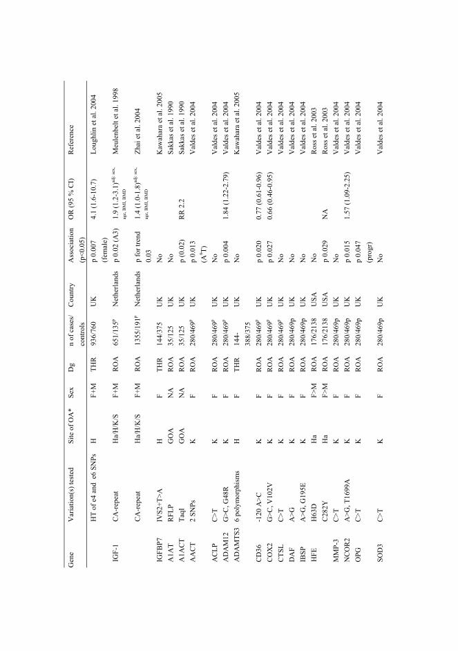
Gen
e V
aria
tion(
s) te
sted
Si
te o
f OA
* Se
x D
g n
of c
ases
/ co
ntro
ls
Cou
ntry
A
ssoc
iatio
n (p
<0.0
5)
OR
(95
% C
I)
Ref
eren
ce
HT
of e
4 an
d e
6 SN
Ps
H
F+M
TH
R
936/
760
UK
p
0.00
7 (f
emal
e)
4.1
(1.6
-10.
7)
Loug
hlin
et a
l. 20
04
CA
-rep
eat
Ha/
H/K
/S
F+M
R
OA
65
1/13
5p N
ethe
rland
s p
0.02
(A3)
1.
9 (1
.2-3
.1)ad
j: se
x,
age,
BM
I, B
MD
Meu
lenb
elt e
t al.
1998
IG
F-1
CA
-rep
eat
Ha/
H/K
/S
F+M
R
OA
13
55/1
91p
Net
herla
nds
p fo
r tre
nd
0.03
1.
4 (1
.0-1
.8)ad
j: se
x,
age,
BM
I, B
MD
Zhai
et a
l. 20
04
IGFB
P7
IVS2
+T>A
H
F
THR
14
4/37
5 U
K
No
K
awah
ara
et a
l. 20
05
A1A
T R
FLP
GO
A
NA
R
OA
35
/125
U
K
No
Sa
kkas
et a
l. 19
90
A1A
CT
TaqI
G
OA
N
A
RO
A
35/1
25
UK
p
(0.0
2)
RR
2.2
Sa
kkas
et a
l. 19
90
AA
CT
2 SN
Ps
K
F R
OA
28
0/46
9p U
K
p 0.
013
(A9 T)
Val
des e
t al.
2004
AC
LP
C>T
K
F
RO
A
280/
469p
UK
N
o
Val
des e
t al.
2004
A
DA
M12
G
>C, G
48R
K
F
RO
A
280/
469p
UK
p
0.00
4 1.
84 (1
.22-
2.79
) V
alde
s et a
l. 20
04
AD
AM
TS3
6 po
lym
orph
ism
s H
F
THR
14
4-38
8/37
5 U
K
No
K
awah
ara
et a
l. 20
05
CD
36
-120
A>C
K
F
RO
A
280/
469p
UK
p
0.02
0 0.
77 (0
.61-
0.96
) V
alde
s et a
l. 20
04
CO
X2
G>C
, V10
2V
K
F R
OA
28
0/46
9p U
K
p 0.
027
0.66
(0.4
6-0.
95)
Val
des e
t al.
2004
C
TSL
C>T
K
F
RO
A
280/
469p
UK
N
o
Val
des e
t al.
2004
D
AF
A>G
K
F
RO
A
280/
469p
U
K
No
V
alde
s et a
l. 20
04
IBSP
A
>G, G
195E
K
F
RO
A
280/
469p
U
K
No
V
alde
s et a
l. 20
04
H63
D
Ha
F>M
R
OA
17
6/21
38
USA
N
o
Ros
s et a
l. 20
03
HFE
C
282Y
H
a F>
M
RO
A
176/
2138
U
SA
p 0.
029
NA
R
oss e
t al.
2003
M
MP-
3 C
>T
K
F R
OA
28
0/46
9p
UK
N
o
Val
des e
t al.
2004
N
CO
R2
A>G
, T16
99A
K
F
RO
A
280/
469p
U
K
p 0.
015
1.57
(1.0
9-2.
25)
Val
des e
t al.
2004
O
PG
C>T
K
F
RO
A
280/
469p
U
K
p 0.
047
(pro
gr)
V
alde
s et a
l. 20
04
SOD
3 C
>T
K
F R
OA
28
0/46
9p
UK
N
o
Val
des e
t al.
2004

Gen
e V
aria
tion(
s) te
sted
Si
te o
f OA
* Se
x D
g n
of c
ases
/ co
ntro
ls
Cou
ntry
A
ssoc
iatio
n (p
<0.0
5)
OR
(95
% C
I)
Ref
eren
ce
TIM
P1
C>T
, L12
4L
K
F R
OA
28
0/46
9p
UK
N
o
Val
des e
t al.
2004
TN
A
A>G
, S10
6G
K
F R
OA
28
0/46
9p
UK
p
0.01
4 (p
rogr
)
Val
des e
t al.
2004
e4+3
7G>A
, R14
4Q
K
F R
OA
28
0/46
9p
UK
N
o
Val
des e
t al.
2004
TN
FAIP
6 e4
+37G
>A, R
144Q
H
F+
M
THR
37
8/76
0 U
K
No
Lo
ughl
in e
t al.
2004
*
Ha
= ha
nd; H
= h
ip; K
= k
nee;
S =
spin
e H
T =
hapl
otyp
e; V
NTR
= v
aria
ble
num
ber o
f tan
dem
repe
ats
RO
A =
radi
olog
ical
OA
; TH
R =
tota
l hip
repl
acem
ent;
TJR
= to
tal j
oint
repl
acem
ent,
eith
er T
HR
or T
KR
; TK
R =
tota
l kne
e re
plac
emen
t p
= po
pula
tion
base
d st
udy
prog
r = p
rogr
essi
on o
f OA
a
Eith
er p
atie
nts w
ith p
rimar
y hi
p O
A p
atie
nts o
r pat
ient
s with
seco
ndar
y O
A d
ue to
hip
dys
plas
ia
adj =
adj
uste
d fo
r, B
MI =
bod
y m
ass i
ndex
; BM
D =
bon
e m
iner
al d
ensi
ty; H
RT
= us
e of
hor
mon
e re
plac
emen
t the
rapy
; MP
= ag
e at
men
opau
se

94
References for the supplemental table 1. Aerssens J, Dequeker J, Peeters J, Breemans S & Boonen S (1998) Lack of association between
osteoarthritis of the hip and gene polymorphisms of VDR, COL1A1, and COL2A1 in postmenopausal women. Arthritis Rheum 41:1946-1950.
Bergink AP, van Meuers JB, Loughlin J, Arp PP, Fang Y, Hofman A, van Leeuwen JPTM, van Duijn CM, Uitterlinden AG & Pols HAP (2003) Estrogen receptor α gene haplotype is associated with radiographic osteoarthritis of the knee in elderly men and women. Arthritis Rheum 48:1913-1922.
Forster T, Chapman K & Loughlin J (2004) Common variants within the interleukin 4 receptor α gene (IL4R) are associated with susceptibility to osteoarthritis. Hum Genet 114:391-395.
Granchi D, Stea S, Sudanese A, Toni A, Baldini & Giunti A (2002) Association of two gene polymorphisms with osteoarthritis secondary to hip dysplasia. Clin Orthop 403:108-117.
Horton WE, Lethbridge-Cejku M, Hochberg MC, Balakir R, Precht P, Plato CC, Tobin JD, Meer L & Doege K (1998) An association between an aggrecan polymorphic allele and bilateral hand osteoarthritis in elderly white men: data from the Baltimore Longitudinal Study of Aging (BLSA). Osteoarthritis Cartilage 6:245-251.
Huanq J, Ushiyama T, Inoue K, Kawasaki T & Hukuda S (2000) Vitamin D receptor gene polymorphisms and osteoarthritis of the hand, hip, and knee: a case-control study in Japan. Rheumatol 2000 39:79-84.
Hull & Pope (1989) Osteoarthritis and cartilage collagen genes. Lancet 8650:1337-1338. Ikeda T, Mabuchi A, Fukuda A, Hiraoka H, Kawakami A, Yamamoto S, Machida H, Takatori Y,
Kawaguchi H, Nakamura K & Igekawa S (2001) Identification of sequence polymorphisms in two sulfation-related genes, PAPSS2 and SLC26A2, and an association analysis with knee osteoarthritis. J Hum Genet 46:538-543.
Ikeda T, Mabuchi A, Fukuda A, Kawakami A, Yamada R, Yamamoto S, Miyoshi K, Haga N, Hiraoka H, Takatori Y, Kawaguchi H, Nakamura K & Igekawa S (2002) Association analysis of single nucleotide polymorphisms in cartilage-specific collagen genes with knee and hip osteoarthritis in the Japanese population. J Bone Miner Res 17:1290-1296.
Kawahara C, Forster T, Chapman K, Carr A & Loughlin J (2005) Genetic association analysis of the IGFBP7, ADAMTS3, and IL8 genes as the potential osteoarthritis susceptibility that maps to chromosome 4. Ann Rheum Dis 64:474-476.
Keen RW, Hart DJ, Lanchbury JS & Spector TD (1997) Association of early osteoarthritis of the knee with a Taq I polymorphism of the vitamin D receptor gene. Arthritis Rheum 40:1444-1449.
Kirk KM, Doege KJ, Hecht JT, Bellamy N & Martin NG (2003) Osteoarthritis of the hands, hips and knees in an Australian twin sample – evidence of association with Aggrecan VNTR polymorphism. Twin Res 6:62-66.
Kizawa H, Kou I, Iida A, Sudo A, Miyamoto Y, Fukuda A, Mabuchi A, Kotani A, Kawakami A, Yamamoto S, Uchida A, Nakamura K, Notoya K, Nakamura Y & Igekawa S (2005). An aspartic acid repeat polymorphism in asporin inhibits chondrogenesis and increases susceptibility to osteoarthritis. Nat Genet, in press (published on-line 9.1.2005).
Loughlin J, Dowling B, Chapman K, Marcelline L, Mustafa Z, Southam L, Ferreira A, Ciesielski C, Carson DA & Corr M (2004) Functional variants within the secreted frizzled-related protein 3 gene are associated with hip osteoarthritis in females. Proc Natl Acad Sci USA 101:9757-9762.
Loughlin J, Dowling B, Mustafa Z & Chapman K (2002b) Association of the interleukin-1 gene cluster on chromosome 2q13 with knee osteoarthritis. Arthritis Rheum 46:1519-1527.
Loughlin J, Dowling B, Mustafa Z, Smith A, Sykes B & Chapman K (2000a) Analysis of the association of the matrilin-1 gene (CRTM) with osteoarthritis: comment on the article by Meulenbelt et al. Arthritis Rheum 43:1423-5.
Loughlin J, Irven C, Athanasou N, Carr A & Sykes B (1995) Differential allelic expression of the type II collagen gene (COL2A1) in osteoarthritic cartilage. Am J Hum Genet 56:1186-1193.

95Loughlin J, Mustafa Z, Dowling B, Southam L, Marcelline L, Räinä SS, Ala-Kokko L & Chapman
K (2002a) Finer linkage mapping of a primary hip osteoarthritis susceptibility locus on chromosome 6. Eur J Hum Genet 10:562-568.
Loughlin J, Sinsheimer JS, Mustafa Z, Carr AJ, Clipsham K, Bloomfield VA, Chitnavis J, Bailey A, Sykes B & Chapman K (2000b) Association analysis of the vitamin D receptor gene, the type I collagen gene COL1A1, and the estrogen receptor gene in idiopathic osteoarthritis. J Rheumatol 27:779-784.
Mabuchi A, Ikeda T, Fukuda A, Koshizuka Y, Hiraoka H, Miyoshi K, Haga N, Kawaguchi H, Kawakami A, Yamamoto S, Takatori Y, Nakamura K & Igekawa S (2001) Identification of sequence polymorphisms of the COMP (cartilage oligomeric matrix protein) gene and association study in osteoarthrosis of the knee and hip joints. J Hum Genet 46:456-462.
Meulenbelt I, Bijkerk C, de Wildt SC, Miedema HS, Valkenburg HA, Breedveld FC, Pols HA, Te Koppele JM, Sloos VF, Hofman A, Slagboom PE & van Duijn CM (1997) Investigation of the association of the CRTM and CRTL1 genes with radiographically evident osteoarthritis in subjects from the Rotterdam study. Arthritis Rheum 40:1760-1765.
Meulenbelt I, Bijkerk C, de Wildt SCM, Miedema HS, Breedveld FC, Pols HAP, Hofman A, van Duijn CM & Slagboom PE (1999) Haplotype analysis of three polymorphisms of the COL2A1 gene and associations with generalised radiological osteoarthritis. Ann Hum Genet 63:393-400.
Meulenbelt I, Bijkerk C, Miedema HS, Breedveld FC, Hofman A, Valkenburg HA, Pols HAP, Slagboom PE & van Duijn CM (1998) A genetic association study of the IGF-1 gene and radiological osteoarthritis in a population-based cohort study (the Rotterdam study). Ann Rheum Dis 57:371-374.
Meulenbelt I, Seymour AB, Nieuwland M, Huizinga TWJ, van Duijn CM & Slagboom PE (2004) Association of the interleukin-1 gene cluster with radiographic signs of osteoarthritis of the hip. Arthritis Rheum 50:1179-1186.
Moos V, Rudwaleit M, Herzog V, Höhlig K, Sieper J & Müller B (2000) Association of genotypes affecting the expression of interleukin-1β or interleukin-1 receptor antagonist with osteoarthritis. Arthritis Rheum 43:2417-2422.
Priestley L, Fergusson C, Ogilvie D, Wordsworth P, Smith R, Pattrick M, Doherty M & Sykes B (1991). A limited association of generalized osteoarthritis with alleles at the type II collagen locus: COL2A1. Br J Rheumatol 30:272-275.
Ross J, Kowalchuk RM, Shaulinsky J, Ross L, Ryan D & Phatak PD (2003) Association of heterozygous hemochromatosis C282Y gene mutation with hand osteoarthritis. J Rheumatol 30:121-125.
Sakkas LI, Macfarlane DG, Bird H, Welsh KI & Panayi GS (1990) Association of osteoarthritis with homozygosity for a 5.8 KB TaqI fragment of the α1-antichymotrypsin gene. Br J Rheumatol 29:245-248.
Southam L, Chapman K & Loughlin J (2003) Genetic association analysis of BMP5 as a potential osteoarthritis susceptibility gene. Rheumatol 42:911-912
Stefánsson SE, Jónsson H, Ingvarsson T, Manolescu I, Jónsson HH, Ólafsdóttir G, Pálsdóttir E, Stefánsdóttir G, Sveinbjörnsdóttir G, Frigge ML, Kong A, Gulcher JR & Stefánsson K (2003) Genomewide scan for hand osteoarthritis: a novel mutation in matrilin-3. Am J Hum Genet 72:1448-1459.
Stern AG, de Carvalho MRC, Buck GA, Adler RA, Rao TPS, Disler D, Moxley G & members of the I-NODAL network (2003) Association of erosive hand osteoarthritis with a single nucleotide polymorphism on the gene encoding interleukin-1 beta. Osteoarthritis Cartilage 11:394-402.
Strusberg I, Sembaj A, Tabares S, Strusberg AM, del Castillo I, Villamar M & Moreno Barral J (2002) Association analysis of genotypic frequencies of matrilin-1 gene in patients with osteoarthritis. Clin Exp Rheumatol 20:543-545.
Uitterlinden AG, Burger H, Huanq Q, Odding E, van Duijn C, Hofman A, Birkenhäger JC, van Leeuwen JPTM & Pols HAP (1997) Vitamin D receptor genotype is associated with radiographic osteoarthritis at the knee. J Clin Invest 100:259-263.
96Uitterlinden AG, Burger H, van Duijn CM, Huang Q, Hofman A, Birkenhager JC, van Leeuwen JP
& Pols HAP (2000) Adjacent genes, for COL2A1 and the vitamin D receptor, are associated with separate features of radiographic osteoarthritis of the knee. Arthritis Rheum 43:1456-1464.
Ushiyama T, Ueyama H, Inoue K, Nishioka J, Ohkubo I & Hukuda S (1998) Estrogen receptor gene polymorphism and generalized osteoarthritis. J Rheumatol 25:134-137.
Valdes AM, Hart DJ, Jones KA, Surdulescu G, Swarbrick P, Doyle DV, Schafer AJ & Spector TD (2004) Association study of candidate genes for the prevalence and progression of knee osteoarthritis. Arthritis Rheum 50:2497-2507.
Vikkula M, Nissila M, Hirvensalo E, Nuotio P, Palotie A, Aho K & Peltonen L (1993). Multiallelic polymorphism of the cartilage collagen gene: no association with osteoarthrosis. Ann Rheum Dis 52:762-764.
Zhai G, Rivadeneira F, Houwing-Duistermaat JJ, Meulenbelt I, Bijkerk C, Hofman A, van Meurs JBJ, Uitterlinden AG, Pols HAP, Slagboom PE & van Duijn CM (2004) Insulin-like growth factor I gene promoter polymorphism, collagen type II α1 (COL2A1) gene, and the prevalence of radiographic osteoarthritis: the Rotterdam study. Ann Rheum Dis 63:544-548.
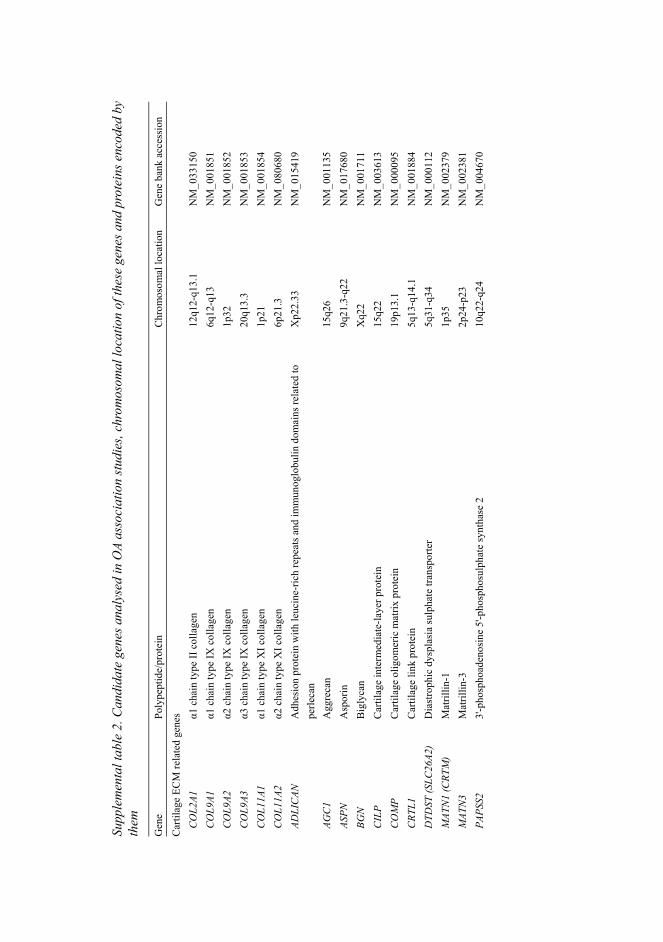
Supp
lem
enta
l tab
le 2
. Can
dida
te g
enes
ana
lyse
d in
OA
asso
ciat
ion
stud
ies,
chro
mos
omal
loca
tion
of th
ese
gene
s and
pro
tein
s enc
oded
by
them
Gen
e Po
lype
ptid
e/pr
otei
n C
hrom
osom
al lo
catio
n G
ene
bank
acc
essi
on
Car
tilag
e EC
M re
late
d ge
nes
C
OL2
A1
α1 c
hain
type
II c
olla
gen
12q1
2-q1
3.1
NM
_033
150
CO
L9A1
α1
cha
in ty
pe IX
col
lage
n 6q
12-q
13
NM
_001
851
CO
L9A2
α2
cha
in ty
pe IX
col
lage
n 1p
32
NM
_001
852
CO
L9A3
α3
cha
in ty
pe IX
col
lage
n 20
q13.
3 N
M_0
0185
3 C
OL1
1A1
α1 c
hain
type
XI c
olla
gen
1p21
N
M_0
0185
4 C
OL1
1A2
α2 c
hain
type
XI c
olla
gen
6p21
.3
NM
_080
680
ADLI
CAN
A
dhes
ion
prot
ein
with
leuc
ine-
rich
repe
ats a
nd im
mun
oglo
bulin
dom
ains
rela
ted
to
perle
can
Xp2
2.33
N
M_0
1541
9
AGC
1 A
ggre
can
15q2
6 N
M_0
0113
5 AS
PN
Asp
orin
9q
21.3
-q22
N
M_0
1768
0 BG
N
Big
lyca
n X
q22
NM
_001
711
CIL
P C
artil
age
inte
rmed
iate
-laye
r pro
tein
15
q22
NM
_003
613
C
OM
P C
artil
age
olig
omer
ic m
atrix
pro
tein
19
p13.
1 N
M_0
0009
5 C
RTL1
C
artil
age
link
prot
ein
5q13
-q14
.1
NM
_001
884
DTD
ST (S
LC26
A2)
Dia
stro
phic
dys
plas
ia su
lpha
te tr
ansp
orte
r 5q
31-q
34
NM
_000
112
MAT
N1
(CRT
M)
Mat
rillin
-1
1p35
N
M_0
0237
9 M
ATN
3 M
atril
lin-3
2p
24-p
23
NM
_002
381
PAPS
S2
3'-p
hosp
hoad
enos
ine
5'-p
hosp
hosu
lpha
te sy
ntha
se 2
10
q22-
q24
NM
_004
670
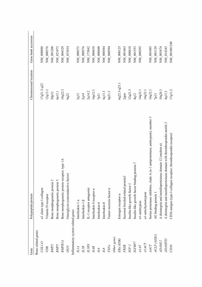
Gen
e Po
lype
ptid
e/pr
otei
n C
hrom
osom
al lo
catio
n G
ene
bank
acc
essi
on
Bon
e re
late
d ge
nes
C
OL1
A1
α1 c
hain
type
I co
llage
n 17
q21.
3-q2
2 N
M_0
0008
8 VD
R V
itam
in D
rece
ptor
12
q13.
1 N
M_0
0037
6 BM
P2
Bon
e m
orph
ogen
etic
pro
tein
2
20p1
2 N
M_0
0120
0 BM
P5
Bon
e m
orph
ogen
etic
pro
tein
5
6p12
.1
NM
_021
073
BM
PR1A
B
one
mor
phog
enet
ic p
rote
in re
cept
or; t
ype
1A
10q2
2.3
NM
_004
329
O
GN
O
steo
glyc
in (o
steo
indu
ctiv
e fa
ctor
) 9q
22
NM
_033
014
Infla
mm
ator
y sy
stem
rela
ted
gene
s
IL
1A
Inte
rleuk
in-1
α
2q13
N
M_0
0057
5 IL
1B
Inte
rleuk
in-1
β
2q14
N
M_0
0057
6
IL1R
N
IL-1
rece
ptor
ant
agon
ist
2q14
.2
NM
_173
842
IL4R
In
terle
ukin
4 re
cept
or α
16
p12.
1 N
M_0
0041
8 IL
6 In
terle
ukin
-6
7p21
N
M_0
0060
0 IL
8 In
terle
ukin
-8
4q13
.3
NM
_000
584
TNFa
Tu
mor
nec
rosi
s fac
tor α
6p
21.3
N
M_0
0059
4 O
ther
gen
es
ER
a (E
SR)
Estro
gen
rece
ptor
α
6q22
.3-q
23.1
N
M_0
0012
5 FR
ZB
Secr
eted
friz
zled
-rel
ated
pro
tein
3 2q
ter
NM
_001
463
IG
F-1
Insu
lin-li
ke g
row
th fa
ctor
-1
12q2
1.3
NM
_000
618
IGFB
P7
Insu
lin-li
ke g
row
th fa
ctor
bin
ding
pro
tein
-7
4q12
N
M_0
0155
3 A1
AT
α1-a
ntitr
ypsi
n 14
q32.
1 N
M_0
0029
5 A1
ACT
α1-a
ntic
hym
otry
psin
14
q32.
1
AAC
T Se
rine
prot
eina
se in
hibi
tor,
clad
e A
(α-1
ant
ipro
tein
ase,
ant
itryp
sin)
, mem
ber 3
14
q32.
1 N
M_0
0108
5 AC
LP (A
EB1)
A
E bi
ndin
g pr
otei
n 1
7p13
N
M_0
0112
9 AD
AM12
A
dis
inte
grin
and
met
allo
prot
eina
se d
omai
n 12
(mel
trin α)
10
q26.
3 N
M_0
0347
4
ADAM
TS3
A d
isin
tegr
in a
nd m
etal
lopr
otei
nase
dom
ain
with
thro
mbo
spon
din
mot
ifs 3
4q
13.3
N
M_0
1424
3
CD
36
CD
36 a
ntig
en (t
ype
I col
lage
n re
cept
or; t
hrom
bosp
ondi
n re
cept
or)
17q1
1.2
NM
_001
0015
48

Gen
e Po
lype
ptid
e/pr
otei
n C
hrom
osom
al lo
catio
n G
ene
bank
acc
essi
on
CO
X2 (P
TGS2
) Pr
osta
glan
din-
endo
pero
xide
synt
hase
2
1q25
N
M_0
0096
3 C
TSL
Cat
heps
in L
9q
21-q
22
NM
_001
912
DAF
D
ecay
-acc
eler
atin
g fa
ctor
for c
ompl
emen
t (C
D55
) 1q
32
NM
_000
574
IBSP
In
tegr
in-b
indi
ng si
alop
rote
in (b
one
sial
opro
tein
) 4q
21-q
25
NM
_004
967
HFE
H
emoc
hrom
atos
is g
ene
6p21
.3
NM
_000
410
MM
P-3
Mat
rix m
etal
lopr
otei
nase
-3
11q2
2.3
NM
_002
422
N
CO
R2
Nuc
lear
rece
ptor
cor
epre
ssor
2
12q2
4 N
M_0
0631
2 O
PG
Tum
or n
ecro
sis f
acto
r sup
erfa
mily
, mem
ber1
1b (o
steo
prog
erin
) 8q
24
NM
_002
546
SOD
3 Su
pero
xide
dis
mut
ase
3, e
xtra
cellu
lar
4p16
N
M_0
0310
2
TIM
P1
Tiss
ue in
hibi
tor o
f met
allo
prot
eina
se 1
X
p11.
3-p1
1.23
N
M_0
0325
4 TN
A (C
LEC
3B)
Tetra
nect
in (p
lasm
inog
en-b
indi
ng p
rote
in)
3p22
-p21
.3
NM
_003
278
TNFA
IP6
Tum
or n
ecro
sis f
acto
r α-in
duce
d pr
otei
n 6
2q23
.3
NM
_007
115