definition of parturitional injury - pedrad > home · · 2013-12-11definition of parturitional...
TRANSCRIPT

1
Parturitional Brain Injury
Thierry A.G.M. Huisman, MD
Director Pediatric Radiology and Pediatric Neuroradiology
Johns Hopkins Hospital
Definition of parturitional injury
• Any condition that affects the fetus adversely
during labor and delivery
• May be caused by:
– Hypoxia and infection (birth injury)
– Mechanical forces (birth trauma)
Definition of parturitional injury
• Any condition that affects the fetus adversely
during labor and delivery
• May be caused by:
– Hypoxia and infection (birth injury)
– Mechanical forces (birth trauma)
• Life starts with a mechanical trauma
– Squeezed together by a muscular wrapping
– Pushed through a narrow, bony canal with multiple bumps
– Getting your neck extended, rotated and pulled
– Life line (umbilical cord) may be compressed
– Possibly additional “medieval” instrumentation
– All of this for many minutes or even hours
Introduction
• Life starts with a mechanical trauma
– Or even worse, within minutes you are squeezed
and “ejected”
Introduction
• Life starts with a “stress” trauma
– And than suddenly lots of light, noise and many
crying/emotional people around you,….
Introduction

2
Tortuguero expedition. www.philsheldon.wordpress.com
Subjects who try to relive the
“I am stuck feeling”
Guettler FV, et al. Magnetic resonance imaging of the active second stage of labour: Proof of principle Eur Radiol 2012;22:2020-2026
„Scientific approach”
Epidemiology
Disability adjusted life year (DALY):
Measure of overall disease burden expressed as number of years lost due to ill-health, disability or early death
• Significant variability across the world
Epidemiology
• Dramatically decreased in last decades
• Birth trauma in 3% of all live births
• Accounts for less than 2% of neonatal deaths
• Even when the injuries are benign, birth trauma
may result in significant anxiety for a family
Reichard R. Birth injury of the cranium and CNS. Brain Pathology 2008;18:565-570
Risk factors for Birth Trauma
• Maternal risk factors
– Diabetes
– Obesity
– Small pelvis
– Large weight gain
– Induction of labor
– Epidural analgesia
– Primiparity
– History of macrosomic
infant
• Infant risk factors
– Macrosomia (>3500g)
– Delayed and prolonged
delivery
– Abnormal presentation
– Instrumented delivery
– Perinatal depression
– Shoulder dystocia
Adapted from: Parker LA, et al. early recognistion and treatment of birth trauma: Injuries to the head and face. Advances in neonatal care, 2005;6:288-297
Kinds of mechanical injury
• Mechanical injury to
– The brain and skull
– The spinal cord
– The head and neck region
• Skin lacerations and hematomas
• Clavicular fracture
• Brachial plexus injury
• Facial nerve injury
• Phrenic nerve injury
• Laryngeal nerve injury
• Nasal injury
www.birthinjury.org

3
Incidence of types of birth trauma
Hughes CA, et al. Birth trauma in the head and neck. Arch Otolaryngol Head neck Surg 1999;125:193-199
Extracranial
hematomas
Extracranial injuries
• Scalp abrasions and
lacerations
– Frequent
– Vaginal and instrumental
delivery (10%)
– Scalp, face, cheek, ear
– Rarely of clinical
significance
www.newborns.stanford.edu
Extracranial injuries
• Caput succedaneum
• Subgaleal hematoma
• Cephalohematoma
Bumps may look very similar, clinical
significance is however very different!!!
www.newborns.stanford.edu
CS SH CH
Extracranial injuries
• Clinical presentation usually allows to
differentiate the various swellings
• Difference is the location, composition and
etiology of the fluid collection
Serous-Sanguineous fluid Serous-Sanguineous fluid
Caput succedaneum
• Serous-sanguinous fluid collection within the scalp
between skin and galea or epicranial aponeurosis
• Results from high pressure exerted on infant‟s head
during labor
• Presenting portion of the scalp, usually the vertex
Caput succedaneum
• Present at delivery, decreases spontaneously within 24-
48h
• Soft swelling, irregular margins, petechiae, purpura
and/or ecchymosis, pitting edema
• Fluid shifts from side to side with head position
• Crosses sutures, often crosses the midline
www.newborns.stanford.edu

4
Primigravida, diabetic mother, shoulder dystocia
Caput succedaneum
+10d
Caput succedaneum
• 20-40% of vacuum extractions ~>
artificial caput also known as “chignon”
• Difficult, prolonged deliveries, PROM (no
amniotic fluid to cushion), primigravidas
• May also be seen intrauterine:
Oligohydramnios, Braxton-Hicks
contractions
• No treatment, imaging used to exclude
other extracranial hematomas
Subgaleal hematoma• Serous-Sanguinous fluid collection between
galea aponeurosis and periosteum
• May be mistaken for caput succedaneum
because swelling (also) crosses sutures
• Not always clinically apparent immediately post
partum, but develops/enlarges over hours-days
Subgaleal hematoma
• Results from tearing of emissary veins
(connects dural sinuses with scalp veins)
• Most often after vacuum assisted delivery
• May also occur spontaneously, or due to skull
fractures or rupture of synchondrosis
Person sent on a special mission
Subgaleal hematoma• Potentially life threatening condition
• Subgaleal space extends from the orbital ridges to the
nuchal ridge, ears and connects into the neck along the
superficial neck fascia (260 ml, term infants)
– No tamponading characteristics
– Blood volume term neonate: 85 mL/kg
~> Hypovolemic shock and death may occur
www.medutah.edu
Subgaleal hematoma
• Bleeding disorders can result in large hematomas and
delayed presentations
– Vitamin K deficiency
– Thrombocytopenia
– Hemophilia
– DIC, consumption coagulopathy
• Neonate may develop hyperbilirubinemia
• Usually resolution in 2-3 weeks, good long term outcome
• Occasionally blood transfusion, blood products or
surgical evacuation

5
Subgaleal hematoma
Hyperdense, crossing sutures, extending into the neck region
Cephalohematoma
• Sanguinous fluid collection between periosteum
and bony calvarium
• Usually not present at birth (unless long labor),
develops within 24h
• Firm, tense mass, does not cross sutures
Cephalohematoma
Ellipsoid, contained blood collection, limited to the bounderies of the sutures
Cephalohematoma
Bilateral, contained by the sutures, some additional subgaleal fluid
Cephalohematoma
• Result from shear forces during birth that tear
emissary and diploic veins resulting in
hemorrhage in the subperiostal space
Cephalohematoma
• Hemorrhage slowly lifts periosteum from calvarium
• Tamponade by periosteum
• Most frequently in parietal location, R:L=2:1 (?)
• May be unilateral or bilateral
• May cross midline in occipital region
• Co-existing CS or SH may obscure suture boundaries
www.newborns.stanford.edu

6
Cephalohematoma• More common in primigravidas, macrosomia,
instrument assisted delivery, prolonged/difficult
labor and deviant position
• May also be present in utero
– Oligohydramnios
– Premature rupture of membranes
• Twice as often in boys than in girls (?)
• Overlying skin is not discolored
• Mass cannot be transilluminated or shifted
• Painful on palpation
Cephalohematoma
• Prognosis is excellent, usually resolves
spontaneously within weeks-months, unless,…
• Complications
– Underlying skull fracture (5-18%)
– Anemia
– Hyperbilirubinemia
– InfectionOften linear fracture
www.mediphotos.blogspot.com
Cephalohematoma
• Infection:
– If infant has local erythema, unexplained fever or
sepsis, cephalohematoma may be source of infection
(E. Coli and Staph aureus)
– Cellulitis, osteomyelitis or meningitis may result
– Scalp electrodes, needle aspiration for decompression
Chen M-H, et al. MRI features of an infected cephalohematoma in a neonate. Journal of Clinical Neuroscience 2006;13:849-852
Cephalohematoma
• Calcification on follow up
~> Surgical augmentation of bony
prominence or molding helmet
Extracranial hematomas
• Problem in daily life
– Often several hematomas affecting multiple
compartments are present simultaneously
– Clear differentiation may be limited
acute cephalohematoma
Reichard R. Birth injury of the cranium and CNS. Brain Pathology 2008;18:565-570
Acute subgaleal hemorrhage
Extracranial hematomas
Subgaleal hematoma + cephalohematoma + subcuatenous edema

7
Extracranial hematomas
Spontaneous recovery on follow up
Differential diagnosis of bumps
Epidermal inclusion cyst Posttraumatic encephalocele
Meningocele
Skull fractures
• Skull fracture must be suspected if there
is an cephalohematoma or intracranial
hemorrhage
• Caused by compression during labor
while skull pushed against maternal
pelvis or due to forceps blades
• Kind of fractures
– Linear fractures
– Depressed fractures
– Occipital osteodiastasis
– Leptomingeal cysts (rare complication)
JAMA 1999;135:697-703
Skull fractures
• Linear fractures
– Usually asymptomatic, heal
without intervention
– Frequently parietal bone
– Cephalohematoma may be
associated
– No relation between size of
cephalohematoma and
presence of fracture
Skull fractures
• Depressed fractures
– Indentation of skull
– Ping pong ball type defect
– Surgical intervention may be required
Skull fractures
• Depressed fractures
– Often associated with additional hematomas
– May obscure the lesion or anatomical boundaries
Ping pong fracture + subgaleal hematoma + SDH

8
Skull fractures
• Leptomeningeal cysts
• Occur along fracture lines
• Meninges are trapped within the fracture line, CSF
pulsations widens fracture line ~> growing fracture
Reichard R. Birth injury of the cranium and CNS. Brain Pathology 2008;18:565-570
Skull fractures
• Occipital osteodiastasis
• Separation of squamous and lateral occipital bone
• Anterior displacement and upward rotation of squamous
portion by suboccipital pressure (breech delivery)
• Posterior fossa SDH, brainstem/cerebellum injury
• Sutural diastasis
• Intracranial edema or hematomas
• DD Non-accidental injury
Skull fractures Caution: skull fractures
Fell from couch while mother was feeding him,…
Fell out of arms of father while walking down staircase,…
• 5-6/10‟000 life births
• Risk factors: Forceps (x6),
vacuum, prolonged delivery,
macrosomia
• EDH, SDH, SAH, IVH
• Parenchymal
contusion/laceration
Intracranial injuries Intracranial injuries

9
Epidural hemorrhage
• Between calvarial bone and
dura mater
• Shear forces and vertical
overriding/molding of skull
bones ~> injury to dura mater
• May be arterial or venous
• Typically associated with
instrument assisted delivery
• Surgical intervention may be
necessary
Epidural hemorrhage
• Frequently associated with
cephalohematoma or skull
fracture with secondary injury
to the middle meningeal artery
• Middle meningeal artery in
neonates not yet embedded
~> EDH rare (2% of all
intracranial hemorrhages)
• Often close to sutures
Epidural hemorrhage
Vertical shear, no fracture
EDH + Cephalohematoma
Subdural hemorrhage
• In virtual space between
dura mater and arachnoidea
• Shear forces tear the bridging
veins and venous sinuses
• Skull molding and sutural
diastasis ~> dural sinus injury
• Supra- and infratentorial
• Along falx and tentorium
• Most common intracranial hemorrhage
• Subtle symptoms <~> irritability,
seizures, bradycardia, full fontanel
Subdural hemorrhage
Dural sinuses may be dense in neonates, blood is usually denser
Subdural hemorrhage
Posterior fossa SDH with
extension into spinal canal and
small ischemic lesion

10
Subarachnoid hemorhage
• Between arachnoidea and
pia mater
• Rupture of small veins bridging
leptomeninges
• Seizures, irritability, apnea,
somnolence, focal neurology
• 2nd most common intracranial
hemorrhage
• Often associated with IVH (reflux)
• Usually good prognosisSkull fracture, subarachnoid hemorrhage, subdural hematoma, intraventricular blood,
extracranial multicompartment hematomas
Subarachnoid hemorhage
Parenchymal hemorrhage
• Isolated parenchymal
hemorrhage is very rare
• In combination with skull fracture
• Due to additional pathology
– Direct trauma (instrumentation)
– Coagulation disorders
– AVM/AVF
• Symptoms and treatment
depend on location
Parenchymal hemorrhage
Skull fracture, subdural hematoma + intraparenchymal hematoma
Associated head, neck, spine injury
• Clavicular fracture
• Brachial plexus injury
• Phrenic nerve injury
• Facial nerve injury
• Laryngeal nerve injury
• Nasal injury
Overall rare but may result in:
• Stridor
• Respiratory distress
• Feeding difficulties
• Cosmetic deformity
Hughes CA, et al. Birth trauma in the Head and neck. Arch Atolaryngol Head Neck Surg 1999;125:193-199
Associated head, neck, spine injury
• Clavicular fracture
– Associated with brachial plexus, phrenic, right
recurrent nerve injury
– 2.7-5.7/1000 births
– Macrosomia and shoulder dystocia
Hughes CA,Arch Atolaryngol Head Neck Surg 1999;125:193-199
11
Associated head, neck, spine injury
• Brachial plexus injury
– 1/1000 births
– Erb palsy (90%): C5/C6
(No Moro reflex)
– Klumpke palsy: C7-T1
(No Moro + no grasp reflex)
– Horner: Sympathetic fibers
of T1
• Spontaeneous recovery is
the rule
Hughes et al. Arch Otolaryngol head Neck Surg 1999;125:193199
Waiters-tip grasp
Associated head, neck, spine injury
Vargas MI, et al. Neuroradiology 2010;52:237-245
DTICISS
• Brachial plexus injury
Associated head, neck, spine injury
• Facial nerve palsy
– In 0.8% of birth trauma
– Forceps delivery (posterior blade)
– 33% spontaneous
– 2M:1F
– Usually spontaneous
recovery in hours-weeks
– No value for imaging other
than to rule out developmental
facial nerve palsy (Moebius) www.newborns.stanford.edu
www.birthinjury.org
Associated head, neck, spine injury
Vialle R, et al. J Materb Fetal Neonatal Med 2007;20:435-450
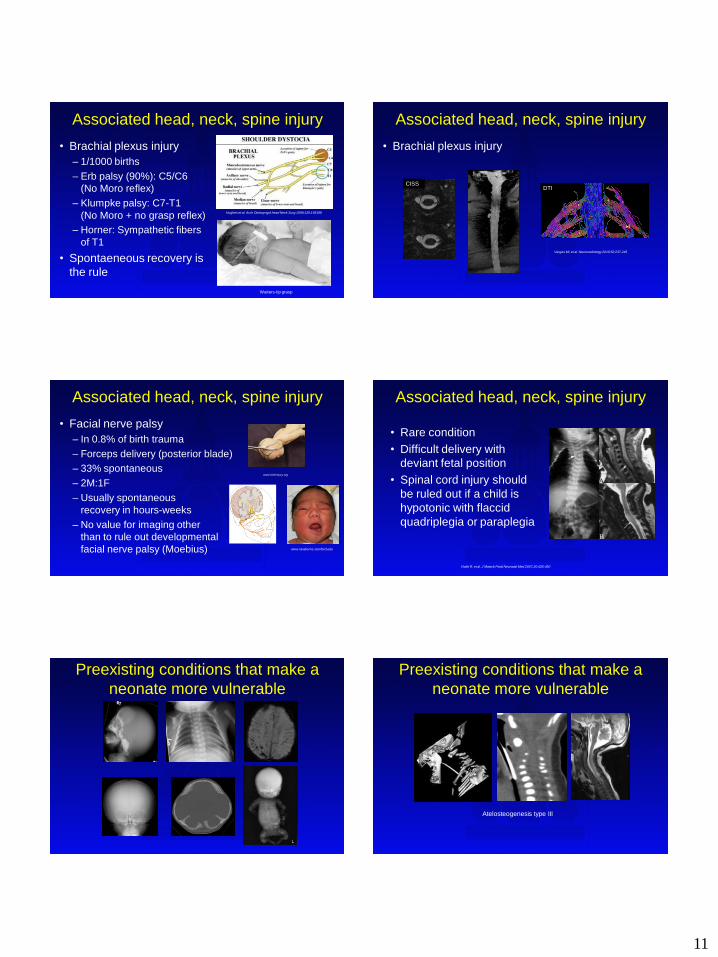
• Rare condition
• Difficult delivery with
deviant fetal position
• Spinal cord injury should
be ruled out if a child is
hypotonic with flaccid
quadriplegia or paraplegia
Preexisting conditions that make a
neonate more vulnerable
Atelosteogenesis type III
Preexisting conditions that make a
neonate more vulnerable
12
Prexisting injury that may
delay/complicate delivery
Early diagnosis may prevent additional injury
Preexisting injury
Child, mother and obstetrician will thank you
Preexisting injury
Child, mother and obstetrician will thank you
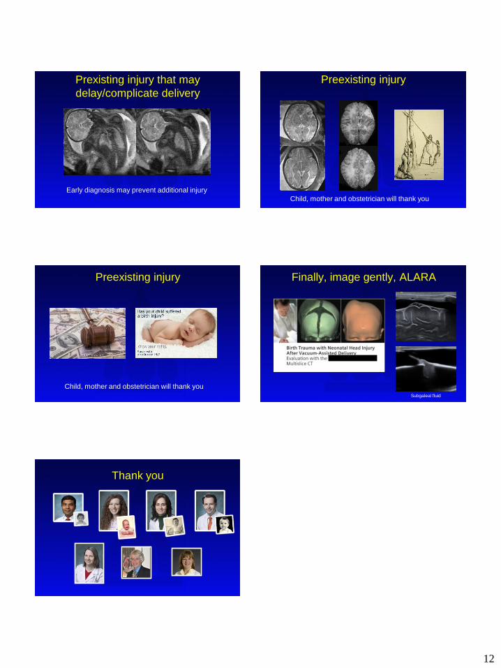
Finally, image gently, ALARA
Subgaleal fluid
Thank you