degree of master of science in dentistry by coursework and
TRANSCRIPT
IMPACTED MANDIBULAR THIRD MOLARS: THE EFFICACY OF
PROPHYLACTIC ANTIBIOTICS AND CHLORHEXIDINE MOUTHWASH IN
PREVENTING POSTOPERATIVE INFECTIONS.
Pooshan Gopee
Degree of Master of Science in Dentistry by coursework and research report
A Research Report submitted to the Faculty of Health Sciences, University of the
Witwatersrand, Johannesburg, in partial fulfilment of the requirements for the degree of
Master of Science in Dentistry.
Johannesburg, 2016

Declaration
I, Pooshan Gopee, declare that this research report is my own work. It is being submitted for the
degree of Master of Science in Dentistry in the University of the Witwatersrand, Johannesburg. It
has not been submitted before for any degree or examination at this or any other university.
S fckday of 20 U .
ii

Dedication
“Aum Shn Ganeshaya Namaha”
To my parents for their valuable guidance, support and encouragement throughout my
studies and to the almighty for his continuous blessing and strength granted to keep
believing and persevering in every task I undertake.

ABSTRACT
Objective: The aim of this study was to investigate:
1) The efficacy of a prophylactic antibiotic regimen compared to a chlorhexidine
mouthwash in reducing postoperative infections in mandibular third molar surgery.
2) The pattern of presentation and the indication for extraction of mandibular third
molars.
Study Design and Method: A total of 100 patients were randomly assigned to two groups
(group 1, 15 ml of chlorhexidine mouthwash for 1 minute before surgery; group 2, 2g
amoxicillin orally 1 hour before surgery). The outcome which included surgical site
infection and other complications was assessed 7 days postoperatively.
Data collected included patients’ age, gender, type of impaction, indication for extraction
and surgical morbidity (postoperative complications). The data were then analysed using
the statistical package STATA 13.1 for Windows.
Results: Of the 100 patients, 4 patients in group 1 and 3 patients in group 2 presented with
surgical wound infection. The infection rate was 8% for group 1 and 6% for group 2 while
the overall infection rate was 7%. No statistically significant difference in surgical wound
infection was found between the 2 groups.
The ages ranged from 18 to 46 years with a mean of 27.75(+/- 5.79). There were 48 males
and 52 females, the male to female ratio being 1:1.08. Mesioangular impaction was the
most common type of impaction recorded (46; 46%), while the most prevalent indication
for extraction was pericoronitis (39; 39%).
IV

Conclusion: In terms of efficacy, this study failed to show that amoxicillin prophylaxis is
more effective than a preoperative chlorhexidine mouthrinse for reducing postoperative
infections in third molar surgery. Hence, antibiotic prophylaxis must not be routinely
administered in non-immunocompromised patients for such procedures.
Patients that presented for mandibular third molar extraction were generally young with an
almost equal distribution in gender. The pattern of presentation as well as the indication for
extraction of mandibular third molars seem to correlate with those found in literature.
v

ACKNOWLEDGEMENTS
I would like to express my sincere gratitude to the following persons:
■ Dr E Rikhotso for his valuable guidance and assistance in completing the research
report.
■ Dr M Kasilembo for his assistance and permission to perform the study at Rob
Ferreira hospital.
■ Dr K L Ntambwe for his continuous encouragement and for allowing me to carry
out the study at Ermelo hospital.
■ My fellow colleagues at Rob Ferreira and Ermelo hospitals for assisting me in the
collection of data.
■ Dr M Mabongo and Dr P Struthers for their valuable time in reading the research
report and making insightful recommendations.
■ Mr E Ngamasana, the biostatistician, for his assistance and contribution in
analysing the data collected.
■ Ms S Lukhoo for her unwavering support and encouragement.
■ To everyone who has contributed, either directly or indirectly, into making this
research report a possibility.
VI

TABLE OF CONTENTS
TITLE PAGE
Page
i
DECLARATION ii
DEDICATION iii
ABSTRACT iv
ACKNOWLEDGEMENTS \i
TABLE OF CONTENTS vii
LIST OF FIGURES X
LIST OF TABLES xi
NOMENCLATURE xii
CHAPTER 1: INTRODUCTION/ BACKGROUND 1
CHAPTER 2: LITERATURE REVIEW 3
2.1 Definition 3
2.2 Development 3
2.3 Aetiology 4
2.4 Classification 5
2.5 Pattern of Presentation 6
2.6 Indications for Removal 7
VII

2.7 Prophylaxis in third molar surgery 8
CHAPTER 3: AIMS AND OBJECTIVES 14
3.1 Aims 14
3.2 Objectives 14
3.3 Hypothesis 15
CHAPTER 4: MATERIALS AND METHODS 16
4.1 Ethical Clearance 16
4.2 Inclusion Criteria 16
4.3 Exclusion Criteria 17
4.4 Study Design 18
4.5 Data Collection 18
4.6 Surgical Data 18
4.7 Data Analysis 21
CHAPTER 5: RESULTS 22
CHAPTER 6: DISCUSSION 34
viii
CHAPTER 7: CONCLUSION 41

APPENDIX 43
APPENDIX A- Ethical Clearance 43
APPENDIX B- Information sheet 44
APPENDIX C- Consent form 45
APPENDIX D- Data Collection sheet 46
REFERENCES 47
IX
LIST OF FIGURES
Figure 5.1 Distribution of the participants by gender. 22
Figure 5.2 Age distribution of the patients by gender. 27
Figure 5.3 Age distribution of participants by type of impaction. 28
Figure 5.4 Gender distribution of patients by type of impaction. 29
Figure 5.5 Type of impaction by infection status. 31
Figure 5.6 Gender disparities associated with infection. 32
Figure 5.7 Flowchart diagram 33
x

LIST OF TABLES
Table 2.1 Summary of the investigations earned out and clinical findings. 12
Table 5.1 Description of the study population. 23
Table 5.2 Association between baseline characteristics of the patients and 25
presence of infection.
Table 5.3 Analysis of indications for extraction by the type of impaction. 30
XI

NOMENCLATURE
American Dental Association =ADA
American Heart Association =AHA
American Academy of Orthopaedic Surgeons =AAOS
British Society for Antimicrobial Chemotherapy =BSAC
World Health Organisation =WHO
Chlorhexidine =CHX
Amoxicillin =AMOX
Human Immunodeficiency Virus =HIV
Standard Deviation =STD
Inter quartile Range =IQR
XII

CHAPTER 1
INTRODUCTION/ BACKGROUND
Removal of impacted teeth is a very common surgical procedure performed by Oral and
Maxillofacial Surgeons as well as general dentists in the South African Public Health
sector.1 Owing to the complexity of this surgical procedure, extensive skills and training
are required in order to reduce the risk of complications that may arise during and after
surgery.1 In addition, adequate knowledge regarding the diagnostic and treatment
modalities is essential in order to achieve optimum results for the patients.
Rob Ferreira Hospital and Ermelo Provincial Hospital are two public hospitals in the
province of Mpumalanga in South Africa where removal of impacted wisdom teeth are
routinely performed by general dentists. Although a generally low complication rate is
associated with such procedures, postoperative surgical site infection has been observed
to remain common in mandibular third molar removal at these institutions. In this
setting, prophylactic antimicrobials are often used haphazardly in an attempt to curb the
incidence of infection.
The unsystematic prescription of antibiotics among dentists has become a serious cause
for concern due to the rapid development of antibiotic resistance. Presently, there is no
consensus with regards to the efficacy of antibiotics in third molar surgery despite a
plethora of studies being available in the literature.2,3 In addition, there is a limited
number of studies that have assessed the effectiveness of prophylactic mouthrinses in
third molar surgery.3
1
Very few studies on impacted third molar teeth have been conducted in South Africa.4-5
The only identified study on antibiotic prophylaxis in third molar surgery was carried
out by Siddiqui et al6 at the University of the Western Cape.
Hence, the rationale behind this study is to generate a pool of data not only in the
demographics and distribution patterns in patients presenting with impacted third molar
teeth but also in treatment outcomes after removal. Such data will be essential in
establishing evidence based clinical guidelines that will further assist in the development
of sound treatment protocols.
2

CHAPTER 2
LITERATURE REVIEW
Impacted Mandibular Third Molars
2.1 Definition
Peterson and Ness1 have defined an impacted tooth as one that cannot erupt into a
normal functioning position during the usual range of expected time. In 2004, Farman
characterized impacted teeth as those that cannot erupt due to some physical obstruction
along the path of eruption.7
Impaction of third molar teeth remains a veiy common pathological condition with a
frequency of occurrence reported to range from 16.7%-68.6%.8 Generally, the eruption
ages are between 17 and 21 years.7 The wide variation in ages may be due to differences
in race, nature of the diet, the force with which the masticatory apparatus is used or
genetic characteristics.7
2.2 Development
Radiographic evidence of third molar tooth starts as early as seven years of age, with
cusp mineralization being complete at around 11 years.1 At this age, the tooth bud is
located in the anterior border of the mandibular ramus with its occlusal surface facing
anterior and upward at an angle of 40-45 degrees.9 The tooth then migrates forward and
3

upward, rotating from a horizontal to mesioangular and ultimately into a vertical
position distal to the second molar.9 The crown formation is completed at around 14
years while the root is fully formed by the age of 20.1,10 Assuming that the tooth has
sufficient space to erupt, then it will be brought into its normal position by about 20
years of age.1 Any failure for the tooth to rotate into its normal position will lead to
impaction.1
2.3 Aetiology
Several theories have been proposed following the high incidence of impacted
mandibular teeth.
One of the most popular theories is a lack of growth of the retromolar space between the
second molar and the ascending ramus.7 The Mendelian theory advocates that the coarse
nature of the diet of primitive men led to increased teeth attrition with a subsequent
decrease in the collective length of teeth, thereby increasing the space needed to
accommodate wisdom teeth.5 Moreover, the unrefined nature of the food as opposed to
what is presently available, resulted in increased masticatory movements and force
which probably stimulated jaw growth.5
Other theories put forward include malposition of the tooth germs, insufficient eruption
force of mandibular third molars and a decrease in the mandibular angulation.7
The causes of impaction have been classified into local and systemic factors.11
4

Among the local factors are: abnormal position of an adjacent tooth, dense overlying
bone, nature of the overlying soft tissues, arch length and tooth size discrepancy,
malposition of the tooth bud, over retained deciduous teeth and unfavourable path of
eruption.11
Systemic causes are subdivided into prenatal and postnatal causes. The prenatal cause is
mainly hereditary and postnatal causes include rickets, anaemia, congenital syphilis,
malnutrition, cleidocranial dysostosis and tuberculosis among others.11
2.4 Classification
Two systems of classification are widely advocated for impacted mandibular third
molars and these are the Pell & Gregory classification (1933) and Winter’s classification
(1926).7,12
The Pell and Gregory classification evaluates the depth of the tooth within the alveolar
bone as well as the distance that exists between the anterior ramus of the mandible and
the distal aspect of the second molar.
Winter’s classification, as described below, assesses the long axis of the impacted 3rd
molar in relation to the long axis of the second molar.1113
Mesioangular: The long axis of the 3rd molar bisects the long axis of the second molar at
or above the occlusal plane.
Distoangular: The long axes of the 2nd and 3rd molars are divergent at the occlusal plane.
Horizontal: The long axis of the 3rd molar bisects that of the second molar at a right angle.
Vertical: The long axes of the 2nd and 3rd molars run parallel to one another.
5
Transverse: The tooth is impacted in a bucco-lingual direction.
Inverted: The crown of the tooth is directed vertically down towards the inferior alveolar
canal.
A study carried out by Almendros Marqes et al12 comparing the two third molar
classification systems concluded that intra-examiner and inter-examiner reproducibility
levels are very high when attempting to classify third molar teeth based on the Winter’s
classification system, as compared to the Pell and Gregory classification system. Hence,
Winter’s classification was used in this study.
2.5 Pattern of Presentation
Several studies have been carried out to determine the pattern of distribution of impacted
mandibular third molars.5,814,15 With regards to gender, most studies have shown no sexual
predilection for impacted third molars, while some studies revealed a higher frequency in
Caucasian, South East Iranian and Singapore Chinese females.1416 As far as the type of
impaction is concerned, Kramer and Williams17 found that mesioangular and horizontal
impactions account for 75% of all mandibular impactions. Hashemipour et al8 revealed
that mesioangular impaction was more common in the mandible (48.3 %). Gtobolorun et
al18 and Tabetze5 similarly noted higher prevalences of mesioangular mandibular
impaction in their studies carried out at the Lagos university hospital and the university of
Limpopo respectively. However, their results were in contrast with those found by
Hugoson and Kugelberg (1988) who noted that vertical impaction was most common.16
6

2.6 Indications for Removal
Removal of impacted third molar teeth has remained a subject of controversy over many
years. Some clinicians advocate for the prophylactic removal of third molars, while others
suggest little justification for the removal of pathology free impacted third molars.19,20
However, as a general principle, once an impacted third molar tooth has been diagnosed,
it has to be evaluated for extraction, periodic monitoring or retention.19
Nordenram et al20 in an investigation of 2630 cases of mandibular third molar extractions,
reported that 60% of the teeth involved had some pathological changes with pericoronitis
being the most common diagnosis.
A prospective study on the indications for third molar surgery done at the Queen
Alexandra hospital in Portsmouth, United Kingdom demonstrated that the commonest
recorded reason for third molar extraction was recurrent pericoronitis (35.7%) followed
by intermittent pain (20.7%), single episode of pericoronitis (17.2%), caries (8.7%) and
periodontal disease (7.1%).21
In contrast, caries and its sequelae (63.2%) was the most common reason for extraction in
a separate review of 1763 cases carried out by Adeyemo et al22.
Taking into account the indications mentioned above, it is generally agreed that if the
potential benefits outweigh the risks involved for third molar surgery, then it is
recommended that the tooth be extracted.
7

The oral cavity harbours a diverse and complex microbial community. Due to the high
number of host bacteria present at the operative site, postoperative complications remain
common during third molar surgery.23 The non-infection related complications usually
include pain, swelling and erythema due to the normal inflammatory process following
trauma while complications that may occur with infection include alveolar osteitis,
necrotic bone, lymphadenopathy (localized/ generalized) and fascial space involvement.24
The infection rate associated with mandibular third molar extraction has been reported to
range from 1-12.6%.24 In order to reduce the incidence of infection, the use of antibiotic
therapy or antimicrobial agents such as chlorhexidine mouthwash has often been advanced
to reduce bacterial contamination at the surgical site.24,25
Antibiotic prophylaxis is described as the prescription of antibiotics in order to prevent
infection at the operative site.3 Various antibiotic regimens have been used with different
timing in administration. In South Africa, the general trend for antibiotic prescription is a
five day postoperative course. Similarly, in Australia between 78% to 90% of dentists
prescribe a five day postoperative course of antibiotic prophylaxis following the removal
of third molars.3
However, there is considerable evidence that antibiotics taken preoperatively may have
significant effects on the rate of postoperative infections, while other studies have also
demonstrated that prophylactic antibiotics may not be of any value in reducing the
incidence of infection.6,25'28
2.7 Prophylaxis in third Molar Surgery
8

In a randomized controlled clinical trial evaluating the efficacy of amoxicillin and
metronidazole in third molar surgery, Pasupathy et al26 failed to demonstrate any
significant advantage over the placebo in reducing postsurgical wound infections.
The results of the study on antibiotic prophylaxis in third molar surgery carried by
Siddiqui et al6 showed that preoperative or postoperative prophylactic antibiotics did not
have any significant effect in reducing postoperative infections and should not be routinely
administered in non-immunocompromised patients.
Arteagoitia at al29 investigated the effect of amoxicillin/ clavulanic acid to prevent
infection following lower impacted mandibular third molar removal. They reported no
significant difference in infection rate in the antibiotic group and the placebo.
Ataoqlu et al30 devised a study involving three groups of patients requiring removal of
lower third molar teeth. The first group received no antibiotic treatment while the second
and third groups were administered a five day preoperative and postoperative course of
amoxicillin respectively. There were no significant differences in postoperative
complications including infection in the groups assessed.
In a prospective, randomized controlled trial, Olusanya et al31 compared the efficacy of a
single pre-emptive bolus of antibiotics to that of a five day postoperative course of
antibiotic regimen. They found no significant difference in the reduction of surgical
morbidity in the two groups.
9

Acute phase protein levels were used as indicators of infection by Bulut et al32. The levels
of C-reactive protein and alpha-1 antitrypsin were measured preoperatively and
postoperatively in patients that received prophylactic antibiotics or placebos. They
concluded that there is no statistically significant difference in the incidence of infection
between the two groups.
Lopez Cedrun et al28 investigated the efficacy of amoxicillin treatment in preventing
postoperative complications in 123 patients. It was concluded that patients who received
preoperative or postoperative amoxicillin prophylaxis showed greater efficacy in
preventing postoperative complications than the placebo group.
Similarly, Monaco et al27 in their evaluation of the removal of third molars in 59 young
patients, demonstrated a statistically significant difference in postoperative complications
between patients that received preoperative amoxicillin prophylaxis and the control group.
In a recent systematic review of randomized controlled trials to assess the effectiveness of
a single dose of preoperative antibiotics during lower third molar extraction, Marcussen
et al33 deduced that a single preoperative bolus of 2g amoxicillin significantly reduced the
incidence of surgical site infection.
While the need for antibiotic therapy in third molar surgery remains controversial, it is
unclear whether the use of antibacterial mouthrinses have an effect in the reduction of
surgical morbidity in third molar surgery. Several studies have claimed their usefulness in
reducing the incidence of alveolar osteitis.34 However, there is a paucity of studies that
have assessed their efficacy in reducing postoperative infections in third molar surgery.
10

The relatively few studies that compared the efficacy of antibacterial mouthrinses to an
antibiotic regimen have all used bacteraemia as a marker for infection.35'37
A pilot study carried out by Tuna et al35 to evaluate the effects of antibacterial mouthrinses
on bacteraemia concluded that bacteraemia was reduced with 0.2% chlorhexidine and 7.5
% Povidone Iodine mouthrinses in third molar surgery.
Duvall36 and colleagues compared the efficacy of a 0.12% chlorhexidine and amoxicillin
in reducing the incidence and magnitude of bacteraemia in third molar extraction. It was
deduced that an oral rinse or systemic antibiotic intervention does not significantly reduce
the incidence and magnitude of bacteraemia.
A study to compare the effectiveness of amoxicillin, clindamycin and a chlorhexidine
mouthrinse in the prevention of post extraction bacteraemia was carried out by Maharaj
et al37. The study revealed a statistically significant difference in bacteraemia between the
amoxicillin and chlorhexidine groups. However, none of the regimens were effective in
preventing post extraction bacteraemia.
11

Table 2.1: Summary of the investigations earned out and the clinical findings thereof.
INVESTIGATOR TYPE OF
STUDY
GROUP SIZE FINDINGS
Siddiqui et a l6,
2010
-Prospective -Randomized -Double blind -Controlled
o Placebo o Preoperative
antibiotics o Postoperative
antibiotics
100 No significant difference in infection rate between groups
Pasupathy et a l26,
2011
-Prospective-Randomized-Controlled
o Placebo o Preoperative
Amoxicillin o Preoperative
Metronidazole
89 No significant difference in infection rate between groups
Monaco et a l21, 2009
-Prospective-Randomized-Controlled
o Placebo o Preoperative
Amoxicillin
59 Significant difference between Control and Amoxicillin groups
Arteogotia et a l29, 2015
-Prospective -Randomized -Double blind -Controlled
o Placebo o Preoperative and
continuing postoperatively
118 No significant difference in infection rate between groups
Ataoqlu et al30, 2008
-Prospective-Randomized-Controlled
o Placebo o Preoperative
Amoxicillin/
Clavulanic acid o Postoperative
Amoxicillin/ Clavulanic acid
150 ■ No significant difference in complications
and infection rate between groups
Olusanya et a l31, 2011
-Prospective-Randomized-Controlled
o Preoperative Amoxicillin/ Metronidazole
o Postoperative Amoxicillin/ Metranidazole
84 No significant difference in surgical morbidity between groups
12

Maharaj et a l32, 2012
-Prospective-Controlled
o Control o Chlorhexidine o Amoxicillin o Clindamycin
160 Significant difference in bacteraemia between the
Control and Chlorhexidine
groups when compared to the Amoxicillin group
Tuna et a l35, 2012
-Prospective-Controlled
o Sterile Saline o Povidone Iodine
o Chlorhexidine
34 Incidence of bacteraemia reduced in the Povidone Iodine andChlorhexidinegroups
Duvall et a l36, 2013
-Prospective-Randomized-Blind-Controlled
o Placebo o Chlorhexidine o Preoperative
Amoxicillin
160 No Statistical difference in reducing bacteraemia between the groups
13

CHAPTER 3
AIMS AND OBJECTIVES
3.1 Aims
This study aims to investigate the efficacy of a prophylactic antibiotic therapy as compared
to a chlorhexidine regimen in preventing postoperative infections in third molar surgery,
and to establish the association between the pattern of presentation of impacted
mandibular third molars with age, gender and indications for extraction.
3.2 Objectives
1. To evaluate the complication that arises due to infection after third molar surgery in
two groups of patients.
2. To provide current local data on the age and gender of patients presenting with
impacted mandibular third molars.
3. To evaluate the type of impacted mandibular third molars based on Winter’s
classification.
4. To determine the indications for the removal of impacted lower third molars.
14

rn
1. There is no difference in efficacy between the preoperative antibiotic therapy and a
regimen of chlorhexidine mouthrinse.
2. There is no gender predilection for patients presenting with impacted mandibular third
molars.
The type of impaction is independent of the age or gender of the patients.
4. Indications for extraction are independent of the type of impaction.
3.3 Hypothesis
15

CHAPTER 4
MATERIALS AND METHODS
4.1 Ethical Clearance (Ethical Considerations/ Issues)
Permission to carry out the study was obtained from the heads of each institution where
the study was undertaken. All information gathered in this study was treated with
confidentiality. Patient file numbers were used instead of names. Since the study
involved clinical assessment and treatment on human subjects, a submission for Ethics
approval was submitted to the Human Research Ethics Committee (HREC) of the
University of the Witwatersrand and granted; Certificate NO: M140435 (Appendix A).
Patients who fulfilled the inclusion criteria were given written and verbal explanation of
the study and were made to sign a consent form (Appendix B) for agreeing to participate
in the study.
4.2 Inclusion Criteria
1. Patients with impacted third molars presenting to Ermelo and Rob Ferreira
hospitals and who gave consent to participate in the study.
2. Patients between the ages of 18-50 years and who are medically competent.
3. Patients undergoing removal of impacted maxillary molars were also included in
the study.
16

4.3 Exclusion Criteria
1. Patients with co morbidities.
2. Patients presenting with third molars that have incomplete root formation.
3. Patients that failed to give consent or withdrew from the study.
INCLUSION AND EXCLUSION CRITERIA
r >POPULATION
v J
Single Bolus of Pre Preoperative Oral rinseemptive antibiotic 1 with 15 ml ofhour before surgery chlorhexidine for 1 min
17
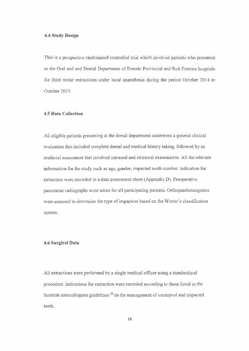
4.4 Study Design
This is a prospective randomized controlled trial which involved patients who presented
to the Oral and and Dental Department of Ermelo Provincial and Rob Ferreira hospitals
for third molar extractions under local anaesthesia during the period October 2014 to
October 2015.
4.5 Data Collection
All eligible patients presenting at the dental department undement a general clinical
evaluation that included complete dental and medical history taking, followed by an
orofacial assessment that involved intraoral and extraoral examination. All the relevant
information for the study such as age, gender, impacted tooth number, indication for
extraction were recorded in a data assessment sheet (Appendix D). Preoperative
panoramic radiographs were taken for all participating patients. Orthopanthomograms
were assessed to determine the type of impaction based on the Winter's classification
system.
4.6 Surgical Data
All extractions were performed by a single medical officer using a standardized
procedure. Indications for extraction were recorded according to those listed in the
Scottish intercollegiate guidelines38 on the management of unerupted and impacted
teeth.
18

To test the effectiveness of the prophylactic regimen in the study, patients were randomly
assigned to two groups (using two sealed envelopes). In the first group (control group),
patients were instructed to rinse with 15ml of 0.2% chlorhexidine (with alcohol)
mouthrinse for one minute prior to the surgery. In the second group (test group), 2g
amoxicillin was administered orally one hour before the surgery.
The Control group consisted of 50 patients; 22 males and 28 females.
The Test group consisted of 50 patients; 26 males and 24 females.
All the operations were of a surgical nature and performed under Local Anaesthesia (2%
lignocaine with 1:80000 adrenaline)
The surgical procedure was earned out based on the university’s protocol on third molar
extraction. Envelope or triangular mucoperiosteal flap elevation with ostectomy and/or
odontectomy was achieved using a surgical scalpel blade no. 15 for best access.
Ostectomy was perfonned using a crosscut tapered fissure bur mounted on a straight
handpiece. The tooth was sectioned appropriately where necessary depending on the
surgeon’s judgement whilst trying to achieve minimal exposure.
Following the removal of the tooth, the surgical site was debrided and irrigated with sterile
water. Primary closure of the flap was achieved using resorbable chromic catgut sutures,
3/0.
19

The time of surgery was recorded as the time span between the first incision and the last
suture placed.
Patients were prescribed 1 g of Paracetamol with codeine and 400mg of Ibuprofen as
analgesics and postoperative instructions of care were given, which included rinsing with
warm saline three times daily starting from the day after surgery.
A recall visit was scheduled seven days later and any postoperative complications were
noted in a questionnaire. Pain, swelling, trismus, alveolitis and surgical site infection were
recorded.
Trismus is defined as an inability to clear an inter incisal distance of at least 2 cm.
Alveolar osteitis is defined as pain that arises 2-5 days after surgery, presence of necrotic
tissue, exposed bone, and absence of clot.23,39
Infection is defined as a purulent discharge at the extraction site with/ without painful
induration.23
If any complication arose before the recall visit was scheduled, the patient was asked to
report back to the hospital to receive the appropriate treatment and postoperative
antibiotics were then prescribed, if required. The complication was recorded on the data
collection sheet.
20

4.7 Data Analysis
A questionnaire was used to capture all relevant data needed for the study. Based on
previous studies, a sample size of 100 patients was used.
Data were captured on an excel spreadsheet which was later imported into Statal3.1 for
further analysis.
Participants were described using frequencies and percentage for categorical predictors
and means and standard deviations for continuous variables.
Associations were investigated using appropriate statistical tests such as the Student's t-
test and Fisher’s exact test for categorical predictors and ANOVA test for equal Variance
to analyze any significant differences inter-groups. Statistical tests used were two sided
and p values <0.01 were considered significant.
2 1

CHAPTER 5
RESULTS
A total number of 110 patients that visited Rob Ferreira and Ermelo provincial hospitals
for third molar surgical extractions were recorded for the study. Out of the 110, 100
patients met the inclusion criteria with 52 Female patients (52%) and 48 (48%) Male
patients. The Female to Male ratio was 1.08:1.
Distribution of participants By Gender
Males Females
Figure 5.1 shows a representation of the participants by gender.
22
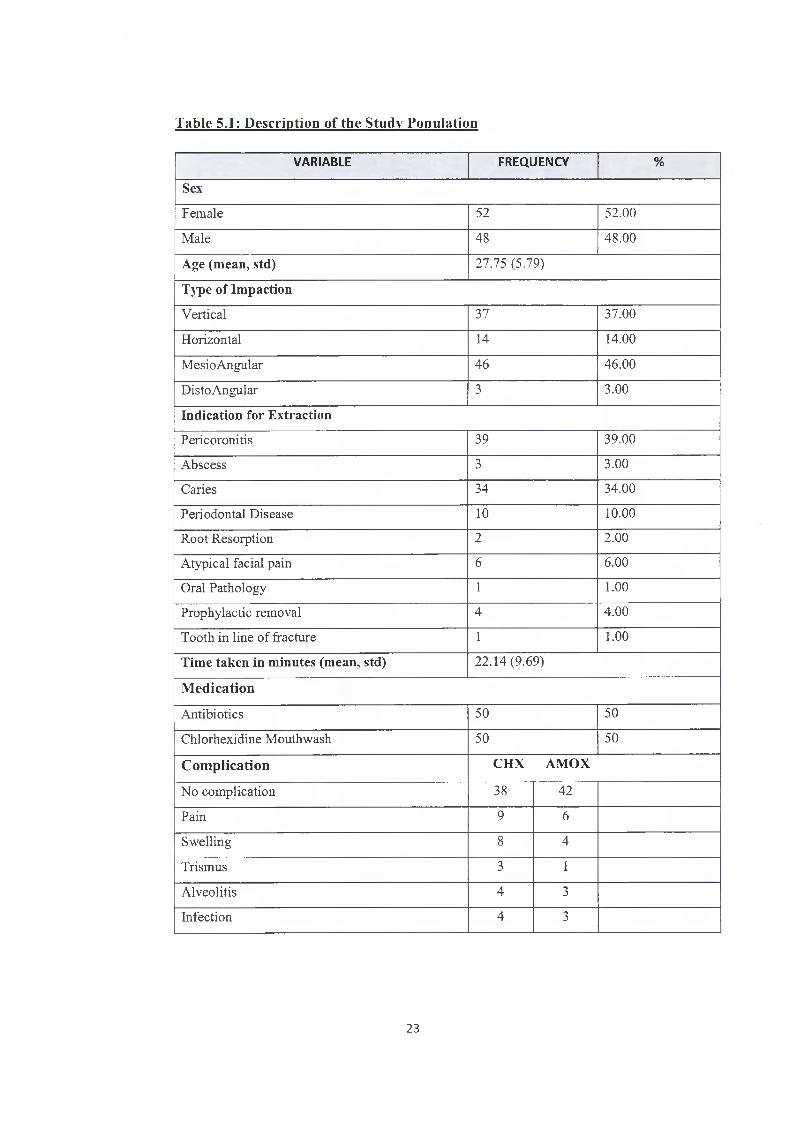
Table 5.1: Description of the Study Population
VARIABLE FREQ UENCY %
SexFemale 52 52.00
Male 48 48.00
Age (mean, std) 27.75 (5.79)
Type of ImpactionVertical 37 37.00
Horizontal 14 14.00
MesioAngular 46 46.00
Disto Angular 3 3.00
Indication for ExtractionPericoronitis 39 39.00
Abscess 3 3.00
Caries 34 34.00
Periodontal Disease 10 10.00
Root Resorption 2 2.00
Atypical facial pain 6 6.00
Oral Pathology 1 1.00
Prophylactic removal 4 4.00
Tooth in line of fracture 1 1.00
Time taken in minutes (mean, std) 22.14(9.69)
Medication
Antibiotics 50 50
Chlorhexidine Mouthwash 50 50
Complication CHX AMOX
No complication 38 42
Pain 9 6
Swelling 8 4
Trismus 3 1
Alveolitis 4 3
Infection 4 3
23

Table 5.1 shows a description of the study population by frequency count. Of the 100
patients who participated into the study, the two types of medication were evenly
distributed with 50% receiving each type of medication.
The frequencies of the various complications occurred, were recorded. Two or more
complications may have presented simultaneously. Alveolitis was recorded in the
presence or absence of infection. For the purpose of this study, the focus was on
infection as a complication.
Mesioangular impactions followed by vertical impactions were the highest angulations
recorded. There were no patients that presented with an inverted type of impaction.
Pericoronitis and caries accounted for most of the indications for extraction. In addition,
no tooth was extracted due to the following indications: Prosthetic Rehabilitation,
Orthodontic treatment or Radiotherapy.
The average time taken to complete the procedure was 22.14 minutes with a standard
deviation of 9.69 minutes. The minimum and maximum times to complete the procedure
were recorded as 5 and 54 minutes respectively.
24

Table 5.2: Association betw een baseline characteristics of the patients and
presence of infection.
Variable Presence of Infection Total p-value
Yes No
Sex
Female 4 (7.69 %) 48 (92.31%) 521.00
Male 3 (6.25%) 45 (93.75%) 48
Age (mean, std) 26.85 (5.21) 27.82 (5.85) 0.67
Type of Impaction
Vertical 1 36 37
0.187Horizontal 1 13 14
MesioAngular 4 42 46
Disto Angular 1 2 3
Indication
Pericoronitis 4 35 39
0.651
Abscess 0 3 3
Caries 1 33 34
Periodontal Disease 1 9 10
Root Resorption 0 2 2
Atypical facial pain 1 5 6
Oral Pathology 0 1 1
Prophylactic Removal 0 4 4
Tooth in line of Fracture 0 1 1
Time taken in minute (median,
IQR)
28 (20-35) 20(15-26) 0.07
Medication
Antibiotic 3 47 50 1.00
Chlorhexidine mouthwash 4 46 50
25

Table 5.2 depicts the association between the presence of infection and the different
variables recorded. The p values calculated show that there is no association between the
presence of infection and the following variables: Gender, Type of impaction,
Indications for extraction. Time taken. Medications used.
26
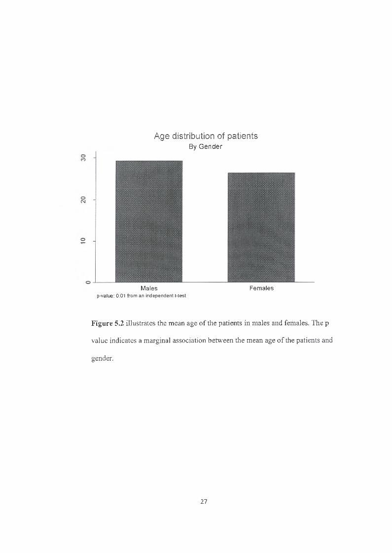
Age distribution of patientsBy Gender
Males Femalesp-value: 0.01 from an independent t-test
Figure 5.2 illustrates the mean age of the patients in males and females. The p
value indicates a marginal association between the mean age of the patients and
gender.
27
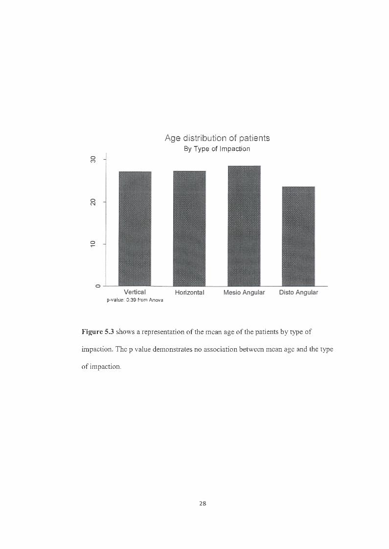
Age distribution of patientsBy Type of Impaction
p-value: 0.39 from Anova
Figure 5.3 shows a representation of the mean age of the patients by type of
impaction. The p value demonstrates no association between mean age and the type
of impaction.
28

Vertical Horizontal
Gender distribution of Participants
By Type of Impaction
G raphs by TYPE OF IMPACTION
Figure 5.4 shows the frequency distribution of males and females that presented
with the different types of impaction. The Fisher’s Exact test indicates a p value
of 0.467 and there is no association between gender and the type of impaction.
29

Table 5.3: Analysis of Indications for Extraction bv the Type of Impaction
IndicationsType of
Impaction
Vertical Horizontal Mesioangular Distoangular Total
Pericoronitis 17 6 13 3 3 9
Abscess 2 - 1 - 3
Caries 14 1 19 - 34
PeriodontalDisease
2 2 6 10
RootResorption
- 1 1 “ 2
Atypical Facial Pain
1 1 4 “ 6
OralPathology
1 - - - 1
ProphylacticRemoval
" 2 2 " 4
Tooth in Line of Fracture
“ 1 - 1
Total 37 14 4 6 3 100
The Fisher’s Exact test shows a p value of 0.070 and there is no association
between the type of impaction and the indications for extraction of third molar
teeth.
30

Type of ImpactionBy Infection Status
No infection Infection
Figure 5.5 shows the different types of impacted teeth associated with their
infection status. The p value from the Fisher’s exact test is 0.651 and demonstrates
no association between the type of impaction and the infection status.
31
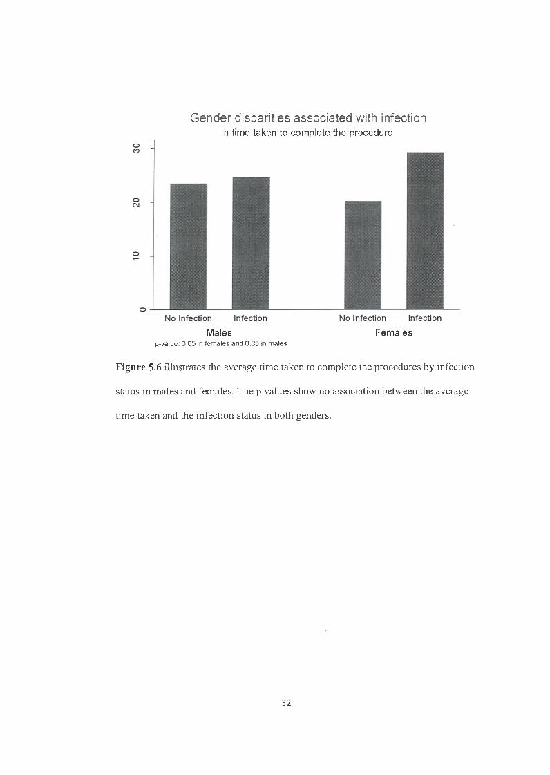
Gender disparities associated with infectionIn time taken to complete the procedure
Males Femalesp-value: 0 .05 in fem ales and 0.85 in males
Figure 5.6 illustrates the average time taken to complete the procedures by infection
status in males and females. The p values show no association between the average
time taken and the infection status in both genders.
32

Flowchart diagram
Figure 5.7 The above flowchart shows a summary of the study and results
obtained.
33

CHAPTER 6
DISCUSSION
This study attempted to compare the efficacy in prophylaxis of two different therapeutic
strategies:
1) A one minute oral rinse with 15 ml of chlorhexidine mouthwash,
2) A single bolus of 2g Amoxicillin, prior to mandibular third molar extraction in healthy
patients.
110 patients were followed. Seven patients did not attend their recall visit and were
excluded from the study. Three patients had an operating time for extraction of above one
hour. Patients with an operating time of one hour and above were not included due to
increased exposure to bacteria within the oral cavity which could compromise the results.
In addition, the procedures were performed in the absence of pericoronitis or active
infection. Therefore, a total of 100 patients completed this study with 50% each receiving
either type of treatment. Each subject had an equal chance of being selected, thus
eliminating any possibility of bias. Furthermore, the assessment of the recall visit was
recorded by independent practitioners who were blinded to the type of treatment
dispensed.
Among the 50 patients, to whom a preoperative rinse of chlorhexidine mouthrinse (control
group) was administered, four patients presented with postoperative infections and in the
group that consumed a bolus of preoperative antibiotics (test group), three patients
34

presented with postoperative infections. The infection rate recorded in this study was 8%
for the chlorhexidine group and 6% for the antibiotic group. The Fisher’s exact test
between the two groups of patients demonstrates a p value > 0.01 indicating no significant
difference in infection rate between the chlorhexidine group and the antibiotics group.
Based on this finding, it can be deduced that the results in terms of efficacy obtained with
either antimicrobial therapy is comparable and therefore an assessment of their mechanism
of action, risk and cost benefits is important before making a recommendation.
Chlorhexidine gluconate is a well-known antimicrobial agent with a broad spectrum of
activity against both Gram positive and Gram negative bacteria, facultative anaerobes and
aerobes, yeasts as well as certain viruses including HIV.40 It exists in the form of cations
and readily binds to the negatively charged particles of the oral mucosa and bacteria
exhibiting an immediate as well as a prolonged antimicrobial effect on a broad spectrum
of bacteria and fungi.40 Its use in the routine disinfection of patients and medical devices
in ICU or in surgical scrubs has been well established.41 In dentistry, chlorhexidine in the
form of an oral rinse, is often used for prophylaxis or in the treatment of oral infections.
In this study, a 0.2% oral rinse of chlorhexidine gluconate was used as a prophylaxis. At
this concentration, chlorhexidine displays bactericidal properties by disrupting the
function of cell membranes and altering the osmotic equilibrium of bacteria.40 The
rationale behind its use in this study was to reduce the oral bacterial load at the surgical
site, thereby decreasing bacteraemia which harbours significant risks for post extraction
infections.42 Organisms that have shown high susceptibility to chlorhexidine include
several species of s ta p h y lo co cc i, s tre p to co c c i and various anaerobes which are important
constituents of the complex oral flora.43
35

Several studies have demonstrated that a preoperative rinse with 0.2% chlorhexidine has
significant antimicrobial effects on the oral flora and post extraction bacteraemia.35-42 In
1997, the AHA recommended the use of an antiseptic mouthwash to reduce bacteraemia
prior to any dental manipulation before modifying it in 2007. In 2006, the British Society
for Antimicrobial Chemotherapy (BSAC) recognized the importance of a preoperative
rinse with a 0.2% chlorhexidine mouthwash before any dental procedures in patients at
high risk of infective endocarditis.44 It has also been suggested that forceful rinsing with
a chlorhexidine mouthwash may actually produce bacteraemia but there is no substantial
evidence to confirm this hypothesis.42
In addition to its broad antibacterial spectrum and substantivity, one of the major
advantages of chlorhexidine is the absence of resistance to the microorganisms it affects.45
The few and rare documented adverse reactions to chlorhexidine include hypersensitivity,
unpleasant taste or tooth discolouration.40 Also, there is no evidence that contradicts the
use of chlorhexidine mouthwash in pregnant or lactating women.40
As opposed to the few studies published on the efficacy of chlorhexidine prophylaxis in
third molar surgery,35,39 there is a plethora of studies on antibiotic prophylaxis.3,26'28
However, its usefulness in preventing postoperative infections continues to remain a
matter of debate. Different treatment protocols and different antibiotics have been used
and conflicting information exists regarding its benefit. Although there are definite
reconunendations by the AHA and ADA/AAOS for antibiotic prophylaxis in the
prevention of infective endocarditis and prosthetic joint infection, the guidelines for
prophylactic antibiotics to prevent surgical site infection in oral and maxillofacial surgery
are less clear.46 It is widely accepted that surgical site infections are caused by the patients’
own endogenous flora and the puipose of prophylaxis is to considerably reduce the
systemic level of the infective pathogens in the tissues at the operative site.47,48
36

The choice of the antibiotics is greatly influenced by the bacteria present at the operative
site. Peterson1 has set forth certain criteria when choosing an antibiotic for prophylaxis.
He advocates that the correct antibiotic with the narrowest antibacterial spectrum must be
selected. In addition, a high enough dosage must be administered at the most appropriate
time and with the shortest exposure. Amoxicillin has been the gold standard for treatment
of infections or prophylaxis in dentistry due to its high efficacy against Gram positive
s tre p to co c c u s and s ta p h y lo c o cc u s species as well as several Gram negative bacteria which
are common isolates in oral infection.49 In addition, its good absorption in the gastro
intestinal tract coupled with its capacity in reaching fast and effective concentrations at
the site it targets has made it an antibiotic of choice.50
A 2g amoxicillin regimen one hour before surgery was used in this study based on the
AHA/ADA/AAOS guidelines.36 Similar protocols were used in several studies with
varying results on the efficacy of the prophylactic treatment.30,51 Ren et al52 performed a
meta-analysis of 23 studies on the effectiveness of antibiotic prophylaxis in third molar
surgery and concluded that when systemic antibiotics are administered before surgery,
they are effective in reducing the frequency of wound infections. In contrast, in a review
of published clinical trials on the efficacy of antibiotic prophylaxis, Oomens et al53
reported that there is a lack of evidence to support the use of prophylactic antibiotics in
lower third molar surgery.
The use of amoxicillin in the prevention of infection in dentistry is a major talking point
due to the development of antibacterial resistance and other adverse reactions such as
anaphylaxis or toxicity.24 The WHO’s 2014 global report on antibiotic resistance
highlighted resistance as a major global threat to worldwide public health with new
resistance mechanisms continuously emerging and spreading globally at an alarming
rate.54 The development of resistance over the years is most probably due to overuse or
37

misuse of antibiotics.24 There is no doubt that the poor antibiotic prescribing practices by
dentists, often motivated by factors ranging from inadequate knowledge to social factors,
contribute significantly towards the fonnation of multiresistant bacterial strains.55
The judicious use of antibiotic prophylaxis based on evidence rather than perception is
imperative. The published infection rate associated with third molar surgery ranges
between 1 %-12.6%.24 The infection rate for amoxicillin and chlorhexidine prophylaxis in
this study is 6% and 8% respectively and falls within the expected rate of infection for
third molar removals. Hence the efficacy of both regimens is comparable.
In view of the potential harmful complications associated with the use of antibiotics for
prophylaxis and the relatively low rate of infection posed by third molar surgery, the use
of amoxicillin is not warranted for such procedures. However, chlorhexidine gluconate
which is a cheap, safe and broad spectrum antiseptic must be recommended to reduce the
oral bacterial count at the surgical site prior to making an incision.56
The demography and socio economic status of the patients seen in Mpumalanga is such
that many of them have never visited the dentist, and the presence of plaque and calculus
in these subjects will be higher than expected. Therefore the administration of a
chlorhexidine mouthrinse prior to extraction is highly recommended.
Moreover, a session of professional scaling would have been desirable before third molar
surgery to decrease the oral bacterial load but due to the limited resources in the public
service and the long waiting list to get an appointment, this treatment plan is not always
feasible.
Finally, the use of chlorhexidine must be an adjunct to proper surgical technique in an
aseptic environment to achieve optimum treatment outcomes.
38

As far as demographics are concerned, the ratio of male to female in this study was 1:1.08,
which shows an almost equal distribution between the two gender groups. This is in
agreement with most studies that have reported no sexual predilection for impacted third
molars.8 Other studies have shown a higher predilection for females and it is believed that
mandibular growth for females stops by the time the third molar starts erupting resulting
in a lack of space for the tooth to erupt.4 814
The most common type of impaction recorded was the mesioangular type (46%) follow'ed
by the vertical type (37%). Mesioangular impactions are most probably caused by the late
development and maturation of the tooth germ with a resulting lack of space for the tooth
to erupt in a normal position.8 The results w'ere similar to those found by Hashemipour et
al8 and Gbotolorun et al18 in Iran and Nigeria respectively. This is in contrast to other
studies that demonstrated the vertical type of impaction to be more common.4,12
Differences in methods of classification for angulation used, variation in genetics between
the populations studied or the type of food consumed in the different geographical areas
may account for the discrepancies.8 This study also demonstrated that no correlation exists
between the type of impaction and the different variables of age and gender.
The mean age of the subjects was 27.75 with a standard deviation of 5.79. The age group
25-30 had the highest proportion of patients with impacted teeth. The delayed
manifestation of patients for impacted teeth removal may be due to a lack of Oral Health
Education. In certain cases there is also the wrong perception that the tooth is still growing
and will erupt eventually until the symptoms become significant. Moreover, due to
apprehension for dental treatment, most patients in Mpumalanga visit the dentist only
when they are inflicted with pain.
39
Pericoronitis followed by caries accounted for the largest distribution of indication for
extractions. These results are comparable to those found by Krishnan et al57 where
pericoronitis was the most frequent reason for extraction followed by caries. The high
prevalence of these pathologies is not uncommon since they usually present with
symptoms of pain which force patients to seek dental assistance. Pericoronitis in the
patients seen is probably exacerbated by the accumulation of bacteria and food under the
operculum. Similarly caries may have been formed due to poor oral hygiene resulting in
a build-up of food and debris that become impacted around the partially erupted tooth.
Hence, the implementation of oral health education in community based programs remains
key to informing patients on the importance of oral hygiene. Also, several steps must be
taken to bring down the different banders that render access to oral health care difficult
for rural residents. In addition, a paradigm shift from interventional dentistry towards
preventive oral health within the hospital setting will undoubtedly assist in curtailing the
burden of oral diseases in the community.
40

CHAPTER 7
CONCLUSION
This study demonstrated that the prophylactic use of a chlorhexidine mouthrinse and
amoxicillin in third molar surgery is equally effective in keeping postoperative infections
to a minimum in medically competent patients. Furthermore, a single dose of antibiotic
prophylaxis failed to show any additional clinical advantage compared to the use of a
chlorhexidine mouthrinse in reducing infections.
Routine administration of amoxicillin in medically competent patients is therefore not
recommended as the risks associated outweigh the benefits. Amoxicillin prophylaxis must
be restricted to cases where the rate of infection is high and the consequences are severe.
The routine prescription of antibiotics motivated by a fear for surgical site infections is
not justifiable and must be strongly discouraged. The focus must rather be on the
application of sound surgical techniques in an aseptic field.
Owing to the lack of oral health awareness among patients seen in the public health service
in Mpumalanga, the value of chlorhexidine mouthwash as a prophylaxis in third molar
surgery remains paramount.
Despite the recognized benefits of chlorhexidine as an antiseptic in dentistry, no studies
that assess its effectiveness in reducing postoperative infection have been conducted.
Although a few studies have demonstrated its efficacy in reducing post extraction
bacteraemia.35,37 There are no investigations so far that establish the relation between
bacteraemia and disease. Moreover, although the results gave clear indications on the
41

potency of chlorhexidine, the sample size used may not reflect statistical differences in
the true population.
Further studies that encompass larger sample size and that take into account the
relationship between bacteraemia and infection are needed to reach a final consensus on
the efficacy of either type of prophylactic regimen.
If anything, it appears like a session of preoperative professional cleaning and a routine
rinse with 0.2% chlorhexidine mouthrinse is more beneficial than antibiotic
administration.
42

APPENDIX
Appendix A
/ W \
R14/49 Dr Pooshan Gopee
HUMAN RESEARCH ETHICS COMMITTEE (MEDICAL)
CLEARANCE CERTIFICATE NO. M140435
NAME: Dr PooBhan Gapee(Principal Investigators
DEPARTMENT: Oral and Maxillofacial SurgeryErmeio Provincial Hospital Rob Ferreira Hospital
PROJECT TITLE: Impacted Mandibular Third Molars: The Efficacy ofProphylactic Antibiotics and Chlorhexidine Mouthwash ,n Preventing Postoperative Infections
DATE CONSIDERED: 25/04/2014
DECISION: Approved unconditionally
CONDITIONS:
SUPERVISOR: Dr E Rikhotso
APPROVED BY: u,Professor P Cleaton-Jones. Co-Chairperson. HREC (Medical)
DATE OF APPROVAL: 12/03/2014This clearance certificate is valid for 5 years from date of approval. Extension may be applied for.
DECLARATION OF INVESTIGATORS
To be completed in duplicate and ONE COPY returned to the Secretary in Room 10004,10th flow. Senate House, University.l/we fully understand the conditions under which I amfwe are authorized to carry out the above-mentioned research and l/we undertake to ensure compliance with these conditions Should any departure be contemplated, from the research protocol as approves l/we undertake to resubmit the application to the Committee [agree to submit a yearly progress report.
Principal Investigator Signature Date
PLEASE QUOTE THE PROTOCOL NUMBER IN ALL ENQUIRIES
43

Appendix B
INFORMATION SHEET
RESEARCH TITLE: Impacted mandibular third molars: The efficacy of prophylactic antibiotics and chlorhexidine mouthwash in preventing postoperative infections.
Dear Patient,
My name is Dr. Pooshan Gopee. I am a postgraduate part-time student in the Department of Oral and Maxillofacial Surgery at the University of the Witwatersrand. As part of my training, I am carrying out a study on impacted mandibular third molars (blocked wisdom teeth) and the effectiveness of preoperative (before surgery) antibiotic use in the prevention of postoperative (after surgery) complications.
Since you present with the condition stated above, I am kindly requesting you to participate in the study and I would be grateful if I could use your clinical records (physical and medical examination and X rays analysis) for the purposes of this study. It is hoped that this study will help provide a better understanding of the above mentioned dental condition and improve the treatment and service delivery to our patients.
This study involves the testing of the efficacy of preoperative antibiotics versus a chlorhexidine mouthwash in the elimination of postoperative infections. The risk for the complete elimination of infections after surgery cannot be guaranteed but appropriate treatment shall be dispensed should any complications arise.
All your personal information will remain confidential as far as possible and will be used for research purposes only. Absolute confidentiality cannot be guaranteed and may be disclosed by law if required. Please note that your participation in this study will be on a purely voluntary basis and you may refuse to participate in the study or you may withdraw from the study at any time without affecting the outcome of your treatment.
If you require any additional information regarding this study, please feel free to contact me on 076 470 3139. Any complaints regarding the study may be addressed to the Human Research Ethics Committee of the University of the Witwatersrand on 011 717 1252.
Thank you.
44

Appendix C
CONSENT FORM
RESEARCH TITLE: Impacted mandibular third molars: The efficacy of prophylactic antibiotics and chlorhexidine mouthwash in preventing postoperative infections.
Details of the study including possible side effects of the drugs to be used as well as implications on my participation in the study have been clearly and fully explained to me. I freely agree to take part in the study and I understand that I have the authority to refuse to participate. I am also allowed to withdraw from the study at any given time without compromising the outcome of treatment.
NAME:
SIGNATURE: DATE:
45
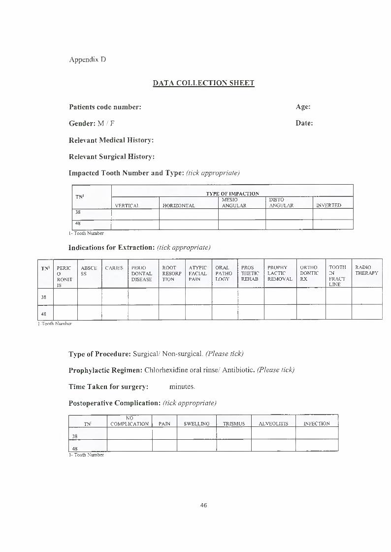
Appendix D
DATA COLLECTION SHEET
Patients code number: Age:
Gender: M / F Date:
Relevant Medical History:
Relevant Surgical History:
Impacted Tooth Number and Type: (tick a p p ro p ria te )
T N 1 T Y P E O F IM P A C T IO N
VERTICAL HORIZONTALMESIOANGULAR
DISTOANGULAR INVERTED
38
48
1- Tooth Number
Indications for Extraction: ( tic k a p p ro p ria te )
T N 1 PERICORONITIS
ABSCESS
CARIES PERIODONTALDISEASE
ROOTRESORPTION
ATYPICFACIALPAIN
ORALPATHOLOGY
PROSTHETICREHAB
PROPHYLACTICREMOVAL
ORTHODONTICRX
TOOTHINFRACTLINE
RADIOTHERAPY
38
48
1-Tooth Number
Type of Procedure: Surgical/Non-surgical. (P lea se tick)
Prophylactic Regimen: Chlorhexidine oral rinse/ Antibiotic. (P lea se tick)
Time Taken for surgery: minutes.
Postoperative Complication: ( tic k a p p ro p ria te )
TN1NO
COMPLICATION PAIN SWELLING TRISMUS ALVEOLITIS INFECTION
38
481- Tooth Number
46

REFERENCES
1. Ness GM, Peterson LJ. Impacted teeth In: Miloro M, Ghali GE, Larsen PE,
Waite PD, editors. Peterson's Principles of Oral and Maxillofacial Surgery. 2nd
edition. Ontario: BC Decker: 2004: Chapter 8: 139-156.
2. Monaco G, Staffolani C, Gatto MR, Checchi L. Antibiotic therapy in impacted
third molar surgery. Eur J Oral Sci. 1999; 107; 437-441.
3. Lawler B, Sambrook PJ, Goss AN. Antibiotic prophylaxis for dento alveolar
surgery: Is it indicated? Aust Dental J. 2005; 50: S54-S59.
4. Qirreish EJ. Radiographic Profile of symptomatic impacted mandibular third
molars in the Western Cape, South Africa. Masters Degree Dissertation. Western
Cape: University of Western Cape; 2005.
5. Tabetze N. Prevalence of Impacted Mandibular third molars at Medunsa Oral
Health Centre. Dissertation for MSC Dentistry. Limpopo: University of
Limpopo; 2012.
6. Siddiqui A, Morkel JA, Zafar S. Antibiotic prophylaxis in third molar surgery : A
randomised double blind placebo-controlled clinical trial using split-mouth
technique. Int J Oral Maxillofac Surg. 2010; 39: 107-114.
7. Juodzbalys G, Daugela P. Mandibular third molar impaction: Review of
literature and a proposal of a classification. J Oral Maxillofac Res. 2013; 4: el.
8. Hashemipour MA, Tahmasbi-Arashlow M, Fahimi-Hanzaei F. Incidence of
impacted mandibular and maxillary third molars: A radiographic study in a South
East Iran population. Med Oral Patol Oral Cir Bucal. 2013; 13: 40-45.
47

9. Pham L, Fang W. Change in inclination and eruption of mandibular third molars
a longitudinal radiographic study among 12 to 21-year-olds. Dissertation for
MSC Dentistry. Oslo: University of Oslo; 2006.
10. Kullman L, Johanson G, Akesson L. Root development of the lower third molar
and its relation to chronological age. Swed Dental J. 1992; 16: 161-167.
11. Balaji S.M. Textbook of Oral and Maxillofacial Surgery. 2nd edition. India:
Elsevier: 2013: Chapter 18: 345-382.
12. Almendros-Marqes N, Berini- Aytes L, Gay-Escoda C. Evaluation of
intraexaminer and interexaminer agreement on classifying lower third molars
according to the systems of Pell and Gregory and of Winter. J Oral and
Maxillofac Surg. 2008; 66: 893-899.
13. Maglione M, Costantinides F, Bazzocchi G. Classification of impacted
mandibular third molars on cone-beam CT images. J Clin Exp Dent. 2015; 7:
e224-231.
14. Quek SL, Tay CK, Tay CH, Toh SL, Lint KC. Pattern of third molar impaction
in a Singapore Chinese population: A retrospective radiographic survey. Int J
Oral Maxillofac Surg. 2003; 32: 548-552.
15. Msagati F, Elison NM, Owibingire S. Pattern of Occurence and treatment of
impacted teeth at the Muhimbili National Hospital, Dar es Salaam, Tanzania.
BMC Oral Health. 2013; 13: 37.
16. Hassan AH. Pattern of third molar impaction in a Saudi population. J Clinical,
Cosmetic and Investig Dent. 2010; 2: 109-111.
48

17. Kramer RM, Williams AC. The incidence of impacted teeth. A survey at Harlem
hospital. Oral Surg Oral Med Oral Pathol. 1970; 29: 237-241.
18. Gbotolorun OM, Olojede AC, Arotiba GT, Ladeinde AL, Akinwande JA,
Bangbose BO. Impacted 3rd molars: Presentation and postoperative complication
at the Lagos University teaching hospital. Nig Q J Hosp Med. 2007; 17: 26-29.
19. Song F, Landes DP, Glenny AM, Shelldon TA. Prophylactic removal of third
molars: an assessment of published reviews. Br Dent J. 1997; 182: 339-346.
20. Nordenram A, Hultin M, Kjellman O, Ramstrom G. Indications for surgical
removal of the mandibular third molar. Study of 2630 cases. Swed Dental J.
1987; 11: 23-29.
21. Pratt CA, Hekmat M, Barnard JDW, Zaki GA. Indications for third molar
surgery. J Royal Coll Surg Edin. 1998; 43: 105-108.
22. Adeyemo WL, James O, Ogunlewe MO, Ladeinde AL, Taiwo OA, Olajede AC.
Indications for extraction of third molars: A review of 1763 cases. Nig Postgrad
Med J. 2008; 15: 42-46.
23. Blondeau F, Daniel N. Extraction of impacted mandibular third molars:
Postoperative complications and their risk factors. J of Can Dent Assoc. 2007;
73: 325a-325e.
24. Martin MV, Kanatas AN, Hardy P. Antibiotic prophylaxis and third molar
surgery. British Dental J. 2005; 198: 327-330.
25. Kaczmarzyk T, Wichlinski J, Stypulkowska J, Zaleska M, Panas M, Woron J.
Single-dose and multi-dose clindamycin therapy fails to demonstrate efficacy in
49

preventing infectious and inflammatory complications in third molar surgery. Int
J Oral Maxillofac Surg. 2007; 36: 417-422.
26. Pasupathy S, Alexander M. Antibiotic prophylaxis in third molar surgery. J
Craniofac Surg. 2011; 22: 551-553.
27. Monaco G, Tavemese L, Agostini R, Marchetti C. Evaluation of antibiotic
prophylaxis in reducing postoperative infection after mandibular third molar
extraction in young patients. J Oral Maxillofac Surg. 2009; 67: 1467-1472.
28. Lopez-Cedmn JL, Pijoan JI, Fernandez S, Santamaria J. Hernandez G. Efficacy
of Amoxicillin treatment in preventing postoperative complications in patients
undergoing third molar surgery: A prospective, randomized, double blind
controlled study. J Oral and Maxillofac Surg. 2011; 69: e5-el4.
29. Arteaogoitia L, Ramos E, Santamaria G, Barbier L, Alvarez J, Santamaria J.
Amoxicillin/clavulanic acid 2000/125 mg to prevent complications due to
infection following completely bone-impacted lower third molar removal: a
clinical trial. Oral Surg Oral Med Oral Pathol Oral Radiol J. 2015; 119: 8-16.
30. Ataoqlu H, Oz YG, Candirli C, Kiziloqlu D. Routine antibiotic prophylaxis is not
necessary during operations to remove third molars. Br J Oral Maxillofac surg.
2008; 46: 133-135.
31. Olusanya AA, Aritiba JT, Fasola OA, Akadiri AO. Prophylaxis versus pre
emptive antibiotics in third molar surgery: a randomised control study. Niger
Postgrad Med J. 2011; 18: 105-110.
50

32. Bulut E, Bulut S, Etikan 1, Koseoglu O. The value of routine antibiotic
prophylaxis in mandibular third molar surgery: acute-phase protein levels as
indicators of infection. J Oral Sci. 2001; 43: 117-122.
33. Marcussen KB, Laulund AS, Jorgensen HL, Pinholt EM. A systematic review on
effect of single-dose preoperative antibiotics at surgical osteotomy extraction of
lower third molars. J Oral Maxillofac Surg. 2015; 15: 1554-1562.
34. Sridhar V, Wali GG, Shyla NN. Evaluation of the perioperative use of 0.2%
Chlorhexidine Gluconate for the prevention of alveolar osteitis after the
extraction of impacted mandibular third molars: A Clinical Study. J Oral
Maxillofac Surg. 2011; 10: 101-111.
35. Tuna A, Delibasi C, Arslan A, Gurol Y, Tazequn Tekkanat Z. Do antibacterial
mouthrinses affect bacteraemia in third molar surgery? A pilot study. Aust
Dental J. 2012; 57: 435-439.
36. Duvall NB, Fisher TD, Hensley D, Hancock RH, Vandewalle KS. The
comparative efficacy of 0.12% Chlorhexidine and Amoxicillin to replace the
incidence and magnitude of bacteraemia during third molar extractions: A
prospective, blind randomised clinical trial. Oral Surg Oral Med Oral Pathol Oral
Radiol. 2013; 115: 752-763.
37. Maharaj B, Coovadia Y, Vayej AC. A comparative study of amoxicillin,
clindamycin and chlorhexidine in the prevention of post extraction bacteraemia. J
Cardiovasc Afr. 2012; 23: 491-494.
38. Scottish Intercollegiate Guidelines Network. Edinburgh, Scotland: 1999.
Management of unerupted and impacted third molar.
51

http://www.maxilofacialchile.cl/web/sitio/images/stories/guias/13.pdffaccessed
on: 25th September 2014],
39. Metin M, Sener IJ. Comparison of two chlorhexidine rinse protocols on the
incidence of alveolar osteitis following surgical removal of impacted third
molars. J Contemp Dent Prac. 2006; 7: 79-86.
40. Yildirim A, Metzler P, Lubbers H, Yildirim V. Digluconate de Chlorhexidine-
histoire, Mecanisme d’action et risques. Swiss Dental J. 2015; 125: 830-831.
41. Karki S, Cheng AC. Impact of non rinse skin cleansing with chlorhexidine
gluconate on prevention of health care associated infections with multi resistant
organisms: a systematic review. Hospital Infection J. 2012; 82: 71-84.
42. Tomas I, Alvarez M, Limeres J. Effect of a Chlorhexidine mouthwash on the risk
of post extraction bacteremia. Infect Control Hosp Epidemiol J. 2007; 28: 577-
582.
43. Me Bain AJ, Bartolo RG, Gilbert P. Effects of a Chlorhexidine Gluconate
mouthwash on the vitality and antimicrobial susceptibility of in vitro oral
bacterial eco systems. Appl Environ Microbiol J. 2003; 69: 4770-4776.
44. Barbosa M, Prada-Lopez I, Alvarez M., Amaral B, Maria de Los Angeles C,
Tomas I. Post-tooth extraction bacteraemia: A randomized clinical trial on the
efficacy of chlorhexidine prophylaxis. A Peer-Reviewed, Open Access J. 2012;
10:e0124249.
45. Watanabe E, Nascimento AP, Guerreiro-Tanomaru JM, Razaboni AM, De
Andrade D, Tanomaru-Filho M. Antiseptic mouthwashes: in vitro antibacterial
activity. Acta odontol Latinoam J. 2015; 18: 180-184.
52

46. Smith JA. Antibiotic prophylaxis to prevent surgical site infection in
Maxillofacial and Oral surgery. In: Bagheri S, Bryan Bell R, Khan H, editors.
Current therapy in Oral and Maxillofacial Surgery. St Louis: Elsevier Saunders:
2011: Chapter 8: 67-78.
47. Mohammed A, Dusara K. Can antibiotic prophylaxis prior to surgical removal of
teeth reduce the incidence of postoperative infections? J of Dentistry, Oral
disorders and therapy. 2014; 1: 5.
48. Carlson MA. Prophylactic antibiotics in surgery. Annual Review of Medicine J.
1993; 44:385-393.
49. Ramu C, Padmanabhan TV. Indications of antibiotic prophylaxis in dental
practice- Review. Asian Pac J Trap Biomed. 2012; 2: 749-754.
50. Sweeney L, Dave J, Chambers PA, Heritage J. Antibiotic resistance in general
dental practice - A cause for concern? Chemotherapy J. 2004; 53: 563-576.
51. Bortoluzzi MC, Capella DL, Barbieri T, Pagliarini M, Cavalieri T, Manfro R. A
single dose of Amoxicillin and Dexamethasone for prevention of postoperative
complications in third molar surgery: A randomized, double blind, placebo
controlled clinical trial. J Clin Med Res. 2013; 5: 26-33.
52. Ren YF, Malmstrom HS. Effectiveness of antibiotic prophylaxis in third molar
surgery: A Meta-Analysis of randomized clinical trials. J Oral Maxillofac Surg.
2007; 65: 1909-1921.
53. Oomens MA, Forouzanfar T. Antibiotic prophylaxis in third molar surgery: a
review, Oral Surg, Oral Med, Oral pathol and Oral Radiol J. 2012; 114: e5-el2.
53

54. World Health Organisation. Antimicrobial Resistance. Fact sheet no 194.
http:/www.who.int/mediacentre/factsheets/fsl94/en/ [Accessed on: 29th May
2014],
55. Dar-Odeh NS, Abu Hammad OA, Al-Omiri MK, Kharaisat AS, Shehabi AA.
Antibiotic prescribing practices by dentists: a review. The Risk Management J.
2010; 6: 301-306.
56. Gusberti FA, Sampathkumar P, Sieqrist BE, Lang NP. Microbiological and
clinical effects of chlorhexidine digluconate and hydrogen peroxide mouthrinses
of developing plaque and gingivitis. Clin Periodontol J. 1988; 15: 60-67.
57. Krishnan B, El Sheikh MH, El Gehani R, Orafi H. Indications for removal of
impacted mandibular molars: a single institutional experience in Lybia. J Oral
Maxillofac Surg. 2009; 8: 246-248.
54