delayed traumatic intrapericardial diaphragmatic …hernia may not appear until viscera incarcerate...
TRANSCRIPT
1/4 J Cardiol Clin Res 1(1): 1147.JSM Clin Cytol Pathol 4: 4
JSM Clinical Cytology and Pathology
Submitted: 15 July 2019 | Accepted: 23 July 2019 | Published: 26 July 2019
*Corresponding author: Manouchehr Aghajanzadeh, Department of Thoracic and General Surgery, Arya Hospital.Guilan University of Medical Sciences, Iran, Email: [email protected]
Copyright: © 2019 Aghajanzadeh M, et al. This is an open-access article distributed under the terms of the Creative Commons Attribution License, which permits unrestricted use, distribution, and reproduction in any medium, provided the original author and source are credited.
Citation: Aghajanzadeh M, Jalali S, Hemmati H, Esmaeili Delshad MS, Samidost P (2019) Delayed Traumatic Intrapericardial Diaphragmatic Her-nia: A Rare Cause of Colon Obstruction. JSM Clin Cytol Pathol 4: 4.
Delayed Traumatic Intrapericardial Diaphragmatic Hernia: A Rare Cause of Colon Obstruction
Manouchehr Aghajanzadeh*, Seyedali Jalali, Hossein Hemmati, Mohammad Sadegh Esmaeili Delshad, and Piroze Samidost
Department of Thoracic and General Surgery, Guilan University of Medical Sciences, Iran
AbstractIntrapericardial Diaphragmatic Hernia (IDH) is the rarest type of adult diaphragmatic hernia and colon obstraction. Only a few cases
have been reported. Blunt trauma has been implicated in most cases, but few cases were reported from a stab wound to the anterior chest. Patients presented immediately or up to 15 to 20 years following trauma with symptoms which include of: intermittent bowel obstruction, strangulation, cardiac dysfunction, dyspnea, palpitations, and even cardiac tamponade. The most unusual site for a diaphragmatic hernia to occur is through the central tendon of the diaphragm into the pericardium. Physical findings included bowel sounds in the chest, decreased heart and lung sounds, and an absent point of maximal cardiac impulse. We present the case of a 67-year-old man who suffered a bowel obstruction when the transverse colon and omentum became incarcerated in the intrapericardial cavity from diaphragmatic hernia. Defect presumably resulted from blunt chest and abdominal trauma received six month earlier. Laparatomy is the preferred approach to surgical repair of IDH. Since the symptoms referable to adult IDH can be incapacitating or life threatening, herniorrhaphy should be performed promptly upon diagnosis. In this very rare case with delayed presentation of intrapericardial diaphragmatic hernia, we want to review the symptoms, recommendations of evaluation and the best approach of treatment and outcome of treatment.
Keywords: Diaphragmatic hernia; Intrapericardial cavity; Complications; Bowel obstruction
IntroductionThe diaphragmatic injury is uncommon and it could be the
consequence of blunt or penetrating thoracoabdominal trauma. It occurs in approximately 5% of blunt trauma patients undergoing laparotomy [1, 2]. Diaphragmatic hernia in adults is almost always secondary to trauma [1,2]. Its incidence is increasing with high speed transportation accidents and civilian warfare and is currently 3-7% in trauma victims [1,2]. Many series of diaphragmatic hernia after blunt and penetrating trauma have mentioned associated central tendon rupture and pericardial laceration, but none has reported intrapericardial diaphragmatic herniation of abdominal viscera (IDH) [1-3]. The difficulties in diagnosing diaphragmatic rupture and herniation at the first encounter can significantly affect morbidity and mortality. Although diaphragmatic injury can be evident like herniation of abdominal organs on chest imaging, in hemodynamically stable patients who are conservatively managed, the rate of initially neglected diaphragmatic injury is between 12 and 66%, and it could be also missed during laparotomy [1-4]. In blunt trauma, most ruptures tend to occur on the left hemidiaphragm specifically
on the poster- lateral side due to structurally weakness of this area as it is the derivative of the pleuroperitoneal membrane [5,6]. The right diaphragm is congenitally stronger and right-sided injuries may not be apparent on initial evaluation because the liver protects the defect preventing intestinal herniation [7,8]. Blunt trauma-related left hemi diaphragmatic ruptures have been reported to range from 68.5 to 87% and it is more frequent than the [1,2]. Many series of diaphragmatic hernia after blunt and penetrating trauma have mentioned associated central tendon rupture and pericardial laceration, but none has reported intrapericardial diaphragmatic herniation of abdominal viscera [3,4,8,9]. Patients may initially present with no symptoms or signs suggesting diaphragmatic injury, but as the time passes, the diaphragmatic rupture tends to grow larger, and herniation of abdominal organs is more likely to occur, particularly on the left side [1,2,8]. Patients with the history of thoracoabdominal blunt trauma complaining of abdominal pain, nausea, and vomiting should be assessed for a possible delayed presentation of diaphragmatic injury [3,5-7] .Failure to diagnose and repair the diaphragm can result in intestinal strangulation and its fatal consequences [5,3]. The earliest symptoms of diaphragmatic hernia may not appear until viscera incarcerate in it years after the causal injury. The most unusual site for a diaphragmatic hernia to occur is through the central tendon of the diaphragm into the pericardium (2,6,7-9) We present a case of 67-year-old man who suffered a bowel obstruction when the transverse colon and omentum became incarcerated in the intrapericardial diaphragmatic hernia. The defect presumably resulted from blunt chest and abdominal trauma which received six month earlier. We want to review the presentation of the delayed intrapericardial diaphragmatic hernia, diagnosis, evaluation and treatment and outcome of management
Case Presentation A 67-year-old male admitted to the emergency department
Case Report © Aghajanzadeh M, et al. 2019

2/4JSM Clin Cytol Pathol 4: 4
with a complaint of abdominal pain with distension, nausea, vomiting and dyspnea and reported constipation but passing of flatus for 5 days. Upon further questioning, he recalled that he had a trauma as a wall of a building wall fallen over his abdomen six month ago and his initial evaluation revealed rib fracture and mild hemothorax and With pelvic rim bone fracture. Other organ of abdomen was intact. With chest –tube and pelvic fixator he discharged in good condition 5 day after admission. In our clinic, on physical examination, the patient was alert and responsive, his vital signs were (HR =104, BR=20, fever 37 and BP=111/60), and dry mucous membranes were noted. Abdominal examination revealed distended abdomen, increased bowel sounds, and generalized tenderness with rebound tenderness and guarding on the epigastric region and breath sound was decreased on the anterior left chest wall. In rectal examination the ampulla of rectum was empty of feces. Fluid resuscitation was started and nasogastric (NG) tube folly catheter was inserted for a patient with a suspected diagnosis of intestinal obstruction. Lab results on admission was Normal range except renal test {WBC –16,000 (mm3), RBC - 5.9 (million/mm3), PLT 285,000 (mm3), Hb 11.9 (g/dl), Blood sugar 125 (mg/dl) Urea 55 (mg/dl) Cr –2.4 (mg/dl) AST –37 (IU/L) ALT –41 (IU/L) Alkaline phosphatase –306 (IU/L) Amylase –100 (IU/L) Lipase 28 ≤ 60U/I}.
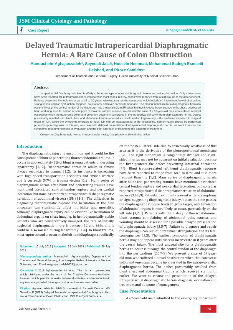
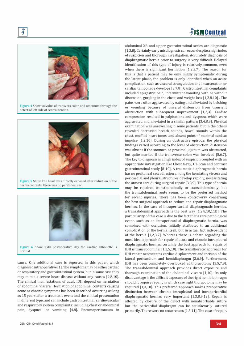
The patient underwent a plane abdominal and chest X-ray. Abdominal X-ray show, multiple distension of small intestine loop and cecum and ascending colon. Continuity of transverse colon was disappeared; this XR shows pelvic fixator too (Figure 1). Chest XR show gas shadows of colon overlying the cardiac silhouette on the left lateral side of heart (Figure 2), and continuity of transvers colon was cut in the epigastric region. Abdominal ultrasonography reported the presence of multiple dilated intestinal loops of abdomen. High midline laparatomy was performed for intestinal obstruction due to diaphragm hernia (Figure 3). Dissection and release of adhesions bound was performed. Intraoperative finding was herniation and volvulus of transvers colon and omentum through the defect of left side of central tendinous portion of diaphragm in to the pericardium cavity (Figure 4). After release of adhesions from pericardium, withdrawal the omentum and transverse colon from the pericardial cavity through a 10 cm transverse rent in the anterior portion of the pericardial diaphragm. The heart was directly exposed after reduction of the hernia contents; there was no peritoneal sac (Figure 5). The defect had thickened, fibrotic edges and was limited solely to the pericardial diaphragm, without pleural involvement. It was primarily closed with interrupted vicryl 1 sutures with a drain left placed in pericardium cavity for prevention of tamponed. The patient referred to the ICU unit for 48 hours. Postoperative serial chest films revealed slow resolution of pneumopericardium such that by the sixth postoperative day the cardiac silhouette was normal (Figure 6). No other complications were found. The patient discharged on day seven post-operative with good condition and in follow-up time was perfect.
DiscussionTraumatic hernia of the diaphragm is a rare but serious
complications of blunt or penetrating thoraco-abdominal
Figure 1 Abdominal X-ray show, multiple distension of small intestine loop and cecum and ascending colon. Pelvic fixator.
Figure 2 Chest roentgenography show gas shadows of colon overlying the cardiac silhouette.
Figure 3 Show High midline laparotomy.
trauma [5,10]. In blunt abdominal trauma the sudden rise in intra-abdominal pressure can cause the diaphragm to rupture in multiple places (the right side, the left side, hiatus or into the pericardium) [5,8]. Diaphragmatic rupture with pericardial involvement is a rare complication [11]. Eight cases have been reported previously [10,8]. Trauma is the major and perhaps only cause of adult IDH blunt trauma [9,5,6,8]. Deceleration injuries during transportation accidents, severe blows to the chest and severe falling and anterior chest wall stab wound are the other
3/4JSM Clin Cytol Pathol 4: 4
abdominal XR and upper gastrointestinal series are diagnostic [1,3,8]. Certainly early misdiagnosis can occur despite a high index of suspicion and thorough investigation. Accurately diagnosis of diaphragmatic hernia prior to surgery is very difficult. Delayed identification of this type of injury is relatively common, even when there is significant herniation [1,2,5,7]. The reason for this is that a patient may be only mildly symptomatic during the latent phase, the problem is only identified when an acute complication, such as visceral strangulation and incarceration or cardiac tamponade develops [3,7,8]. Gastrointestinal complaints included epigastric pain, intermittent vomiting with or without distension, gurgling in the chest, and weight loss [1,2,8,10] . The pains were often aggravated by eating and alleviated by belching or vomiting because of visceral distension from transient obstruction with subsequent improvement [1,2,3]. Cardiac compression resulted in palpitations and dyspnea, which were aggravated and alleviated in a similar pattern [3,4,8,9]. Physical examination was unrevealing in some patients, but in the others revealed decreased breath sounds, bowel sounds within the chest, muffled heart tones, and absent point of maximal cardiac impulse [1,2,10]. During an obstructive episode, the physical findings varied according to the level of obstruction: distension was absent if the stomach or proximal jejunum was obstructed, but quite marked if the transverse colon was involved [5,6,7]. The key to diagnosis is a high index of suspicion coupled with an appropriate investigation like Chest X-ray, CT-Scan and contrast gastrointestinal study [8-10]. A traumatic diaphragmatic hernia has no peritoneal sac; adhesion among the herniating viscera and pericardial and pleural structures develop rapidly, necessitating the utmost care during surgical repair [3,8,9]. This type of hernia may be repaired transthoracically or transabdominally, but the transabdominal route seems to be the preferred method for recent injuries. There has been controversy concerning the best surgical approach to reduce and repair diaphragmatic hernias. In the case of intrapericardial diaphragmatic hernias, a transabdominal approach is the best way [1,2,8,10,11�]. The particularity of this case is due to the fact that a rare pathological event, such as an intrapericardial diaphragmatic hernia, was combined with occlusion, initially attributed to an additional complication of the hernia itself, but in actual fact independent of the hernia [1,2,3,7]. Whereas there is debate regarding the most ideal approach for repair of acute and chronic intrapleural diaphragmatic hernias, certainly the best approach for repair of IDH is transabdominal [1,2,5,10]. The transthoracic approach for IDH repair necessitates cardiac displacement and incision of the lateral pericardium and hemidiaphragm [3,6,9]. Furthermore, IDH has been completely overlooked at thoracotomy [3,5,7,9]. The transabdominal approach provides direct exposure and thorough examination of the abdominal viscera [1,10]. Its only disadvantage is the difficult exposure of the right hemidiaphragm should it require repair, in which case right thoracotomy may be required [1,3,10]. This preferred approach makes preoperative distinction between chronic intrapleural and intrapericardial diaphragmatic hernias very important [1,3,8,9,12]. Repair is affected by closure of the defect with nonabsorbable suture but the pericardial diaphragm can be satisfactorily sutured primarily. There were no recurrences [1,5,11]. The ease of repair,
Figure 4 Show volvulus of transvers colon and omentum through the defect of left side of central tendon.
Figure 5 Show The heart was directly exposed after reduction of the hernia contents; there was no peritoneal sac.
Figure 6 Show sixth postoperative day the cardiac silhouette is normal.
cause. One additional case is reported in this paper, which diagnosed intraoperative [1]. The symptoms may be either cardiac or respiratory and gastrointestinal system, but in some case they may mimic a severe heart disease without any causes [9,8,10]. The clinical manifestations of adult IDH depend on herniation of abdominal viscera. Herniation of abdominal contents causing acute or chronic symptoms has been described occurring as long as 15 years after a traumatic event and the clinical presentation in different type, and can include gastrointestinal, cardiovascular and respiratory system complaints: including chest or abdominal pain, dyspnea, or vomiting [4,8]. Pneumoperitoneum in

4/4JSM Clin Cytol Pathol 4: 4
low complication rate, and lack of recurrence certainly justify correction of the defect with laparatomy
ConclusionWe report an unusual post-traumatic intrapericardial
diaphragmatic hernia presenting with intestinal occlusion due to volvulus of transvers colon and omentum. In our opinion, the particularity of this case is due to the fact that a rare pathological event, such as an intrapericardial diaphragmatic hernia, was initially attributed to an additional complication of the hernia itself, but in fact independent of the hernia and a consequence of a previous laparotomy and pelvic rim fixation. Both in first admission upper gastrointestinal barium and Thoraco-abdomonial CT-Scan didn’t show the IDH. Only laparotomy exploration found the key of such a strange diagnostic dilemma. An adhesion, caused by the previous laparatomy, which was strangulating a volvulus of transvers colon and omentum in the diaphragm defect.
References1. F Al Hadi, M Haid, I Dafallah. Delayed Intrapericardial
transdiaphragmatic herniation: An Unusual Cause of Cardiohepatic Adhesions. Thor Cardiovasc Surg. 2007; 11: 2.
2. Cihan Ağalar1, Sami Benli, Tufan Egeli, Mucahit Ozbilgin, Isil Basara. Laparoscopic Repair of Delayed Traumatic Diaphragmatic Hernia with Mesh. Trauma Acute Care. 2017; 6: 63.
3. Alessandro Bini, Fabio Davoli, Nicola Cassanelli, Giampiero Dolci, Giulia Luciano, Franco Stella. Unusual delayed presentation of post-traumatic intrapericardial hernia associated with intestinal occlusion. Ann Ital Chir. 2010; 81: 45-47.
4. Kuzuc A, Isik B, Baysal T, Soysal O, Ulutas H. Traumatic intrapericardial diaphragmatic hernia. Radiography. 2007; 13: 169-171.
5. Daniel J Beless, Brian C. Organ. Delayed presentation of intrapericardial diaphragmatic hernia, an unusual case of colon obstruction. Ann Emerg Med. 1991; 20: 415-417.
6. Hon Chi Suen, Sundeep Das, Ronald L Palmer, Cami L Watkins, Ajit S Nagra. Intrapericardial colonic herniation. J Thorac Cardiovasc Surg. 2006; 13: 919-920.
7. Delliturri A, Chiba S1, Brichkov I, Sherwinter D. Laparoscopic repair of a peritoneopericardial diaphragmatic hernia after a convergent procedure for the treatment of atrial fibrillation. J Thorac Dis. 2017; 9: E767-E770.
8. Bini A, Davoli F, Cassanelli N, Dolci G, Luciano G, Stella F. Unusual delayed presentation of post-traumatic intrapericardial hernia associated with intestinal occlusion. Ann Ital Chir. 2010; 81: 45-47.
9. Suen HC, Das S, Palmer RL, Watkins CL, Nagra AS. Intrapericardial colonic herniation. J Thorac Cardiovasc Surg. 2006; 131: 919-920.
10. Reina A, Vidana E, Soriano P, Orte A, Ferrer M, Herrera E, et al. Traumatic intrapericardial diaphragmatic hernia: Case report and literature review. Injury. Int J Care Injured. 2001; 32: 153-156.
11. Manouchehr Aghajanzade, Bahareh Hesamifard, Milad Sarafi, Omid Mosafaee Rad. Rare emergency presentation in delayed diaphragmatic hernia andtotal orpartial reconstruction of hemidiaphragm with prolenemesh: in five cases. Int J Scien Res. 2017; 46.
12. Aghajanzadeh M, Hemmati H, Delshad MSE, Samidost P, Mosaffaei O, Rafiei E. Rare Presentations and Repair of Delayed Traumatic Diaphragmatic Rupture: Report of 39 Cases Over 10 Years. Clin Surg. 2018; 3: 1859.