deleterious effects of plasminogen activators in neonatal...
TRANSCRIPT
Neurobiology
Deleterious Effects of Plasminogen Activators inNeonatal Cerebral Hypoxia-Ischemia
Faisal Adhami,*† Dan Yu,*‡ Wei Yin,§
Aryn Schloemer,* Kevin A. Burns,* Guanghong Liao,*Jay L. Degen,* Jun Chen,§ and Chia-Yi Kuan*From the Division of Developmental Biology and Neurology,*
Cincinnati Children’s Hospital Medical Center, Cincinnati, Ohio;
the Physician Scientist Training Program,† University of
Cincinnati, College of Medicine, Cincinnati, Ohio; the
Department of Neurology,§ University of Pittsburgh School of
Medicine, Pittsburgh, Pennsylvania; and the Department of
Pediatrics,‡ West China Second University Hospital, Sichuan
University, Chengdu, Sichuan, China
The immature brains of newborns often respond differ-ently from the brains of adults when exposed to similarinsults. Previous studies have indicated that although hy-poxia-ischemia (HI) induces persistent thrombosis inadult brains, it only modestly impairs blood perfusion innewborn brains. Here, we used the Vannucci model of HIencephalopathy to study age-related responses to cerebralHI in rat pups. We found that HI triggered fibrin deposi-tion and impaired blood perfusion in both neonatal andadult brains. However, these effects were only transient inneonatal brains (<4 hours) and were accompanied byacute induction of both tissue-type and urinary-type plas-minogen activators (tPA and uPA), which was not ob-served in adult brains subjected to the same insult. Inter-estingly, activation of the plasminogen system persistedup to 24 hours in neonatal brains, long after the clearanceof fibrin-rich thrombi. Furthermore, astrocytes and mac-rophages outside blood vessels expressed tPA after HI,suggesting the possibility of tPA/plasmin-mediated cyto-toxicity. Consistent with this hypothesis, injection of �2-antiplasmin into cerebral ventricles markedly amelioratedHI-induced damage to neurofilaments and white matteroligodendrocytes, providing a dose-response reduction ofbrain injury after 7 days of recovery. Conversely, ventric-ular injection of tPA increased HI-induced brain damage.Together, these results suggest that tPA/plasmin induc-tion, which may contribute to acute fibrinolysis, is a crit-ical component of extravascular proteolytic damage inimmature brains, representing a new therapeutic targetfor the treatment of HI encephalopathy. (Am J Pathol2008, 172:1704–1716; DOI: 10.2353/ajpath.2008.070979)
Hypoxic-ischemic encephalopathy (HIE) is a leadingcause of mortality and long-term neurological morbidity inpremature infants.1 Previous studies have indicated theimmature brain of newborns has a higher susceptibility tohypoxia-ischemia (HI) than adult brains.2 The increasedvulnerability to HI in newborn brains can be attributed toseveral reasons, including a greater risk of energy failure,the immaturity of blood-brain-barrier (BBB) and whitematter, a high susceptibility to glutamate excitotoxicity,3
low expression of the glutamate transporters,4 as well asother unknown mechanisms. These neonate-specific re-sponses are important considerations in designing effec-tive therapies of HIE in infants.5
Accumulating evidence demonstrates the tissue-typeplasminogen activator (tPA) has complex functions in thenervous system.6 By converting plasminogen to activefibrinolytic plasmin, tPA is currently the only Food andDrug Administration-approved specific therapy for acuteischemic stroke. However, tPA can also induce metallo-proteases to damage the BBB, leading to hemorrhagiccomplications.7 Moreover, the brain parenchyma has alow level of tPA and plasminogen expression that is mark-edly increased by seizures and excitotoxins.8,9 Becauseplasmin is a broad-spectrum protease, the excitotoxin-induced endogenous tPA/plasmin can degrade the ex-tracellular matrix, activate protease-activated receptors,and cause neuronal degeneration.10,11 Further, tPA hasplasmin-independent mechanisms to stimulate micro-glia12 and enhance N-methyl-D-aspartate receptor-medi-ated excitotoxicity.13 Hence, tPA may have both protec-tive (thrombolytic) and deleterious (neurotoxic) effects inischemic brain.
Supported by the National Institutes of Health (grants NS44315 andNS59668 to C.-Y.K. and NS45048 to J.C.).
F.A., D.Y., and W.Y. contributed equally to this study.
Accepted for publication March 13, 2008.
D.Y. was a Bang Bao Scholar sponsored by Procter and Gamble, Inc.
Present address of G.L.: Department of Basic Medical Sciences, WesternUniversity of Health Sciences, Pomona, CA.
Address reprint requests to Chia-Yi Kuan, M.D., Ph.D., Division ofDevelopmental Biology and Division of Neurology, Cincinnati Children’sHospital Research Foundation, TCHRF S3.421, 3333 Burnet Ave., Cincin-nati, OH 45229. E-mail: alex.kuan @cchmc.org.
The American Journal of Pathology, Vol. 172, No. 6, June 2008
Copyright © American Society for Investigative Pathology
DOI: 10.2353/ajpath.2008.070979
1704
Although the risk of tPA neurotoxicity associated withthrombolytic therapy is well-recognized, the roles of endog-enous tPA and plasmin in ischemic brain injury are unclear.This uncertainty is partly because, except in animal modelsof permanent cerebral ischemia, transient cerebral isch-emia does not significantly induce the expression nor activ-ity of endogenous tPA and plasmin.14–16 Moreover, al-though initial studies suggested tPA-null mice had smallerinfarct volumes than wild-type mice after the suture model oftransient focal ischemia, a later study using the geneticbackground-matched wild-type and tPA-null mice revealedopposite results in similar experiments.17–19 Even with aphotochemically induced thrombosis model of stroke, stud-ies using wild-type and mutant mice indicated that tPA hasparadoxical functions in protecting against large thrombi,but increasing the damage by small thrombi.20 Hence, theroles of endogenous tPA and plasmin(ogen) are uncertainin adult ischemic brain injury, and almost unexplored inneonatal HIE.
The present study was stimulated by our recent publica-tion showing while HI only transiently impairs cerebral per-fusion in newborns,21 it produces persistent occlusive fibrindeposition in adult brains.22 In light of the contrasting effectsof HI in newborn versus adult brains, we hypothesized thatneonatal brains have an age-specific response to HIthrough activation of the endogenous plasminogen system.To test this hypothesis, we examined the thrombosis-fibrin-olysis responses and the tPA/plasmin activity in rat pupssubjected to the Vannucci model of cerebral HI.2 Results ofthese experiments supported our hypothesis, but also indi-cated persistent extravascular induction of the tPA/plasminsystem is a critical factor of brain injury in this paradigm.These results shed new insight into the mechanism of braininjury in neonatal HIE in infants.
Materials and Methods
Animal Surgery
Wistar rat pups were used for the Vannucci model ofneonatal HI as previously described.2 Briefly, 7-day-oldrat pups were anesthetized with 3% isoflurane mixed withambient air under spontaneous inhalation, and the rightcommon carotid artery was ligated. After a 1-hour recov-ery period, the pups were placed in glass chamberscontaining a humidified atmosphere of 10% oxygen and90% nitrogen and submerged in a 37°C water bath. After1.5 hours of hypoxia, the pups were returned to their damfor the indicated time. Wistar rats, 8 to 12 weeks of age,were used for adult HI model as previously described.22
Intracerebroventricular injections were done immediatelyafter, 2 hours, or 4 hours after hypoxia with 10 �l volumesof �2-antiplasmin (BioDesign International, Saco, ME),saline, tPA [50 �g recombinant tPA (Activase; Genen-tech, South San Francisco, CA) and 3.85 mg L-Arginine in10 �l], and tPA-vehicle (3.85 mg L-arginine in 10 �l) witha Hamilton syringe at 2.0 mm rostral and 1.5 mm lateral tothe right from the Lamda suture, and at a depth of 2.0 mmfrom the surface of the brain, as previously described.23
These animal procedures were approved by the Institu-
tional Animal Care and Use Committee and conform tothe National Institutes of Health Guide for Care and Useof Laboratory Animals.
tPA-Null Mice
Homozygous tPA-null mice backcrossed to the C57BL/6strain were used in the present study. These mice weregenerated as previously described,24 and obtained fromThe Jackson Laboratory, Bar Harbor, ME (stock no.002508).
Assessment of Brain Damage
After 7 days of recovery from HI, pups were sacrificedunder deep anesthesia by transcardiac perfusion withsaline and then 4% paraformaldehyde. After postfixation,brains were taken through sucrose gradients and frozenin tissue-freezing medium, then cut in coronal sections 50 �min thickness. Eight evenly spaced sections were taken torepresent the whole brain and stained with cresyl violet.Every section was analyzed with ImageJ software (Na-tional Institutes of Health, Bethesda, MD) for area ofsurviving cortex, striatum, and hippocampus on lesionand contralateral sides as indicated by Nissl stain. Thepercentage of tissue loss was calculated at every rele-vant section for each structure as a ratio of lesion tocontralateral areas, and this was averaged over eightsections for each animal.
Histology
Brain sections were obtained as above for lesion analy-sis, but with 30-�m-thick sections. Immunohistochemistrywas performed using rat polyclonal antibodies againstP-selectin (BD Pharmingen, Franklin Lakes, NJ), Glycop-rotein IIb (BD Pharmingen), mouse monoclonal antibod-ies against CD11b (OX42 antibody; Serotec, Oxford, UK),myelin basic protein (Chemicon, Temecula, CA) and neu-rofilament (clone 2H3; Hybridoma Bank, University ofIowa, Ames, IA), as well as, rabbit polyclonal antibodiesagainst tPA (Molecular Innovations, Novi, MI) and fibrin-(ogen).22 Biotinylated secondary antibodies and strepta-vidin conjugated to Alexa Fluor 488 or Alexa Fluor 594(Molecular Probes, Eugene, OR) were used to amplify theimmuno signals. For mouse primary antibodies, a highlycross-absorbed anti-mouse secondary antibody (Molec-ular Probes) was used because of cross reactivity with ratIgG. Goat biotinylated anti-rat (H�L; Vector Laboratories,Burlingame, CA) was used to label IgG leakage. The termi-nal deoxynucleotidyl transferase-mediated dUTP nick end-labeling (TUNEL) was performed as described.22
Fluorescein Isothiocyanate (FITC)-DextranPerfusion Analysis
Cerebral perfusion analysis was performed as previouslypublished.22 Briefly, FITC-dextran [5 mg in 100 �l ofphosphate-buffered saline (PBS) of 2 � 106 MW; Sigma,
Plasminogen Activators in Neonatal HIE 1705AJP June 2008, Vol. 172, No. 6
St. Louis, MO] was injected with a small-bore needle intothe left ventricle of pups 1 or 4 hours after ischemia/hypoxia. After 2 minutes, the animal was rapidly decap-itated and the brain was removed and postfixed for 48hours. Sections were cut as above and analyzed withfluorescence microscopy. For quantification, images ob-tained were analyzed using ImageJ software. An index ofperfusion was measured in each hemisphere in an areaexcluding the midline (matching the maximal possibleinfarction) in two caudal coronal sections covering thehippocampus in each animal. The number of FITC pixelsper number of total pixels in maximal infarct area ofipsilateral hemisphere was first determined. The datafrom the lesion sides were then normalized to the averageof data from the contralateral side at the correspondingtime point. Presented are the lesion and the contralateraldata at 1 hour and 4 hours after HI, with P values reportedrelative to each time point’s contralateral data set.
Reverse Transcriptase-Polymerase ChainReaction (RT-PCR) Analysis
Total RNA of the hippocampus was isolated from thelesion, contralateral, and untreated hemispheres usingTRIzol reagent (Invitrogen, Carlsbad, CA) according tothe manufacturer’s instruction. Moloney murine leukemiavirus reverse transcriptase (M-MLV RT, Invitrogen) andoligo dT18 primer were used for cDNA synthesis. Forquantification, optical intensity was measured on the pre-sented images using ImageJ and normalized to G3PDHfor each time point. The primer pairs used for PCR anal-ysis were as follows: tPA: 5�-CTCTGACTTCGTCTGC-CAG-3� and 5�-TGTCATTGCTTCATGTTGTC-3�; uPA: 5�-CTACACCAAAGGCTGTGGC-3� and 5�-GCCATTACAG-CAGGAGATAG-3�; PAI-1: 5�-ATCCTGCCTAAGTTCTC-TCT-3� and 5�-ATTGTCTCTGTCGGGTTGTG-3�; matrixmetalloprotease (MMP)-2: 5�-GGACAGTGACACCACGT-GAC-3� and 5�-ACTCATTCCCTGCGAAGAACA-3�; MMP-9:5�-GCACGCTGTCCTTTCTTGTTGG-3� and 5�-CCCGAC-GCACAGTAAGCATTC-3�.
Immunoblot Analyses
Brain samples for immunoblots were homogenized inTLB buffer (containing 20 mmol/L Tris, pH 7.4, 137mmol/L NaCl, 25 mmol/L �-glycerophosphate, 25 mmol/LNa-pyrophosphate, 2 mmol/L ethylenediaminetetraaceticacid, 1 mmol/L Na3VO4, 1% Triton X-100, 10% glycerol, 1mmol/L phenylmethyl sulfonyl fluoride, 0.7% protease in-hibitor cocktail). The proteins were separated by stan-dard sodium dodecyl sulfate-polyacrylamide gel electro-phoresis procedures, electrotransferred onto a polyvinylidenedifluoride microporous membrane (Bio-Rad, Hercules,CA), and immunoblotted with designated antibodies fol-lowed by enhanced chemiluminescence detection (Am-ersham Biosciences, Buckinghamshire, UK). The anti-bodies used are: anti-occludin (Zymed, South SanFrancisco, CA) and anti-�-actin (Sigma).
tPA/uPA Zymogram
For detection of tPA/uPA, plasminogen and casein wereadded to the sodium dodecyl sulfate-polyacrylamide gelelectrophoresis gel. Protein samples were extracted us-ing RIPA buffer and mixed with an equal volume of thesample buffer (0.5 mol/L Tris-HCl, pH 6.8, 100% glycerol,0.05% bromophenol blue, 10% sodium dodecyl sulfate)and run at low voltage for up to 5 hours. After electro-phoresis, the gel was rinsed twice in 2.5% Triton X-100 for30 minutes each at room temperature, followed by anincubation in glycine buffer (0.1 mol/L glycine, pH 8.0) at37°C overnight. The gel was then stained with Coomassieblue solution and destained overnight to reveal the lyticzones of protease activity. Recombinant human tPA (Ac-tivase, Genentech) was used as the positive control toreveal the tPA band in zymogram. Quantification wasdone on the presented image using ImageJ to determinethe area of signal for each time point.
MMP Zymogram
For detection of the activity of MMP-2 and MMP-9, 0.15%porcine skin gelatin was added to the sodium dodecylsulfate-polyacrylamide gel electrophoresis gel. Proteinsamples were extracted and ran as mentioned above.The gels were washed twice with 2.5% Triton X-100, thenincubated in reaction buffer (50 mmol/L Tris, pH 7.5, 200mmol/L NaCl, 5 mmol/L CaCl2) at 37°C overnight, thenstained and destained as mentioned above. PurifiedMMP-2 (M-1552, Sigma) and MMP-9 (M-7942, Sigma)were used for positive controls. Quantification was doneon the presented image using ImageJ to determine thearea of signal for each time point.
In Situ Gel Zymogram Analysis
Brains were obtained at 4 hours after hypoxia fromdeeply anesthetized pups and snap-frozen in isopentanecooled with dry ice. Sections were then obtained with acryostat, and mounted onto poly-L-lysine (Sigma) coated,positive-charged slides kept on dry ice to prevent tissuedecay. After thawing the slides, they were coated with 300�l of a mixture of 2% milk, 1% agar in PBS kept at 50 to 60°Cto which was added just before coating plasminogen (80�g/ml, Hematological Technologies, Essex Junction, VT),tPA-stop (17 �mol/L; American Diagnostica, Stamford, CT),or amiloride (0.1 mmol/L, Sigma). Pictures were taken withdark-field illumination of each section before and after de-veloping at 37°C in a humid chamber for 2.5 hours.
In Situ Hybridization
The in situ probe of tPA was designed roughly based onan Allen (mouse) brain atlas riboprobe (RP_050614_03_A10),but adjusted for rat. Using two primers matched to rat tPAtranscript (forward: 5�-GCTGACGTGGGAATACTGTG-AC-3�, reverse: 5�-TCCTTCTGCCCACAGCCGAG-3�), a760-bp RT-PCR product was cloned directly into PCRII(Invitrogen), linearized, and purified. In vitro transcription
1706 Adhami et alAJP June 2008, Vol. 172, No. 6
was performed using 2 �g of linearized DNA at 37°C for 5hours with digoxigenin incorporation to obtain sense andanti-sense probes. This was then ethanol-precipitated andseparated on an agarose gel for verification. Brain sectionswere obtained as mentioned above for histology. Sectionswere first refixed in 4% paraformaldehyde, washed in PBS,and treated for 2 minutes with proteinase K (10 �g/ml;Lambda Biotech, San Diego, CA). These were then washedwith triethanolamine (0.125 mol/L, Sigma) and then treatedtwice for 5 minutes with acetic anhydride in triethanolamine(0.25% in 0.125 mol/L). Next, the sections were washed inPBS, refixed in 4% paraformaldehyde, washed again, takenthrough ethanol dehydration, and briefly dried. Hybridiza-tion was then performed at 55°C in a humid chamber over-night with 5 �g/ml of antisense and 12 �g/ml of sense probein a mix of 50% formamide, 10% dextran sulfate, 1� Den-hardt’s solution, 250 �g/ml yeast tRNA, 0.3 mol/L NaCl,20 mmol/L Tris-HCl, pH 8, 5 mmol/L ethylenediaminetet-raacetic acid, 10 mmol/L Na2HPO4, pH 7.2, and 1% n-laurylsarcosine, briefly preheated to 80°C. The next day,slides were washed in 50% formamide/2� standard salinecitrate (SSC) at 65°C for 30 minutes, washed in RNasebuffer (0.5 mol/L NaCl, 10 mmol/L Tris-HCl, pH 7.5), treatedwith RNase A (20 �g/ml in RNase buffer) at 37°C for 30minutes, washed again with 50% formamide/2� SSC at65°C, 2� SSC at 37°C, 0.1� SSC at 37°C, maleic acidbuffer (0.1 mol/L maleic acid, 0.2 mol/L NaOH, 0.15 mol/LNaCl, pH 7.5) plus 0.1% Tween-20 at room temperature,and then blocked with a solution of 2% blocking reagent(no. 1096176; Roche, Basel Switzerland) � 10% heat-inac-tivated normal goat serum in MAB for 1 hour at room tem-perature. Sheep anti-digoxigenin primary antibody (Roche)was then added to the block and incubated overnight at4°C in a humid chamber. Afterward, slides were washedeight times with MAB � 0.3% Tween-20 for 30 minuteseach, 100 mmol/L NaCl/100 mmol/L Tris-HCl, pH 9.5, twicefor 20 minutes, and then covered with BM purple (Roche)with levamisole (2 mmol/L). This was allowed to develop atroom temperature in a dark, humid chamber for 3 to 7 daysto get appropriate signal/background ratio, replacing theBM purple/levamisole solution daily. When finished, slideswere washed twice in PBS, dipped in water, dried thor-oughly, coverslipped in DPX mounting medium (Sigma),and analyzed with light microscopy.
Statistical Analysis
Values are represented as mean � SE. Quantitative datawere compared between the lesion side and contralateralside, or between the treatment group and the vehiclegroup using Microsoft (Redmond, WA) Excel’s two-sam-ple t-test assuming equal variance.
Results
Neonatal HI Induces Transient Thrombosis andAcute BBB Permeability
A previous study using autoradiography reported theVannucci model of HI does not induce a significant re-
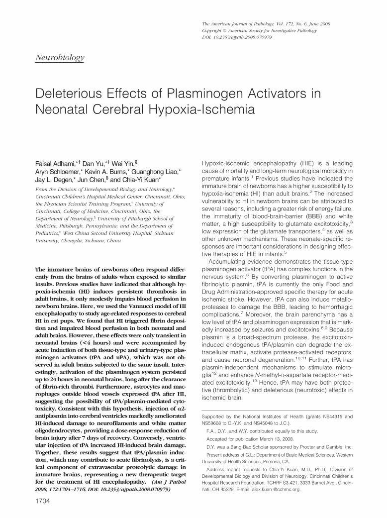
duction of cerebral blood flow in rat pups within the first24 hours after the insult.21 To confirm this observation, weused fluorescein-conjugated dextran (FITC-dextran) as atracer of blood perfusion to examine the brains of 7-day-old rat pups at 1 and 4 hours after the Vannucci model ofHI.2 This analysis revealed patches of vascular obstruc-tion in the hippocampus and the cerebral cortex on thecarotid-occluded side of brain at 1 hour of recovery (Fig-ure 1, A and B). The perfusion index (the ratio of FITC-filled pixels to those of the total area) is at 53% of thecontralateral side (P � 0.0001 by t-test, n � 8) (Figure1Q). However, at 4 hours of recovery, the patches ofvascular obstructions disappeared, and the cerebral per-fusion index rose to 74% of the contralateral side (P �0.03, n � 8) (Figure 1, C, D, and Q). These resultsconfirmed the previous report,21 indicating that HI onlycauses a brief period of vascular obstruction in the new-born brain. The HI-induced transient hypoperfusion innewborn brains is in contrast to its effect of persistentdeficits of cerebral perfusion in adult brains.22
In adult brains, the cerebral blood flow reduction isprimarily caused by HI-induced fibrin and platelet depo-sition inside the blood vessels.22 Thus, we set out toexamine whether HI has the same effects in the newbornbrain. To do so, rat pups were sacrificed at 1 and 4 hoursafter HI by transcardial perfusion of saline—to remove theintravascular fibrinogen and platelets—and fixative, fol-lowed by processing the brains for immunohistochemicalanalysis. This analysis revealed unilateral fibrin(ogen)and platelet (detected by antibodies against the platelet-specific integrin subunit �IIb) deposition in the bloodvessels at 1 hour of recovery on the carotid-occlusionside of brain (Figure 1, E, F, I, and J; n � 5). However, theextent of intravascular fibrin(ogen) and platelet deposi-tion markedly diminished at 4 hours of recovery (Figure 1,G, H, K, and L; n � 5), consistent with the recovery ofcerebral perfusion at this time (Figure 1, C and D).
Besides thrombosis-fibrinolysis events, we also usedimmunocytochemistry and immunoblot analyses to ex-amine the timing of HI-induced BBB damage in newbornbrains. Immunocytochemistry revealed immunoglobulinstaining, an indication of increased BBB permeability,23,25 at 4hours on the carotid-occluded side of brain (Figure 1, Mand N), which was intensified at 24 hours (Figure 1, Oand P). Moreover, immunoblot showed the reduction ofoccludin, an endothelial tight junction-associated protein,at 4 hours after HI (Figure 1R). Together, these resultssuggest HI induces progressive BBB damage and rapidthrombosis in neonatal brains, similar to its effect inadults,22 but the immature brain has a countermecha-nism to rapidly remove the thrombi.
Neonatal HI Triggers Persistent Induction of thePlasminogen System
To search for the mechanism of acute recovery of cere-bral blood flow and progressive damage to BBB in HI-challenged neonatal brains, we used RT-PCR, zymogra-phy, and in situ zymography to examine the expressionand activity of tPA and urinary-type plasminogen activa-
Plasminogen Activators in Neonatal HIE 1707AJP June 2008, Vol. 172, No. 6
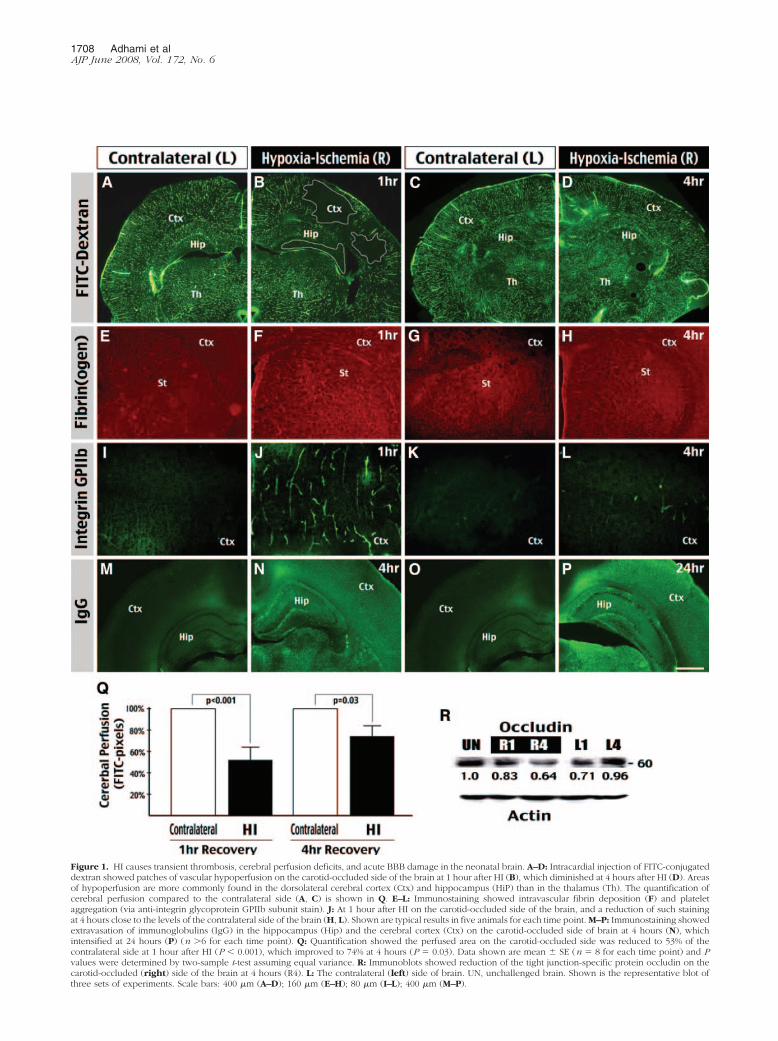
Figure 1. HI causes transient thrombosis, cerebral perfusion deficits, and acute BBB damage in the neonatal brain. A–D: Intracardial injection of FITC-conjugateddextran showed patches of vascular hypoperfusion on the carotid-occluded side of the brain at 1 hour after HI (B), which diminished at 4 hours after HI (D). Areasof hypoperfusion are more commonly found in the dorsolateral cerebral cortex (Ctx) and hippocampus (HiP) than in the thalamus (Th). The quantification ofcerebral perfusion compared to the contralateral side (A, C) is shown in Q. E–L: Immunostaining showed intravascular fibrin deposition (F) and plateletaggregation (via anti-integrin glycoprotein GPIIb subunit stain). J: At 1 hour after HI on the carotid-occluded side of the brain, and a reduction of such stainingat 4 hours close to the levels of the contralateral side of the brain (H, L). Shown are typical results in five animals for each time point. M–P: Immunostaining showedextravasation of immunoglobulins (IgG) in the hippocampus (Hip) and the cerebral cortex (Ctx) on the carotid-occluded side of brain at 4 hours (N), whichintensified at 24 hours (P) (n �6 for each time point). Q: Quantification showed the perfused area on the carotid-occluded side was reduced to 53% of thecontralateral side at 1 hour after HI (P � 0.001), which improved to 74% at 4 hours (P � 0.03). Data shown are mean � SE (n � 8 for each time point) and Pvalues were determined by two-sample t-test assuming equal variance. R: Immunoblots showed reduction of the tight junction-specific protein occludin on thecarotid-occluded (right) side of the brain at 4 hours (R4). L: The contralateral (left) side of brain. UN, unchallenged brain. Shown is the representative blot ofthree sets of experiments. Scale bars: 400 �m (A–D); 160 �m (E–H); 80 �m (I–L); 400 �m (M–P).
1708 Adhami et alAJP June 2008, Vol. 172, No. 6
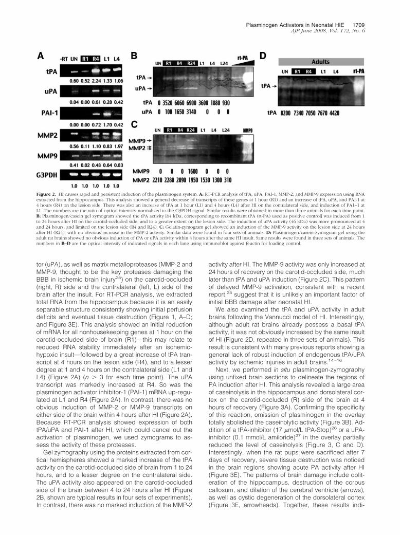
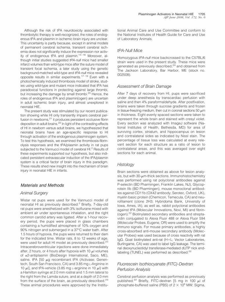
tor (uPA), as well as matrix metalloproteases (MMP-2 andMMP-9, thought to be the key proteases damaging theBBB in ischemic brain injury25) on the carotid-occluded(right, R) side and the contralateral (left, L) side of thebrain after the insult. For RT-PCR analysis, we extractedtotal RNA from the hippocampus because it is an easilyseparable structure consistently showing initial perfusiondeficits and eventual tissue destruction (Figure 1, A–D;and Figure 3E). This analysis showed an initial reductionof mRNA for all nonhousekeeping genes at 1 hour on thecarotid-occluded side of brain (R1)—this may relate toreduced RNA stability immediately after an ischemic-hypoxic insult—followed by a great increase of tPA tran-script at 4 hours on the lesion side (R4), and to a lesserdegree at 1 and 4 hours on the contralateral side (L1 andL4) (Figure 2A) (n � 3 for each time point). The uPAtranscript was markedly increased at R4. So was theplasminogen activator inhibitor-1 (PAI-1) mRNA up-regu-lated at L1 and R4 (Figure 2A). In contrast, there was noobvious induction of MMP-2 or MMP-9 transcripts oneither side of the brain within 4 hours after HI (Figure 2A).Because RT-PCR analysis showed expression of bothtPA/uPA and PAI-1 after HI, which could cancel out theactivation of plasminogen, we used zymograms to as-sess the activity of these proteases.
Gel zymography using the proteins extracted from cor-tical hemispheres showed a marked increase of the tPAactivity on the carotid-occluded side of brain from 1 to 24hours, and to a lesser degree on the contralateral side.The uPA activity also appeared on the carotid-occludedside of the brain between 4 to 24 hours after HI (Figure2B, shown are typical results in four sets of experiments).In contrast, there was no marked induction of the MMP-2
activity after HI. The MMP-9 activity was only increased at24 hours of recovery on the carotid-occluded side, muchlater than tPA and uPA induction (Figure 2C). This patternof delayed MMP-9 activation, consistent with a recentreport,25 suggest that it is unlikely an important factor ofinitial BBB damage after neonatal HI.
We also examined the tPA and uPA activity in adultbrains following the Vannucci model of HI. Interestingly,although adult rat brains already possess a basal tPAactivity, it was not obviously increased by the same insultof HI (Figure 2D, repeated in three sets of animals). Thisresult is consistent with many previous reports showing ageneral lack of robust induction of endogenous tPA/uPAactivity by ischemic injuries in adult brains.14–16
Next, we performed in situ plasminogen-zymographyusing unfixed brain sections to delineate the regions ofPA induction after HI. This analysis revealed a large areaof caseinolysis in the hippocampus and dorsolateral cor-tex on the carotid-occluded (R) side of the brain at 4hours of recovery (Figure 3A). Confirming the specificityof this reaction, omission of plasminogen in the overlaytotally abolished the caseinolytic activity (Figure 3B). Ad-dition of a tPA-inhibitor (17 �mol/L tPA-Stop)26 or a uPA-inhibitor (0.1 mmol/L amiloride)27 in the overlay partiallyreduced the level of caseinolysis (Figure 3, C and D).Interestingly, when the rat pups were sacrificed after 7days of recovery, severe tissue destruction was noticedin the brain regions showing acute PA activity after HI(Figure 3E). The patterns of brain damage include oblit-eration of the hippocampus, destruction of the corpuscallosum, and dilation of the cerebral ventricle (arrows),as well as cystic degeneration of the dorsolateral cortex(Figure 3E, arrowheads). Together, these results indi-
Figure 2. HI causes rapid and persistent induction of the plasminogen system. A: RT-PCR analysis of tPA, uPA, PAI-1, MMP-2, and MMP-9 expression using RNAextracted from the hippocampus. This analysis showed a general decrease of transcripts of these genes at 1 hour (R1) and an increase of tPA, uPA, and PAI-1 at4 hours (R4) on the lesion side. There was also an increase of tPA at 1 hour (L1) and 4 hours (L4) after HI on the contralateral side, and induction of PA1–1 atL1. The numbers are the ratio of optical intensity normalized to the G3PDH signal. Similar results were obtained in more than three animals for each time point.B: Plasminogen/casein gel zymogram showed the tPA activity [64 kDa; corresponding to recombinant tPA (rt-PA) used as positive control] was induced from 1to 24 hours after HI on the carotid-occluded side, and to a greater extent on the lesion side. The induction of uPA activity (46 kDa) was more pronounced at 4and 24 hours, and limited on the lesion side (R4 and R24). C: Gelatin-zymogram gel showed an induction of the MMP-9 activity on the lesion side at 24 hoursafter HI (R24), with no obvious increase in the MMP-2 activity. Similar data were found in four sets of animals. D: Plasminogen/casein-zymogram gel using theadult rat brains showed no obvious induction of tPA or uPA activity within 4 hours after the same HI insult. Same results were found in three sets of animals. Thenumbers in B–D are the optical intensity of indicated signals in each lane using immunoblot against �-actin for loading control.
Plasminogen Activators in Neonatal HIE 1709AJP June 2008, Vol. 172, No. 6
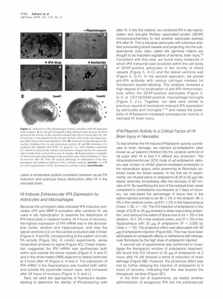
cated a remarkable spatial correlation between acute PAinduction and eventual tissue destruction after HI in theneonatal brain.
HI Induces Extravascular tPA Expression byAstrocytes and Macrophages
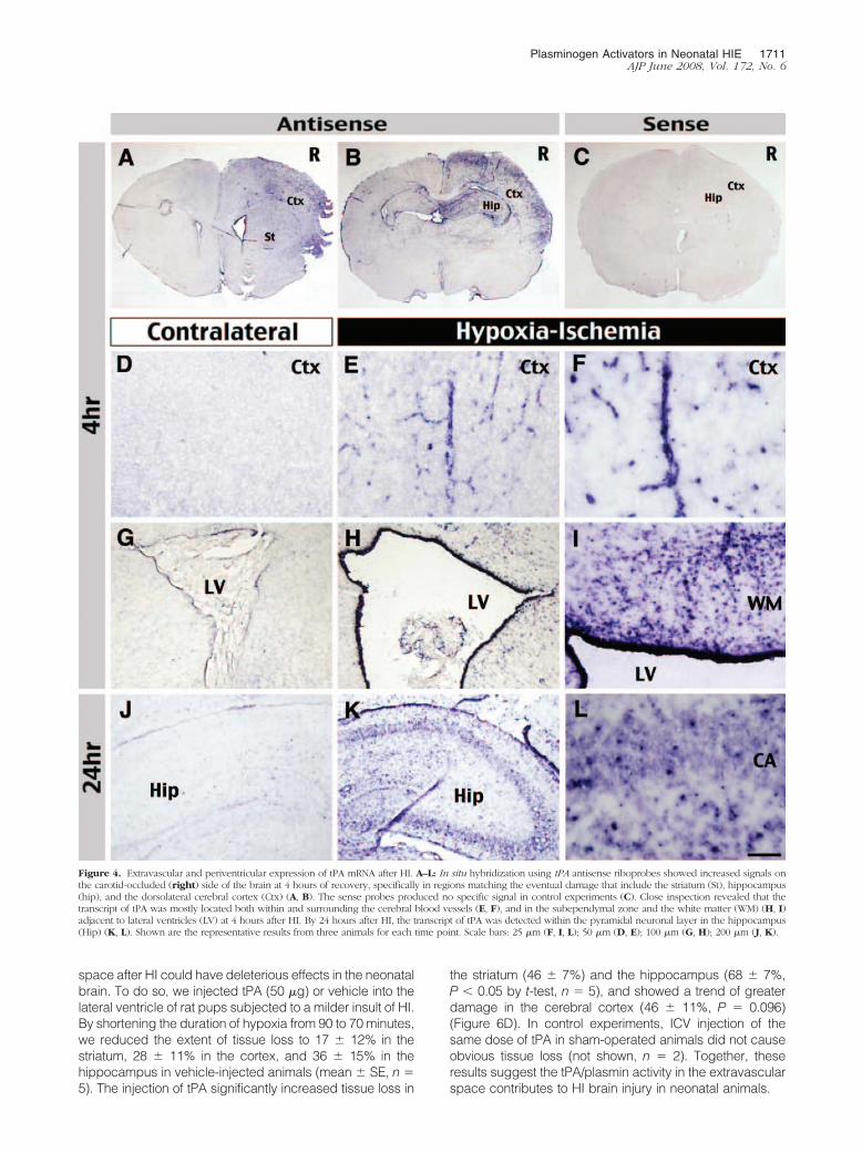
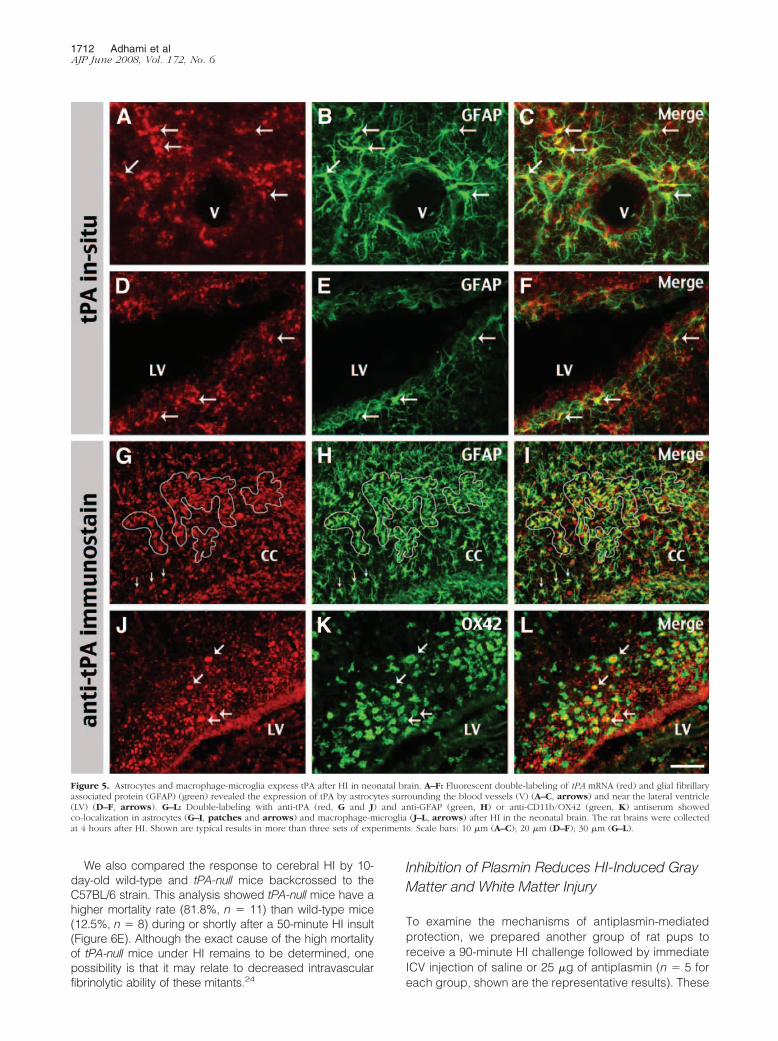
Because the zymogram data indicated tPA induction pre-cedes uPA and MMP-9 activation after cerebral HI, weused in situ hybridization to examine the distribution oftPA transcripts in newborn brains. At 4 hours of recovery,the highest expression of tPA mRNA was in the dorsolat-eral cortex, striatum and hippocampus, and near thelateral ventricle (LV) on the carotid-occluded side of brain(Figure 4, A and B), corresponding to the pattern of in situPA activity (Figure 3A). In control experiments, senseriboprobes showed no signal (Figure 4C). Closer inspec-tion suggested the tPA mRNA was synthesized bothwithin and outside the blood vessels (Figure 4, E and F)and in the white matter (WM) adjacent to lateral ventriclesat 4 hours after HI (Figure 4, H and I). The expression oftPA mRNA in the hippocampus was located both withinand outside the pyramidal neuron layer, and increasedafter 24 hours of recovery (Figure 4, K and L).
Next, we used two approaches of fluorescent double-labeling to determine the identity of tPA-producing cells
after HI. In the first method, we combined tPA in situ hybrid-ization and anti-glial fibrillary associated protein (GFAP)immunocytochemistry to test whether astrocytes expresstPA after HI. This is because astrocytes with extensive end-feet surrounding blood vessels and projecting into the sub-ependymal zone (also called the germinal matrix) arethought to be important regulators of ischemic brain injury.28
Consistent with this view, we found many instances inwhich tPA transcript was localized within the cell bodyof GFAP-positive astrocytes in the vicinity of bloodvessels (Figure 5, A–C) and the lateral ventricle wall(Figure 5, D–F). In the second approach, we pairedanti-tPA antibody with various cell-type markers forfluorescent double-labeling. This analysis revealed ahigh degree of co-localization of anti-tPA immunoreac-tivity within the GFAP-positive astrocytes (Figure 5,G–I) or CD11b/OX42-positive macrophage-microglia(Figure 5, J–L). Together, our data were similar toprevious reports of excitotoxin-induced tPA expressionby astrocytes and microglia,9,10 and raised the possi-bility of tPA/plasmin-mediated extravascular toxicity inneonatal HI brain injury.
tPA/Plasmin Activity Is a Critical Factor of HIBrain Injury in Neonates
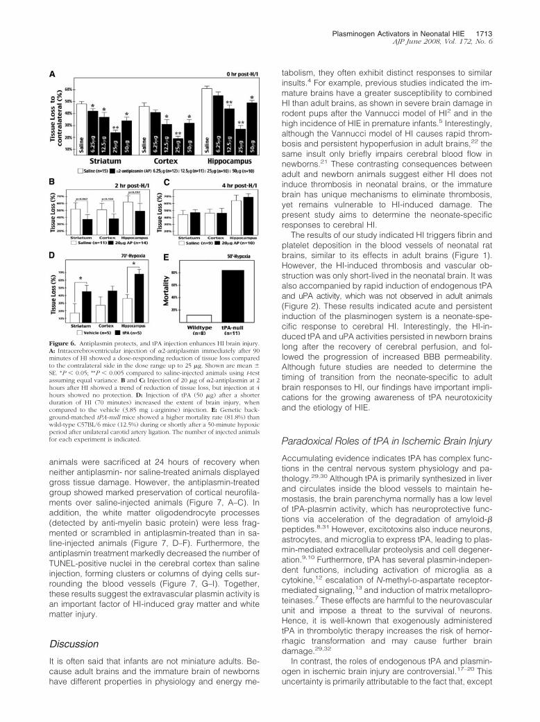
To test whether the HI-induced tPA/plasmin activity contrib-utes to brain damage, we injected �2-antiplasmin (alsoknown as �2 plasmin inhibitor) into the cerebral ventricle ofrat pups after HI to test if it offered any protection. Theintracerebroventricular (ICV) route of �2-antiplasmin deliv-ery was chosen to inhibit plasmin-mediated proteolysis inthe extracellular space, while preserving its fibrinolytic po-tential inside the blood vessels. In the first set of experi-ments, we infused saline or antiplasmin (6.25 to 50 �g) intolateral ventricles immediately after the recovery of 90 min-utes of HI. By quantifying the size of the residual brain areascompared to contralateral counterparts at 7 days of recov-ery, we calculated the percentage of tissue loss in thesaline-injected animals to be 48 � 2% in the striatum, 46 �3% in the cerebral cortex, and 61 � 2% in the hippocampus(mean � SE, n � 15). The ICV-injection of antiplasmin in therange of 6.25 to 25 �g showed a dose-responding protec-tion, and reduced the extent of tissue loss to 24 � 3% in thestriatum, 19 � 3% in the cerebral cortex, and 27 � 3% in thehippocampus with 25 �g of antiplasmin (P � 0.005 byt-test, n � 10). The protective effect was attenuated with 50�g of antiplasmin injection (Figure 6A). This may have beenattributable to nonspecific effects or interference with intravas-cular fibrinolysis by the high dose of antiplasmin injected.
A second set of experiments was performed to inves-tigate the therapeutic window of antiplasmin treatment.We found that ICV infusion of 20 �g of antiplasmin at 2hours after HI still showed a trend of reduction of braindamage (Figure 6B). However, the protective effect waslost by further delaying the injection of antiplasmin to 4hours of recovery, indicating that this was beyond thetherapeutic window (Figure 6C).
In the third set of experiments, we tested whetheradministration of exogenous tPA into the extravascular
Figure 3. Induction of the plasminogen system correlates with HI-damagedbrain regions. A: In situ gel zymogram using unfixed brain sections showedincreased PA activity on the carotid-occluded side (R) at 4 hours after HI. ThePA activity is concentrated in the dorsal cerebral cortex (Ctx) and hippocam-pus (Hip) than in the basal thalamus (Th). B: Omission of plasminogen in theoverlay abolished the in situ proteolysis activity. C and D: Inclusion of asynthetic tPA inhibitor tPA-STOP (17 �mol/L) or a uPA inhibitor amiloride(0.1 mmol/L) reduced the extent of proteolysis. Images shown are represen-tative results from at least three sets of samples. E: Representative examplesof Nissl stain indicating damage in the posterior part of the forebrain at 7 daysof recovery after HI. Note the typical pathology of obliteration of the hip-pocampus and unilateral dilation of the cerebral ventricle (arrows), as wellas cystic degeneration of the dorsolateral cerebral cortex (arrowheads).
1710 Adhami et alAJP June 2008, Vol. 172, No. 6
space after HI could have deleterious effects in the neonatalbrain. To do so, we injected tPA (50 �g) or vehicle into thelateral ventricle of rat pups subjected to a milder insult of HI.By shortening the duration of hypoxia from 90 to 70 minutes,we reduced the extent of tissue loss to 17 � 12% in thestriatum, 28 � 11% in the cortex, and 36 � 15% in thehippocampus in vehicle-injected animals (mean � SE, n �5). The injection of tPA significantly increased tissue loss in
the striatum (46 � 7%) and the hippocampus (68 � 7%,P � 0.05 by t-test, n � 5), and showed a trend of greaterdamage in the cerebral cortex (46 � 11%, P � 0.096)(Figure 6D). In control experiments, ICV injection of thesame dose of tPA in sham-operated animals did not causeobvious tissue loss (not shown, n � 2). Together, theseresults suggest the tPA/plasmin activity in the extravascularspace contributes to HI brain injury in neonatal animals.
Figure 4. Extravascular and periventricular expression of tPA mRNA after HI. A–L: In situ hybridization using tPA antisense riboprobes showed increased signals onthe carotid-occluded (right) side of the brain at 4 hours of recovery, specifically in regions matching the eventual damage that include the striatum (St), hippocampus(hip), and the dorsolateral cerebral cortex (Ctx) (A, B). The sense probes produced no specific signal in control experiments (C). Close inspection revealed that thetranscript of tPA was mostly located both within and surrounding the cerebral blood vessels (E, F), and in the subependymal zone and the white matter (WM) (H, I)adjacent to lateral ventricles (LV) at 4 hours after HI. By 24 hours after HI, the transcript of tPA was detected within the pyramidal neuronal layer in the hippocampus(Hip) (K, L). Shown are the representative results from three animals for each time point. Scale bars: 25 �m (F, I, L); 50 �m (D, E); 100 �m (G, H); 200 �m (J, K).
Plasminogen Activators in Neonatal HIE 1711AJP June 2008, Vol. 172, No. 6
We also compared the response to cerebral HI by 10-day-old wild-type and tPA-null mice backcrossed to theC57BL/6 strain. This analysis showed tPA-null mice have ahigher mortality rate (81.8%, n � 11) than wild-type mice(12.5%, n � 8) during or shortly after a 50-minute HI insult(Figure 6E). Although the exact cause of the high mortalityof tPA-null mice under HI remains to be determined, onepossibility is that it may relate to decreased intravascularfibrinolytic ability of these mitants.24
Inhibition of Plasmin Reduces HI-Induced GrayMatter and White Matter Injury
To examine the mechanisms of antiplasmin-mediatedprotection, we prepared another group of rat pups toreceive a 90-minute HI challenge followed by immediateICV injection of saline or 25 �g of antiplasmin (n � 5 foreach group, shown are the representative results). These
Figure 5. Astrocytes and macrophage-microglia express tPA after HI in neonatal brain. A–F: Fluorescent double-labeling of tPA mRNA (red) and glial fibrillaryassociated protein (GFAP) (green) revealed the expression of tPA by astrocytes surrounding the blood vessels (V) (A–C, arrows) and near the lateral ventricle(LV) (D–F, arrows). G–L: Double-labeling with anti-tPA (red, G and J) and anti-GFAP (green, H) or anti-CD11b/OX42 (green, K) antiserum showedco-localization in astrocytes (G–I, patches and arrows) and macrophage-microglia (J–L, arrows) after HI in the neonatal brain. The rat brains were collectedat 4 hours after HI. Shown are typical results in more than three sets of experiments. Scale bars: 10 �m (A–C); 20 �m (D–F); 30 �m (G–L).
1712 Adhami et alAJP June 2008, Vol. 172, No. 6
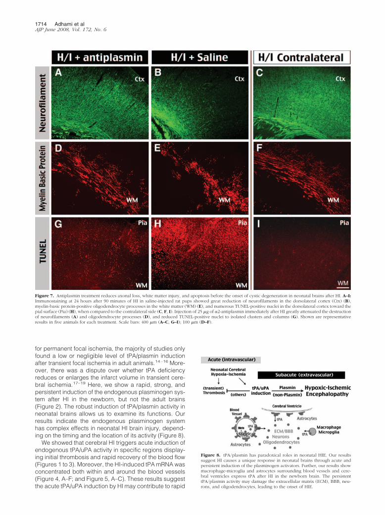
animals were sacrificed at 24 hours of recovery whenneither antiplasmin- nor saline-treated animals displayedgross tissue damage. However, the antiplasmin-treatedgroup showed marked preservation of cortical neurofila-ments over saline-injected animals (Figure 7, A–C). Inaddition, the white matter oligodendrocyte processes(detected by anti-myelin basic protein) were less frag-mented or scrambled in antiplasmin-treated than in sa-line-injected animals (Figure 7, D–F). Furthermore, theantiplasmin treatment markedly decreased the number ofTUNEL-positive nuclei in the cerebral cortex than salineinjection, forming clusters or columns of dying cells sur-rounding the blood vessels (Figure 7, G–I). Together,these results suggest the extravascular plasmin activity isan important factor of HI-induced gray matter and whitematter injury.
Discussion
It is often said that infants are not miniature adults. Be-cause adult brains and the immature brain of newbornshave different properties in physiology and energy me-
tabolism, they often exhibit distinct responses to similarinsults.4 For example, previous studies indicated the im-mature brains have a greater susceptibility to combinedHI than adult brains, as shown in severe brain damage inrodent pups after the Vannucci model of HI2 and in thehigh incidence of HIE in premature infants.5 Interestingly,although the Vannucci model of HI causes rapid throm-bosis and persistent hypoperfusion in adult brains,22 thesame insult only briefly impairs cerebral blood flow innewborns.21 These contrasting consequences betweenadult and newborn animals suggest either HI does notinduce thrombosis in neonatal brains, or the immaturebrain has unique mechanisms to eliminate thrombosis,yet remains vulnerable to HI-induced damage. Thepresent study aims to determine the neonate-specificresponses to cerebral HI.
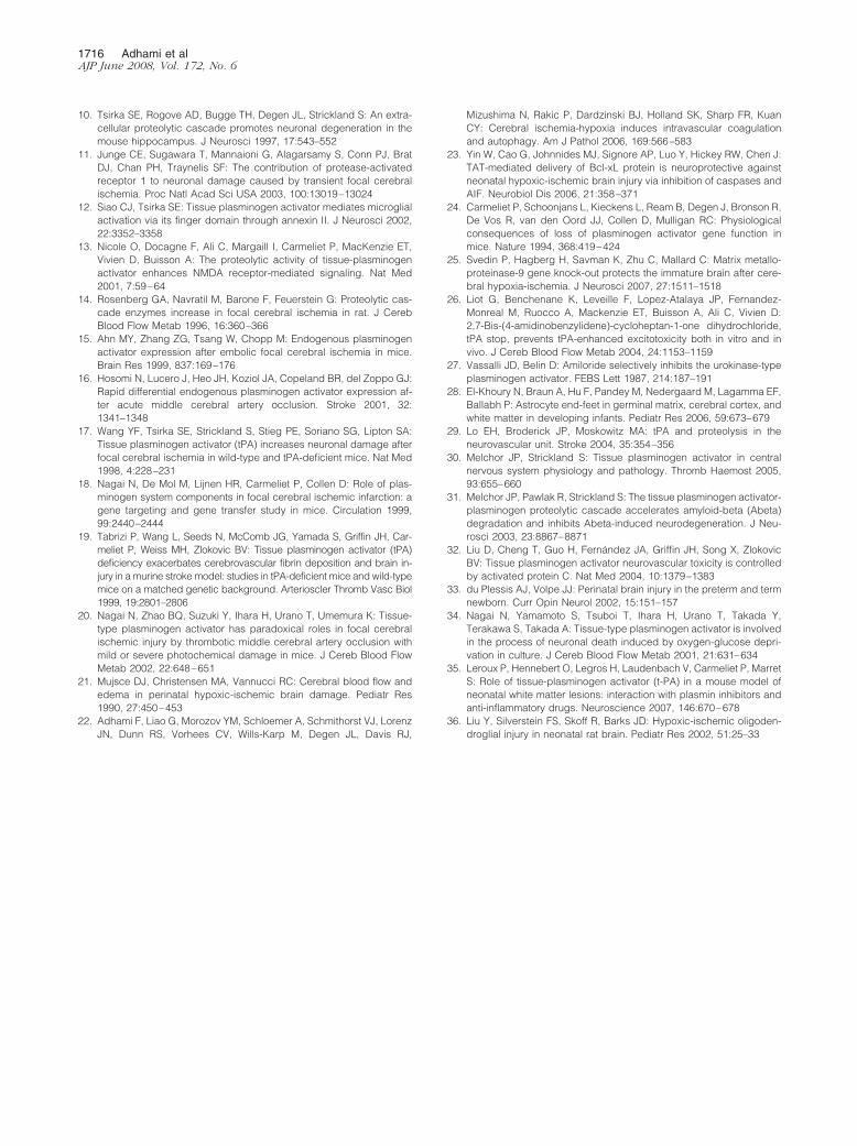
The results of our study indicated HI triggers fibrin andplatelet deposition in the blood vessels of neonatal ratbrains, similar to its effects in adult brains (Figure 1).However, the HI-induced thrombosis and vascular ob-struction was only short-lived in the neonatal brain. It wasalso accompanied by rapid induction of endogenous tPAand uPA activity, which was not observed in adult animals(Figure 2). These results indicated acute and persistentinduction of the plasminogen system is a neonate-spe-cific response to cerebral HI. Interestingly, the HI-in-duced tPA and uPA activities persisted in newborn brainslong after the recovery of cerebral perfusion, and fol-lowed the progression of increased BBB permeability.Although future studies are needed to determine thetiming of transition from the neonate-specific to adultbrain responses to HI, our findings have important impli-cations for the growing awareness of tPA neurotoxicityand the etiology of HIE.
Paradoxical Roles of tPA in Ischemic Brain Injury
Accumulating evidence indicates tPA has complex func-tions in the central nervous system physiology and pa-thology.29,30 Although tPA is primarily synthesized in liverand circulates inside the blood vessels to maintain he-mostasis, the brain parenchyma normally has a low levelof tPA-plasmin activity, which has neuroprotective func-tions via acceleration of the degradation of amyloid-�peptides.8,31 However, excitotoxins also induce neurons,astrocytes, and microglia to express tPA, leading to plas-min-mediated extracellular proteolysis and cell degener-ation.9,10 Furthermore, tPA has several plasmin-indepen-dent functions, including activation of microglia as acytokine,12 escalation of N-methyl-D-aspartate receptor-mediated signaling,13 and induction of matrix metallopro-teinases.7 These effects are harmful to the neurovascularunit and impose a threat to the survival of neurons.Hence, it is well-known that exogenously administeredtPA in thrombolytic therapy increases the risk of hemor-rhagic transformation and may cause further braindamage.29,32
In contrast, the roles of endogenous tPA and plasmin-ogen in ischemic brain injury are controversial.17–20 Thisuncertainty is primarily attributable to the fact that, except
Figure 6. Antiplasmin protects, and tPA injection enhances HI brain injury.A: Intracerebroventricular injection of �2-antiplasmin immediately after 90minutes of HI showed a dose-responding reduction of tissue loss comparedto the contralateral side in the dose range up to 25 �g. Shown are mean �SE. *P � 0.05; **P � 0.005 compared to saline-injected animals using t-testassuming equal variance. B and C: Injection of 20 �g of �2-antiplasmin at 2hours after HI showed a trend of reduction of tissue loss, but injection at 4hours showed no protection. D: Injection of tPA (50 �g) after a shorterduration of HI (70 minutes) increased the extent of brain injury, whencompared to the vehicle (3.85 mg L-arginine) injection. E: Genetic back-ground-matched tPA-null mice showed a higher mortality rate (81.8%) thanwild-type C57BL/6 mice (12.5%) during or shortly after a 50-minute hypoxicperiod after unilateral carotid artery ligation. The number of injected animalsfor each experiment is indicated.
Plasminogen Activators in Neonatal HIE 1713AJP June 2008, Vol. 172, No. 6
for permanent focal ischemia, the majority of studies onlyfound a low or negligible level of tPA/plasmin inductionafter transient focal ischemia in adult animals.14–16 More-over, there was a dispute over whether tPA deficiencyreduces or enlarges the infarct volume in transient cere-bral ischemia.17–19 Here, we show a rapid, strong, andpersistent induction of the endogenous plasminogen sys-tem after HI in the newborn, but not the adult brains(Figure 2). The robust induction of tPA/plasmin activity inneonatal brains allows us to examine its functions. Ourresults indicate the endogenous plasminogen systemhas complex effects in neonatal HI brain injury, depend-ing on the timing and the location of its activity (Figure 8).
We showed that cerebral HI triggers acute induction ofendogenous tPA/uPA activity in specific regions display-ing initial thrombosis and rapid recovery of the blood flow(Figures 1 to 3). Moreover, the HI-induced tPA mRNA wasconcentrated both within and around the blood vessels(Figure 4, A–F; and Figure 5, A–C). These results suggestthe acute tPA/uPA induction by HI may contribute to rapid
Figure 7. Antiplasmin treatment reduces axonal loss, white matter injury, and apoptosis before the onset of cystic degeneration in neonatal brains after HI. A–I:Immunostaining at 24 hours after 90 minutes of HI in saline-injected rat pups showed great reduction of neurofilaments in the dorsolateral cortex (Ctx) (B),myelin-basic protein-positive oligodendrocyte processes in the white matter (WM) (E), and numerous TUNEL-positive nuclei in the dorsolateral cortex toward thepial surface (Pia) (H), when compared to the contralateral side (C, F, I). Injection of 25 �g of �2-antiplasmin immediately after HI greatly attenuated the destructionof neurofilaments (A) and oligodendrocyte processes (D), and reduced TUNEL-positive nuclei to isolated clusters and columns (G). Shown are representativeresults in five animals for each treatment. Scale bars: 400 �m (A–C, G–I); 100 �m (D–F).
Figure 8. tPA/plasmin has paradoxical roles in neonatal HIE. Our resultssuggest HI causes a unique response in neonatal brains through acute andpersistent induction of the plasminogen activators. Further, our results showmacrophage-microglia and astrocytes surrounding blood vessels and cere-bral ventricles express tPA after HI in the newborn brain. The persistenttPA/plasmin activity may damage the extracellular matrix (ECM), BBB, neu-rons, and oligodendrocytes, leading to the onset of HIE.
1714 Adhami et alAJP June 2008, Vol. 172, No. 6

fibrinolysis in neonatal brains. Consistent with this view,tPA-null mice show a higher mortality rate than wild-typemice during or shortly after HI (81.8% versus 12.5%,Figure 6E). Future studies are needed to compare theresponse to HI among several strains of mice with amutation in components of the plasminogen system (in-cluding tPA-null, uPA-null, and plasminogen-nullmice10,24) to determine their relative functions for acutefibrinolysis in neonatal brains.
Although the induction of tPA/plasmin in the acutephase after HI may be protective, our study providedstrong evidence that their persistent activity is a criticalfactor of brain injury in newborns (Figure 8). First, there isa high degree of spatial correlation between HI-inducedtPA expression and brain regions that are most suscep-tible to this insult, including the hippocampus, theperiventricular white matter, and the dorsolateral cerebralcortex (Figures 3 and 4). Furthermore, the double-label-ing data indicate perivascular-periventricular astro-cytes28 and microphage-microglia are principle tPA-pro-ducing cell types after HI (Figure 5). This pattern of tPAexpression, similar to the situation in excitotoxin-chal-lenge,9,10 suggests the persistent tPA/plasmin activityafter HI may induce extravascular proteolysis leading tobrain damage. Consistent with this hypothesis, weshowed that inhibition of extravascular proteolysis, bycerebroventricular injection of antiplasmin in the range of6.25 to 25 �g, provided dose-responding reduction ofbrain damage at 7-day recovery of HI (Figure 6). The lessefficacy of 50 �g of antiplasmin in ventricular injectionmay be attributable to nonspecific effects or drug diffu-sion into blood vessels at this high dose. Furthermore, weshowed that ventricular injection of antiplasmin markedlyprotects against HI-induced neurofilament and oligoden-drocyte damage before the onset of gross tissue degen-eration (Figure 7). In contrast, ventricular injection of tPAsignificantly amplified the extent of HI brain damage (Fig-ure 6). Together, these results strongly suggest persis-tent activation of endogenous tPA/plasmin is a criticalfactor of HI-induced brain damage through extravascularproteolysis.
Extracellular Proteolysis in HIE
HIE attributable to placenta insufficiency or birth as-phyxia is an important cause of perinatal mortality andlong-term neurological morbidity, including cerebralpalsy, mental retardation, and epilepsy.1,33 Previousstudies on the etiology of HIE focused on the risk ofenergy failure and the cell death signaling pathwayswithin neurons. However, except for hypothermia ther-apy, none of these neuroprotective strategies have beentranslated to clinical practice against HIE.4,5 Conse-quently, the current therapy of HIE remains mainly sup-portive, and directed toward the systemic complications.Hence, better understanding the pathological mecha-nism of HIE and development of new therapies remainsan urgent issues in neonatology.
In this regard, the present study is significant becauseit suggests a novel mechanism and a new therapeutic
target in HIE, namely tPA/plasmin-mediated extracellularproteolysis. Although this hypothesis is currently basedon studies using a rodent model of neonatal HIE (theVannucci model),2 several lines of evidence suggest it isrelevant to clinical settings. First, imaging studies rarelyidentify thrombosis in infants diagnosed with HIE, similarto the rapid recovery of cerebral blood flow after HI in theanimal model.21 Moreover, the characteristic pathologi-cal finding of HIE in humans is cystic encephalomalacia,which is more consistent with an extracellular proteolysis-based mechanism of tissue injury than apoptosis-basedcell elimination that does not typically lead to cystic cav-itations.33 Furthermore, the locations of tPA transcript inthe Vannucci model correlate with the white matter le-sions (periventricular leukomalacia) and germinal matrixhemorrhage often observed in the HIE infants.1 Finally,tPA has been shown to increase oxygen-glucose depri-vation-induced neuronal death in vitro,34 and injection oftPA into the white matter induces oligodendrocyte dam-age in neonatal brains.35,36 Together, these circumstan-tial evidences suggest our findings are not limited to therodent model, but may be relevant to HIE in clinicalsettings.
In conclusion, we suggest future studies are warrantedto examine whether infants diagnosed with HIE have anelevated level of tPA and plasmin activity in the brain orthe cerebrospinal fluid. If so, anti-tPA or anti-plasmintreatment in the extravascular space of neonatal brainmay offer direct brain protection against this devastatingdisorder.
Acknowledgments
We thank Drs. Ton DeGrauw and Joe Broderick for criti-cal reading of the manuscript and Ms. Wei-Lan Weng fortechnical assistance.
References
1. Ferriero DM: Neonatal brain injury. N Engl J Med 2004, 351:1985–19952. Rice JE III, Vannucci RC, Brierley JB: The influence of immaturity on
hypoxic-ischemic brain damage in the rat. Ann Neurol 1981, 19:131–1413. McDonald JW, Silverstein FS, Cardona D, Hudson C, Chen R,
Johnston MV: Systemic administration of MK-801 protects againstN-methyl-D-aspartate- and quisqualate-mediated neurotoxicity inperinatal rats. Neuroscience 1990, 36:589–599
4. Vannucci SJ, Hagberg H: Hypoxia-ischemia in the immature brain.J Exp Biol 2004, 207:3149–3154
5. Vexler ZS, Sharp FR, Feuerstein GZ, Ashwal S, Thoresen M, Yager JY,Ferriero DM: Translational stroke research in the developing brain.Pediatr Neurol 2006, 34:459–463
6. Kaur J, Zhao Z, Klein GM, Lo EH, Buchan AM: The neurotoxicity oftissue plasminogen activator? J Cereb Blood Flow Metab 2004,24:945–963
7. Tsuji K, Aoki T, Tejima E, Arai K, Lee SR, Atochin DN, Huang PL,Wang X, Montaner J, Lo EH: Tissue plasminogen activator promotesmatrix metalloproteinase-9 upregulation after focal cerebral ischemia.Stroke 2005, 36:1954–1959
8. Sappino AP, Madani R, Huarte J, Belin D, Kiss JZ, Wohlwend A,Vassalli JD: Extracellular proteolysis in the adult murine brain. J ClinInvest 1993, 92:679–685
9. Tsirka SE, Gualandris A, Amaral DG, Strickland S: Excitotoxin-in-duced neuronal degeneration and seizure are mediated by tissueplasminogen activator. Nature 1995, 377:340–344
Plasminogen Activators in Neonatal HIE 1715AJP June 2008, Vol. 172, No. 6
10. Tsirka SE, Rogove AD, Bugge TH, Degen JL, Strickland S: An extra-cellular proteolytic cascade promotes neuronal degeneration in themouse hippocampus. J Neurosci 1997, 17:543–552
11. Junge CE, Sugawara T, Mannaioni G, Alagarsamy S, Conn PJ, BratDJ, Chan PH, Traynelis SF: The contribution of protease-activatedreceptor 1 to neuronal damage caused by transient focal cerebralischemia. Proc Natl Acad Sci USA 2003, 100:13019–13024
12. Siao CJ, Tsirka SE: Tissue plasminogen activator mediates microglialactivation via its finger domain through annexin II. J Neurosci 2002,22:3352–3358
13. Nicole O, Docagne F, Ali C, Margaill I, Carmeliet P, MacKenzie ET,Vivien D, Buisson A: The proteolytic activity of tissue-plasminogenactivator enhances NMDA receptor-mediated signaling. Nat Med2001, 7:59–64
14. Rosenberg GA, Navratil M, Barone F, Feuerstein G: Proteolytic cas-cade enzymes increase in focal cerebral ischemia in rat. J CerebBlood Flow Metab 1996, 16:360–366
15. Ahn MY, Zhang ZG, Tsang W, Chopp M: Endogenous plasminogenactivator expression after embolic focal cerebral ischemia in mice.Brain Res 1999, 837:169–176
16. Hosomi N, Lucero J, Heo JH, Koziol JA, Copeland BR, del Zoppo GJ:Rapid differential endogenous plasminogen activator expression af-ter acute middle cerebral artery occlusion. Stroke 2001, 32:1341–1348
17. Wang YF, Tsirka SE, Strickland S, Stieg PE, Soriano SG, Lipton SA:Tissue plasminogen activator (tPA) increases neuronal damage afterfocal cerebral ischemia in wild-type and tPA-deficient mice. Nat Med1998, 4:228–231
18. Nagai N, De Mol M, Lijnen HR, Carmeliet P, Collen D: Role of plas-minogen system components in focal cerebral ischemic infarction: agene targeting and gene transfer study in mice. Circulation 1999,99:2440–2444
19. Tabrizi P, Wang L, Seeds N, McComb JG, Yamada S, Griffin JH, Car-meliet P, Weiss MH, Zlokovic BV: Tissue plasminogen activator (tPA)deficiency exacerbates cerebrovascular fibrin deposition and brain in-jury in a murine stroke model: studies in tPA-deficient mice and wild-typemice on a matched genetic background. Arterioscler Thromb Vasc Biol1999, 19:2801–2806
20. Nagai N, Zhao BQ, Suzuki Y, Ihara H, Urano T, Umemura K: Tissue-type plasminogen activator has paradoxical roles in focal cerebralischemic injury by thrombotic middle cerebral artery occlusion withmild or severe photochemical damage in mice. J Cereb Blood FlowMetab 2002, 22:648–651
21. Mujsce DJ, Christensen MA, Vannucci RC: Cerebral blood flow andedema in perinatal hypoxic-ischemic brain damage. Pediatr Res1990, 27:450–453
22. Adhami F, Liao G, Morozov YM, Schloemer A, Schmithorst VJ, LorenzJN, Dunn RS, Vorhees CV, Wills-Karp M, Degen JL, Davis RJ,
Mizushima N, Rakic P, Dardzinski BJ, Holland SK, Sharp FR, KuanCY: Cerebral ischemia-hypoxia induces intravascular coagulationand autophagy. Am J Pathol 2006, 169:566–583
23. Yin W, Cao G, Johnnides MJ, Signore AP, Luo Y, Hickey RW, Chen J:TAT-mediated delivery of Bcl-xL protein is neuroprotective againstneonatal hypoxic-ischemic brain injury via inhibition of caspases andAIF. Neurobiol Dis 2006, 21:358–371
24. Carmeliet P, Schoonjans L, Kieckens L, Ream B, Degen J, Bronson R,De Vos R, van den Oord JJ, Collen D, Mulligan RC: Physiologicalconsequences of loss of plasminogen activator gene function inmice. Nature 1994, 368:419–424
25. Svedin P, Hagberg H, Savman K, Zhu C, Mallard C: Matrix metallo-proteinase-9 gene knock-out protects the immature brain after cere-bral hypoxia-ischemia. J Neurosci 2007, 27:1511–1518
26. Liot G, Benchenane K, Leveille F, Lopez-Atalaya JP, Fernandez-Monreal M, Ruocco A, Mackenzie ET, Buisson A, Ali C, Vivien D:2,7-Bis-(4-amidinobenzylidene)-cycloheptan-1-one dihydrochloride,tPA stop, prevents tPA-enhanced excitotoxicity both in vitro and invivo. J Cereb Blood Flow Metab 2004, 24:1153–1159
27. Vassalli JD, Belin D: Amiloride selectively inhibits the urokinase-typeplasminogen activator. FEBS Lett 1987, 214:187–191
28. El-Khoury N, Braun A, Hu F, Pandey M, Nedergaard M, Lagamma EF,Ballabh P: Astrocyte end-feet in germinal matrix, cerebral cortex, andwhite matter in developing infants. Pediatr Res 2006, 59:673–679
29. Lo EH, Broderick JP, Moskowitz MA: tPA and proteolysis in theneurovascular unit. Stroke 2004, 35:354–356
30. Melchor JP, Strickland S: Tissue plasminogen activator in centralnervous system physiology and pathology. Thromb Haemost 2005,93:655–660
31. Melchor JP, Pawlak R, Strickland S: The tissue plasminogen activator-plasminogen proteolytic cascade accelerates amyloid-beta (Abeta)degradation and inhibits Abeta-induced neurodegeneration. J Neu-rosci 2003, 23:8867–8871
32. Liu D, Cheng T, Guo H, Fernandez JA, Griffin JH, Song X, ZlokovicBV: Tissue plasminogen activator neurovascular toxicity is controlledby activated protein C. Nat Med 2004, 10:1379–1383
33. du Plessis AJ, Volpe JJ: Perinatal brain injury in the preterm and termnewborn. Curr Opin Neurol 2002, 15:151–157
34. Nagai N, Yamamoto S, Tsuboi T, Ihara H, Urano T, Takada Y,Terakawa S, Takada A: Tissue-type plasminogen activator is involvedin the process of neuronal death induced by oxygen-glucose depri-vation in culture. J Cereb Blood Flow Metab 2001, 21:631–634
35. Leroux P, Hennebert O, Legros H, Laudenbach V, Carmeliet P, MarretS: Role of tissue-plasminogen activator (t-PA) in a mouse model ofneonatal white matter lesions: interaction with plasmin inhibitors andanti-inflammatory drugs. Neuroscience 2007, 146:670–678
36. Liu Y, Silverstein FS, Skoff R, Barks JD: Hypoxic-ischemic oligoden-droglial injury in neonatal rat brain. Pediatr Res 2002, 51:25–33
1716 Adhami et alAJP June 2008, Vol. 172, No. 6