dennis j. dowling, do, faaofiles.academyofosteopathy.org › convo › direct...anterior pelvic...
TRANSCRIPT

1
Direct techniques to treat sacrum
American Academy of OsteopathyConvocation
PHYSICIAN STUDENTThursday, March 18, 2010 Friday, March 19, 2010
2:30 – 4:00 PM 8:00 – 9:30 AM4:30 – 6:00 PM 10:00 – 11:30 AM
Direct techniques to treat sacrum and pelvis somatic dysfunction
(HVLA, MET)
Dennis J. Dowling, DO, FAAO
DIAGNOSISANTERIOR PELVIC ROTATION
FINDINGS ‐ RIGHT
+ STANDING FLEXION TEST RIGHT
RIGHT ASIS LOW – RIGHT PSIS HIGH
TREATMENT
MUSCLE ENERGY
DIAGNOSISANTERIOR PELVIC ROTATION
FINDINGS ‐ RIGHT
+ STANDING FLEXION TEST RIGHT
RIGHT ASIS LOW – RIGHT PSIS HIGH
TREATMENT
FACILITATED POSITIONAL RELEASE
•Physician places cephalad hand on inferior ASIS of the leg that is flexed and externally rotated•Other hand is on the patient’s anterior‐medial knee of the same side
•The patient is instructed to slide that foot down along the opposite leg while the physician resists with both the hand on the ASIS and the hand on the knee (can be repeated)
DIAGNOSISANTERIOR PELVIC ROTATION
FINDINGS ‐ RIGHT
+ STANDING FLEXION TEST RIGHT
RIGHT ASIS LOW – RIGHT PSIS HIGH
TREATMENT
STILL TECHNIQUE
Anterior Ilium/Anterior Innominate (Right) –Patient supine
1. Anterior Ilium/Anterior Innominate (Right) – Patient supine 2. 1. Patient position: supine.3. 2. Physician position: standing at the side of the patient, typically on the same side,
facing the patient.4. 3. Technique:5. The physician places the pad of his index or middle finger of his monitoring cephalad hand
(the hand that is closer to the patient’s head) medial to the posterior superior iliac spine (PSIS) as a means of following the iliosacral motion (The left finger contacts the patient's medial to PSIS on the right).
6. The physician uses his other hand to grasp the patient’s leg on the dysfunctional side and flexes the patient’s hip and knee until motion is felt at the monitoring finger (The physician’s right hand grasps and flexes the patient’s right leg in this example) (Figure 64physician s right hand grasps and flexes the patient s right leg in this example). (Figure 64‐1A)
7. The physician abducts the patient’s knee of the affected side and externally rotates the leg until motion and soft tissue relaxation is felt at the monitoring finger. (Figure 64‐1B) A modification is for the physician to insert the forearm of his caudad arm from lateral to medial beneath the patient’s knee and places his hand on the patient’s anterior thigh. This can add torsion to the modifying and localizing forces.
8. The physician puts downward compression with his caudad (right) arm or shoulder from the patient’s knee through his femur, towards the pelvis and hip, and directed towards sacroiliac joint until further soft tissue is noted at the monitoring finger.
9. While maintaining the compression, the patient’s leg is gently carried through adduction in an arc across the midline and with increased hip flexion by engaging the sacroiliac barrier (rotating the innominate/ilum posteriorly).
10. The patient's hip and knee are brought back to the neutral position by extending both and then the iliosacral somatic dysfunction is reassessed.

2
A
B
Anterior Ilium/Anterior Innominate(Right) – Patient supine
B
DIAGNOSISANTERIOR PELVIC ROTATION
FINDINGS ‐ RIGHT
+ STANDING FLEXION TEST RIGHT
RIGHT ASIS LOW – RIGHT PSIS HIGH
TREATMENT
HVLA
ANTERIOR ROTATIONANTERIOR ROTATION
•HIGH PSIS
ANTERIOR ROTATIONANTERIOR ROTATION
•Place top leg off table
•Thrusting arm posterior/superior to ischial tuberosity
•Direction of THRUST (posterior)
ANTERIOR ROTATIONANTERIOR ROTATION
•Place top leg off table
•Thrusting arm posterior/superior to ischial tuberosity
•Direction of THRUST (posterior)
DIAGNOSISPOSTERIOR PELVIC ROTATION
FINDINGS ‐ RIGHT
+ STANDING FLEXION TEST RIGHT
RIGHT ASIS HIGH – RIGHT PSIS LOW
TREATMENT
MUSCLE ENERGY

3
DIAGNOSISPOSTERIOR PELVIC ROTATION
FINDINGS ‐ RIGHT
+ STANDING FLEXION TEST RIGHT
RIGHT ASIS HIGH – RIGHT PSIS LOW
TREATMENT
FACILITATED POSITIONAL RELEASE
•Physician places cephalad hand on ischialtuberosity of the leg that is flexed and externally rotated•Other hand is on the patient’s anterior‐medial knee of the same side
•The patient is instructed to slide that foot down along the opposite leg while the physician resists with both the hand on the ischial tuberosity and the hand on the knee (can be repeated)
DIAGNOSISPOSTERIOR PELVIC ROTATION
FINDINGS ‐ RIGHT
+ STANDING FLEXION TEST RIGHT
RIGHT ASIS LOW – RIGHT PSIS HIGH
TREATMENT
STILL TECHNIQUE
Posterior Innominate /Posterior Ilium (Right) – Patient supine
• Posterior Innominate /Posterior Ilium (Right) – Patient supine • 1. Patient position: supine.• 2. Physician position: standing at the side of the patient, typically on the same side, facing the
patient.• 3. Technique:• The physician places the pad of his index or middle finger of his monitoring cephalad hand (the hand
that is closer to the patient’s head) medial to the posterior superior iliac spine (PSIS) as a means of following the iliosacral motion (The left finger contacts the patient's medial to PSIS on the right).
• The physician uses his other hand to grasp the patient’s leg on the dysfunctional side and fully flexes th ti t’ hi ( t th 90 d ) d k til ti i f lt t th it i fi Ththe patient’s hip (greater than 90 degrees) and knee until motion is felt at the monitoring finger. The knee is adducted (The physician’s right hand grasps and flexes the patient’s right leg in this example).
• The physician puts downward compression with his caudad (right) arm or shoulder from the patient’s knee through his femur, towards the pelvis and hip, and directed towards sacroiliac joint until further soft tissue is noted at the monitoring finger. (Figure 64‐2A).
• The physician abducts the patient’s knee of the affected side and externally rotates the leg until motion and soft tissue relaxation is felt at the monitoring finger.
• The physician then uses his abdomen or hip to maintain compression through the femur. (Figure 64‐2B).
• While maintaining the compression and external rotation, the patient’s leg is gently carried through extension and to the neutral position.
• The iliosacral somatic dysfunction is reassessed.
A
B
Posterior Innominate /Posterior Ilium (Right) –Patient supine
B
DIAGNOSISPOSTERIOR PELVIC ROTATION
FINDINGS ‐ RIGHT
+ STANDING FLEXION TEST RIGHT
RIGHT ASIS HIGH – RIGHT PSIS LOW
TREATMENT
HVLA

4
POSTERIOR POSTERIOR ROTATIONROTATION
•LOW PSIS
POSTERIOR POSTERIOR ROTATIONROTATION
•Place top leg into position
•Thrusting arm posterior/inferior to ischial tuberosity
•Direction of THRUST (superior)
ANTERIOR ROTATIONANTERIOR ROTATION
•Place top leg off table
•Thrusting arm posterior/superior to ischial tuberosity
•Direction of THRUST (posterior)
DIAGNOSISUPSLIPPED INNOMINATE
FINDINGS ‐ RIGHT
+ STANDING FLEXION TEST RIGHT
RIGHT ASIS HIGH – RIGHT PSIS HIGH
TREATMENT
STILL TECHNIQUE
Upslipped Innominate (Right) – Patient supine
• Upslipped Innominate (Right) – Patient supine • Patient position: supine.• Physician position: initially standing at the foot of the table.• Technique:• The physician grasps the ankle on the dysfunctional side with both hands and
externally rotates and compresses the leg towards the hip and iliosacral joint. (Figure 64‐3)
• The physician then internally rotates the patient’s leg on the involved side until just prior to the ASIS lifting upwards.
• The physician then progressively reduces the compression and transitions to traction until localized to the involved joint (Occasionally, a slighter sharper long axis tug can be placed to encourage downslip of the somatic dysfunction).
• The Still technique for a posteriorly rotated innominate/ilium is applied; followed by:
• The Still technique applied for an anteriorly rotated innominate/ilium.• This series is designed to “ratchet” the upslipped innominate/ilium into place.• The dysfunction is reassessed
Upslipped Innominate (Right) – Patient supine

5
DIAGNOSISUPSLIPPED INNOMINATE
FINDINGS ‐ RIGHT
+ STANDING FLEXION TEST RIGHT
RIGHT ASIS HIGH – RIGHT PSIS HIGH
TREATMENT
HVLA
R SUPERIOR ILIAC SHEARR SUPERIOR ILIAC SHEAR
•Patient supine
•Patient supine – patient holds onto sides of table
•Select superior shear side (ASIS & PSIS side higher)
R SUPERIOR ILIAC SHEARR SUPERIOR ILIAC SHEAR
•Superior ILIAC side
•Patient supine – patient holds onto sides of table
•Select superior shear side (ASIS & PSIS side higher)
•Physician stands at foot of table
R SUPERIOR ILIAC SHEARR SUPERIOR ILIAC SHEAR
•Patient supine – patient holds onto sides of table
•Select superior shear side (ASIS & PSIS side higher)
•Physician stands at foot of table
R SUPERIOR ILIAC SHEARR SUPERIOR ILIAC SHEAR
•Physician grasps affected leg with both hands above ankle
•Patient supine – patient holds onto sides of table
•Select superior shear side (ASIS & PSIS side higher)
•Physician stands at foot of table
R SUPERIOR ILIAC SHEARR SUPERIOR ILIAC SHEAR
•Physician grasps affected leg with both hands above ankle
•Leg is lifted slightly (hip flexion), internally rotated, and slight adduction is introduced

6
•Patient is instructed to take a deep breath
•(Patient can be instructed to give a deep cough)
•Physician performs a sharp tug along leg
R SUPERIOR ILIAC SHEARR SUPERIOR ILIAC SHEAR
DIAGNOSISDOWNSLIPPED INNOMINATE
FINDINGS ‐ RIGHT
+ STANDING FLEXION TEST RIGHT
RIGHT ASIS LOW – RIGHT PSIS LOW
TREATMENT
STILL TECHNIQUE
Downslipped Innominate (Right) – Patient supine
• Downslipped Innominate (Right) – Patient supine • Patient position: supine.• Physician position: initially standing at the foot of the table.• Technique:
– The physician grasps the ankle on the dysfunctional side with both hands and externally rotates and tractions the leg towards the physician by pulling parallel to the tableparallel to the table.
– The physician then internally rotates the patient’s leg on the involved side until just prior to the ASIS lifting upwards.
– The physician then progressively reduces the traction and transitions to compression until localized to the involved joint
– The Still technique for an anteriorly rotated innominate/ilium is applied; followed by:
– The Still technique applied for a posteriorly rotated innominate/ilium.– This series is designed to “ratchet” the downslipped innominate/ilium into
place.
– The dysfunction is reassessed
Downslipped Innominate (Right) –Patient supine
•Patient stands on foot of side of inferior iliac shear
•The patient pis instructed to hop on one foot

7

8
DIAGNOSISPUBIC DYSFUNCTION
PUBIC RESTRICTION
TREATMENT
MUSCLE ENERGY AMHVLA
PUBIC DYSFUNCTIONPUBIC DYSFUNCTION
•Patient supine
PUBIC DYSFUNCTIONPUBIC DYSFUNCTION
•Patient supine
PUBIC DYSFUNCTIONPUBIC DYSFUNCTION
•Patient supine
•Knees and hips bent; feet together and flat on table
PUBIC DYSFUNCTIONPUBIC DYSFUNCTION
•Patient supine
•Knees and hips bent; feet together and flat on table
•Physician at side of table

9
PUBIC DYSFUNCTIONPUBIC DYSFUNCTION
•Patient supine
•Knees and hips bent; feet together and flat on table
•Physician at side of table – holds outside of patient’s knees
Patient pushes knees laterally (abduction) against isometric resistance provided by physician
PUBIC DYSFUNCTIONPUBIC DYSFUNCTION
•Patient supine
•Knees and hips bent; feet together and flat on table
•Physician at side of table – holds outside of patient’s knees – puts fist between patient’s knees
Patient pushes knees medially (adduction) against isometric resistance provided by physician
PUBIC DYSFUNCTIONPUBIC DYSFUNCTION
•Puts 2 fists between patient’s knees
Patient pushes knees medially (adduction) against isometric resistance provided by physician
PUBIC DYSFUNCTIONPUBIC DYSFUNCTION
•Puts forearm between patient’s knees
Patient pushes knees medially (adduction) against isometric resistance provided by physician
PUBIC DYSFUNCTIONPUBIC DYSFUNCTION
•Patient’s legs are externally rotated (knees abducted) to barrier
•Puts hands against patient’s knees
Patient pushes knees medially (adduction) against isometric resistance provided by physician
PUBIC DYSFUNCTIONPUBIC DYSFUNCTION
•At the conclusion of the previous effort, the patient is instructed to relax completely
•A slight exaggeration of external rotation/abduction is introduced

10
DIAGNOSISSACRAL DYSFUNCTIONS
FINDINGS – RIGHT DEEP SULCUS
L on L; L on R; R Unilateral; ;
TREATMENT
“jiggle the doodad”
DIAGNOSISSACRAL DYSFUNCTIONS
FINDINGS – RIGHT DEEP SULCUS
L on L; L on R; R Unilateral; ;
TREATMENT
HVLA
Description of Supine HVLA technique for Sacroiliac dysfunction•Patient supine•The side of the deeper sulcus is as close to the edge of the table as possible•Physician stands on side of the deeper sacral sulcus•The physician faces the patient•The physicians positions the patient•positioning the patient’s legs•the cephalad hand holds the patient’s pelvis•the caudad hand pushes the patient’s legs side towards the opposite table side (away from the deep sulcus)•positioning the patient’s torso•the caudad hand holds the patient’s pelvis•the cephalad hand pushes the patient at the shoulders towards the opposite side of the table (away from the deep sulcus side)•The patient is instructed to •lace his fingers together•place them behind his neck as low down the neck as possible•bring his elbows together•The physician brings his cephalad hand
th ti t•across the patient•over the patient’s el;bow on the opposite side•then medially through the opening at the patient’s elbow•rests the dorsal aspect of his hand on the patient’s upper sternum•The physician places his caudad hand•on the patient’s ASIS of the opposite side•The physician places his cephalad knee•next to the patient’s shoulder on the same side (•Procedure•with the cephalad arm in place•roll the patent towarsd the deep sulcus side•when the patient’s shoulder is planted on the table, the physician’s knee can be removed•as the rolling occurs, the physican maintains the pelvis in place by holding onto the ASIS•as the limit of rotation is reached, the patient is asked to inhale•on the exhalation, a final rotary motion is introduced towards the physician with the cephalad hand•the hand that is holding the ASIS maintains the patient’s pelvis on the table (no thrust through here)•the patient is returned to neutral•the findings are reassessed
R UNILATERAL SACRAL SHEAR, L on L , L on RR UNILATERAL SACRAL SHEAR, L on L , L on R
•Patient supine

11
R UNILATERAL SACRAL SHEAR, L on L , L on RR UNILATERAL SACRAL SHEAR, L on L , L on R
•Patient supine
•Select deep sulcus side
•DEEP SULCUS SIDE
R UNILATERAL SACRAL SHEAR, L on L , L on RR UNILATERAL SACRAL SHEAR, L on L , L on R
•Patient supine
•Select deep sulcus side
•Sidebend patient completely away
R UNILATERAL SACRAL SHEAR, L on L , L on RR UNILATERAL SACRAL SHEAR, L on L , L on R
•Patient supine
•Select deep sulcus side
•Sidebend patient completely away
•Patient laces fingers together behind neck
R UNILATERAL SACRAL SHEAR, L on L , L on RR UNILATERAL SACRAL SHEAR, L on L , L on R
•Physician stands on side of deep sulcus
R UNILATERAL SACRAL SHEAR, L on L , L on RR UNILATERAL SACRAL SHEAR, L on L , L on R
•Physician stands on side of deep sulcus
•Hand that is closer to patient’s feet holds opposite ASIS
R UNILATERAL SACRAL SHEAR, L on L , L on RR UNILATERAL SACRAL SHEAR, L on L , L on R
•Physician stands on side of deep sulcus
•Hand that is closer to patient’s feet holds opposite ASIS

12
R UNILATERAL SACRAL SHEAR, L on L , L on RR UNILATERAL SACRAL SHEAR, L on L , L on R
•Physician stands on side of deep sulcus
•Hand that is closer to patient’s feet holds opposite ASIS
•Hand that is closer to patient’s head reaches across…
R UNILATERAL SACRAL SHEAR, L on L , L on RR UNILATERAL SACRAL SHEAR, L on L , L on R
•Physician stands on side of deep sulcus
•Hand that is closer to patient’s feet holds opposite ASIS
•Hand that is closer to patient’s head reaches across and is inserted through opening of opposite arm
Back of hand is on patient’s sternum
R UNILATERAL SACRAL SHEAR, L on L , L on RR UNILATERAL SACRAL SHEAR, L on L , L on R
•Upper body is rotated towards the deep sulcus side while the opposite ASIS is held in position DIAGNOSIS
SACRAL TORSIONS
FINDINGS – LEFT DEEP SULCUS
POSTERIOR RIGHT ILA
R on L
TREATMENT
HVLA
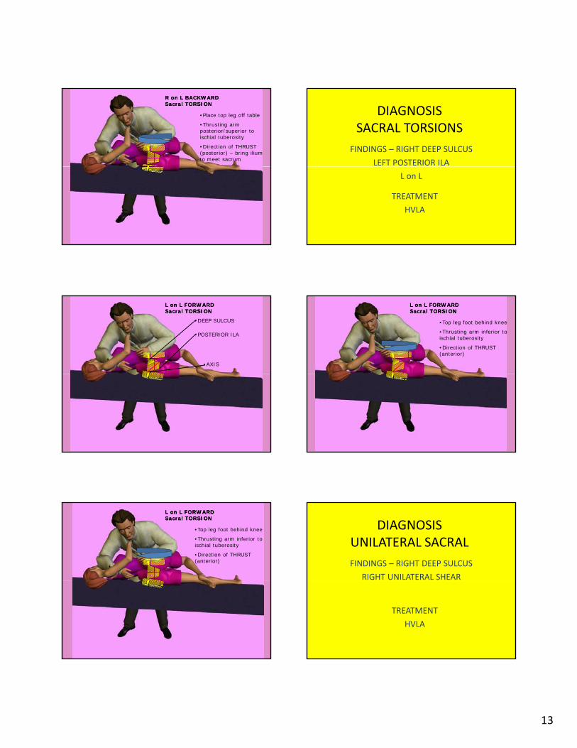
R on L BACKWARD R on L BACKWARD Sacral TORSIONSacral TORSION
•SHALLOW SULCUS
•AXIS
•POSTERIOR ILA
R on L BACKWARD R on L BACKWARD Sacral TORSIONSacral TORSION
•Place top leg off table
•Thrusting arm posterior/superior to ischial tuberosity
•Direction of THRUST (posterior) – bring ilium to meet sacrum
13
R on L BACKWARD R on L BACKWARD Sacral TORSIONSacral TORSION
•Place top leg off table
•Thrusting arm posterior/superior to ischial tuberosity
•Direction of THRUST (posterior) – bring ilium to meet sacrum
DIAGNOSISSACRAL TORSIONS
FINDINGS – RIGHT DEEP SULCUS
LEFT POSTERIOR ILA
L on L
TREATMENT
HVLA
L on L FORWARD L on L FORWARD Sacral TORSIONSacral TORSION
•DEEP SULCUS
•AXIS
•POSTERIOR ILA
L on L FORWARD L on L FORWARD Sacral TORSIONSacral TORSION
•Top leg foot behind knee
•Thrusting arm inferior to ischial tuberosity
•Direction of THRUST (anterior)
L on L FORWARD L on L FORWARD Sacral TORSIONSacral TORSION
•Top leg foot behind knee
•Thrusting arm inferior to ischial tuberosity
•Direction of THRUST (anterior)
DIAGNOSISUNILATERAL SACRAL
FINDINGS – RIGHT DEEP SULCUS
RIGHT UNILATERAL SHEAR
TREATMENT
HVLA

14
•Patient prone
SACRAL DYSFUNCTION SACRAL DYSFUNCTION –– UNILATERAL SACRAL SHEARUNILATERAL SACRAL SHEAR
•Patient prone
•Physician at side of table – on dysfunction side
•Physician’s hand that is closer to the patient’s head –thenar eminence on posterior/inferior ILA; fingertips at SI joint (medially to PSIS at same side)
SACRAL DYSFUNCTION SACRAL DYSFUNCTION –– UNILATERAL SACRAL SHEARUNILATERAL SACRAL SHEAR
•Physician’s hand that is closer to the patient’s feet grasps the same sided leg above the ankle; the leg is externally rotated and abducted
SACRAL DYSFUNCTION SACRAL DYSFUNCTION –– UNILATERAL SACRAL SHEARUNILATERAL SACRAL SHEAR
•Muscle Energy treatment for a UNILATERAL UNILATERAL SACRAL SHEARSACRAL SHEAR is performed
•Physician’s hand that is closer to the patient’s head:
•The thenar eminence on posterior/inferior ILA presses anteriorly and a cephalad thrust is directed towards the monitoring fingers
SACRAL DYSFUNCTION SACRAL DYSFUNCTION –– UNILATERAL SACRAL SHEARUNILATERAL SACRAL SHEAR
DIAGNOSISUNILATERAL SACRAL
FINDINGS – RIGHT DEEP SULCUS
RIGHT UNILATERAL SHEAR
TREATMENT
HVLA
R UNILATERAL R UNILATERAL SACRAL SHEARSACRAL SHEAR
•DEEP SULCUS
•POSTERIOR ILA

15
R UNILATERAL R UNILATERAL SACRAL SHEARSACRAL SHEAR
•Top leg foot behind knee
•Thrusting arm inferior to ischial tuberosity
•Direction of THRUST (anterior) – to bring ilium to the sacrum
TREATED THE SAMETREATED THE SAME• Posteriorly Rotated Ilium
• Forward Sacral Torsion
• Unilateral Sacral Shear
TREATED THE SAMETREATED THE SAME• Anteriorly Rotated Ilium
• Backward Sacral Torsion