deorphanization of the human leukocyte tyrosine …deorphanization of the human leukocyte tyrosine...
TRANSCRIPT

Deorphanization of the human leukocyte tyrosinekinase (LTK) receptor by a signaling screen of theextracellular proteomeHongbing Zhang, Lily I. Pao, Aileen Zhou, Arthur D. Brace, Robert Halenbeck, AmyW. Hsu, Thomas L. Bray, Kevin Hestir,Elizabeth Bosch, Ernestine Lee, Gang Wang, Haixia Liu, Brian R. Wong, W. Michael Kavanaugh, and Lewis T. Williams1
Five Prime Therapeutics Inc., South San Francisco, CA 94080
Edited by K. Christopher Garcia, Stanford University, Stanford, CA, and approved September 26, 2014 (received for review June 25, 2014)
There are many transmembrane receptor-like proteins whoseligands have not been identified. A strategy for finding ligandswhen little is known about their tissue source is to screen eachextracellular protein individually expressed in an array format byusing a sensitive functional readout. Taking this approach, we havescreened a large collection (3,191 proteins) of extracellular proteinsfor their ability to activate signaling of an orphan receptor, leukocytetyrosine kinase (LTK). Only two related secreted factors, FAM150Aand FAM150B (family with sequence similarity 150 member A andmember B), stimulated LTK phosphorylation. FAM150A binds LTKextracellular domain with high affinity (KD = 28 pM). FAM150A stim-ulates LTK phosphorylation in a ligand-dependent manner. This strat-egy provides an efficient approach for identifying functional ligandsfor other orphan receptors.
leukocyte tyrosine kinase | extracellular protein | library screening |FAM150A | orphan receptor
Many biological processes and pathogenic conditions involveligand/receptor signaling. There are numerous transmem-
brane receptor-like proteins whose ligands have not been iden-tified. Most known ligand–receptor interactions were discoveredby painstaking protein purification, genetic approaches, or se-quence homology. Strategies using cDNA library expression—such as secretion trapping (1), mammalian expression cloning (2),yeast signaling display (3), and λ phage binding display (4)—havealso identified ligands for orphan receptors. However, theseapproaches are limited by the unknown comprehensiveness of thecDNA library and by the fact that the libraries are biased towardhigh-abundance transcripts and not focused on the extracellularproteome. An alternative strategy is to screen secreted factorsthat are individually expressed in an array format in which there isno bias in expression of each protein based on the abundance ofits cDNA. The advantages of this approach for deorphanizationof receptors are that it is extremely sensitive and selective andthat it covers the majority of the extracellular proteome. Althougharrayed proteins have been used to identify receptor partners withlow-affinity, high-avidity interactions (5–7), few studies have useda cell-signaling measurement to study “classic” high-affinity ligand–receptor interactions.Leukocyte tyrosine kinase (LTK) is a receptor tyrosine kinase
that was identified in 1988 (8). The extracellular domain (ECD)of LTK has no known domain structure except a glycine-richregion, leading to a proposal that LTK is not activated bya protein ligand. LTK has a close homolog named anaplasticlymphoma kinase (ALK) (9). The LTK pathway has been im-plicated in autoimmunity, neuronal development, and cancer.One study showed that gain-of-function polymorphism of Ltkhas been associated with systemic lupus erythematosus (SLE)pathogenesis (10). Ltk is expressed throughout the adult hip-pocampus, and mouse knockout studies indicated that both Ltkand Alk are involved in adult neurogenesis with some functionalredundancy (11). Loss-of-function studies showed that LTK may
have function in mammalian neuronal development. In addition,a high-throughput kinase expression study showed that LTKexpression is increased in acute myeloid leukemia patients (12).It is likely that LTK signaling is involved in multiple biologicaland pathological processes. The identification of the ligand(s)for LTK should help decipher its function.In this study, we have taken the strategy of screening most of
the extracellular proteome by using a large collection of in-dividually expressed secreted proteins, tested for their ability toactivate signaling of an orphan receptor, LTK. We set up a sig-naling assay for LTK and screened the extracellular proteome(3,191 extracellular proteins) for its ligand(s). We identified tworelated ligands [family with sequence similarity 150 member A andmember B (FAM150A and FAM150B)] that stimulate LTKphosphorylation. FAM150A binds LTK with high affinity (28 pM).The finding will further facilitate revealing the biological functionof the LTK signaling pathway, both in physiological and patho-logical conditions. A similar strategy could be used to deorphanizeligands of other types of receptors.
ResultsAn Extracellular Proteome Signaling Screen Identified FAM150A andFAM150B as LTK Ligands. The lack of information regarding thecellular source of the LTK ligand has added to the difficulty in itsidentification. We have taken the approach of screening thousands
Significance
Secreted factors and their cell-surface receptors play im-portant roles in the communication between cells in normaland pathological conditions. There are many transmembranereceptor-like proteins whose ligands have not been identified(also known as orphan receptors). Knowledge of the ligandshould help in understanding the biological role of the re-ceptor. We used a strategy of screening the extracellular pro-teome, one protein at a time, to identify ligands for suchreceptors. We discovered the ligands for the orphan receptorleukocyte tyrosine kinase. To our knowledge, this is the firstcase in which secreted factor ligands were identified for anorphan receptor with this technique. This approach is especiallyvaluable when little is known about the ligand.
Author contributions: H.Z., B.R.W., W.M.K., and L.T.W. designed research; H.Z., L.I.P., A.Z.,A.D.B., R.H., A.W.H., T.L.B., E.B., G.W., and H.L. performed research; L.I.P., A.D.B., R.H.,A.W.H., K.H., E.B., E.L., G.W., H.L., B.R.W., and W.M.K. contributed new reagents/analytictools; A.Z., A.D.B., A.W.H., T.L.B., K.H., E.L., and G.W. analyzed data; and H.Z., L.I.P., A.D.B.,A.W.H., T.L.B., E.L., and L.T.W. wrote the paper.
Conflict of interest statement: The authors are employees and/or officers of and ownstock in Five Prime Therapeutics, Inc.
This article is a PNAS Direct Submission.
Freely available online through the PNAS open access option.1To whom correspondence should be addressed. Email: [email protected].
This article contains supporting information online at www.pnas.org/lookup/suppl/doi:10.1073/pnas.1412009111/-/DCSupplemental.
www.pnas.org/cgi/doi/10.1073/pnas.1412009111 PNAS | November 4, 2014 | vol. 111 | no. 44 | 15741–15745
BIOCH
EMISTR
Y
Dow
nloa
ded
by g
uest
on
Mar
ch 1
, 202
0
of extracellular proteins derived from numerous human tissues fortheir ability to activate LTK. Using similar methods that have beendescribed (13), we constructed full-length cDNAs encoding 3,191extracellular proteins, including “classically secreted” proteins(1,795), soluble ECDs of type I transmembrane proteins (962),soluble ECDs of type ll transmembrane proteins (335), and solubleECDs of glycosylphosphatidylinositol-anchored proteins (99). Theproteins encoded by each cDNA were produced by describedmethods (13) and formatted for screening (see below).To test each extracellular protein for its ability to activate
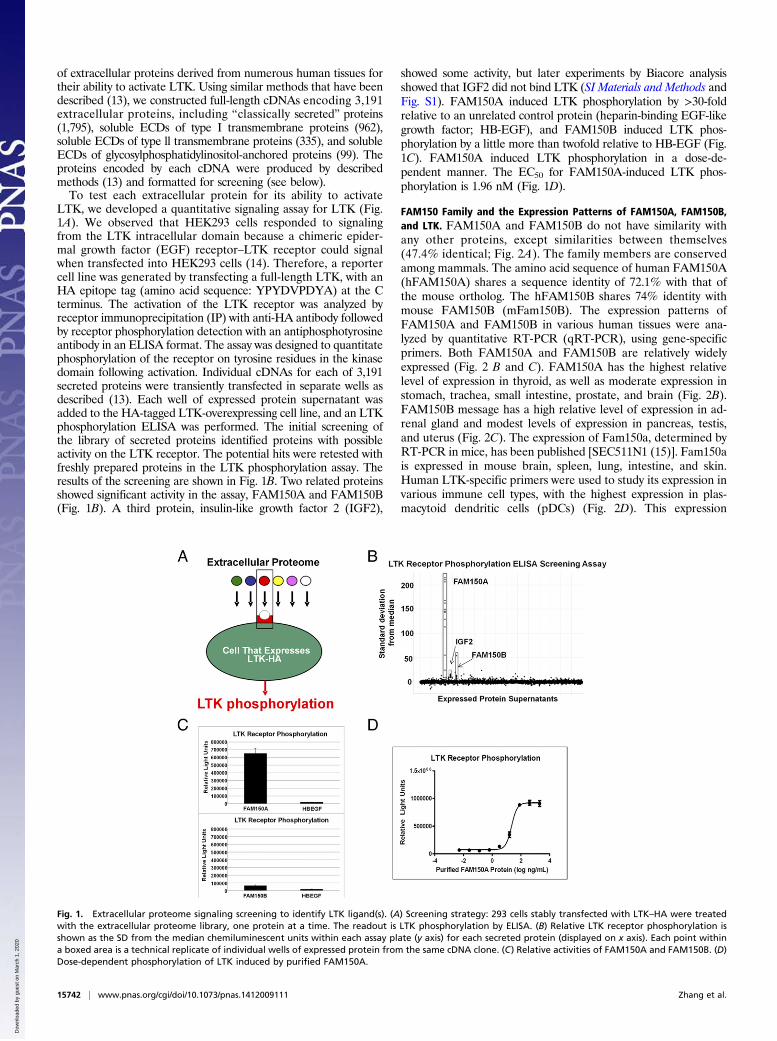
LTK, we developed a quantitative signaling assay for LTK (Fig.1A). We observed that HEK293 cells responded to signalingfrom the LTK intracellular domain because a chimeric epider-mal growth factor (EGF) receptor–LTK receptor could signalwhen transfected into HEK293 cells (14). Therefore, a reportercell line was generated by transfecting a full-length LTK, with anHA epitope tag (amino acid sequence: YPYDVPDYA) at the Cterminus. The activation of the LTK receptor was analyzed byreceptor immunoprecipitation (IP) with anti-HA antibody followedby receptor phosphorylation detection with an antiphosphotyrosineantibody in an ELISA format. The assay was designed to quantitatephosphorylation of the receptor on tyrosine residues in the kinasedomain following activation. Individual cDNAs for each of 3,191secreted proteins were transiently transfected in separate wells asdescribed (13). Each well of expressed protein supernatant wasadded to the HA-tagged LTK-overexpressing cell line, and an LTKphosphorylation ELISA was performed. The initial screening ofthe library of secreted proteins identified proteins with possibleactivity on the LTK receptor. The potential hits were retested withfreshly prepared proteins in the LTK phosphorylation assay. Theresults of the screening are shown in Fig. 1B. Two related proteinsshowed significant activity in the assay, FAM150A and FAM150B(Fig. 1B). A third protein, insulin-like growth factor 2 (IGF2),
showed some activity, but later experiments by Biacore analysisshowed that IGF2 did not bind LTK (SI Materials and Methods andFig. S1). FAM150A induced LTK phosphorylation by >30-foldrelative to an unrelated control protein (heparin-binding EGF-likegrowth factor; HB-EGF), and FAM150B induced LTK phos-phorylation by a little more than twofold relative to HB-EGF (Fig.1C). FAM150A induced LTK phosphorylation in a dose-de-pendent manner. The EC50 for FAM150A-induced LTK phos-phorylation is 1.96 nM (Fig. 1D).
FAM150 Family and the Expression Patterns of FAM150A, FAM150B,and LTK. FAM150A and FAM150B do not have similarity withany other proteins, except similarities between themselves(47.4% identical; Fig. 2A). The family members are conservedamong mammals. The amino acid sequence of human FAM150A(hFAM150A) shares a sequence identity of 72.1% with that ofthe mouse ortholog. The hFAM150B shares 74% identity withmouse FAM150B (mFam150B). The expression patterns ofFAM150A and FAM150B in various human tissues were ana-lyzed by quantitative RT-PCR (qRT-PCR), using gene-specificprimers. Both FAM150A and FAM150B are relatively widelyexpressed (Fig. 2 B and C). FAM150A has the highest relativelevel of expression in thyroid, as well as moderate expression instomach, trachea, small intestine, prostate, and brain (Fig. 2B).FAM150B message has a high relative level of expression in ad-renal gland and modest levels of expression in pancreas, testis,and uterus (Fig. 2C). The expression of Fam150a, determined byRT-PCR in mice, has been published [SEC511N1 (15)]. Fam150ais expressed in mouse brain, spleen, lung, intestine, and skin.Human LTK-specific primers were used to study its expression invarious immune cell types, with the highest expression in plas-macytoid dendritic cells (pDCs) (Fig. 2D). This expression
Fig. 1. Extracellular proteome signaling screening to identify LTK ligand(s). (A) Screening strategy: 293 cells stably transfected with LTK–HA were treatedwith the extracellular proteome library, one protein at a time. The readout is LTK phosphorylation by ELISA. (B) Relative LTK receptor phosphorylation isshown as the SD from the median chemiluminescent units within each assay plate (y axis) for each secreted protein (displayed on x axis). Each point withina boxed area is a technical replicate of individual wells of expressed protein from the same cDNA clone. (C) Relative activities of FAM150A and FAM150B. (D)Dose-dependent phosphorylation of LTK induced by purified FAM150A.
15742 | www.pnas.org/cgi/doi/10.1073/pnas.1412009111 Zhang et al.
Dow
nloa
ded
by g
uest
on
Mar
ch 1
, 202
0
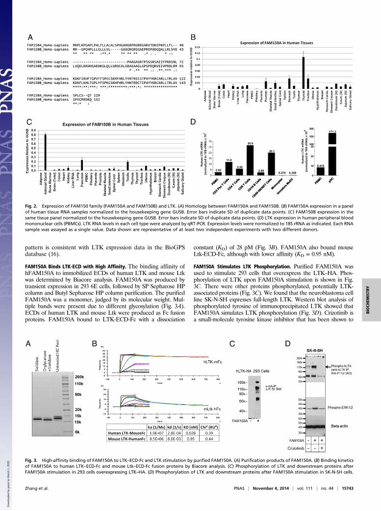
pattern is consistent with LTK expression data in the BioGPSdatabase (16).
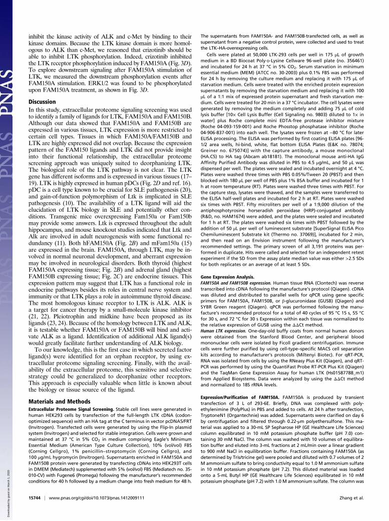
FAM150A Binds LTK-ECD with High Affinity. The binding affinity ofhFAM150A to immobilized ECDs of human LTK and mouse Ltkwas determined by Biacore analysis. FAM150A was produced bytransient expression in 293 6E cells, followed by SP Sepharose HPcolumn and Butyl Sepharose HP column purification. The purifiedFAM150A was a monomer, judged by its molecular weight. Mul-tiple bands were present due to different glycosylation (Fig. 3A).ECDs of human LTK and mouse Ltk were produced as Fc fusionproteins. FAM150A bound to LTK-ECD-Fc with a dissociation
constant (KD) of 28 pM (Fig. 3B). FAM150A also bound mouseLtk-ECD-Fc, although with lower affinity (KD = 0.95 nM).
FAM150A Stimulates LTK Phosphorylation. Purified FAM150A wasused to stimulate 293 cells that overexpress the LTK–HA. Phos-phorylation of LTK upon FAM150A stimulation is shown in Fig.3C. There were other proteins phosphorylated, potentially LTK-associated proteins (Fig. 3C). We found that the neuroblastoma cellline SK-N-SH expresses full-length LTK. Western blot analysis ofphosphorylated tyrosine of immunoprecipitated LTK showed thatFAM150A simulates LTK phosphorylation (Fig. 3D). Crizotinib isa small-molecule tyrosine kinase inhibitor that has been shown to
Fig. 2. Expression of FAM150 family (FAM150A and FAM150B) and LTK. (A) Homology between FAM150A and FAM150B. (B) FAM150A expression in a panelof human tissue RNA samples normalized to the housekeeping gene GUSB. Error bars indicate SD of duplicate data points. (C) FAM150B expression in thesame tissue panel normalized to the housekeeping gene GUSB. Error bars indicate SD of duplicate data points. (D) LTK expression in human peripheral bloodmononuclear cells (PBMCs). LTK RNA levels in each cell type were analyzed by qRT-PCR. Expression levels were normalized to 18S rRNA as indicated. Each RNAsample was assayed as a single value. Data shown are representative of at least two independent experiments with two different donors.
Fig. 3. High-affinity binding of FAM150A to LTK–ECD-Fc and LTK stimulation by purified FAM150A. (A) Purification products of FAM150A. (B) Binding kineticsof FAM150A to human LTK–ECD-Fc and mouse Ltk–ECD-Fc fusion proteins by Biacore analysis. (C) Phosphorylation of LTK and downstream proteins afterFAM150A stimulation in 293 cells overexpressing LTK–HA. (D) Phosphorylation of LTK and downstream proteins after FAM150A stimulation in SK-N-SH cells.
Zhang et al. PNAS | November 4, 2014 | vol. 111 | no. 44 | 15743
BIOCH
EMISTR
Y
Dow
nloa
ded
by g
uest
on
Mar
ch 1
, 202
0
inhibit the kinase activity of ALK and c-Met by binding to theirkinase domains. Because the LTK kinase domain is more homol-ogous to ALK than c-Met, we reasoned that crizotinib should beable to inhibit LTK phosphorylation. Indeed, crizotinib inhibitedthe LTK receptor phosphorylation induced by FAM150A (Fig. 3D).To explore downstream signaling after FAM150A stimulation ofLTK, we measured the downstream phosphorylation events afterFAM150A stimulation. ERK1/2 was found to be phosphorylatedupon FAM150A treatment, as shown in Fig. 3D.
DiscussionIn this study, extracellular proteome signaling screening was usedto identify a family of ligands for LTK, FAM150A and FAM150B.Although our data showed that FAM150A and FAM150B areexpressed in various tissues, LTK expression is more restricted tocertain cell types. Tissues in which FAM150A/FAM150B andLTK are highly expressed did not overlap. Because the expressionpattern of the FAM150 ligands and LTK did not provide insightinto their functional relationship, the extracellular proteomescreening approach was uniquely suited to deorphanizing LTK.The biological role of the LTK pathway is not clear. The LTKgene has different isoforms and is expressed in various tissues (17–19). LTK is highly expressed in human pDCs (Fig. 2D and ref. 16).pDC is a cell type known to be crucial for SLE pathogenesis (20),and gain-of-function polymorphism of Ltk is implicated in SLEpathogenesis (10). The availability of a LTK ligand will aid theelucidation of LTK biology in SLE and potentially other con-ditions. Transgenic mice overexpressing Fam150a or Fam150bmay provide some answers. Ltk is expressed throughout the adulthippocampus, and mouse knockout studies indicated that Ltk andAlk are involved in adult neurogenesis with some functional re-dundancy (11). Both hFAM150A (Fig. 2B) and mFam150a (15)are expressed in the brain. FAM150A, through LTK, may be in-volved in normal neuronal development, and aberrant expressionmay be involved in neurological disorders. Both thyroid (highestFAM150A expressing tissue; Fig. 2B) and adrenal gland (highestFAM150B expressing tissue; Fig. 2C) are endocrine tissues. Thisexpression pattern may suggest that LTK has a functional role inendocrine pathways besides its roles in central nerve system andimmunity or that LTK plays a role in autoimmune thyroid disease.The most homologous kinase receptor to LTK is ALK. ALK isa target for cancer therapy by a small-molecule kinase inhibitor(21, 22). Pleiotrophin and midkine have been proposed as itsligands (23, 24). Because of the homology between LTK and ALK,it is testable whether FAM150A or FAM150B will bind and acti-vate ALK as a ligand. Identification of additional ALK ligand(s)would greatly facilitate further understanding of ALK biology.To our knowledge, this is the first case in which secreted factor
ligand(s) were identified for an orphan receptor, by using ex-tracellular proteome signaling screening. Finally, with the avail-ability of the extracellular proteome, this sensitive and selectivestrategy could be generalized to deorphanize other receptors.This approach is especially valuable when little is known aboutthe biology or tissue source of the ligand.
Materials and MethodsExtracellular Proteome Signal Screening. Stable cell lines were generated inhuman HEK293 cells by transfection of the full-length LTK cDNA (codon-optimized sequence) with an HA tag at the C terminus in vector pcDNA5/FRT(Invitrogen). Transfected cells were generated by using the Flip-In plasmidsystem (Invitrogen) and selected for stable integration. Cells were grown andmaintained at 37 °C in 5% CO2 in medium comprising Eagle’s MinimumEssential Medium (American Type Culture Collection), 10% (vol/vol) FBS(Corning Cellgro), 1% penicillin–streptomycin (Corning Cellgro), and100 μg/mL hygromycin (Invitrogen). Supernatants enriched in FAM150A andFAM150B protein were generated by transfecting cDNAs into HEK293T cellsin DMEM (Mediatech) supplemented with 5% (vol/vol) FBS (Mediatech no. 35-010-CV) with Fugene6 (Promega) following the manufacturer’s recommendedconditions for 40 h followed by a medium change into fresh medium for 48 h.
The supernatants from FAM150A- and FAM150B-transfected cells, as well assupernatant from a negative control protein, were collected and used to treatthe LTK–HA-overexpressing cells.
Cells were plated at 50,000 LTK-293 cells per well in 175 μL of growthmedium in a BD Biocoat Poly-D-Lysine Cellware 96-well plate (no. 356461)and incubated for 24 h at 37 °C in 5% CO2. Serum starvation in minimumessential medium (MEM) (ATCC no. 30-2003) plus 0.1% FBS was performedfor 24 h by removing the culture medium and replacing it with 175 μL ofstarvation medium. Cells were treated with the enriched protein expressionsupernatants by removing the starvation medium and replacing it with 100μL of a 1:1 mix of expressed protein supernatant and fresh starvation me-dium. Cells were treated for 20 min in a 37 °C incubator. The cell lysates weregenerated by removing the medium completely and adding 75 μL of coldlysis buffer [10× Cell Lysis Buffer (Cell Signaling no. 9803) diluted to 1× inwater] plus Roche complete mini EDTA-free protease inhibitor mixture(Roche 04-093-159-001) and Roche Phosstop phosphatase inhibitor (Roche04-906-837-001) into each well. The lysates were frozen at −80 °C for laterELISA processing. The ELISA was performed by first coating ELISA plates [96-1/2 area wells, hi-bind, white, flat bottom ELISA Plates (E&K no. 78074;Greiner no. 675074)] with the capture antibody, a mouse monoclonal(HA.C5) to HA tag (Abcam ab18181). The monoclonal mouse anti-HA IgGAffinity Purified Antibody was diluted in PBS to 4.5 μg/mL, and 50 μL wasdispensed per well. The plates were sealed and incubated overnight at 4 °C.Plates were washed three times with PBS 0.05%/Tween 20 (PBST) and thenblocked with 180 μL per well of PBS plus 1% BSA buffer and incubated for 1h at room temperature (RT). Plates were washed three times with PBST. Forthe capture step, lysates were thawed, and the samples were transferred tothe ELISA half-well plates and incubated for 2 h at RT. Plates were washedsix times with PBST. Fifty microliters per well of a 1:9,000 dilution of theantiphosphotyrosine horseradish peroxidase (HRP)-conjugated antibody(R&D, no. HAM1674) were added, and the plates were sealed and incubatedfor 1 h at RT. The plates were washed six times with PBST followed by theaddition of 50 μL per well of luminescent substrate [SuperSignal ELISA PicoChemiluminescent Substrate kit (Thermo no. 37069)], incubated for 2 min,and then read on an Envision instrument following the manufacturer’srecommended settings. The primary screen of all 3,191 proteins was per-formed in duplicate. Hits were called and selected for an independent retestexperiment if the SD from the assay plate median value was either >2.5 SDsfor both replicates or an average of at least 5 SDs.
Gene Expression Analysis.FAM150A and FAM150B expression. Human tissue RNA (Clontech) was reversetranscribed into cDNA following the manufacture’s protocol (Qiagen). cDNAwas diluted and distributed to parallel wells for qPCR using gene specificprimers for FAM150A, FAM150B, or β-glucuronidase (GUSB) (Qiagen) andSYBR Green reagent (Qiagen). qPCR was performed following the manu-facture’s recommended protocol for a total of 40 cycles of 95 °C 15 s, 55 °Cfor 30 s, and 72 °C for 30 s Expression within each tissue was normalized tothe relative expression of GUSB using the ΔΔCt method.Human LTK expression. One-day-old buffy coats from normal human donorswere obtained from the Stanford Blood Center, and peripheral bloodmononuclear cells were isolated by Ficoll gradient centrifugation. Immunecells were further purified by using cell-type–specific MACS cell separationkits according to manufacturer’s protocols (Miltenyi Biotec). For qRT-PCR,RNA was isolated from cells by using the RNeasy Plus Kit (Qiagen), and qRT-PCR was performed by using the QuantiFast Probe RT-PCR Plus Kit (Qiagen)and the TaqMan Gene Expression Assay for human LTK (Hs01587788_m1)from Applied Biosystems. Data were analyzed by using the ΔΔCt methodand normalized to 18S rRNA levels.
Expression/Purification of FAM150A. FAM150A is produced by transienttransfection of 3 L of 293-6E. Briefly, DNA was complexed with poly-ethylenimine (PolyPlus) in PBS and added to cells. At 24 h after transfection,TryptoneN1 (Organtechnie) was added. Supernatants were clarified on day 6by centrifugation and filtered through 0.22-μm polyethersulfone. This ma-terial was applied to a 30-mL SP Sepharose HP (GE Healthcare Life Sciences)column equilibrated in 10 mM potassium phosphate buffer (pH 7.0) con-taining 30 mM NaCl. The column was washed with 10 volumes of equilibra-tion buffer and eluted into 3-mL fractions at 2 mL/min over a linear gradientto 900 mM NaCl in equilibration buffer. Fractions containing FAM150A (asdetermined by Tris/tricine gel) were pooled and diluted with 0.7 volumes of 2M ammonium sulfate to bring conductivity equal to 1.0 M ammonium sulfatein 10 mM potassium phosphate (pH 7.2). This diluted material was loadedonto a 5-mL Butyl HP (GE Healthcare Life Sciences) equilibrated in 10 mMpotassium phosphate (pH 7.2) with 1.0 M ammonium sulfate. The column was
15744 | www.pnas.org/cgi/doi/10.1073/pnas.1412009111 Zhang et al.
Dow
nloa
ded
by g
uest
on
Mar
ch 1
, 202
0

washed with 10 volumes of equilibration buffer, and bound protein waseluted by using a linear gradient to 0 mM ammonium sulfate in 10 mM po-tassium phosphate (pH 7.2) over 20 column volumes, collecting 1.5 mL perfraction at a rate of 1 mL/min. Fractions were pooled by analysis of Tris/tricinegel and dialyzed against 30 volumes of PBS. Following dialysis, protein wasaliquotted and frozen at −80 °C for subsequent analysis.
LTK ECD-Fc fusion proteins were expressed transiently in 293-6E cells asdescribed above. Day 6 harvest supernatants were clarified by centrifugationand filtration before loading onto a 5-mL HiTrap Protein A HP (GE HealthcareLife Sciences) column equilibrated in PBS with 500 mM NaCl. The column waswashedwith equilibrationbuffer andelutedby using a linear gradient to 100mMglycine (pH 2.7), with 500 mM NaCl over 20 column volume collecting 1.5 mL perfraction at 2 mL/min. Elution fractions are collected into 150 μL of 1.0 M Tris(pH 8.0) to neutralize the pH. Elution fractions were pooled based on gel anal-ysis; the pooled protein was dialyzed overnight against PBS, filter sterilized, andstored at −80 °C.
Purified Fam150A was deglycosylated and/or desialylated by using theProzyme Deglycosylation Kit (Prozyme; catalog no. 80110) using recom-mended nondenaturing protocols therein. Briefly, 10 μL of 250 mM sodiumphosphate (pH 7.0) was added to 50 μg of Fam150A protein. Then, 1 μL ofSialidase A and 1 μL of O-Glycanase were added, or 1 μL of Sialidase A wasadded to the protein/buffer solution. Water was added to bring the finalvolume to 100 μL. Samples were incubated at RT for 1 h, then at 4 °C for48 h. A total of 8 μg of each reaction was mixed with 20 mM iodoacetamide,loaded on a Criterion Tricine Gel (Bio-Rad Laboratories), and run at 125 V for90 min. Following electrophoresis, the gel was stained by using CoomassieBlue R250 dye and destained per standard protocols.
Determination of FAM150A Binding Constants for Human and Mouse LTK ECD-Fc Fusions. Binding kinetics of FAM150A to human and mouse LTK ECD-Fcfusion proteins were determined by using the Biacore T100 Surface PlasmonResonance instrument (GE Healthcare Life Sciences). Purified LTK-Fc fusionswere captured on a CM4 sensor chip immobilized with Protein A (ThermoScientific). We used 10 mMHepes-buffered saline (pH 7.4) with 0.05% Tween
20 (HBSP) (GE Healthcare Life Sciences) for dilution of all samples and as therunning buffer for data collection. Capture levels of the ECD-Fcs were adjustedto 300–500 response units to ensure that binding values would be muchgreater than levels of nonspecific binding to the reference flow cell. FAM150Awas injected at eight concentrations (100, 33.3, 11.1, 3.7, 1.2, 0.41, 0.13, 0.046,and 0 nM) for 120 s each. Dissociation was followed for an additional 120 or600 s. The association and dissociation constants and affinity of FAM150A forLTK ECD-Fc fusions was calculated by using the Biacore T100 Evaluation soft-ware package using the 1:1 binding model with standard double referencing.BMPR1A ECD-Fc was included as a negative binding control, and no non-specific binding of FAM150A was observed.
FAM150A Induce Phosphorylation of LTK. The 293 cells expressing LTK or SK-N-SH cells (ATCC) were seeded at 5 × 10⁶ cells per well in six-well culture platesin DMEM with 10% FBS and grown overnight at 37 °C. The culture mediumwas replaced with starvation medium (DMEM, 0.1% FBS), and the cells werestarved for 24 h at 37 °C. FAM150A (200 ng/mL), with or without 1 μM kinaseinhibitor crizotinib (catalog no. S1068, Selleckchem), was then added to thecells for 20 min. At the end of the incubation, cells were washed with coldPBS, and 250 μL of cell lysis buffer (catalog no. 9803S, Cell Signaling Tech-nology) containing protease inhibitor mixture (catalog no. P8340; Sigma-Aldrich) and phosphatase inhibitor mixture 2 (catalog no. 5726, Sigma-Aldrich) was added to each well. Cell lysate was immunoprecipitated witha sheep anti-human LTK affinity purified polyclonal antibody (R&D Systems)overnight, and the immunoprecipitate was separated on a reducing SDS/PAGE gel. Tyrosine phosphorylation was detected by blotting with a mouseantiphosphotyrosine monoclonal antibody conjugated to HRP (R&D Sys-tems), and the signal was developed according to the manufacturer’sinstructions. Whole-cell lysate was run on a separate reducing SDS/PAGE geland probed for ERK1/2 phosphorylation by using an anti–phospho-p44/42MAPK (ERK1/2) (Thr-202/Tyr-204) antibody (Cell Signaling Technology).Β-actin was detected as a loading control by using an anti–β-actin antibodyconjugated to HRP (Abcam).
1. Davis S, et al. (1996) Isolation of angiopoietin-1, a ligand for the TIE2 receptor, by
secretion-trap expression cloning. Cell 87(7):1161–1169.2. Gale NW, et al. (1996) Elk-L3, a novel transmembrane ligand for the Eph family of
receptor tyrosine kinases, expressed in embryonic floor plate, roof plate and hind-
brain segments. Oncogene 13(6):1343–1352.3. Kinoshita N, Minshull J, Kirschner MW (1995) The identification of two novel ligands
of the FGF receptor by a yeast screening method and their activity in Xenopus de-
velopment. Cell 83(4):621–630.4. Brandenberger R, et al. (2001) Identification and characterization of a novel extra-
cellular matrix protein nephronectin that is associated with integrin alpha8beta1 in
the embryonic kidney. J Cell Biol 154(2):447–458.5. Özkan E, et al. (2013) An extracellular interactome of immunoglobulin and LRR
proteins reveals receptor-ligand networks. Cell 154(1):228–239.6. Bushell KM, Söllner C, Schuster-Boeckler B, Bateman A, Wright GJ (2008) Large-scale
screening for novel low-affinity extracellular protein interactions. Genome Res 18(4):
622–630.7. Haddick PC, et al. (2014) Defining the ligand specificity of the deleted in colorectal
cancer (DCC) receptor. PLoS ONE 9(1):e84823.8. Ben-Neriah Y, Bauskin AR (1988) Leukocytes express a novel gene encoding a putative
transmembrane protein-kinase devoid of an extracellular domain. Nature 333(6174):
672–676.9. Palmer RH, Vernersson E, Grabbe C, Hallberg B (2009) Anaplastic lymphoma kinase:
Signalling in development and disease. Biochem J 420(3):345–361.10. Li N, et al. (2004) Gain-of-function polymorphism in mouse and human Ltk: Im-
plications for the pathogenesis of systemic lupus erythematosus. Hum Mol Genet
13(2):171–179.11. Weiss JB, et al. (2012) Anaplastic lymphoma kinase and leukocyte tyrosine kinase:
Functions and genetic interactions in learning, memory and adult neurogenesis.
Pharmacol Biochem Behav 100(3):566–574.
12. Muller-Tidow C, et al. (2004) High-throughput analysis of genome-wide receptor ty-rosine kinase expression in human cancers identifies potential novel drug targets. ClinCancer Res 10(4):1241–1249.
13. Lin H, et al. (2008) Discovery of a cytokine and its receptor by functional screening ofthe extracellular proteome. Science 320(5877):807–811.
14. Ueno H, et al. (1995) An epidermal growth factor receptor-leukocyte tyrosine kinasechimeric receptor generates ligand-dependent growth signals through the Ras sig-naling pathway. J Biol Chem 270(34):20135–20142.
15. Tang T, et al. (2010) A mouse knockout library for secreted and transmembraneproteins. Nat Biotechnol 28(7):749–755.
16. Wu C, et al. (2009) BioGPS: An extensible and customizable portal for querying andorganizing gene annotation resources. Genome Biol 10(11):R130.
17. Haase VH, et al. (1991) Alternatively spliced ltk mRNA in neurons predicts a receptorwith a larger putative extracellular domain. Oncogene 6(12):2319–2325.
18. Krolewski JJ, Dalla-Favera R (1991) The ltk gene encodes a novel receptor-type pro-tein tyrosine kinase. EMBO J 10(10):2911–2919.
19. Toyoshima H, et al. (1993) Differently spliced cDNAs of human leukocyte tyrosinekinase receptor tyrosine kinase predict receptor proteins with and without a tyrosinekinase domain and a soluble receptor protein. Proc Natl Acad Sci USA 90(12):5404–5408.
20. Craft JE (2011) Dissecting the immune cell mayhem that drives lupus pathogenesis. SciTransl Med 3(73):ps9.
21. Shaw AT, Engelman JA (2013) ALK in lung cancer: Past, present, and future. J ClinOncol 31(8):1105–1111.
22. Webb TR, et al. (2009) Anaplastic lymphoma kinase: Role in cancer pathogenesis andsmall-molecule inhibitor development for therapy. Expert Rev Anticancer Ther 9(3):331–356.
23. Stoica GE, et al. (2001) Identification of anaplastic lymphoma kinase as a receptor forthe growth factor pleiotrophin. J Biol Chem 276(20):16772–16779.
24. Stoica GE, et al. (2002) Midkine binds to anaplastic lymphoma kinase (ALK) and acts asa growth factor for different cell types. J Biol Chem 277(39):35990–35998.
Zhang et al. PNAS | November 4, 2014 | vol. 111 | no. 44 | 15745
BIOCH
EMISTR
Y
Dow
nloa
ded
by g
uest
on
Mar
ch 1
, 202
0