deoxyribonucleic acid renaturation kinetics and ... · vererbungslehre, 89, 103 (1958). ......
TRANSCRIPT
Vol. 6, 1973 DNA Renaturation Kinetics 293
short piece of quartz fiber 2 or 3 pm in diameter. A small tissue sample ( 2 x 10⁻⁸ g or less) is easily picked up with this tool and transferred to the bal- ance tip. A similar quartz-tipped “hair point” is also used for transferring weighed samples into the oil wells for analysis.
Specificity. The specificity of the overall analysis rests largely on the first enzymatic step. As in any enzymatic analysis this specificity depends on the properties and purity of the enzymes used and on the conduct of the assay. Increasing sensitivity does not increase the specificity problem and may make it less. For example, a t higher dilutions relatively less enzyme may be required with less danger from con- taminants. Similarly, with amplification by cycling the “signal” is increased relative to the “noise” (tis- sue blank).
Performance and Applications. The microanaly- tical system described is not only unlimited in theory but has been shown in practice to be capable of very high sensitivity and quite satisfactory precision over a ten-million-fold range in sample size ( 1 0 - 9 to 10-l6 mol). The following are typical examples. In the 10-lo mol range Karlsson and coworkers13 have made extensive studies of muscle metabolites in small human biopsy samples, and numerous en- zymes have been measured in different segments of single kidney t u b u l e ~ . l ~ > ~ 5 Metabolite levels have
(13) J. Karlsson,ActaPhysiol. Scand., Suppl., 358,7 (1971). (14) H. Mattenheimer in “Current Problems in Clinical Biochemistry,”
U. C. Dubach, Ed., Hans Huber, Berne, Switzerland, 1968, p 119. (15) U. C. Dubach and U. Schmidt in “Recent Advances in Quantita-
tive Histo- and Cytochemistry,” U. C. Dubach and U. Schmidt, Ed., Hans Huber, Berne, Switzerland 1971, p 312.
been measured in discrete layers of mouse cerebel- lum16 (10-11-10-12 mol), in different layers of retina (10-1240-13 mol),ll and in single mouse pancreatic islets (10-12-10-14 In one studyl8 single nerve cell bodies from mouse spinal cord were ana- lyzed for ATP, phosphocreatine, glucose, and glyco- gen under a number of different experimental condi- tions. The sample sizes ranged from 1 X to 3 X g dry weight and the amounts of the four substances measured were all in the 10-l2 mol range. Mrs. Elizabeth Barbehenn in this laboratory is currently measuring metabolite levels ranging down to 2 X l O - l 5 mol in single mouse ova. Mea- surements have been made of NAD in single nerve cell nuclei in the 10-16 mol range.lg In an unpub- lished study, Dr. Frank A. Welsh determined the ac- tivity of single molecules of glucose 6-phosphate de- hydrogenase. This required the measurement of
mol of enzyme product and a 10,000,000-fold amplification by two cycling steps.
This performance exceeds that attainable a t pres- ent with methods based on other analytical princi- ples. Of these, radioactive tracer methods are per- haps the most sensitive and may be used for compar- ison with the above. In the case of labeled amino acids having the highest available specific activity, the limit for accurate assay is about 10-12 mol with 14C, mol with 3H, and mol with 35S (methionine).
(16) P. D. Gatfield, 0. H. Lowry, D. W. Schulz, and J. V. Passonneau, J.
(17) F. Matschinsky,J. Neurochem., 13,143 (1966). (18) J . V. Passonneau, and 0. H. Lowry, in ref 15. (19) T. Kat0 and 0. H. Lowry, J. Bid. Chem., in press.
Neurochem., 13,185 (1966).
Deoxyribonucleic Acid Renaturation Kinetics and Hybridization. Probes to the Structure of the Eukaryotic Chromosome
John E. Hearst* and Michael Botchan
Depar tment of Chemistry , University o f California, Berkeley, California 94720
Received January 18, 1973
The cell nucleus is defined as that part of a living cell containing the genetic information or, in molecu- lar terms, the DNA. In bacteria and in blue-green algae the nucleus is not separated by a nuclear mem- brane from the rest of the cell, the cytoplasm. Under the microscope the nucleus appears amorphous and spread throughout the cell. Cells without nuclear
John E. Hearst was born in Vienna, Austria, in 1935. He was educated at Yale University and at the California Institute of Technology, where he took his Ph.D. with Jerome Vinograd. After a year of postdoctoral re- search with Walter Stockmayer at Dartmouth College (1961-1962), he began teaching at University of California, Berkeley. His matn re- search interests have been nucleic acid physical chemistry and more re- cently the molecular genetics of the higher organism.
Michael Botchan was a graduate student at Berkeley until 1972 and is presently at the Cold Spring Harbor Laboratory.
membranes are called prokaryotes. Until approxi- mately 5 years ago the major discoveries of molecular biology were made in experiments on bacteria and their viruses.
Eukaryotes are cells with nuclear membranes. Such cells are found in most of the remaining single- cell organisms not mentioned above, such as fungi, protozoa, and most algae. Eukaryotic cells are also found in all higher plants and animals. The molecu- lar biology of the eukaryotes is only now emerging as an area of useful experimentation. There are many similarities with the prokaryote, for the central dogma of molecular biology is common to all cells: DNA codes for DNA, DNA codes for RNA, and RNA codes for protein. The differences between the two
294 Hearst and Botchan Accounts of Chemical Research
classes of cells, however, are the stimulus for most of the studies presently undertaken. The multicellular organism is far more complex than the single-cell or- ganism because its cells become differentiated or committed to a particular characteristic class or function.
There are other differences between the prokar- yotes and the eukaryotes, many of which deal with the life cycles of the cells and their mechanisms of cell division. Eukaryotes undergo a clearly defined cell cycle during most of which a cell nucleus con- taining the DNA of the cell is discernible with a light microscope. Just preceding cell division, the nuclear membrane disappears, and the contents of the nucle- us simultaneously condenses into metaphase chro- mosomes. Cell division continues with the split of the chromosomes into two equal halves by the spin- dle apparatus, the loss of the condensed state of the metaphase chromosomes, and the pinching off of the cell membrane. When division is complete, each new cell contains half of the nuclear material of the me- taphase cell.
Such a division cycle is far more complicated than the cycle of prokaryotes. I t is, therefore, not surpris- ing that substantial differences can be found in the properties of eukaryotic DNA relative to prokaryotic DNA. One of the most striking differences is that eu- karyotes contain DNA of a very simple repeating pattern.l.2 A sequence of as few as six bases is re- peated millions of times in the total set of chromo- somes known as the g e n ~ m e . ~ . ~ These simple se- quences, in many cases, have been localized in or near the centromeres of metaphase chromosomes.5,6 The centromere is the point of attachment of spindle fibres and is the point a t which the chromosomes are parted in division. I t frequently appears more highly condensed in microscope spreads of metaphase chro- mosomes, staining darker than the remainder of the chromosome. This suggests that simple DNA se- quences are important in determining the state of condensation of the DNA in chromosomes. At the molecular level the more condensed portions of the chromosomes contain more tightly packaged DNA, probably in a supercoil structure with protein.
The chromosome of the higher organism is a com- plex of DNA and protein (and perhaps RNA), and the molecular basis for the highly condensed state is not yet understood. What is known, however, from years of cytogenetics, is that the condensed state of portions of chromosomes is not a unique property of centromeres but is found in other regions of the ge- nome as well.7,8 Some regions of the genome, includ- ing sometimes whole chromosomes, remain tightly condensed in the cell nucleus. Such regions are called heterochromatic and are distinguished from the regions which do not remain condensed, which are called euchromatic. All heterochromatin is now believed to contain repetitious sequences of DNA.
(1) M. Waringand R. J. Britten, Science. 151. 791 (1966). (2) R. J. Britten and D. E. Kohne, Science, 161,529 (19681. (3) E. M. Southern, Nature (London) , 227,794 (1970) (4) P. M. B. Walker, Progr. Biophys. M o l Biol., 23, 145 (1971). ( 5 ) M. L. Pardue and J. G. Gall, Science, 168, 1356 (1970). ( 6 ) K. W. Jones, iVature /London), 225, 912 (1970). ( 7 ) C. P. Swanson, "Cytology and Cytogenetics," Prentice-Hall, Engle-
(8) S . W. Brown, Science, 151,417 (1966). wood Cliffs, N. J., 1957.
Cytogeneticists9 also believe heterochromatic regions are relatively inert with respect to the expression of genes.10.11
Many translocation mutants in Drosophila mela- nogaster suggest that there are positional effects re- lated to gene activity: the nearer to heterochromatin the less active the gene. We therefore have proposed a coarse genetic control mechanism12 which depends upon the physical state of condensation of the chro- matin and which acts upon relatively long stretches of the genome, 20 to 100 genes for example. We dis- tinguish this mechanism from fine control which operates a t the level of one to five genes and has been extensively studied in prokaryotes.
There are two very broad features of this proposal which are directly related to the nature of the higher organism and its evolution. First. special genes may be located in heterochromatin. Since genetic cross- ing-over is rarely observed in heterochromatic re- gions, such genes would be partially protected from evolutionary changes by virtue of their special posi- tion in heterochromatin. The genes in heterochroma- tin may also be expressed a t a special time in the cell cycle. We suggest13 that genes in heterochroma- tin will be found in proximity to simple-sequence DNA. The second proposal, made by Kenneth Jonesx4 of Edinburgh, is that speciation comes about by the emergence of a new distribution of simple se- quences in the genome and therefore a new control of neighboring genes by positional effects. Many 0thers3,~ have speculated upon the importance of simple sequences to speciation as well, because very closely related species are observed t o have entirely different simple sequences.
The major interest of this laboratory15316 in the last three years has been the distribution of simple sequences between different chromosomes and the organization and function of these sequences within a chromosome. We have concentrated our efforts on the DNA of the fruit fly, Drosophila melanogaster, because of the wealth of classical genetic information available for this organism.
DNA Renaturation Kinetics The two strands of the Watson-Crick duplex can
be separated either by heating a solution of DNA to about 100" or by raising the pH of the solution to 13. This process is called denaturation. At pH 7 and at 60" or 70", complementary regions of DNA can re- form the double-stranded duplex, or be renatured. This renaturation reaction is a second-order reaction, the rate of which depends on the concentration of complementary pairs of nucleotide sequences in the solution.2 The rate of this reaction is readily moni- tored in a spectrophotometer by recording the hypo- chromicity a t 260 nm associated with the re-forma- tion of helix. The initial native DNA is generally fragmented by sonication to single strand, mol wt 1.5
(9) W. K. Baker, Adcan. Genet. , 14, 133 (1968). (10) H. Swift. Genetics, Suppl , 61,452 (19691. (11) D. Lindsley, 2. Vererbungslehre, 89, 103 (1958). (12) J. E. Hearst and M . Botchan, Annu. Rec. Blochen., 39. 151 (19701. (13) M. Botchan, Thesis. University of California, Berkeley, 1972. (14) K. Jones, private communication. (15) M. Botchan, R. Kram, C. W. Schmid. and J . E. Hearst. Proc 'Vat
(16) R. Kram. M. Botchan, and J. E. Hearst. J. M o l . Biol., 64, 103 Acad . Sei. C. S I G8,1125 (1971).
(1972).
Vol. 6, 1973 DNA Renaturation Kinetics 295
I 1.701
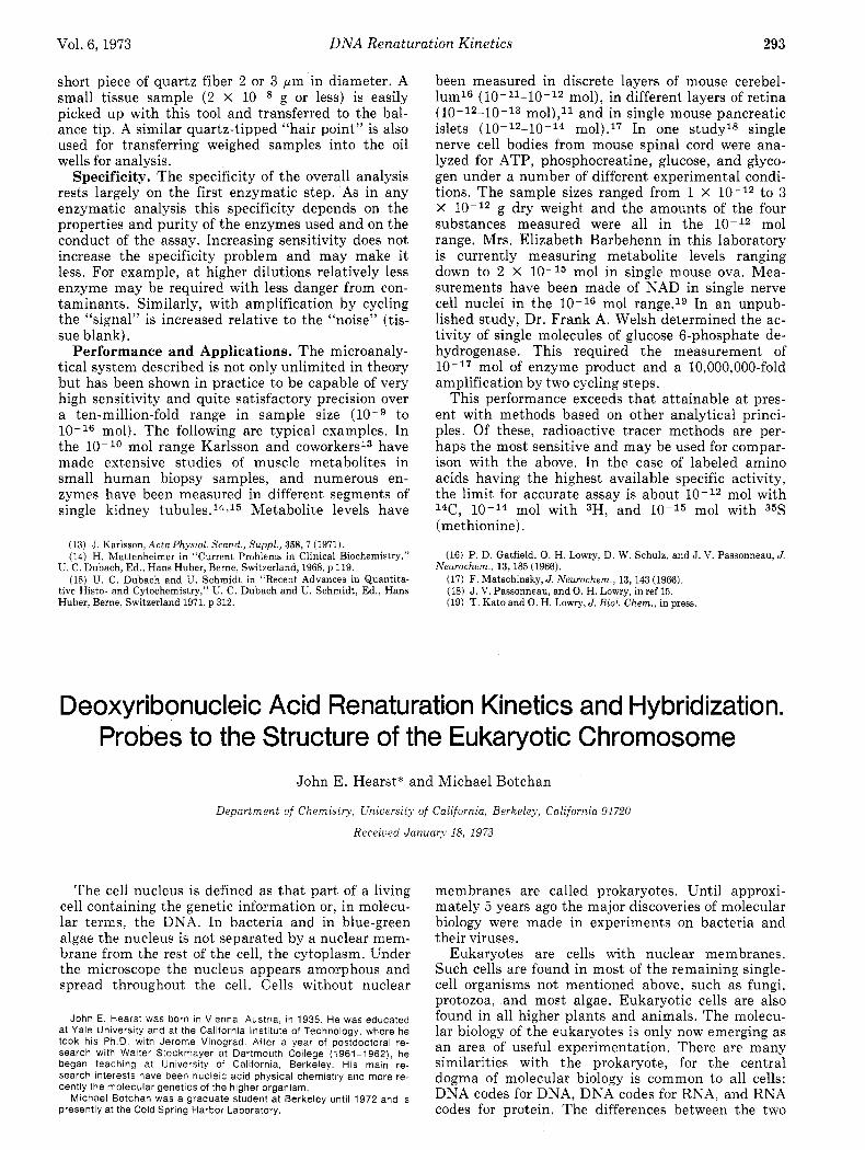
P Figure 1. Neutral CsCl buoyant-density profile for DNA isolated from Drosophila melanogaster embryos. The double-strand mo- lecular weight of this DNA is 30 X 106. (a) Total cellular DNA; (b) the main band DNA of isolated nuclei.
X 105, before studies of renaturation kinetics are performed, thus eliminating the variable of DNA molecular weight as a complicating parameter.17 If all the DNA fragments contain unique sequences, the rate of renaturation a t a given weight concentra- tion of DNA is proportional to the size of the genome since the concentration of any one unique fragment is inversely proportional to the size of the genome from which it originated. Thus, T4 bacteriophage DNA renatures 20 times faster than E. coli DNA a t some standard condition because there is 20 times as much DNA in an E. coli bacterium as there is in a T4 phage and all nucleotide sequences in both orga- nisms are unique.
If the organism contains repeated DNA sequences, these sequences will renature more rapidly than the unique sequences. In the case of 106 copies of a re- peated DNA sequence per genome, these sequences will renature 106 times more rapidly if the repeated sequence is longer than the length of helix required for stability a t the standard renaturation conditions (about 20 base pairs). Such large spreads in rates of renaturation have been observed in the DNA of al- most all eukaryotes studied. The genome has arbi- trarily been divided into three classes: unique se- quences, intermediate sequences renaturing a t rates suggesting 10 to 1000 copies per genome, and the very rapidly renaturing or simple sequences which renature a t rates suggesting 103 to l o 7 copies per ge- nome. We have concentrated on simple DNA se- quences in this laboratory.
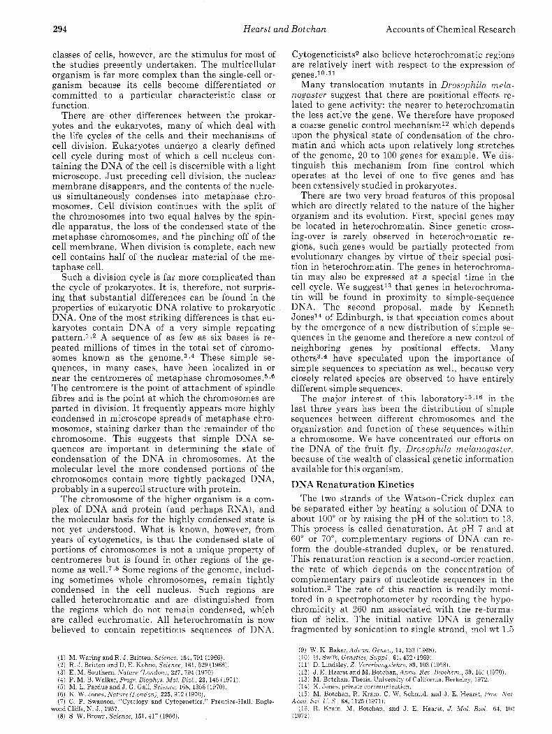
Any kinetic class of DNA may be separated from the others by use of hydroxyapatite which binds double-stranded DNA but not single strands in 0.12 M phosphate buffer. The double-stranded DNA is readily eluted from the hydroxyapatite using a 0.45 M phosphate buffer. We have renatured Drosophila melanogaster DNA for very short times and isolated the double-stranded product on hydroxyapatite. We call this material isolated harrDNA for hydroxyapa- tite-isolated rapidly renatured DNA.
CsCl Density Profiles Frequently, the simple-sequence DNA occurs in
large enough blocks and has a base composition (GC content) different enough from the remaining DNA
Single- Strand Molecular Weight of Starting Material
1.704
HARRD Yields (% of Total DNA)
1.727
3.8x IO6 (c) 14%
1.36x10⁶ (b) I7O2 I I - 12%
1.691
Figure 2. Buoyant-density profiles of hydroxyapatite-isolated rapidly renaturing DNA from different single-strand molecular weights of the starting DNA. Marker DNAs are E. coli [ISNIDNA ( p = 1.727 g/ml) and M . I$sodeikticus DNA ( p = 1.731 g/ml). The figure also contains the yield of rapidly renaturing DNA from the hydroxyapatite.
so that banding the partially fragmented DNA in the ultracentrifuge in a CsCl density gradient results in the separation of the simple DNA from the bulk of the DNA in a density satellite. Figure l a shows the CsCl density profile of total cellular DNA of mol wt 30 X 106 from Drosophila melanogaster embryos. The profile shows the bulk of the DNA bands a t a density of 1.701. The satellite a t density p = 1.672 represents 2-370 of the total DNA, and its origin is unknown, although it has been shown to be partially associated with the Y chromosome. The satellite a t p = 1.680 representing 3-490 of the total is mitochon- drial DNA and thus cytoplasmic in origin.l3 The sat- ellite a t p = 1.687 representing 7-990 of the total DNA has been localized to the centric heterochroma- tin by Gall, Cohen, and Polanl8 using in situ hybri- dization, a technique described below. It is thus nu- clear in origin.
We have isolated harrDNA from main-band Drosophila melanogaster DNA with different single- strand molecular weights16 and determined the den- sity profile of the product. Main-band DNA, Figure lb , is DNA from which all satellites have been re- moved. Figure 2 shows the results of this experiment. For sonicated DNA, the harrDNA appears as a sin- gle peak a t p = 1.691 and a yield of 8-970. The fact that this density would be easily resolved in Figure l b suggests that the simple sequences are attached to a more dense DNA and that their length is shorter than mol wt 15 X 106 single strand. A complete analysis of the profiles obtained a t higher molecular weights suggests that the simple sequences come in blocks of mol wt 0.75 to 3 million interspersed by more complex sequences of mol wt 1 to 2 million sin- gle strand.
Since renaturation is a rather complex process yielding double-stranded product with mismatching
(17) J . G. Wetmur and N. Davidson, J. Mol. Biol., 31,349 (1968). (18) J. G. Gall, E. H. Cohen, and M. L. Polan, Chromosoma, 33, 319
(1971).
296 Hearst and Botchan Accounts of Chemical Research
26,000
22,000
Table I Comparison of % of DNA Hybridized to in Vivo RNA by
C S Z S O ~ and Hydroxyapatite Methods
70 hydridization
2,000 A A
0 5 IO 15 20 25 30 35 Fraction Number
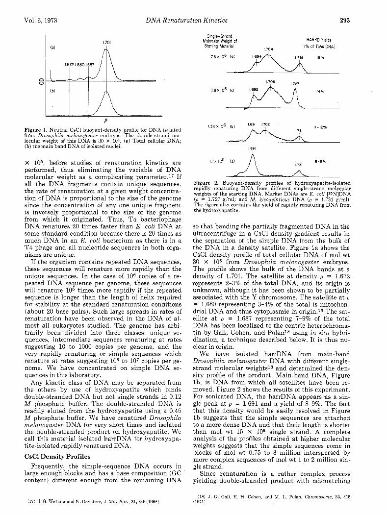
Figure 3. The location of sonicated harrDNA sequences in a na- tive DNA CsCl gradient. [32P]RNA complementary to harrDNA was incubated with filter papers containing the DNA from each fraction. (A) Hybridized [32P]DNA; (*) [3H]DNA counts from starting fractions.
and free ends, an additional experiment on native DNA which demonstrated the bimodal distribution in density seemed advisable. Figure 3 shows the re- sults of banding total nuclear DNA (including the satellites) in a preparative CsCl gradient.13 The solid circles show the distribution of DNA (3H labeled, mol wt 7 X 10⁶) in the gradient. Labeled 32P RNA complementary to sonicated harrDNA(the p = 1.691 peak in Figure 2) was synthesized using RNA poly- merase, 32P-labeled nucleotide triphosphates, and harrDNA as a template. The labeled RXA was hy- bridized to the various DNA fractions which were denatured and mounted on nitrocellulose filters.lg This localizes the harrDNA sequences in the native DNA density profile. There are essentially no counts coincident with the p = 1.687 satellite. A 25% peak occurs a t p = 1.691, the density of pure simple se- quence DNA, while the major portion of the [32P]RNA hybridizing ability is found in the light portion of the main band ( p = 1.695-1.705). This shows that in this native DSA most of the simple harrDNA sequences are attached to a heavier DXA a t 7 X lo6 molecular weight.
Localization of harrDNA in the Salivary Chromosome
Salivary cells of Drosophila melanogaster are poly- tene; that is, each cell contains many copies of the haploid genome (more than 1000 copies). The sali- vary chromosome has a banded structure in which each of these 1000 copies of DNA are lined up side by side. It is partially these unique banded struc- tures which make the fruit fly such an attractive or- ganism for the geneticist. All of the centromeres and centric heterochromatin are connected in the sali- vary cell a t the chromocenter. Thus localization of a sequence to the salivary chromocenter is equivalent to localizing the sequence to the centric heterochro- matin of the normal diploid cell.
If 3H labeled RXA complementary to harrDNA is synthesized using RNA polymerase, i t can be hybri- dized to a partially denatured chromosome on a glass slide. A ribonuclease treatment degrades any bound RNA unprotected by its hybridization to
(19) D. Gillespie and S. Spiegelman, J. Mol. B i d , 12,829 (1965).
[RNA]. mg/ml Cs2SO4 HAP Type of DNA
1 0 15 0
4 0 39 0
10 0 58 0
3 18 8 2
49 6 5
70 8
A B
control A B
control A B
control
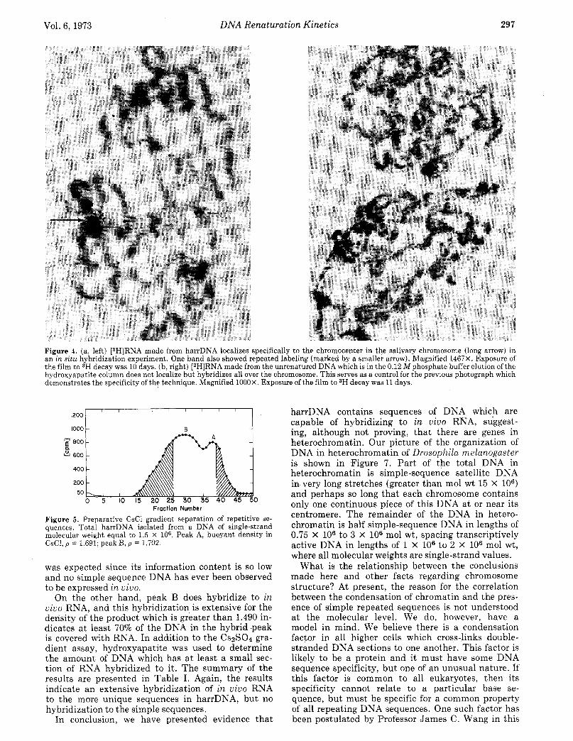
DNA. The RNA is then located by autoradiography. The results of this experiment for harrDNA are shown in Figure 4a. Figure 4b is a control demon- strating the specificity of the technique. Clearly our harrDNA comes from centric heterochromatin. We mentioned earlier that Gall, et al.,18 have shown that the 1.687 satellite also is in centric heterochro- matin.
I n Vivo Expression of the harrDNA The next step toward our eventual goal of demon-
strating coarse control is the demonstration that there is some DNA in heterochromatin which is transcribed (used as a template for making comple- mentary RNA).
The hybridization experiments designed13 to dem- onstrate the existence of an i n vivo RNA comple- mentary to our harrDNA were performed in large RNA excess in solution. 3H-labeled DNA of a specif- ic activity of 1.5 X l o 5 cpm/pg was isolated from a Drosophila melanogaster tissue culture line obtained from I. Schneider. The density profile of DNA isolat- ed from this line is identical with that isolated from embryos. The unlabeled RNA was isolated from em- bryos and extensively purified to eliminate any contaminating protein, polysaccharide, and DNA.
Labeled harrDNA was isolated at a single-strand molecular weight of 1.5 X 10⁶. Figure 5 shows the re- sulting density profile in a CsCl preparative gradi- ent. Fractions as indicated by the cross-hatched areas of Figure 5 were further purified by additional bandings in CsCl gradients. This yielded a sample of purified simple sequence DKA (peak A, p = 1.691) and a sample of simple sequence DNA with more unique sequences attached (peak B, p = 1.702).
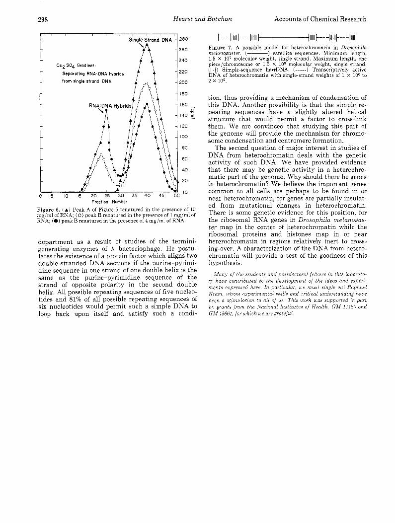
These two fractions were then hybridized with the i n vivo RNA in vast excess so that the harrDNA had insufficient time to renature with itself but ample time to react with the complementary RNA se- quences if present. The product was treated with ri- bonuclease to destroy any single-stranded RNA and banded in a CszS04 density gradient. Pure RSA- DNA hybrids have a density of 1.49-1.51 in CszS04 while denatured DNA has a density of 1.445, so if an appreciable portion of any piece of DNA hybridizes to the RNA its density will be shifted to a higher value and be resolved. Figure 6 shows the result of this experiment. It is clear that peak A, or simple- sequence DNA, does not hybridize any RNA. This
Vol. 6, 1973 DNA Renaturation Kinetics 297
Figure 4. (a, left) [3H]RNA made from harrDNA localizes specifically to the chromocenter in the salivary chromosome (long arrow) in an in situ hybridization experiment. One band also showed repeated labeling (marked by a smaller arrow). Magnified 1467X. Exposure of the film to 3H decay was 10 days. (b, right) [3H]RNA made from the unrenatured DNA which is in the 0.12 Mphosphate buffer elution of the hydroxyapatite column does not localize but hybridizes all over the chromosome. This serves as a control for the previous photograph which demonstrates the specificity of the technique. Magnified 1OOOX. Exposure of the film to 3H decay was 11 days.
1000 !- R J
Fraction Number
Figure 5. Preparative CsCl gradient separation of repetitive se- quences. Total harrDNA isolated from a DNA of single-strand molecular weight equal to 1.5 X lo6. Peak A, buoyant density in CsCl, p = 1.691; peak B, p = 1.702.
was expected since its information content is so low and no simple sequence DNA has ever been observed to be expressed i n vivo.
On the other hand, peak B does hybridize to in vivo RNA, and this hybridization is extensive for the density of the product which is greater than 1.490 in- dicates a t least 70% of the DNA in the hybrid.peak is covered with RNA. In addition to the C S ~ S O ~ gra- dient assay, hydroxyapatite was used to determine the amount of DNA which has a t least a small sec- tion of RNA hybridized to it. The summary of the results are presented in Table I. Again, the results indicate an extensive hybridization of i n vivo RNA to the more unique sequences in harrDNA, but no hybridization to the simple sequences.
In conclusion, we have presented evidence that
harrDNA contains sequences of DNA which are capable of hybridizing to in vivo RNA, suggest- ing, although not proving, that there are genes in heterochromatin. Our picture of the organization of DNA in heterochromatin of Drosophila melanogaster is shown in Figure 7. Par t of the total DNA in heterochromatin is simple-sequence satellite DNA in very long stretches (greater than mol wt 15 X lo6) and perhaps so long that each chromosome contains only one continuous piece of this DNA a t or near its centromere. The remainder of the DNA in hetero- chromatin is half simple-sequence DNA in lengths of 0.75 X lo6 to 3 X lo6 mol wt, spacing transcriptively active DNA in lengths of 1 X lo6 to 2 X lo6 mol wt, where all molecular weights are single-strand values.
What is the relationship between the conclusions made here and other facts regarding chromosome structure? At present, the reason for the correlation between the condensation of chromatin and the pres- ence of simple repeated sequences is not understood a t the molecular level. We do, however, have a model in mind. We believe there is a condensation factor in all higher cells which cross-links double- stranded DNA sections to one another. This factor is likely to be a protein and it must have some DNA sequence specificity, but one of an unusual nature. If this factor is common to all eukaryotes, then its specificity cannot relate to a particular base se- quence, but must be specific for a common property of all repeating DNA sequences. One such factor has been postulated by Professor James C. Wang in this
298 Hearst and Botchan Accounts of Chemical Research
r l , I I , I I I
Single Strand DNA
"A I \
Cs2 SO4 Gradient: I f t Separating RNA: DNA hybrids
from single strand DNA
?a0
5 0
!40
2 0
!OO
180
160 ~
140 5 I20
I O 0
80
60
40
20
10
"
Fraction Number
Figure 6. (A) Peak A of Figure 5 renatured in the presence of 10 mg/ml of RNA; ( 0 ) peak B renatured in the presence of 1 mg/ml of RNA; (0) peak B renatured in the presence of 4 mg/ml of RNA.
department as a result of studies of the termini- generating enzymes of X bacteriophage. He postu- lates the existence of a protein factor which aligns two double-stranded DXA sections if the purine-pyrimi- dine sequence in one strand of one double helix is the same as the purine-pyrimidine sequence of the strand of opposite polarity in the second double helix. All possible repeating sequences of five nucleo- tides and 81% of all possible repeating sequences of six nucleotides would permit such a simple DSA to loop back upon itself and satisfy such a condi-
Figure 7. A possible model for heterochromatin in Drosophila melarzogaster. (- ) satellite sequences. Minimum length, 1.5 X lo7 molecular weight, single strand. Maximum length, one piece/chromosome or 1.5 X lo9 molecular weight, single strand. ( 1 1 I) Simple-sequence harrDNA. ( - - - - - - ) Transcriptively active DNA of heterochromatin with single-strand weights of 1 X lo6 to 2 x 10⁶.
tion, thus providing a mechanism of condensation of this DNA. Another possibility is that the simple re- peating sequences have a slightly altered helical structure that would permit a factor to cross-link them. We are convinced that studying this part of the genome will provide the mechanism for chromo- some condensation and centromere formation.
The second question of major interest in studies of DNA from heterochromatin deals with the genetic activity of such DNA. We have provided evidence that there may be genetic activity in a heterochro- matic part of the genome. Why should there be genes in heterochromatin? We believe the important genes common to all cells are perhaps to be found in or near heterochromatin, for genes are partially insulat- ed from mutational changes in heterochromatin. There is some genetic evidence for this position, for the ribosomal RNA genes in Drosophila melanogas- ter map in the center of heterochromatin while the ribosomal proteins and histones map in or near heterochromatin in regions relatively inert to cross- ing-over. A characterization of the DNA from hetero- chromatin will provide a test of the goodness of this hypothesis.
M a n ) of t he s tudents and postdoctoral fe l lous cn this laborato- ry habe contributed t o the development of t he ideas and experz- men t s expressed here I n particular, &e m u s t single out Raphael K r a m , uhose experimental skills and critical understanding have been a s t imulat ion to all of us Thzs uiork &as supported in part bq grants from the A'atzonal Inst i tutes of Heal th , G M 11180 and G M 15661, for u ~ h i c h ule are grateful