dependence on the lazaro phosphatidic acid phosphatase for the maximum light response
TRANSCRIPT
Current Biology 16, 723–729, April 4, 2006 ª2006 Elsevier Ltd All rights reserved DOI 10.1016/j.cub.2006.02.057
ReportDependence on the LazaroPhosphatidic Acid Phosphatasefor the Maximum Light Response
Young Kwon1 and Craig Montell1,*1Department of Biological Chemistry andDepartment of NeuroscienceThe Johns Hopkins University School of MedicineBaltimore, Maryland 21205
Summary
The Drosophila phototransduction cascade serves asa paradigm for characterizing the regulation of sen-
sory signaling and TRP channels in vivo [1, 2]. Activa-tion of these channels requires phospholipase C (PLC)
and may depend on subsequent production of diacyl-glycerol (DAG) and downstream metabolites [3, 4].
DAG could potentially be produced through a secondpathway involving the combined activities of a phos-
pholipase D (PLD) [5] and a phosphatidic acid (PA)phosphatase (PAP). However, a role for a PAP in the
regulation of TRP channels has not been described.Here, we report the identification of a PAP, referred
to as Lazaro (Laza). Mutations in laza caused a reduc-tion in the light response and faster termination kinet-
ics. Loss of laza suppressed the severity of the pheno-type caused by mutation of the DAG kinase, RDGA [6,
7], indicating that Laza functions in opposition to
RDGA. We also showed that the retinal degenerationresulting from overexpression of the PLD [5] was sup-
pressed by elimination of Laza. These data demon-strate a requirement for a PLD/PAP-dependent path-
way for achieving the maximal light response. Thegenetic interactions with both rdgA and Pld indicate
that Laza functions in the convergence of both PLC-and PLD-coupled signaling in vivo.
Results and Discussion
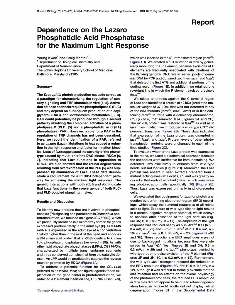
To identify new proteins that are involved in phosphoi-nositide (PI) signaling and participate in Drosophila pho-totransduction, we focused on a gene (CG11440), whichwe previously identified in a microarray screen for genesexpressed predominately in the adult eye [8]. CG11440mRNA is expressed in the adult eye at a concentration15-fold higher than in the rest of the head and encodesa 334 amino acid protein that is >35% identical to knownlipid phosphate phosphatases (reviewed in [9]). As withother lipid phosphate phosphatases (LPPs), CG11440 ischaracterized by multiple transmembrane segmentsand three conserved domains that form the catalytic do-main. An LPP would be predicted to catalyze the reversereaction promoted by RDGA (Figure 1A).
To address a potential function for the CG11440(referred to as lazaro, laza; see figure legends for an ex-planation of the gene name) in phototransduction, weobtained a P element insertion line, GE27043 (GenExel),
*Correspondence: [email protected]
which was inserted in the 50 untranslated region (lazaGE;Figure 1B). We created a null mutation in laza by genet-ically mobilizing the P element, because excisions of Pelements are frequently associated with deletions ofthe flanking genomic DNA. We screened pools of geno-mic DNA by PCR and obtained two lines (laza1 and laza2)that deleted the first ATG and additional portions of thecoding region (Figure 1B). In addition, we retained onerevertant line in which the P element excised precisely(lazaRE).
We raised antibodies against the C-terminal regionof Laza and identified a protein of 42 kDa (predicted mo-lecular weight of 37 kDa) that was not detected in anyof the laza mutants (lazaGE, laza1, laza2) or in flies con-taining lazaGE in trans with a deficiency chromosome,Df(3L)ED230, that removed laza (Figures 2A and 2B).The 42 kDa protein was restored in lazaRE as well as inlaza1 flies in which we introduced a wild-type CG11440genomic transgene (Figure 2B). These data indicatedthat expression of the Laza protein was disrupted inlazaGE, laza1, and laza2. Protein levels of other photo-transduction proteins were unchanged in each of thelines studied (Figure 2C).
To evaluate whether the Laza protein was expressedin the retina, we used a biochemical approach becausethe antibodies were ineffective for immunostaining. Wedetected Laza exclusively in extracts from wild-typeheads but not bodies (Figure 2D). Moreover, the Lazaprotein was absent in head extracts prepared from amutant lacking eyes (sine oculis, so) and was greatly re-duced in the heads of a mutant (glass), which were miss-ing photoreceptor cells specifically [10] (Figure 2E).Thus, Laza was expressed primarily in photoreceptorcells.
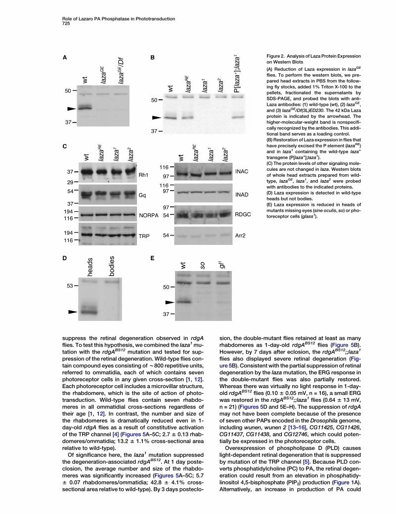
We evaluated the requirement for laza for phototrans-duction by performing electroretinogram (ERG) record-ings, which assay the summed responses of all retinalcells to light. Exposure of wild-type flies to light resultsin a corneal negative receptor potential, which decaysto baseline after cessation of the light stimulus (Fig-ure 3A; 10.3 6 0.7 mV, n = 17). The amplitude of the lightresponse was reduced nearly 40% in lazaGE flies (6.5 60.4 mV, n = 29) and 3-fold in laza1 (3.7 6 0.4 mV, n =19) and laza2 flies (2.8 6 0.3 mV, n = 23) (Figures 3B–3Dand 3H). These reductions in ERG amplitudes were notdue to background mutations because they were ob-served in lazaGE/Df flies (Figures 3E and 3H; 5.9 60.3 mV, n = 25) and the lazaGE phenotype reverted towild-type upon precise excision of the P element (Fig-ures 3F and 3H; 10.1 6 0.2 mV, n = 19). Furthermore,the wild-type laza+ transgene rescued the reduction inthe ERG amplitude (Figures 3G–3H; 10.9 6 0.3 mV, n =12). Although it was difficult to formally exclude that thelaza mutation had no effects on the overall physiologyof the photoreceptor cells, the reduced ERG amplitudein laza flies did not appear to be due to retinal degener-ation because 7-day-old adults did not display retinaldegeneration (Figure S1 in the Supplemental Data
Current Biology724
Figure 1. Phosphoinositide Signaling and the
laza Genomic Region
(A) PI cycle. Activation of PLC (NORPA),
which occurs following light stimulus, leads
to hydrolysis of PIP2 and generation of DAG
and IP3. The PIP2 is regenerated through
a multistep cycle initiated with the phosphor-
ylation of DAG by the DAG kinase (RDGA) to
produce PA. The proposed site of action of
Laza is indicated, and it is in opposition to
RDGA. PA is also produced from PC through
the action of PLD. The following abbre-
viations are used: CDS, CDP-diacylglycerol
synthase; DAG, diacylglycerol; DGK, DAG ki-
nase; IP3, inositol 1,4,5-trisphosphate; Laza,
Lazaro; NORPA, No Receptor Potential A;
PA, phosphatidic acid; PAP, phosphatidic
acid phosphatase; PC, phosphatidylcholine;
PI, phosphatidylinositol; PITP, PI-transfer
protein; PIP, phosphatidylinositol-phosphate;
PIP2, phosphatidylinositol 4,5-bisphosphate;
PLC, phospholipase C; PLD, phospholipase
D; RDGA, Retinal Degeneration A; and RDGB,
Retinal Degeneration B.
(B) Schematic representation of the laza mu-
tations. A P element insertion (GE27043) was
used to generate the deletions in laza1 and
laza2 through imprecise excision of the trans-
posable element. The 1.1 and 0.6 kb deletions
in laza1 (2317 to +1381) and laza2 (2309 to
+913), respectively, are indicated.
available online) and the ERGs were performed on flies<7 days old. As is the consequence of most mutationsthat affect phototransduction, in older flies (R15 daysold) the laza mutation resulted in retinal degeneration,which was light-dependent (data not shown). Neverthe-less, the decreased ERG responsiveness in the laza flieswas not age dependent (Figure 3I).
To assess whether the decreased amplitude in lazawas more likely a reflection of a lower sensitivity to lightor a decreased maximal obtainable amplitude, we mea-sured the ERG responses at three different intensities.Over the 130-fold intensity range tested, both wild-type and laza flies showed a linear intensity-responsiverelationship when the amplitudes were plotted againstthe logs of the light intensities (Figure 3J). However,in the laza mutant, the plot is shifted down. If therewas a reduction strictly in the sensitivity of the light re-sponse, the lines generated from the wild-type andlaza intensity-responsive relationships should havebeen parallel. This was not the case, because the ampli-tude of the laza response increased less sharply than inwild-type (Figure 3J). These data are most consistentwith a defect in the maximum light response in the mu-tant; however, the data do not allow us to assesswhether or not there is a defect in the sensitivity to light.If laza operates in a pathway downstream of PLD (Fig-ure 1A), then there would be expected to be overlapsin the pld and laza mutant phenotypes. Interestingly,in the pld null mutant, there is also a defect in the maxi-mum achievable ERG amplitude [5].
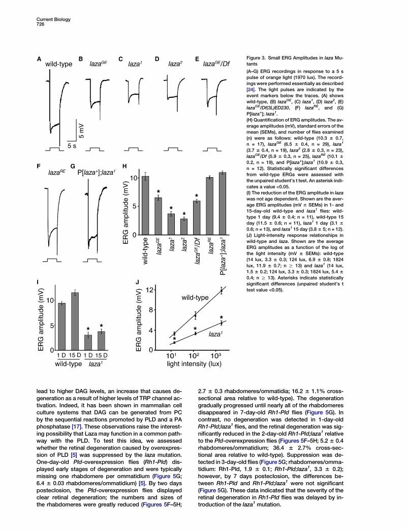
Mutations in rdgA, which encodes a diacylglycerol(DAG) kinase [11], prolong termination kinetics [4]. Totest whether elimination of laza might accelerate de-activation kinetics, consistent with the predicted bio-chemical function (Figure 1A), we assayed the time re-quired for an 80% return to the baseline after cessationof the photoresponse. We found that the termination ofthe photoresponse was approximately 4-fold faster inthe laza1 (0.29 6 0.02 s, n = 19) and laza2 (0.25 6 0.03 s,n = 18) mutant flies than in wild-type (1.24 6 0.11 s, n =20) (Figures 4A–4C and 4G). Introduction of the genomicrescue transgene in the laza1 mutant backgroundrestored the deactivation kinetics to that of wild-type(Figures 4D and 4G; 1.29 6 0.11 s, n = 16).
The apparent increase in the speed of termination ofthe photoresponse in the laza mutants may have beendue to the smaller ERG amplitude. Therefore, we ex-posed wild-type flies to a lower light intensity so thatthe amplitudes of the wild-type and laza ERGs were com-parable (Figures 4B, 4C, and 4E). Under these condi-tions, the time required for 80% termination of the photo-response was 2-fold shorter in the laza1 (0.29 6 0.02 s,n = 19) and laza2 (0.25 6 0.03 s, n = 18) mutant fliesthan in wild-type (0.53 6 0.04 s, n = 20) (Figures 4B, 4C,4E, and 4H). Thus, the more rapid termination in laza flieswas the reverse of that observed in rdgA flies.
If laza functions in the regulation of the photoresponseby catalyzing the PI-cycle reaction that is the reciprocalof that catalyzed by RDGA (Figure 1A), then it is plausiblethat introduction of laza into an rdgA background would
Role of Lazaro PA Phosphatase in Phototransduction725
Figure 2. Analysis of Laza Protein Expression
on Western Blots
(A) Reduction of Laza expression in lazaGE
flies. To perform the western blots, we pre-
pared head extracts in PBS from the follow-
ing fly stocks, added 1% Triton X-100 to the
pellets, fractionated the supernatants by
SDS-PAGE, and probed the blots with anti-
Laza antibodies: (1) wild-type (wt), (2) lazaGE,
and (3) lazaGE/Df(3L)ED230. The 42 kDa Laza
protein is indicated by the arrowhead. The
higher-molecular-weight band is nonspecifi-
cally recognized by the antibodies. This addi-
tional band serves as a loading control.
(B) Restoration of Laza expression in flies that
have precisely excised the P element (lazaRE)
and in laza1 containing the wild-type laza+
transgene (P[laza+];laza1).
(C) The protein levels of other signaling mole-
cules are not changed in laza. Western blots
of whole head extracts prepared from wild-
type, lazaGE, laza1, and laza2 were probed
with antibodies to the indicated proteins.
(D) Laza expression is detected in wild-type
heads but not bodies.
(E) Laza expression is reduced in heads of
mutants missing eyes (sine oculis, so) or pho-
toreceptor cells (glass1).
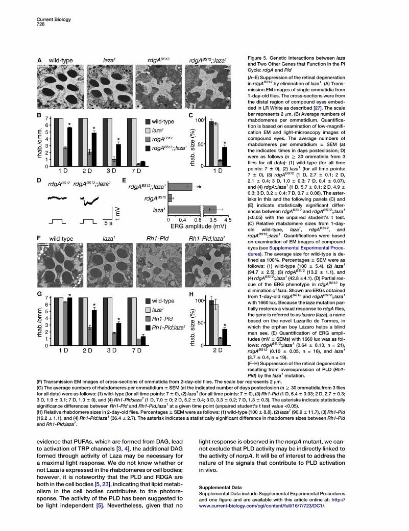
suppress the retinal degeneration observed in rdgAflies. To test this hypothesis, we combined the laza1 mu-tation with the rdgABS12 mutation and tested for sup-pression of the retinal degeneration. Wild-type flies con-tain compound eyes consisting of w800 repetitive units,referred to ommatidia, each of which contains sevenphotoreceptor cells in any given cross-section [1, 12].Each photoreceptor cell includes a microvillar structure,the rhabdomere, which is the site of action of photo-transduction. Wild-type flies contain seven rhabdo-meres in all ommatidial cross-sections regardless oftheir age [1, 12]. In contrast, the number and size ofthe rhabdomeres is dramatically reduced even in 1-day-old rdgA flies as a result of constitutive activationof the TRP channel [4] (Figures 5A–5C; 2.7 6 0.13 rhab-domeres/ommatidia; 13.2 6 1.1% cross-sectional arearelative to wild-type).
Of significance here, the laza1 mutation suppressedthe degeneration-associated rdgABS12. At 1 day poste-closion, the average number and size of the rhabdo-meres was significantly increased (Figures 5A–5C; 5.76 0.07 rhabdomeres/ommatidia; 42.8 6 4.1% cross-sectional area relative to wild-type). By 3 days posteclo-
sion, the double-mutant flies retained at least as manyrhabdomeres as 1-day-old rdgABS12 flies (Figure 5B).However, by 7 days after eclosion, the rdgABS12;;laza1
flies also displayed severe retinal degeneration (Fig-ure 5B). Consistent with the partial suppression of retinaldegeneration by the laza mutation, the ERG response inthe double-mutant flies was also partially restored.Whereas there was virtually no light response in 1-day-old rdgABS12 flies (0.10 6 0.05 mV, n = 16), a small ERGwas restored in the rdgABS12;;laza1 flies (0.64 6 13 mV,n = 21) (Figures 5D and 5E–H). The suppression of rdgAmay not have been complete because of the presenceof seven other PAPs encoded in the Drosophila genome,including wunen, wunen 2 [13–16], CG11425, CG11426,CG11437, CG11438, and CG12746, which could poten-tially be expressed in the photoreceptor cells.
Overexpression of phospholipase D (PLD) causeslight-dependent retinal degeneration that is suppressedby mutation of the TRP channel [5]. Because PLD con-verts phosphatidylcholine (PC) to PA, the retinal degen-eration could result from an elevation in phosphatidy-linositol 4,5-bisphosphate (PIP2) production (Figure 1A).Alternatively, an increase in production of PA could
Current Biology726
Figure 3. Small ERG Amplitudes in laza Mu-
tants
(A–G) ERG recordings in response to a 5 s
pulse of orange light (1970 lux). The record-
ings were performed essentially as described
[24]. The light pulses are indicated by the
event markers below the traces. (A) shows
wild-type, (B) lazaGE, (C) laza1, (D) laza2, (E)
lazaGE/Df(3L)ED230, (F) lazaRE, and (G)
P[laza+]; laza1.
(H) Quantification of ERG amplitudes. The av-
erage amplitudes (mV), standard errors of the
mean (SEMs), and number of flies examined
(n) were as follows: wild-type (10.3 6 0.7,
n = 17), lazaGE (6.5 6 0.4, n = 29), laza1
(3.7 6 0.4, n = 19), laza2 (2.8 6 0.3, n = 23),
lazaGE/Df (5.9 6 0.3, n = 25), lazaRE (10.1 6
0.2, n = 19), and P[laza+];laza1 (10.9 6 0.3,
n = 12). Statistically significant differences
from wild-type ERGs were assessed with
the unpaired student’s t test. An asterisk indi-
cates a value <0.05.
(I) The reduction of the ERG amplitude in laza
was not age dependent. Shown are the aver-
age ERG amplitudes (mV 6 SEMs) in 1- and
15-day-old wild-type and laza1 flies: wild-
type 1 day (9.4 6 0.4; n = 11), wild-type 15
day (11.5 6 0.6; n = 11), laza1 1 day (3.1 6
0.6; n = 13), and laza1 15 day (3.8 6 5; n = 12).
(J) Light-intensity response relationships in
wild-type and laza. Shown are the average
ERG amplitudes as a function of the log of
the light intensity (mV 6 SEMs): wild-type
(14 lux, 3.3 6 0.3; 124 lux, 6.9 6 0.8; 1824
lux, 11.9 6 0.7; n R 13) and laza1 (14 lux,
1.5 6 0.2; 124 lux, 3.3 6 0.3; 1824 lux, 5.4 6
0.4; n R 13). Asterisks indicate statistically
significant differences (unpaired student’s t
test value <0.05).
lead to higher DAG levels, an increase that causes de-generation as a result of higher levels of TRP channel ac-tivation. Indeed, it has been shown in mammalian cellculture systems that DAG can be generated from PCby the sequential reactions promoted by PLD and a PAphosphatase [17]. These observations raise the interest-ing possibility that Laza may function in a common path-way with the PLD. To test this idea, we assessedwhether the retinal degeneration caused by overexpres-sion of PLD [5] was suppressed by the laza mutation.One-day-old Pld-overexpression flies (Rh1-Pld) dis-played early stages of degeneration and were typicallymissing one rhabdomere per ommatidium (Figure 5G;6.4 6 0.03 rhabdomeres/ommatidium) [5]. By two daysposteclosion, the Pld-overexpression flies displayedclear retinal degeneration; the numbers and sizes ofthe rhabdomeres were greatly reduced (Figures 5F–5H;
2.7 6 0.3 rhabdomeres/ommatidia; 16.2 6 1.1% cross-sectional area relative to wild-type). The degenerationgradually progressed until nearly all of the rhabdomeresdisappeared in 7-day-old Rh1-Pld flies (Figure 5G). Incontrast, no degeneration was detected in 1-day-oldRh1-Pld;laza1 flies, and the retinal degeneration was sig-nificantly reduced in the 2-day-old Rh1-Pld;laza1 relativeto the Pld-overexpression flies (Figures 5F–5H; 5.2 6 0.4rhabdomeres/ommatidium; 36.4 6 2.7% cross-sec-tional area relative to wild-type). Suppression was de-tected in 3-day-old flies (Figure 5G; rhabdomeres/omma-tidium: Rh1-Pld, 1.9 6 0.1; Rh1-Pld;laza1, 3.3 6 0.2);however, by 7 days posteclosion, the differences be-tween Rh1-Pld and Rh1-Pld;laza1 were not significant(Figure 5G). These data indicated that the severity of theretinal degeneration in Rh1-Pld flies was delayed by in-troduction of the laza1 mutation.
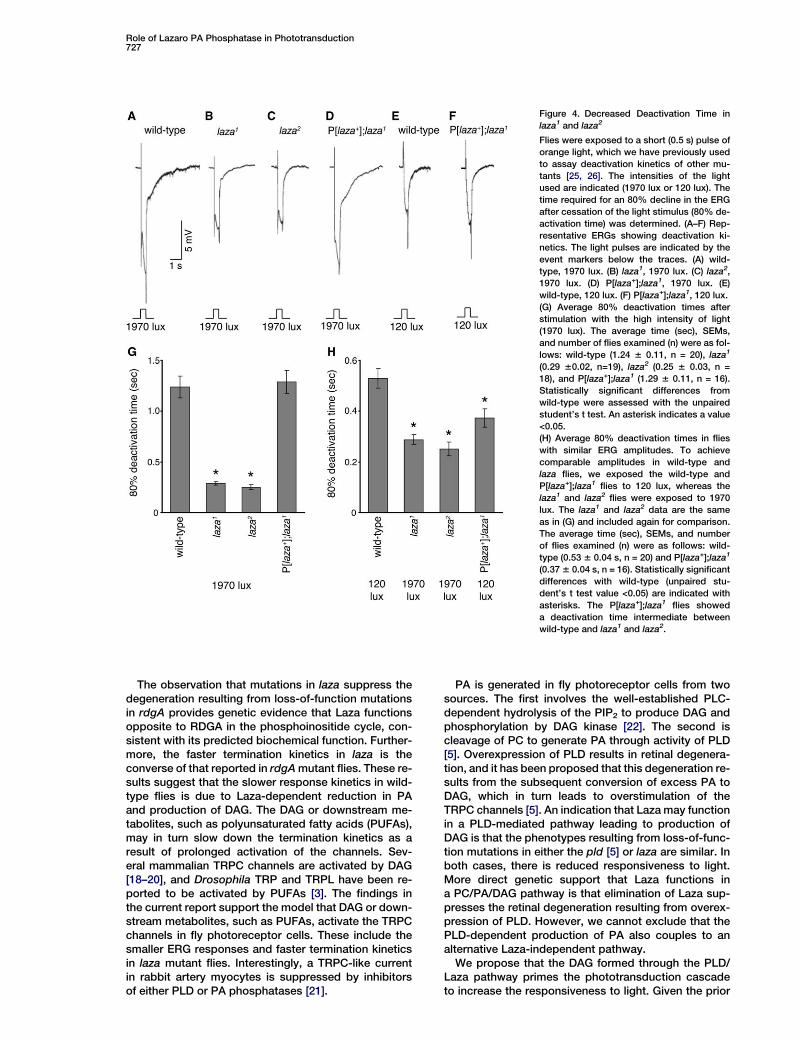
Role of Lazaro PA Phosphatase in Phototransduction727
Figure 4. Decreased Deactivation Time in
laza1 and laza2
Flies were exposed to a short (0.5 s) pulse of
orange light, which we have previously used
to assay deactivation kinetics of other mu-
tants [25, 26]. The intensities of the light
used are indicated (1970 lux or 120 lux). The
time required for an 80% decline in the ERG
after cessation of the light stimulus (80% de-
activation time) was determined. (A–F) Rep-
resentative ERGs showing deactivation ki-
netics. The light pulses are indicated by the
event markers below the traces. (A) wild-
type, 1970 lux. (B) laza1, 1970 lux. (C) laza2,
1970 lux. (D) P[laza+];laza1, 1970 lux. (E)
wild-type, 120 lux. (F) P[laza+];laza1, 120 lux.
(G) Average 80% deactivation times after
stimulation with the high intensity of light
(1970 lux). The average time (sec), SEMs,
and number of flies examined (n) were as fol-
lows: wild-type (1.24 6 0.11, n = 20), laza1
(0.29 60.02, n=19), laza2 (0.25 6 0.03, n =
18), and P[laza+];laza1 (1.29 6 0.11, n = 16).
Statistically significant differences from
wild-type were assessed with the unpaired
student’s t test. An asterisk indicates a value
<0.05.
(H) Average 80% deactivation times in flies
with similar ERG amplitudes. To achieve
comparable amplitudes in wild-type and
laza flies, we exposed the wild-type and
P[laza+];laza1 flies to 120 lux, whereas the
laza1 and laza2 flies were exposed to 1970
lux. The laza1 and laza2 data are the same
as in (G) and included again for comparison.
The average time (sec), SEMs, and number
of flies examined (n) were as follows: wild-
type (0.53 6 0.04 s, n = 20) and P[laza+];laza1
(0.37 6 0.04 s, n = 16). Statistically significant
differences with wild-type (unpaired stu-
dent’s t test value <0.05) are indicated with
asterisks. The P[laza+];laza1 flies showed
a deactivation time intermediate between
wild-type and laza1 and laza2.
The observation that mutations in laza suppress thedegeneration resulting from loss-of-function mutationsin rdgA provides genetic evidence that Laza functionsopposite to RDGA in the phosphoinositide cycle, con-sistent with its predicted biochemical function. Further-more, the faster termination kinetics in laza is theconverse of that reported in rdgA mutant flies. These re-sults suggest that the slower response kinetics in wild-type flies is due to Laza-dependent reduction in PAand production of DAG. The DAG or downstream me-tabolites, such as polyunsaturated fatty acids (PUFAs),may in turn slow down the termination kinetics as aresult of prolonged activation of the channels. Sev-eral mammalian TRPC channels are activated by DAG[18–20], and Drosophila TRP and TRPL have been re-ported to be activated by PUFAs [3]. The findings inthe current report support the model that DAG or down-stream metabolites, such as PUFAs, activate the TRPCchannels in fly photoreceptor cells. These include thesmaller ERG responses and faster termination kineticsin laza mutant flies. Interestingly, a TRPC-like currentin rabbit artery myocytes is suppressed by inhibitorsof either PLD or PA phosphatases [21].
PA is generated in fly photoreceptor cells from twosources. The first involves the well-established PLC-dependent hydrolysis of the PIP2 to produce DAG andphosphorylation by DAG kinase [22]. The second iscleavage of PC to generate PA through activity of PLD[5]. Overexpression of PLD results in retinal degenera-tion, and it has been proposed that this degeneration re-sults from the subsequent conversion of excess PA toDAG, which in turn leads to overstimulation of theTRPC channels [5]. An indication that Laza may functionin a PLD-mediated pathway leading to production ofDAG is that the phenotypes resulting from loss-of-func-tion mutations in either the pld [5] or laza are similar. Inboth cases, there is reduced responsiveness to light.More direct genetic support that Laza functions ina PC/PA/DAG pathway is that elimination of Laza sup-presses the retinal degeneration resulting from overex-pression of PLD. However, we cannot exclude that thePLD-dependent production of PA also couples to analternative Laza-independent pathway.
We propose that the DAG formed through the PLD/Laza pathway primes the phototransduction cascadeto increase the responsiveness to light. Given the prior
Current Biology728
Figure 5. Genetic Interactions between laza
and Two Other Genes that Function in the PI
Cycle: rdgA and Pld
(A–E) Suppression of the retinal degeneration
in rdgABS12 by elimination of laza1. (A) Trans-
mission EM images of single ommatidia from
1-day-old flies. The cross-sections were from
the distal region of compound eyes embed-
ded in LR White as described [27]. The scale
bar represents 2 mm. (B) Average numbers of
rhabdomeres per ommatidium. Quantifica-
tion is based on examination of low-magnifi-
cation EM and light-microscopy images of
compound eyes. The average numbers of
rhabdomeres per ommatidium 6 SEM (at
the indicated times in days posteclosion; D)
were as follows (n R 30 ommatidia from 3
flies for all data): (1) wild-type (for all time
points: 7 6 0), (2) laza1 (for all time points:
7 6 0), (3) rdgABS12 (1 D, 2.7 6 0.1; 2 D,
2.1 6 0.4; 3 D, 1.0 6 0.3; 7 D, 0.4 6 0.07),
and (4) rdgA;;laza1 (1 D, 5.7 6 0.1; 2 D, 4.9 6
0.3; 3 D, 3.2 6 0.4; 7 D, 0.7 6 0.06). The aster-
isks in this and the following panels (C) and
(E) indicate statistically significant differ-
ences between rdgABS12 and rdgABS12;;laza1
(<0.05) with the unpaired student’s t test.
(C) Relative rhabdomere sizes from 1-day-
old wild-type, laza1, rdgABS12, and
rdgABS12;;laza1. Quantifications were based
on examination of EM images of compound
eyes (see Supplemental Experimental Proce-
dures). The average size for wild-type is de-
fined as 100%. Percentages 6 SEM were as
follows: (1) wild-type (100 6 5.4), (2) laza1
(94.7 6 2.5), (3) rdgABS12 (13.2 6 1.1), and
(4) rdgABS12;;laza1 (42.8 64.1). (D) Partial res-
cue of the ERG phenotype in rdgABS12 by
elimination of laza. Shown are ERGs obtained
from 1-day-old rdgABS12 and rdgABS12;;laza1
with 1660 lux. Because the laza mutation par-
tially restores a visual response to rdgA flies,
the gene is referred to as lazaro (laza), a name
based on the novel Lazarillo de Tormes, in
which the orphan boy Lazaro helps a blind
man see. (E) Quantification of ERG ampli-
tudes (mV 6 SEMs) with 1660 lux was as fol-
lows: rdgABS12;;laza1 (0.64 6 0.13, n = 21),
rdgABS12 (0.10 6 0.05, n = 16), and laza1
(3.7 6 0.4, n = 19).
(F–H) Suppression of the retinal degeneration
resulting from overexpression of PLD (Rh1-
Pld) by the laza1 mutation.
(F) Transmission EM images of cross-sections of ommatidia from 2-day-old flies. The scale bar represents 2 mm.
(G) The average numbers of rhabdomeres per ommatidium 6 SEM (at the indicated number of days posteclosion (n R 30 ommatidia from 3 flies
for all data) were as follows: (1) wild-type (for all time points: 7 6 0), (2) laza1 (for all time points: 7 6 0), (3) Rh1-Pld (1 D, 6.4 6 0.03; 2 D, 2.7 6 0.3;
3 D, 1.9 6 0.1; 7 D, 1.0 6 0), and (4) Rh1-Pld;laza1 (1 D, 7.0 6 0; 2 D, 5.2 6 0.4; 3 D, 3.3 6 0.2; 7 D, 1.3 6 0.3). The asterisks indicate statistically
significance differences between Rh1-Pld and Rh1-Pld;laza1 at a given time point (unpaired student’s t test value <0.05).
(H) Relative rhabdomere sizes in 2-day-old flies. Percentages 6 SEM were as follows: (1) wild-type (100 6 8.8), (2) laza1 (90.9 6 11.7), (3) Rh1-Pld
(16.2 6 1.1), and (4) Rh1-Pld;laza1 (36.4 6 2.7). The asterisk indicates a statistically significant difference in rhabdomere sizes between Rh1-Pld
and Rh1-Pld;laza1.
evidence that PUFAs, which are formed from DAG, leadto activation of TRP channels [3, 4], the additional DAGformed through activity of Laza may be necessary fora maximal light response. We do not know whether ornot Laza is expressed in the rhabdomeres or cell bodies;however, it is noteworthy that the PLD and RDGA areboth in the cell bodies [5, 23], indicating that lipid metab-olism in the cell bodies contributes to the photore-sponse. The activity of the PLD has been suggested tobe light independent [5]. Nevertheless, given that no
light response is observed in the norpA mutant, we can-not exclude that PLD activity may be indirectly linked tothe activity of norpA. It will be of interest to address thenature of the signals that contribute to PLD activationin vivo.
Supplemental Data
Supplemental Data include Supplemental Experimental Procedures
and one figure and are available with this article online at: http://
www.current-biology.com/cgi/content/full/16/7/723/DC1/.
Role of Lazaro PA Phosphatase in Phototransduction729
Acknowledgments
We thank Heather Shim for help with fly work, Michael Sepanski and
Michael Delannoy for preparing sections of compound eyes, Dr.
Emiko Suzuki for rdgA alleles, Dr. Michael A. Frohman for Pld flies,
and Dr. Michael Kottgen for helpful comments on the manuscript.
This work was supported by grants to C.M. from the National Eye
Institute (EY08117 and EY10852).
Received: December 15, 2005
Revised: February 16, 2006
Accepted: February 17, 2006
Published online: March 2, 2006
References
1. Montell, C. (1999). Visual transduction in Drosophila. Annu. Rev.
Cell Dev. Biol. 15, 231–268.
2. Montell, C. (2005). The TRP superfamily of cation channels. Sci.
STKE 2005, re3.
3. Chyb, S., Raghu, P., and Hardie, R.C. (1999). Polyunsaturated
fatty acids activate the Drosophila light-sensitive channels
TRP and TRPL. Nature 397, 255–259.
4. Raghu, P., Usher, K., Jonas, S., Chyb, S., Polyanovsky, A., and
Hardie, R.C. (2000). Constitutive activity of the light-sensitive
channels TRP and TRPL in the Drosophila diacylglycerol kinase
mutant, rdgA. Neuron 26, 169–179.
5. LaLonde, M.M., Janssens, H., Rosenbaum, E., Choi, S.Y., Ger-
gen, J.P., Colley, N.J., Stark, W.S., and Frohman, M.A. (2005).
Regulation of phototransduction responsiveness and retinal de-
generation by a phospholipase D-generated signaling lipid.
J. Cell Biol. 169, 471–479.
6. Hotta, Y., and Benzer, S. (1970). Genetic dissection of the Dro-
sophila nervous system by means of mosaics. Proc. Natl.
Acad. Sci. USA 67, 1156–1163.
7. Harris, W.A., and Stark, W.S. (1977). Heriditary retinal degenera-
tion in Drosophila melanogaster: A mutant defect associated
with the phototransduction process. J. Gen. Physiol. 69, 261–
291.
8. Xu, H., Lee, S.J., Suzuki, E., Dugan, K.D., Stoddard, A., Li, H.S.,
Chodosh, L.A., and Montell, C. (2004). A lysosomal tetraspanin
associated with retinal degeneration identified via a genome-
wide screen. EMBO J. 23, 811–822.
9. Brindley, D.N. (2004). Lipid phosphate phosphatases and re-
lated proteins: Signaling functions in development, cell division,
and cancer. J. Cell. Biochem. 92, 900–912.
10. Moses, K., Ellis, M.C., and Rubin, G.M. (1989). The glass gene
encodes a zinc-finger protein required by Drosophila photore-
ceptor cells. Nature 340, 531–536.
11. Masai, I., Okazaki, A., Hosoya, T., and Hotta, Y. (1993). Drosoph-
ila retinal degeneration A gene encodes an eye-specific diacyl-
glycerol kinase with cysteine-rich zinc-finger motifs and ankyrin
repeats. Proc. Natl. Acad. Sci. USA 90, 11157–11161.
12. Pak, W.L. (1994). Retinal degeneration mutants of Drosophila. In
Molecular Genetics of Inherited Eye Disorders, A.F. Wright and
B. Jay, eds. (Chur, Switzerland: Harwood Academic Publishers),
pp. 29–52.
13. Hanyu-Nakamura, K., Kobayashi, S., and Nakamura, A. (2004).
Germ cell-autonomous Wunen2 is required for germline devel-
opment in Drosophila embryos. Development 131, 4545–4553.
14. Renault, A.D., Sigal, Y.J., Morris, A.J., and Lehmann, R. (2004).
Soma-germ line competition for lipid phosphate uptake regu-
lates germ cell migration and survival. Science 305, 1963–1966.
15. Zhang, N., Zhang, J., Purcell, K.J., Cheng, Y., and Howard, K.
(1997). The Drosophila protein Wunen repels migrating germ
cells. Nature 385, 64–67.
16. Starz-Gaiano, M., Cho, N.K., Forbes, A., and Lehmann, R. (2001).
Spatially restricted activity of a Drosophila lipid phosphatase
guides migrating germ cells. Development 128, 983–991.
17. Sciorra, V.A., and Morris, A.J. (1999). Sequential actions of phos-
pholipase D and phosphatidic acid phosphohydrolase 2b gener-
ate diglyceride in mammalian cells. Mol. Biol. Cell 10, 3863–
3876.
18. Lucas, P., Ukhanov, K., Leinders-Zufall, T., and Zufall, F. (2003).
A diacylglycerol-gated cation channel in vomeronasal neuron
dendrites is impaired in TRPC2 mutant mice: Mechanism of
pheromone transduction. Neuron 40, 551–561.
19. Hofmann, T., Obukhov, A.G., Schaefer, M., Harteneck, C., Gu-
dermann, T., and Schultz, G. (1999). Direct activation of human
TRPC6 and TRPC3 channels by diacylglycerol. Nature 397,
259–263.
20. Okada, T., Inoue, R., Yamazaki, K., Maeda, A., Kurosaki, T., Ya-
makuni, T., Tanaka, I., Shimizu, S., Ikenaka, K., Imoto, K., et al.
(1999). Molecular and functional characterization of a novel
mouse transient receptor potential protein homologue TRP7.
Ca2+-permeable cation channel that is constitutively activated
and enhanced by stimulation of G protein-coupled receptor.
J. Biol. Chem. 274, 27359–27370.
21. Albert, A.P., Piper, A.S., and Large, W.A. (2005). Role of phos-
pholipase D and diacylglycerol in activating constitutive
TRPC-like cation channels in rabbit ear artery myocytes.
J. Physiol. 566, 769–780.
22. Bloomquist, B.T., Shortridge, R.D., Schneuwly, S., Perdew, M.,
Montell, C., Steller, H., Rubin, G., and Pak, W.L. (1988). Isolation
of a putative phospholipase C gene of Drosophila, norpA, and its
role in phototransduction. Cell 54, 723–733.
23. Masai, I., Suzuki, E., Yoon, C.S., Kohyama, A., and Hotta, Y.
(1997). Immunolocalization of Drosophila eye-specific diacylgyl-
cerol kinase, rdgA, which is essential for the maintenance of the
photoreceptor. J. Neurobiol. 32, 695–706.
24. Lee, S.J., and Montell, C. (2001). Regulation of the rhodopsin
protein phosphatase, RDGC, through interaction with calmodu-
lin. Neuron 32, 1097–1106.
25. Wang, T., Xu, H., Oberwinkler, J., Gu, Y., Hardie, R.C., and Mon-
tell, C. (2005). Light activation, adaptation, and cell survival func-
tions of the Na+/Ca2+ exchanger CalX. Neuron 45, 367–378.
26. Lee, S.J., Xu, H., Kang, L.W., Amzel, L.M., and Montell, C. (2003).
Light adaptation through phosphoinositide-regulated transloca-
tion of Drosophila visual arrestin. Neuron 39, 121–132.
27. Porter, J.A., and Montell, C. (1993). Distinct roles of the Drosoph-
ila ninaC kinase and myosin domains revealed by systematic
mutagenesis. J. Cell Biol. 122, 601–612.