derivation of induced pluripotent stem cells from pig … of induced pluripotent stem cells from pig...
TRANSCRIPT
Derivation of induced pluripotent stem cells from pigsomatic cellsToshihiko Ezashia, Bhanu Prakash V. L. Telugua, Andrei P. Alexenkoa, Shrikesh Sachdevb, Sunilima Sinhaa,and R. Michael Robertsa,b,1
Divisions of aAnimal Sciences and bBiochemistry, University of Missouri, Columbia, MO 65211
Contributed by R. Michael Roberts, May 13, 2009 (sent for review April 15, 2009)
For reasons that are unclear the production of embryonic stem cellsfrom ungulates has proved elusive. Here, we describe inducedpluripotent stem cells (iPSC) derived from porcine fetal fibroblastsby lentiviral transduction of 4 human (h) genes, hOCT4, hSOX2,hKLF4, and hc-MYC, the combination commonly used to create iPSCin mouse and human. Cells were cultured on irradiated mouseembryonic fibroblasts (MEF) and in medium supplemented withknockout serum replacement and FGF2. Compact colonies of alka-line phosphatase-positive cells emerged after �22 days, providingan overall reprogramming efficiency of �0.1%. The cells expressedporcine OCT4, NANOG, and SOX2 and had high telomerase activity,but also continued to express the 4 human transgenes. Unlikehuman ESC, the porcine iPSC (piPSC) were positive for SSEA-1, butnegative for SSEA-3 and -4. Transcriptional profiling on Affymetrix(porcine) microarrays and real time RT-PCR supported the conclu-sion that reprogramming to pluripotency was complete. One cellline, ID6, had a normal karyotype, a cell doubling time of �17 h,and has been maintained through >220 doublings. The ID6 lineformed embryoid bodies, expressing genes representing all 3 germlayers when cultured under differentiating conditions, and tera-tomas containing tissues of ectoderm, mesoderm, and endodermorigin in nude mice. We conclude that porcine somatic cells can bereprogrammed to form piPSC. Such cell lines derived from individ-ual animals could provide a means for testing the safety andefficacy of stem cell-derived tissue grafts when returned to thesame pigs at a later age.
iPS � reprogramming � OCT4
P luripotent cells from the mouse were first described in 1981 (1).Such cells, which are usually known as embryonic stem cells
(ESC), are most commonly derived from either the inner cell mass(ICM) of the blastocyst or from the early epiblast. The cells are trulypluripotent in the sense that they have the potential to differentiateinto all of the cell types found in the body and, under appropriateculture conditions, to proliferate more or less indefinitely (2, 3).Their discovery provided a new and powerful model for studyingthe programs that drive differentiation and senescence. Crucially,murine ESC also afforded a means for ‘‘knocking out’’ and ‘‘knock-ing in’’ genes in mice by homologous recombination, becausemutated cells with the genetic alteration can be introduced intoblastocysts to give rise to chimeric mice that, in turn, can pass on themutated gene through their germline (4, 5). The derivation ofhuman ESC from blastocysts, more than 15 years after the firstdescription of ESC in the mouse (6), presaged their recent deriva-tion from other species, including the dog (7), cat (8), and rat (9, 10).This discovery also heralded the prospects of using ESC in regen-erative medicine. Despite their undoubted promise as sources oftissue transplants, many road blocks remain to using human ESC asa source of transplant material, especially as a means to test theefficacy of therapies and the safety of the transferred cells inanimals whose anatomy and physiology better resemble the humanthan the mouse (11–15). The pig is a potentially useful model in thisregard because of similarities in organ size, immunology, and wholeanimal physiology (16–18).
For reasons that are unclear, the establishment of porcine ESCfrom ICM of blastocysts and the epiblast of slightly olderembryos has proven to be elusive. There has been a similar lackof success with other ungulate species. The earliest reportsannouncing the derivation of ESC-like cells from ICM of pigsappeared in the early 1990s (19–21), but these ESC-like cells andmany others since then, including ones for cattle, goat, andsheep, as well as for pig, have failed to meet the full criteria todefine them as ESC (11–13). There are a number of reasons thatmight explain the problems encountered, including choice of thewrong stage of embryo development to establish the cultures,inappropriate culture and cell passage conditions, and contam-ination by more vigorously growing cells, such as the endodermand trophectoderm of the blastocyst from which the culture wasderived. Attempts to create pluripotent cells from embryonicgerm cells have also run into difficulties. As a result, this field ofresearch has languished without major breakthroughs for wellover a decade. These difficulties, as well as the potential value ofESC from ungulate species, have been discussed at length inseveral recent reviews (11, 13–15, 22).
Many recent papers (23–29) have reported the generation ofinduced pluripotent stem cells (iPSC), with properties almostindistinguishable from those of ESC, by transgenic manipulation ofmouse and human somatic cells. In most of these examples, thesame 4 ‘‘reprogramming genes’’ were able to establish the iPSC (25,26). In addition, the efficiency of the overall process could beimproved by the inclusion of various small molecules and growthfactors (30). The major focus of this report describes the derivationof porcine-induced pluripotent stem cells (piPSC) based on thesame strategy as that used for the mouse and human, namelyectopic expression of reprogramming genes in somatic cells by theuse of lentiviral vectors. An important justification for establishingsuch a technology is that the ability to derive iPSC from a particularpig, conduct tissue transplantation on the same animal at a latertime, and then follow the success of the transplant over the courseof months or even years would be a particularly valuable advance.Finally, the ability to provide iPSC from animals with valuable traitswould provide a permanent source of cells for clonal propagationthat would likely avoid the inefficiencies and problems arising fromsomatic cell nuclear transfer (SCNT), where many of the clonedoffspring die or are developmentally abnormal even if they surviveto term (31).
ResultsGeneration of piPSC. Four reprogramming genes (OCT4, SOX2,KLF4, and c-MYC), each integrated into separate lentiviral
Author contributions: T.E., B.P.V.L.T., S. Sachdev, and R.M.R. designed research; T.E.,B.P.V.L.T., A.P.A., and S. Sinha performed research; T.E., B.P.V.L.T., and R.M.R. analyzeddata; and T.E., B.P.V.L.T., and R.M.R. wrote the paper.
The authors declare no conflict of interest.
Freely available online through the PNAS open access option.
Data deposition: The data reported in this paper have been deposited in the GeneExpression Omnibus (GEO) database, www.ncbi.nlm.nih.gov/geo (accession no. GSE15472).
1To whom correspondence should be addressed. E-mail: [email protected].
This article contains supporting information online at www.pnas.org/cgi/content/full/0905284106/DCSupplemental.
www.pnas.org�cgi�doi�10.1073�pnas.0905284106 PNAS � July 7, 2009 � vol. 106 � no. 27 � 10993–10998
APP
LIED
BIO
LOG
ICA
LSC
IEN
CES
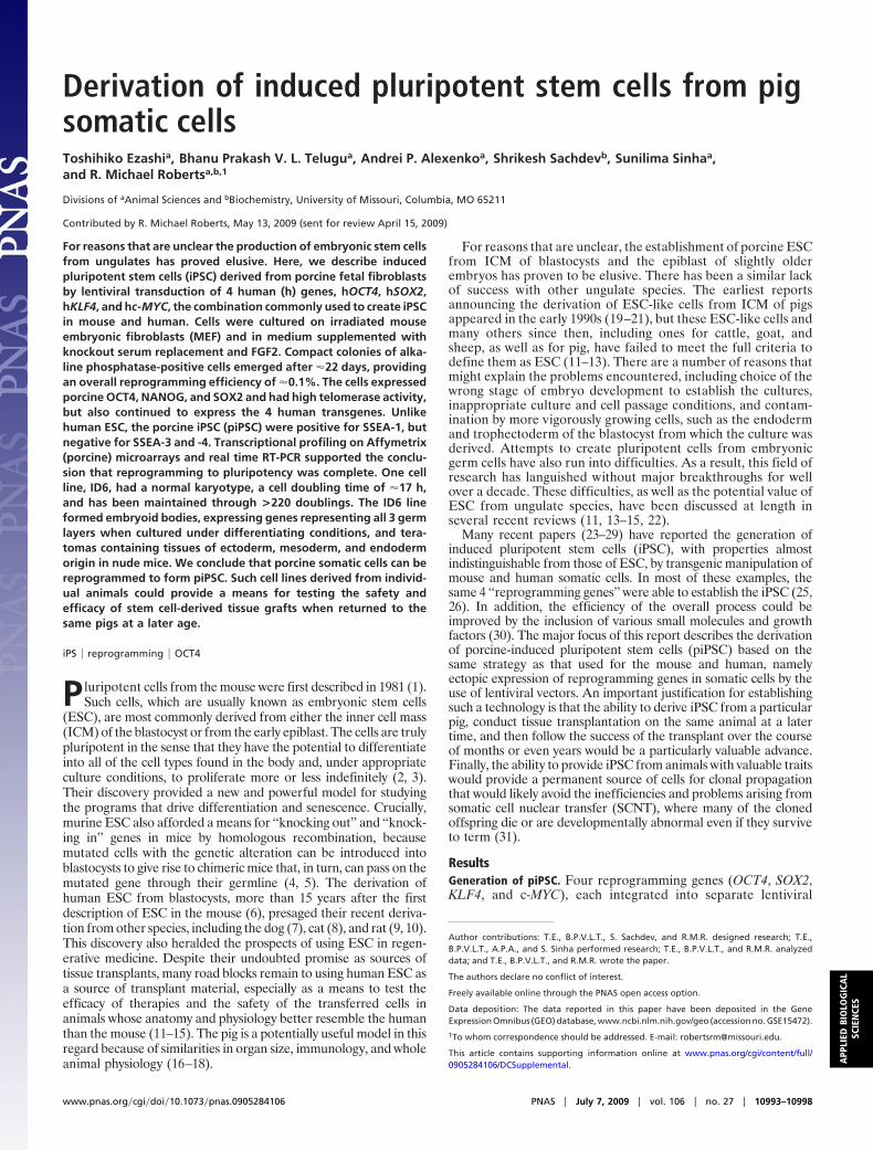
vectors, were introduced into porcine fetal fibroblasts (PFF) andPFF expressing enhanced green fluorescent protein (EGFP)(Fig. 1A and Fig. S1). Twenty-two days after transduction,compact colonies comprised of cells, which were positive foralkaline phosphatase (AP), stage-specific embryonic antigen(SSEA)1, and OCT4, were noted (Fig. 1 B–F). Individualcolonies were mechanically dissociated by using a pulled Pasteurpipette. A colony’s component cells were transferred to freshmedium in 24-well plates coated with irradiated MEF. After �4days, well-developed secondary colonies (Fig. 1C), resemblinghESC colonies, formed. The cells exhibited a high nuclear tocytoplasm ratio with prominently visible nucleoli (Fig. 1D). Thisprotocol was used to isolate a large number of clonal lines (65under 20% O2 conditions, 96 under 4% O2), each derived froma single ESC-like colony. Similar to hESC, areas of large,f lattened cells, presumably undergoing spontaneous differenti-ation, became evident within some colonies, particularly if thecells were not passaged within 5 days (Fig. 1G).
All of the cell lines grew at similar rates, requiring subcultureat a roughly 1:10 ratio every 4–5 days. The ID6 line, the only oneto be carefully examined, showed a doubling time of �17 h.
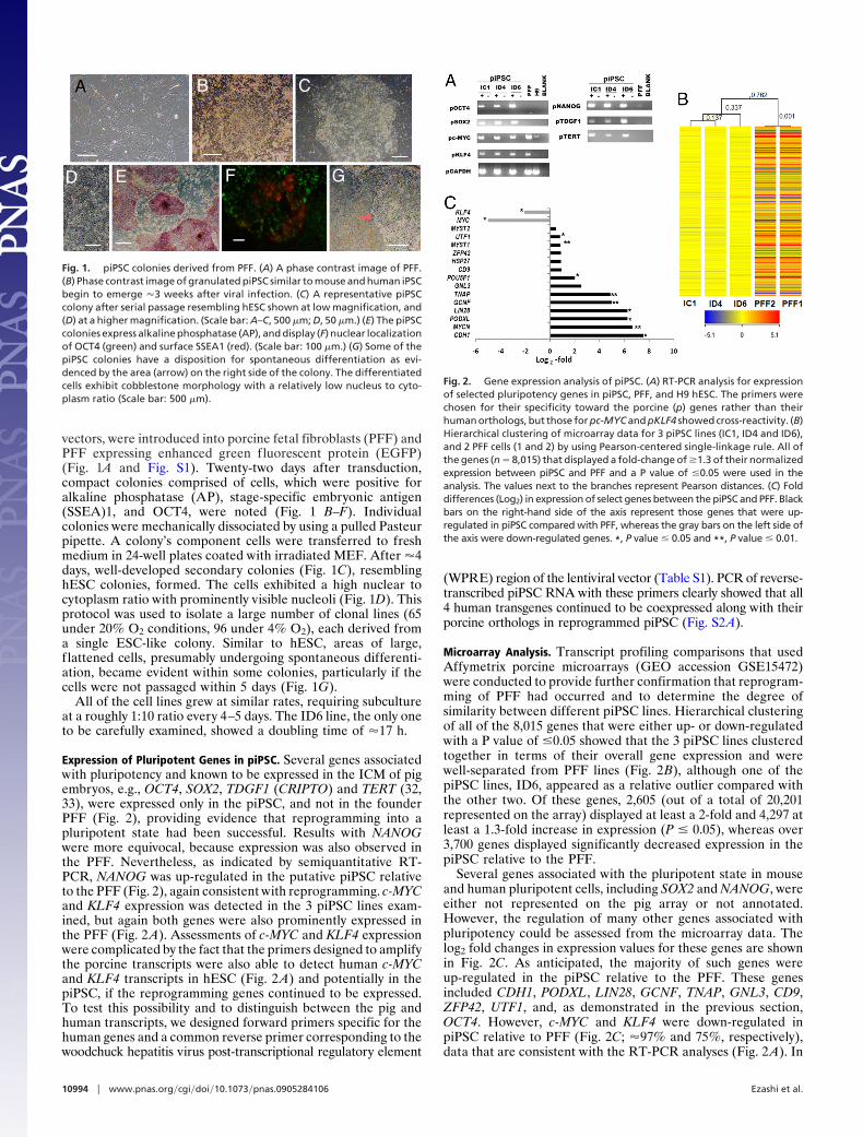
Expression of Pluripotent Genes in piPSC. Several genes associatedwith pluripotency and known to be expressed in the ICM of pigembryos, e.g., OCT4, SOX2, TDGF1 (CRIPTO) and TERT (32,33), were expressed only in the piPSC, and not in the founderPFF (Fig. 2), providing evidence that reprogramming into apluripotent state had been successful. Results with NANOGwere more equivocal, because expression was also observed inthe PFF. Nevertheless, as indicated by semiquantitative RT-PCR, NANOG was up-regulated in the putative piPSC relativeto the PFF (Fig. 2), again consistent with reprogramming. c-MYCand KLF4 expression was detected in the 3 piPSC lines exam-ined, but again both genes were also prominently expressed inthe PFF (Fig. 2 A). Assessments of c-MYC and KLF4 expressionwere complicated by the fact that the primers designed to amplifythe porcine transcripts were also able to detect human c-MYCand KLF4 transcripts in hESC (Fig. 2 A) and potentially in thepiPSC, if the reprogramming genes continued to be expressed.To test this possibility and to distinguish between the pig andhuman transcripts, we designed forward primers specific for thehuman genes and a common reverse primer corresponding to thewoodchuck hepatitis virus post-transcriptional regulatory element
(WPRE) region of the lentiviral vector (Table S1). PCR of reverse-transcribed piPSC RNA with these primers clearly showed that all4 human transgenes continued to be coexpressed along with theirporcine orthologs in reprogrammed piPSC (Fig. S2A).
Microarray Analysis. Transcript profiling comparisons that usedAffymetrix porcine microarrays (GEO accession GSE15472)were conducted to provide further confirmation that reprogram-ming of PFF had occurred and to determine the degree ofsimilarity between different piPSC lines. Hierarchical clusteringof all of the 8,015 genes that were either up- or down-regulatedwith a P value of �0.05 showed that the 3 piPSC lines clusteredtogether in terms of their overall gene expression and werewell-separated from PFF lines (Fig. 2B), although one of thepiPSC lines, ID6, appeared as a relative outlier compared withthe other two. Of these genes, 2,605 (out of a total of 20,201represented on the array) displayed at least a 2-fold and 4,297 atleast a 1.3-fold increase in expression (P � 0.05), whereas over3,700 genes displayed significantly decreased expression in thepiPSC relative to the PFF.
Several genes associated with the pluripotent state in mouseand human pluripotent cells, including SOX2 and NANOG, wereeither not represented on the pig array or not annotated.However, the regulation of many other genes associated withpluripotency could be assessed from the microarray data. Thelog2 fold changes in expression values for these genes are shownin Fig. 2C. As anticipated, the majority of such genes wereup-regulated in the piPSC relative to the PFF. These genesincluded CDH1, PODXL, LIN28, GCNF, TNAP, GNL3, CD9,ZFP42, UTF1, and, as demonstrated in the previous section,OCT4. However, c-MYC and KLF4 were down-regulated inpiPSC relative to PFF (Fig. 2C; �97% and 75%, respectively),data that are consistent with the RT-PCR analyses (Fig. 2 A). In
A B C
D E F G
Fig. 1. piPSC colonies derived from PFF. (A) A phase contrast image of PFF.(B) Phase contrast image of granulated piPSC similar to mouse and human iPSCbegin to emerge �3 weeks after viral infection. (C) A representative piPSCcolony after serial passage resembling hESC shown at low magnification, and(D) at a higher magnification. (Scale bar: A–C, 500 �m; D, 50 �m.) (E) The piPSCcolonies express alkaline phosphatase (AP), and display (F) nuclear localizationof OCT4 (green) and surface SSEA1 (red). (Scale bar: 100 �m.) (G) Some of thepiPSC colonies have a disposition for spontaneous differentiation as evi-denced by the area (arrow) on the right side of the colony. The differentiatedcells exhibit cobblestone morphology with a relatively low nucleus to cyto-plasm ratio (Scale bar: 500 �m).
Fig. 2. Gene expression analysis of piPSC. (A) RT-PCR analysis for expressionof selected pluripotency genes in piPSC, PFF, and H9 hESC. The primers werechosen for their specificity toward the porcine (p) genes rather than theirhuman orthologs, but those for pc-MYC and pKLF4 showed cross-reactivity. (B)Hierarchical clustering of microarray data for 3 piPSC lines (IC1, ID4 and ID6),and 2 PFF cells (1 and 2) by using Pearson-centered single-linkage rule. All ofthe genes (n � 8,015) that displayed a fold-change of �1.3 of their normalizedexpression between piPSC and PFF and a P value of �0.05 were used in theanalysis. The values next to the branches represent Pearson distances. (C) Folddifferences (Log2) in expression of select genes between the piPSC and PFF. Blackbars on the right-hand side of the axis represent those genes that were up-regulated in piPSC compared with PFF, whereas the gray bars on the left side ofthe axis were down-regulated genes. *, P value � 0.05 and **, P value � 0.01.
10994 � www.pnas.org�cgi�doi�10.1073�pnas.0905284106 Ezashi et al.
contrast to c-MYC, mRNA for MYCN was barely detectable inPFF, but displayed a 102-fold up-regulation in piPSC.
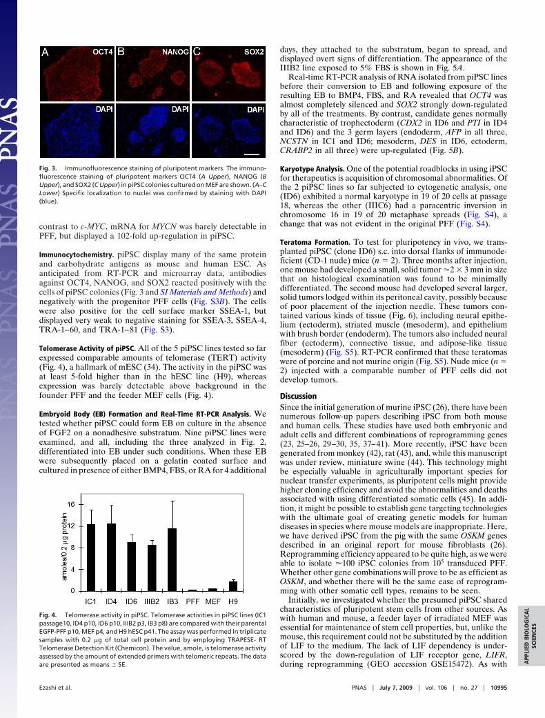
Immunocytochemistry. piPSC display many of the same proteinand carbohydrate antigens as mouse and human ESC. Asanticipated from RT-PCR and microarray data, antibodiesagainst OCT4, NANOG, and SOX2 reacted positively with thecells of piPSC colonies (Fig. 3 and SI Materials and Methods) andnegatively with the progenitor PFF cells (Fig. S3B). The cellswere also positive for the cell surface marker SSEA-1, butdisplayed very weak to negative staining for SSEA-3, SSEA-4,TRA-1–60, and TRA-1–81 (Fig. S3).
Telomerase Activity of piPSC. All of the 5 piPSC lines tested so farexpressed comparable amounts of telomerase (TERT) activity(Fig. 4), a hallmark of mESC (34). The activity in the piPSC wasat least 5-fold higher than in the hESC line (H9), whereasexpression was barely detectable above background in thefounder PFF and the feeder MEF cells (Fig. 4).
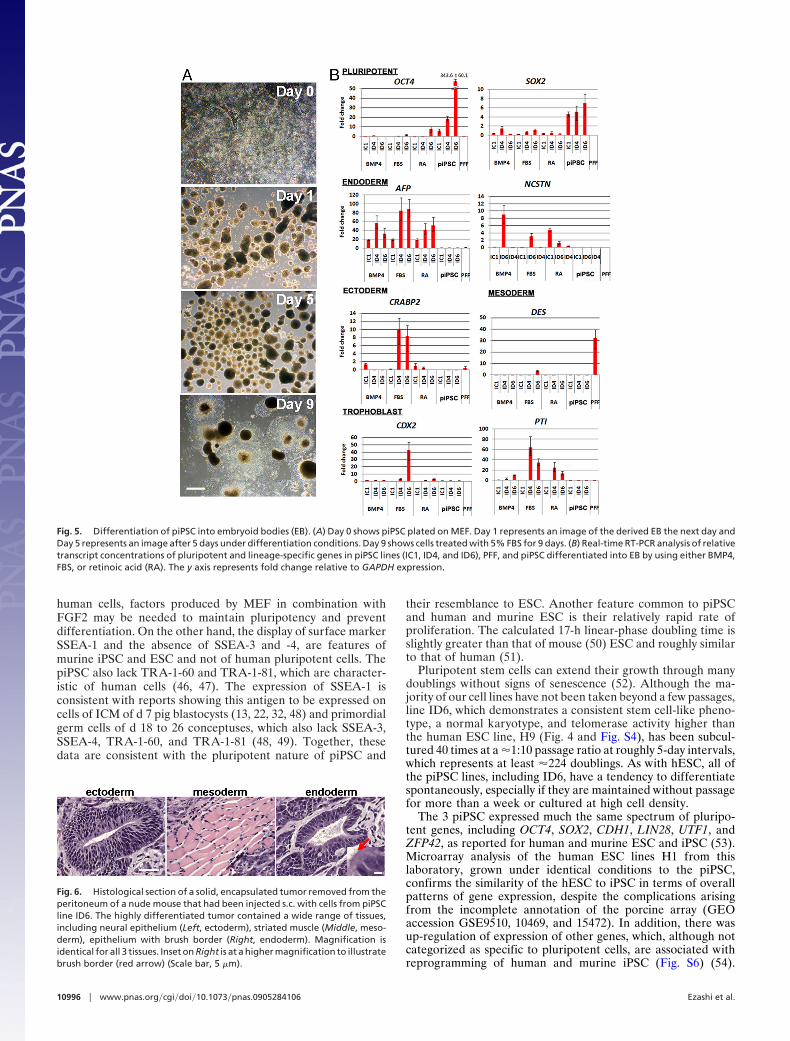
Embryoid Body (EB) Formation and Real-Time RT-PCR Analysis. Wetested whether piPSC could form EB on culture in the absenceof FGF2 on a nonadhesive substratum. Nine piPSC lines wereexamined, and all, including the three analyzed in Fig. 2,differentiated into EB under such conditions. When these EBwere subsequently placed on a gelatin coated surface andcultured in presence of either BMP4, FBS, or RA for 4 additional
days, they attached to the substratum, began to spread, anddisplayed overt signs of differentiation. The appearance of theIIIB2 line exposed to 5% FBS is shown in Fig. 5A.
Real-time RT-PCR analysis of RNA isolated from piPSC linesbefore their conversion to EB and following exposure of theresulting EB to BMP4, FBS, and RA revealed that OCT4 wasalmost completely silenced and SOX2 strongly down-regulatedby all of the treatments. By contrast, candidate genes normallycharacteristic of trophectoderm (CDX2 in ID6 and PTI in ID4and ID6) and the 3 germ layers (endoderm, AFP in all three,NCSTN in IC1 and ID6; mesoderm, DES in ID6, ectoderm,CRABP2 in all three) were up-regulated (Fig. 5B).
Karyotype Analysis. One of the potential roadblocks in using iPSCfor therapeutics is acquisition of chromosomal abnormalities. Ofthe 2 piPSC lines so far subjected to cytogenetic analysis, one(ID6) exhibited a normal karyotype in 19 of 20 cells at passage18, whereas the other (IIIC6) had a paracentric inversion inchromosome 16 in 19 of 20 metaphase spreads (Fig. S4), achange that was not evident in the original PFF (Fig. S4).
Teratoma Formation. To test for pluripotency in vivo, we trans-planted piPSC (clone ID6) s.c. into dorsal f lanks of immunode-ficient (CD-1 nude) mice (n � 2). Three months after injection,one mouse had developed a small, solid tumor �2 � 3 mm in sizethat on histological examination was found to be minimallydifferentiated. The second mouse had developed several larger,solid tumors lodged within its peritoneal cavity, possibly becauseof poor placement of the injection needle. These tumors con-tained various kinds of tissue (Fig. 6), including neural epithe-lium (ectoderm), striated muscle (mesoderm), and epitheliumwith brush border (endoderm). The tumors also included neuralfiber (ectoderm), connective tissue, and adipose-like tissue(mesoderm) (Fig. S5). RT-PCR confirmed that these teratomaswere of porcine and not murine origin (Fig. S5). Nude mice (n �2) injected with a comparable number of PFF cells did notdevelop tumors.
DiscussionSince the initial generation of murine iPSC (26), there have beennumerous follow-up papers describing iPSC from both mouseand human cells. These studies have used both embryonic andadult cells and different combinations of reprogramming genes(23, 25–26, 29–30, 35, 37–41). More recently, iPSC have beengenerated from monkey (42), rat (43), and, while this manuscriptwas under review, miniature swine (44). This technology mightbe especially valuable in agriculturally important species fornuclear transfer experiments, as pluripotent cells might providehigher cloning efficiency and avoid the abnormalities and deathsassociated with using differentiated somatic cells (45). In addi-tion, it might be possible to establish gene targeting technologieswith the ultimate goal of creating genetic models for humandiseases in species where mouse models are inappropriate. Here,we have derived iPSC from the pig with the same OSKM genesdescribed in an original report for mouse fibroblasts (26).Reprogramming efficiency appeared to be quite high, as we wereable to isolate �100 iPSC colonies from 105 transduced PFF.Whether other gene combinations will prove to be as efficient asOSKM, and whether there will be the same ease of reprogram-ming with other somatic cell types, remains to be seen.
Initially, we investigated whether the presumed piPSC sharedcharacteristics of pluripotent stem cells from other sources. Aswith human and mouse, a feeder layer of irradiated MEF wasessential for maintenance of stem cell properties, but, unlike themouse, this requirement could not be substituted by the additionof LIF to the medium. The lack of LIF dependency is under-scored by the down-regulation of LIF receptor gene, LIFR,during reprogramming (GEO accession GSE15472). As with
Fig. 3. Immunofluorescence staining of pluripotent markers. The immuno-fluorescence staining of pluripotent markers OCT4 (A Upper), NANOG (BUpper), and SOX2 (C Upper) in piPSC colonies cultured on MEF are shown. (A–CLower) Specific localization to nuclei was confirmed by staining with DAPI(blue).
Fig. 4. Telomerase activity in piPSC. Telomerase activities in piPSC lines (IC1passage10, ID4 p10, ID6 p10, IIIB2 p3, IB3 p8) are compared with their parentalEGFP-PFF p10, MEF p4, and H9 hESC p41. The assay was performed in triplicatesamples with 0.2 �g of total cell protein and by employing TRAPESE- RTTelomerase Detection Kit (Chemicon). The value, amole, is telomerase activityassessed by the amount of extended primers with telomeric repeats. The dataare presented as means � SE.
Ezashi et al. PNAS � July 7, 2009 � vol. 106 � no. 27 � 10995
APP
LIED
BIO
LOG
ICA
LSC
IEN
CES
human cells, factors produced by MEF in combination withFGF2 may be needed to maintain pluripotency and preventdifferentiation. On the other hand, the display of surface markerSSEA-1 and the absence of SSEA-3 and -4, are features ofmurine iPSC and ESC and not of human pluripotent cells. ThepiPSC also lack TRA-1-60 and TRA-1-81, which are character-istic of human cells (46, 47). The expression of SSEA-1 isconsistent with reports showing this antigen to be expressed oncells of ICM of d 7 pig blastocysts (13, 22, 32, 48) and primordialgerm cells of d 18 to 26 conceptuses, which also lack SSEA-3,SSEA-4, TRA-1-60, and TRA-1-81 (48, 49). Together, thesedata are consistent with the pluripotent nature of piPSC and
their resemblance to ESC. Another feature common to piPSCand human and murine ESC is their relatively rapid rate ofproliferation. The calculated 17-h linear-phase doubling time isslightly greater than that of mouse (50) ESC and roughly similarto that of human (51).
Pluripotent stem cells can extend their growth through manydoublings without signs of senescence (52). Although the ma-jority of our cell lines have not been taken beyond a few passages,line ID6, which demonstrates a consistent stem cell-like pheno-type, a normal karyotype, and telomerase activity higher thanthe human ESC line, H9 (Fig. 4 and Fig. S4), has been subcul-tured 40 times at a �1:10 passage ratio at roughly 5-day intervals,which represents at least �224 doublings. As with hESC, all ofthe piPSC lines, including ID6, have a tendency to differentiatespontaneously, especially if they are maintained without passagefor more than a week or cultured at high cell density.
The 3 piPSC expressed much the same spectrum of pluripo-tent genes, including OCT4, SOX2, CDH1, LIN28, UTF1, andZFP42, as reported for human and murine ESC and iPSC (53).Microarray analysis of the human ESC lines H1 from thislaboratory, grown under identical conditions to the piPSC,confirms the similarity of the hESC to iPSC in terms of overallpatterns of gene expression, despite the complications arisingfrom the incomplete annotation of the porcine array (GEOaccession GSE9510, 10469, and 15472). In addition, there wasup-regulation of expression of other genes, which, although notcategorized as specific to pluripotent cells, are associated withreprogramming of human and murine iPSC (Fig. S6) (54).
Fig. 5. Differentiation of piPSC into embryoid bodies (EB). (A) Day 0 shows piPSC plated on MEF. Day 1 represents an image of the derived EB the next day andDay 5 represents an image after 5 days under differentiation conditions. Day 9 shows cells treated with 5% FBS for 9 days. (B) Real-time RT-PCR analysis of relativetranscript concentrations of pluripotent and lineage-specific genes in piPSC lines (IC1, ID4, and ID6), PFF, and piPSC differentiated into EB by using either BMP4,FBS, or retinoic acid (RA). The y axis represents fold change relative to GAPDH expression.
Fig. 6. Histological section of a solid, encapsulated tumor removed from theperitoneum of a nude mouse that had been injected s.c. with cells from piPSCline ID6. The highly differentiated tumor contained a wide range of tissues,including neural epithelium (Left, ectoderm), striated muscle (Middle, meso-derm), epithelium with brush border (Right, endoderm). Magnification isidentical for all 3 tissues. Inset on Right is at a higher magnification to illustratebrush border (red arrow) (Scale bar, 5 �m).
10996 � www.pnas.org�cgi�doi�10.1073�pnas.0905284106 Ezashi et al.
Further evidence that the PFF had been efficiently repro-grammed is the very low expression of the differentiation-associated, fibroblast-specific gene, THY1, in piPSC, which israpidly down-regulated when murine fibroblasts are repro-grammed (54). To our surprise, porcine c-MYC and KLF4 weredown-regulated, whereas MYCN was strongly up-regulated inpiPSC compared with PFF (Fig. 2B), but examination of themicroarray data of others (23, 54) revealed that a similarphenomenon has been observed in reprogramming of murineand human cells. Finally, pNANOG was up-regulated duringreprogramming, although it was already expressed in the paren-tal PFF cells, an observation consistent with previous reportsthat NANOG is not a reliable marker for pluripotency in pig (32).
Until recently most iPSC have been generated by usingintegrating retroviral vectors, leading in some cases, at least, tothe continued expression of the transgenes in the reprogrammedcells (29, 55–59). As the ectopically expressed reprogramminggenes may cause imbalance in the pluripotency network and eveninfluence the ability of reprogrammed cells to differentiateoptimally (29, 54, 59), ideally the genes should either not bepermitted to integrate in the first place (60–62) or excised oncereprogramming has been completed (59, 63). A more recentstrategy has been to provide the reprogramming proteins them-selves directly to the targeted cell type (64) Because retroviruseswere used in our experiments, not unexpectedly, transcripts forthe 4 transgenes remained detectable in the piPSC (Fig. S2), justas they did in the iPSC established from PFF of miniature swine(44). Although such expression is not desirable, an impairedability to differentiate was not a feature of iPSC described here,which were fully capable of generating EB expressing genesindicative of the 3 germ layers and trophoblast (Fig. 5B) and ofproducing highly differentiated teratomas in immune-compromised mice (Fig. 6 and Fig. S5). Nevertheless, if thesecells are to be tested in pigs to mimic therapeutic applications inhuman patients, it may be necessary to ensure that transgeneexpression is eliminated completely from the donor lines used byusing one or other of the strategies described above.
Materials and MethodsCell Culture and Lentiviral Transduction of Human Factors. The ORFs of thehuman (h) SOX2, hKLF4, and hc-MYC were cloned into the FUGW lentiviralvector (65), and the hOCT4 cDNA was cloned into pSIN18cPPt.hEF1a.EGFP.WPRE (66) (Fig. S1). Pseudovirus was produced in human293FT cells (Invitrogen) by transfection with each lentiviral vector along withthe VSV-G envelope (pMD2.G) and packaging vector (psPAX2) (67) by usingLipofectamine and PLUS reagents (Invitrogen). Titered virus (68) was used toinfect the target cells (PFF or EGFP-PFF; 1 � 105 cells/35-mm dish). On day 2after infection, the cells were dispersed with trypsin and transferred to 10-cmplates seeded with irradiated MEF. Subsequently, cells were maintained on aculture medium standardized for human ESC (69) containing 4 ng/ml humanFGF2 (Peprotech).
Alkaline Phosphatase (AP) Staining and Immunocytochemistry (ICC). AP stainingwas performed with the AP detection kit (Chemicon). For immunocytochem-istry, cells were fixed in 2% paraformaldehyde in PBS for 20 min at roomtemperature (RT), washed, and exposed to either 5% goat or donkey serum(Sigma), 1% BSA (Jackson-Immunoresearch), and 0.1% Triton X-100 (Fisher) inPBS for 30 min. The cells were then incubated with primary antibody (SIMaterials and Methods) overnight at 4 °C. After washing, the cells wereincubated with secondary antibody (SI Materials and Methods). Nuclei werestained with 6 ng/ml DAPI (Invitrogen).
Transcriptional Profiling by Microarray. RNA from 3 piPSC and 2 PFF lines wasextracted by using STAT-60 (IsoTex Diagnostics). Ten-�g of each sample wassubjected to double-stranded cDNA synthesis and labeling by using the Ge-
neChip Expression 3�-Amplification IVT Labeling Kit (Affymetrix), according tomanufacturer’s instructions. Biotin-labeled cRNA was fragmented and hybrid-ized to the Affymetrix GeneChip Porcine Genome Array. Microarrays werescanned (GeneChip Scanner 3000; Affymetrix), and data were assembled byMicroArray Suite 5.0 software (Affymetrix). The output data were analyzedwith GeneSpring GX analysis software (Agilent). The CEL files for each arraywere normalized, and false discovery rates were calculated by T test unpaired,unequal variance with asymptotic p-values and Benjamini-Hochberg multipletesting correction options. Fold-changes in pairwise comparison of the piPSCand PFF data were calculated based on the unadjusted data means to obtaina list of 8,015 genes with an acceptable fold-change of 1.3. Clustering wasperformed by the Pearson-centered similarity measure and single linkage rule.Because the porcine array remains poorly annotated, additional annotationwas performed by using the information of Tsai et al. (70) (Gene ExpressionOmnibus accession no. GPL6472) and an annotated file provided by Dr. C. K.Tuggle (Iowa State University, Ames, IA) (36).
Embryoid Body (EB) Formation. Colonies were detached from the MEF feederlayer by manual dissection and transferred into hESC medium without FGF2(differentiation medium) in low attachment plates (Corning). After 5 days ofculture, the EB were transferred onto adherent, gelatin-coated tissue-culturedishes and were cultured in differentiation medium supplemented with either10 ng/ml BMP4 (R & D Systems), 5% FBS, or 0.5 �M all-trans retinoic acid (RA;Sigma) and harvested for RNA isolation 9 days later.
Real-Time RT-PCR Analysis. Real-time RT-PCR was performed on RNA extractedfrom EB (at day 9), undifferentiated piPSC, and PFF to assess the relativeconcentrations of genes from the 3 germ layers [AFP and NCSTN (endoderm),CRABP2 (ectoderm), and DES (mesoderm)] as well as pluripotent (OCT4 andSOX2), and trophoblast genes (CDX2 and PTI). A reference sample constitutedby pooling RNA from all of the samples was also used in the analysis. Three �gof DNase-treated RNA was used in the first strand cDNA synthesis (qScriptcDNA synthesis kit; Quanta Biosciences). Validated primer sets for real-timePCR analysis are listed in Table S1. Expression of candidate genes relative toGAPDH in the test was determined with SYBR Green PCR Master Mix (AppliedBiosystems) on an ABI PRISM 7500 Real Time System (Applied Biosystems). Theconditions for real-time RT-PCR were as follows: 50 °C, 2 min; 95 °C,10 min;followed by 40 amplification cycles (95 °C,15 s; 60 °C (62 °C for OCT4), 30 s).The reaction was terminated by an elongation and data acquisition step at75 °C for 1 min. The relative expression ratio of target genes was calculated by��Ct method. The specificity of the PCR products amplified was confirmed bydissociation curve analysis.
Telomerase Activity Assay. Telomerase activity in 0.2-�g cell protein fromrepresentative piPSC, MEFs, and HESC was determined by using TRAPEZE RTTelomerase Detection Kit (Chemicon) with Platinum TaqDNA polymerase(Invitrogen) according to manufacturer’s instructions.
Teratoma Formation. 106 ID6 cells from a confluent 35-mm dish were injecteds.c. into dorsal flanks of two 4-week-old CD-1 nude mice, Crl: CD-1-Foxn1nu
(Charles River). Tumors were harvested 3 months after injection, dissected,and fixed in 4% paraformaldehyde. Paraffin-embedded tissue was sectionedand stained with hematoxylin and eosin. All animal experiments were ap-proved by the Institutional Animal Care and Use Committee under ProtocolNo. 4467. Control animals were injected with an equivalent number of PFFcells.
ACKNOWLEDGMENTS. We thank Dr. Randall Prather and Lee Spate for provid-ing EGFP-PFF and PFF, Drs. Michal Gropp and Benjamin Reubinoff (HadassahUniversity Medical Center, Jerusalem, Israel) for the pSIN18.cPPT,hEF1a.EGFP.WPRE plasmid, Dr. David Baltimore (California Institute of Technol-ogy, Pasadena, CA) for the FUGW and VSVG envelope plasmids, Dr. Didier Trono(Ecole Polytechnique Federale de Lausanne, Lausanne, Switzerland) for the ps-PAX2 packaging plasmid, and Dr. Christopher K. Tuggle (Iowa State University,Ames, IA) for information on annotation of porcine EST sequences. Kristin Whit-worth assisted in the microarray analysis and Dr. Kei Kuroki (Veterinary MedicalDiagnostic Laboratory, University of Missouri, Columbia, Missouri) provided his-tological analysis of the teratomas. This work was supported by National Insti-tutes of Health Grants HD42201 and HD21896 (to R.M.R), and Missouri LifeSciences Trust Fund Grant 00022147 (to T.E.).
1. Evans MJ, Kaufman MH (1981) Establishment in culture of pluripotential cells frommouse embryos. Nature 292:154–156.
2. Evans M (2005) Embryonic stem cells: A perspective. Novartis Found Symp 265:98–103,and discussion (2005) 265:103–106, 265:122–128.
3. Wobus AM, Boheler KR (2005) Embryonic stem cells: Prospects for developmentalbiology and cell therapy. Physiol Rev 85:635–678.
4. Thomas KR, Folger KR, Capecchi MR (1986) High frequency targeting of genes tospecific sites in the mammalian genome. Cell 44:419–428.
Ezashi et al. PNAS � July 7, 2009 � vol. 106 � no. 27 � 10997
APP
LIED
BIO
LOG
ICA
LSC
IEN
CES

5. Doetschman T, et al. (1987) Targetted correction of a mutant HPRT gene in mouseembryonic stem cells. Nature 330:576–578.
6. Thomson JA, et al. (1998) Embryonic stem cell lines derived from human blastocysts.Science 282:1145–1147.
7. Hatoya S, et al. (2006) Isolation and characterization of embryonic stem-like cells fromcanine blastocysts. Mol Reprod Dev 73:298–305.
8. Yu X, et al. (2008) Isolation and characterization of embryonic stem-like cells derivedfrom in vivo-produced cat blastocysts. Mol Reprod Dev 75:1426–1432.
9. Buehr M, et al. (2008) Capture of authentic embryonic stem cells from rat blastocysts.Cell 135:1287–1298.
10. Li P, et al. (2008) Germline competent embryonic stem cells derived from rat blastocysts.Cell 135:1299–1310.
11. Vackova I, Ungrova A, Lopes F (2007) Putative embryonic stem cell lines from pigembryos. J Reprod Dev 53:1137–1149.
12. Keefer CL, Pant D, Blomberg L, Talbot NC (2007) Challenges and prospects for theestablishment of embryonic stem cell lines of domesticated ungulates. Anim ReprodSci 98:147–168.
13. Brevini TA, et al. (2007) Porcine embryonic stem cells: Facts, challenges and hopes.Theriogenology 68(Suppl 1):S206–S213.
14. Talbot NC, Blomberg le A (2008) The pursuit of ES cell lines of domesticated ungulates.Stem Cell Rev 4:235–254.
15. Hall V (2008) Porcine embryonic stem cells: A possible source for cell replacementtherapy. Stem Cell Rev 4:275–282.
16. Prather RS, et al. (2003) Transgenic swine for biomedicine and agriculture. Theriog-enology 59:115–123.
17. Brandl U, et al. (2007) Transgenic animals in experimental xenotransplantation models:Orthotopic heart transplantation in the pig-to-baboon model. Transplant Proc 39:577–578.
18. Piedrahita JA, Mir B (2004) Cloning and transgenesis in mammals: Implications forxenotransplantation. Am J Transplant 4(Suppl 6):43–50.
19. Notarianni E, Laurie S, Moor RM, Evans MJ (1990) Maintenance and differentiation inculture of pluripotential embryonic cell lines from pig blastocysts. J Reprod Fertil Suppl41:51–56.
20. Piedrahita JA, Anderson GB, Bondurant RH (1990) On the isolation of embryonic stemcells: Comparative behavior of murine, porcine, and ovine embryos. Theriogenology34:879–901.
21. Strojek RM, Reed MA, Hoover JL, Wagner TE (1990) A method for cultivating morpho-logically undifferentiated embryonic stem cells from porcine blastocysts. Theriogenol-ogy 33:901–913.
22. Brevini TA, et al. (2007) Derivation and characterization of pluripotent cell lines frompig embryos of different origins. Theriogenology 67:54–63.
23. Lowry WE, et al. (2008) Generation of human induced pluripotent stem cells fromdermal fibroblasts. Proc Natl Acad Sci USA 105:2883–2888.
24. Nakagawa M, et al. (2008) Generation of induced pluripotent stem cells without Mycfrom mouse and human fibroblasts. Nat Biotech 26:101–106.
25. Takahashi K, et al. (2007) Induction of pluripotent stem cells from adult humanfibroblasts by defined factors. Cell 131:861–872.
26. Takahashi K, Yamanaka S (2006) Induction of pluripotent stem cells from mouseembryonic and adult fibroblast cultures by defined factors. Cell 126:663–676.
27. Park I-H, et al. (2008) Reprogramming of human somatic cells to pluripotency withdefined factors. Nature 451:141–146.
28. Kim JB, et al. (2008) Pluripotent stem cells induced from adult neural stem cells byreprogramming with two factors. Nature 454:646–650.
29. Yu J, et al. (2007) Induced pluripotent stem cell lines derived from human somatic cells.Science 318:1917–1920.
30. Amabile G, Meissner A (2009) Induced pluripotent stem cells: Current progress andpotential for regenerative medicine. Trends Mol Med 15:59–68.
31. Prather RS, Sutovsky P, Green JA (2004) Nuclear remodeling and reprogramming intransgenic pig production. Exp Biol Med 229:1120–1126.
32. Blomberg L, Schreier LL, Talbot NC (2008) Expression analysis of pluripotency factors inthe undifferentiated porcine inner cell mass and epiblast during in vitro culture. MolReprod Dev 75:450–463.
33. Magnani L, Cabot RA (2008) In vitro and in vivo derived porcine embryos possesssimilar, but not identical, patterns of Oct4, Nanog, and Sox2 mRNA expression duringcleavage development Mol Reprod Dev 75:1726–1735.
34. Armstrong L, et al. (2000) mTert expression correlates with telomerase activity duringthe differentiation of murine embryonic stem cells. Mech Dev 97:109–116.
35. Aoi T, et al. (2008) Generation of pluripotent stem cells from adult mouse liver andstomach cells. Science 321:699–702.
36. Wang Y, et al. (2008) Analysis of porcine transcriptional response to Salmonellaenterica serovar Choleraesuis suggests novel targets of NFkappaB are activated in themesenteric lymph node. BMC Genomics 9:437.
37. Park IH, et al. (2008) Generation of human-induced pluripotent stem cells. Nat Protoc3:1180–1186.
38. Maherali N, et al. (2007) Directly reprogrammed fibroblasts show global epigeneticremodeling and widespread tissue contribution. Cell Stem Cell 1:55–70.
39. Wernig M, et al. (2007) In vitro reprogramming of fibroblasts into a pluripotentES-cell-like state. Nature 448:318–324.
40. Okita K, Ichisaka T, Yamanaka S (2007) Generation of germline-competent inducedpluripotent stem cells. Nature 448:313–317.
41. Feng B, et al. (2009) Reprogramming of fibroblasts into induced pluripotent stem cellswith orphan nuclear receptor Esrrb. Nat Cell Biol 11:197–203.
42. Liu H, et al. (2008) Generation of induced pluripotent stem cells from adult rhesusmonkey fibroblasts. Cell Stem Cell 3:587–590.
43. Liao J, et al. (2009) Generation of induced pluripotent stem cell lines from adult ratcells. Cell Stem Cell 4:11–15.
44. Esteban MA, et al. (April 17, 2009) Generation of induced pluripotent stem cell linesfrom tibetan miniature pig. J Biol Chem, 10.1074/jbc.M109.008938.
45. Oback B, Wells DN (2007) Donor cell differentiation, reprogramming, and cloningefficiency: Elusive or illusive correlation? Mol Reprod Dev 74:646–654.
46. Draper JS, Pigott C, Thomson JA, Andrews PW (2002) Surface antigens of humanembryonic stem cells: Changes upon differentiation in culture. J Anat 200:249–258.
47. O’Connor MD, et al. (2008) Alkaline phosphatase-positive colony formation is a sen-sitive, specific, and quantitative indicator of undifferentiated human embryonic stemcells. Stem Cells 26:1109–1116.
48. Wianny F, Perreau C, Hochereau de Reviers MT (1997) Proliferation and differentiationof porcine inner cell mass and epiblast in vitro. Biol Reprod 57:756–764.
49. Takagi Y, Talbot NC, Rexroad CE, Jr, Pursel VG (1997) Identification of pig primordialgerm cells by immunocytochemistry and lectin binding. Mol Reprod Dev 46:567–580.
50. Udy GB, Parkes BD, Wells DN (1997) ES cell cycle rates affect gene targeting frequencies.Exp Cell Res 231:296–301.
51. Becker KA, et al. (2006) Self-renewal of human embryonic stem cells is supported by ashortened G1 cell cycle phase. J Cell Physiol 209:883–893.
52. Zeng X (2007) Human embryonic stem cells: Mechanisms to escape replicative senes-cence? Stem Cell Rev 3:270–279.
53. Ginis I, et al. (2004) Differences between human and mouse embryonic stem cells. DevBiol 269:360–380.
54. Sridharan R, et al. (2009) Role of the murine reprogramming factors in the inductionof pluripotency. Cell 136:364–377.
55. Dimos JT, et al. (2008) Induced pluripotent stem cells generated from patients with ALScan be differentiated into motor neurons. Science 321:1218–1221.
56. Ebert AD, et al. (2009) Induced pluripotent stem cells from a spinal muscular atrophypatient. Nature 457:277–280.
57. Hockemeyer D, et al. (2008) A drug-inducible system for direct reprogramming ofhuman somatic cells to pluripotency. Cell Stem Cell 3:346–353.
58. Park IH, et al. (2008) Disease-specific induced pluripotent stem cells. Cell 134:877–886.59. Soldner F, et al. (2009) Parkinson’s disease patient-derived induced pluripotent stem
cells free of viral reprogramming factors. Cell 136:964–977.60. Okita K, et al. (2008) Generation of mouse induced pluripotent stem cells without viral
vectors. Science 322:949–953.61. Stadtfeld M, et al. (2008) Induced pluripotent stem cells generated without viral
integration. Science 322:945–949.62. Yu J, et al. (2009) Human induced pluripotent stem cells free of vector and transgene
sequences. Science 324:797–801.63. Yusa K, Rad R, Takeda J, Bradley A (2009) Generation of transgene-free induced
pluripotent mouse stem cells by the piggyBac transposon. Nat Methods 6:363–369.64. Zhou H, et al. (2009) Generation of induced pluripotent stem cells using recombinant
proteins. Cell Stem Cell 4(5):381–384.65. Lois C, et al. (2002) Germline transmission and tissue-specific expression of transgenes
delivered by lentiviral vectors. Science 295:868–872.66. Gropp M, et al. (2003) Stable genetic modification of human embryonic stem cells by
lentiviral vectors. Mol Ther 7:281–287.67. Szulc J, et al. (2006) A versatile tool for conditional gene expression and knockdown.
Nat Methods 3:109–11668. Lizee G, et al. (2003) Real-time quantitative reverse transcriptase-polymerase chain
reaction as a method for determining lentiviral vector titers and measuring transgeneexpression. Hum Gene Ther 14:497–507.
69. Amit M, et al. (2000) Clonally derived human embryonic stem cell lines maintainpluripotency and proliferative potential for prolonged periods of culture. Dev Biol227:271.
70. Tsai S, et al. (2006) Annotation of the Affymetrix porcine genome microarray. AnimGenet 37:423–424.
10998 � www.pnas.org�cgi�doi�10.1073�pnas.0905284106 Ezashi et al.