dermatology for the small animal practitioner (made easy series)
TRANSCRIPT
Dermatologyfor theS mall Anima lPra ctitioner
Ral£ S. MuellerD r. rne d .ver.,MACYSc, D ;pA YO, FACYSc
Made Easy SeriesTeton NevvMedia
.. I··'•• . 1Compliments of Eukanuba· ••
VETERINARY DIETS lAMS '~
Dermatology fo r the Small Anima l P ractitio n e r
by R a lf S . Mue lle r, Dr. med.vet .• MACVSc, OipAC VO, FAC V Sc
Teton NewMedia Jackson , W yoming 83001
Executive Editor: Carroll C . Cann
Develo pment Editor: S usan L. Hunsbe rger
Editor: Cynthia J. Roa ntree
Design and Layo ut: Anita Ba ran Sykes
Printer: Grand Teton Lithograp h y, Jackson, WY
Teto n NewMedia
P.O. Bo x 4833
125 South King Street
Jackson, WY 83001
1-888-770-3 165
http://w\Vw.teto nnm.com
Interior pho t ographs by RalfS. Mue lle r (unless o therwise n o ted)
Copyright © 2000 Teto n NewMed ia
At! rights reserved. This book is protected by copyr igh t. No part of thi s book may be reprod uced in an y fo rm o r for :-lny mf'.::ms , inr.lLKling pho rncopying, n r LLrili zed hy any information storage and re tri eva l system s witho ut written permission fro m the copyri ght o wner.
The authors a nd publisher h ave made eve ry effort to provide an accurate refe re n ce te xt. H owever, they sh a t! n o t be h e ld responsible for prob lem.s arisi.ng from e rrors o r o miss io n s, o r fro m misunderstandings o n the part of the reader.
PRINTED IN THE UNITED STATES O F AMERICA
IS BN # 1-893441-06-7
Print numbe r 5 4 3 2 1
Library o f Congress Catalogin g~ in ~ Publicat i on Data
Mueller, Ra lf S. Dermato logy fo r the smat! animal practition e r I Ralf S. Muelle r.
p. cm . ~~ (Made easy series) ISBN 1-893441-06-7 (a lk. pape ,)
1. Veterinary dermatology. 2 . Dogs~~Diseases~~Treatment . 3. Cats~~Diseases~~Treatmen.t. I. Title . I I. Made easy series (Jackson, Wyo.)
S F992.S55 M83 2000 636.089'65--dc21
00-059976

Dedication
To m y wife , pa rme r, and best fr iend, Son ya Bette n ay, whose contin
ued love, support, a nd feedback h as helped Il"le tre m e ndo usly over d1.e
years pe rson a ll y and pro fess io n a ll y. With o ut h e r this book wo uLd not
have been poss ib le.
To m y pa re nts , Irmi and S ig i, wh o encouraged a nd suppo rted me
w h e n I le ft Ge rtTla n y to le a rn Ill.o re abo ll t ve te ri n a ry d e rm a t o l.ogy
and wh o yea rs later Looked after the childre n to g ive m e tim e to
write this book .
To m y c hildre n , An ya a nd Flo rian, wh o se inte rest a nd pe rs iste n ce in
lea rning a re a constant !5ource of arnazenle nt a nd inspi ra ti o n to m e.
To m y m.e nto rs, Pe te r Ihrke and T o n y S t a nna rd , a nd to a ll th e co l
leagues w h ose suppo rt a llowed me to d e velo p the kno w led ge and
expe ri e nce that I h o pe w ill m a ke this book usefu l in SIn a l! a nilna l
prac tice . S pecific thanks to Drs. C arol Fo il , Ga il Kunkle, Ke rr ie La y,
H e le n Po we r, Dav id Ro bson , a n d Linda Vogel n est, a nd the ed ito ria l
team w ith C indy Roan tree, S usan Hunsbe rge r, a n d R a y Luke n s fo r
the ir input a nd to D rs. Son ya Be ttena y, Peter Ih rke , Th ie rry O li vry,
W a yne R osenkrantz, and Michael S hi pst o n e fo r prov iding some o f
the pi c t u res .
•
Preface
Dear Colleagues,
Veterinary derm:=ttology plays an itupo rra nt role in slna ll a ninla i
p ract ice. S kin proble lTIS a re the m ost freq ue nt presenting compla int
in ma n y practices but due to th.e il' o fte n c hron ic nature m ay ca lise
fru s tra tion for veterina ri a ns , clie nts, a nd patients a li ke.
Thi s book is a prac tica l. introductio n to veterina ry dernl a to logy for
the busy snla il a nim a l prac titio n e r. It w ill h e lp YO LI to di.agn ose a nd
tnanage skin diseases seen eve ry day in your practice a n d a llo w you
to pe rfo rm a so lid workup in pa tients with ra re o r complicated skin
diseases that Ill.ay require further read ing, referra l to, o r advice from
a veterinary dennatologist.
M ost o f a ll , I h ope it wi ll a llow you to e njoy your dermatology
cases, provide good se rvice, and improve the quality of life in
your pa tie nts.
Beca use your opini o n s a nd concerns are impo rtant in 111aking this
boo k ITIOS t usefu l for th e s lTla ll anima l prac t iti o ner. I wou ld apprec i;
ate it if you wou ld m a i l o r e mail [TIe a n y c ritic isms o r suggestio n s
fo r inclus io n in future ed itio n s.
W a rm regards.
Ra lf S. Mueller, Dr. m ed.vet. , MACVSc, DipACVD, FACVSc
Department o f C lin ical Sciences College o f Veter inary M edicine and Bio l11eclical Sc ie n ces Colo rado S tate Uni vers ity Fo r t Collins, CO 80526, USA Emai l: nnueller@vth .co lostate.edu
•
Table of Contents
Section 1"How To"Dermatologic History 2
D ermato lo gic Ex amination 11
S pec if ic T ests in Small A nim al Dermato logy 21Cytology 21Superficia l S kin Scra p ings 26Deep S kin Scrap ings 28Wood's Lamp Exam ination 30Fungal C u lt u re 32Tricho gram 36Bio psy 38Serum T esting for A llergen-specif ic IgE 4 2Bacter ia l C u ltu re 43P atch T esting 44
D iagnost ic Trials 46E lim inat ion D iet 46In sec t C ontrol Tria l. 4 8Scabie s T reatm e nt T rial. 4 9
Section 2 The Approach to CommonDermatologic Presentations
The Pruritic Dog 53
T he Dog with P apules, Pustules and C rusts 58
The Dog w ith A lopecia 69
The D o g w ith Nod ules 76
T h e Dog with Nasal D ermatitis 83
The Cat w ith M il iary D ermatitis 87
The Cat with Nonin flammatory A lopecia 92
The C a t w ith Le sions of the E o sino philicGranuloma Complex 96
The C a t with N odules .
T he P atient with O t it is Ext e rna
Section 3 Treatments
100
107
S hampoo The ra py of V arious Skin Condit ions 115
T reatment of Bacter ial Infe c tion . . . . . . . . .. 118
Treatment o f P ru r itus 123A lle rgen-specific Im mun o the rapy 123Antih istamines 125Esse ntia l Fa t t y A c ids .. _ _ . . 128G lucoc o rtic o id s 129
T re a tment of Fu ngal Infec t io n s 130
Ectoparas it icid a l Agents 133Insec t Con t rol Tria ls and Individ ual M anagement o fPatients w ith Flea -b ite Hyperse n sitiv ity 136
Immuno su p pre ssi v e T herapy. . . . . . . . . . . . . 141
Treatment of A lo p ecia d u e toH orm o n a l D iseases and Foll icu lar Dysplasia . 144
Appendices 145
A. Breed Predile c tions . 145
B. Q uest ion naire. . . 149
Recommended Readings 15 1
General Principles
The main goal of this book is to provide a readily useable reference for veterinary dennatology
that allows the thorough and logical workup of a patient seen for
skin disease. It also provides therapeutic protocols for the Inost COln~
moll. dennatologic problern.s. There are three sections to this book.
The first covers how to take a dennatologic history, interpret the
results of this history in light of the clinical findings, and decide on
and perfo nn necessary tests. The second explains the approach to
certain conunon dermatologic problell.1.s in small ani11.l.al practice.
The last SUll.1.rnarizes therapeutic options for specific conditions.
Some Helpful Hints Scattered throughout the text, you will find the foHowing symbols
to help you focus all. what is really im.portanr:
./ This is a routine feature of the subject discussed.
h We wiH use this selectively. This is a key point to understanding
this particular topic.
~ Stop. This does [lot look inl.portant, but it can really Iuake
a difference.
fj7:c. SOlnething serious will happen if you do not relnember thi.s,
possibly resulting in the loss of both patient and client.
* Drugs and Diseases luarked with an asterisk and a colored
screen in the tables i.n Sections 2 and 3, are potentially
difficult and/or are dangerous. You may consider referral
to a veterinary specialist or seek further advise from a
colleague with more know ledge about the drug or disease.
Section 1
"How To"
2
In this section, I discuss key questions important in taking a der
Lnatologic history and their implications, as well as specific der
Lnatologic lesions and what they tell us. F urthennore, I introduce
various tests ilnportant in veterinary dermatology, give their
indications, explain necessary techniques in'detail, and discuss
the interpretation of the resu 1 ts.
Dermatologic History Clinical signs for various skin diseases aTe very sin1.ilar and the
etiology of a patient's problelnmay not be apparent based solely on the findings of a clinical eX3lTlination. A thorough history
will typica lly provide clues in regard to the cause of the skin dis
order and a llow the veterinarian to prioritize time-cons ..... lming
and frequently costly laboratory tests needed to confinn the diag
nosis . I prefer lny clients to fill out a questionnaire in the waiting
[OOIn which we then review t ogether during the consultation.
This decreases the time needed to extract a good history from the owner, helps ensure a complete history independent of stress
levels and tiine constraints, and allows the client to think about
her or his pet's skin problem for a little while without unneces
sarily delaying the appointment schedule. A salnple of a denna
tology questionnaire is enclosed in the Appendix. It is ilnportant
to phrase questions appropriately, because many owners leave out
pertinent facts either because they are not aware of cheir rele
vance or b ecause they think these facts may not be well received
by the veterinarian. Som.etimes, it is necessary to ask the same
question several times in different ways to obtain lTleaningful
answers. I cannot overemphasize the ilTlportance of taking a good
and efficient d ermatologic history, which requires tremendous
knowl e dge, experience, practice, and effective comLnunication skiLLs. To teach this is beyond the scope of this book. However,
do d iscuss SOUl.e crucial questions and their implications in more
d e tail.
Question: What is the breed of the patient?
Relevance
../ SOlne breeds are predisposed to certain skin diseases and it
lnay be worthwhile to keep a list of such breed predispositions
in easy reach .
../ A list of reported breed predisposition is given in the
Appendix. But beware, breed predispositions may vary with
geographic location!
Question: How old was the patient when clinical signs were first recognized?
Relevance
./ Very young anilTIals (puppies and kittens) are more COIUlTIOnly presented with congenital and hereditary defects, ectopara~ sites such as Sarcoptes scabiei, Otodectes cynotis, or Demodex canis, infections with bacteria (im_petigo) or fungi (denuatophytosis) or, in dogs, canine juvenile sterile granulo~ ITlatous dermatitis and lYluphadenitis .
./ Young adult dogs are rnore commonly affected by demodicosis, atopic denuatitis, and flea~bite hypersensitivity, as well as idiopathic seborrhea and follicular dysplasia .
./ In lTIiddle age, horn"lonal diseases becorne a significant consid~ eration, although allergies still occur in a significant nurnber of anintals, particularly in cats .
./ Neoplastic diseases are m.ore cOlnnlonly seen in older animals.
Question: How long has the disease been present and how did it progress?
Relevance
./ Acute onset of severe pruritus is frequently associated with scabies. Food adverse reaction Iuay also have an explosive onset .
./ If pruritus was the first initial sign and lesions occurred later, then atopy or food-adverse reaction are most likely. Pruritus with lesions that occur at approxim.ately the same tilne may be due -to a wide variety of causes .
./' Chronic nonlesional pruritus is typically due to atopic der~ Inatitis or food adverse reaction, possibly complicated by sec~ ondary infections. Scabies incognito may also cause nonlesLonal pruritus .
./ If cutaneous signs have been present for years without the development of concurrent systeluic signs, endocrine disorders are unlikely .
./ Nonpruritic alopecia for years without systeluic signs points towards alopecia and follicular dysplasias or hereditary alopecia .
./ The presence of chronic wounds alone or associated with draining tracts necessitates the search for an infectious organism.
3
4
Diagnostic procedures: Scabies treatment trial, skin scrapings elilnination diet, cytology, bacterial culture, fungal culture, b iopsy.
Question: Where on the body did the problem start?
Relevance Tables 1 ~ 1 and 1 ~2 outline typically affected sites of certain diseases.
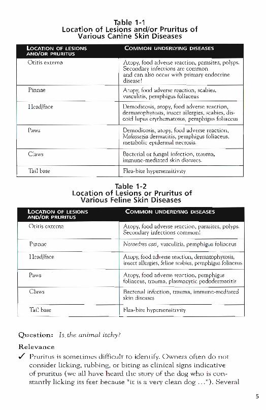
Table 1-1 location of lesions and/or Pruritus of
Various Canine Skin Diseases
LOCATION OF LESIONS COMMON UNDERLYING DISEASES AND/OR PRURITUS
Otitis externa Atopy, food adverse reaction, parasites, polyps. Secondary infections are common and can also occur with primary endocrine disease!
Pinnae Atopy, food adverse reaction, scabies, vasculitis, pemphigus foliaceus
Head/face Demodicosis, atopy, food adverse reaction, dermatophytosis, insect allergies, scabies, dis-coid lupus erythematosus, pemphigus foliaceus
Paws Demodicosis, atopy, food adverse reaction, Malillsezia dermatitis, pemphigus foliaceus, metabolic epidermal necrosis.
Claws Bacterial or fungal infection, trauma, immune-mediated skin diseases .
Tail base Flea-bite hypersensitivity
Table 1-2 Location of lesions or Pruritus of
Various Feline Skin Diseases
lOCATiON OF LESIONS AND/OR PRURITUS
Otitis externa
Pinnae
Head/face
Paws
Claws
Tail base
Question: Is, the animal itchy?
Relevance
COMMON UNDERLYING DISEASES
Atopy, food adverse reaction, parasites, polyps. Secondary infections common~
Notoedres wti, vasculitis, pemph igus foliaceus
Atopy, food adverse reaction, dermatophytosis, insect allergies, feline scabies, pernphigus foliaceus
Atopy, food adverse reaction, pemphigus foliaceus, trauma, plasmacytic pododermatitis
Bacterial infection, trauma, immune~medi()ted skin diseases
Flea-bire hypersensi ti vity
./ Pruritus is sometimes difficult to identify. Owners often do not consider licking, rubbing, or biting as clinical signs indica tive of pruritus (we all have h eard the story of the dog who is constantly licking its feet because lIit is a very clean dog ... "). Severa l
5
6
routine questions may be needed to identify pruritus in SOITle patients: Are they licking or chewing their paws? Are they rubbing their faces? Do they scoot on their rear ends? Are they scratching their anTlpits?
./ The presence of pruritus with skin lesions does not help ITluch in discovering the etiology of the pruritus, given that ITlany skin diseases cause pruritlls. However, pruritus without lesions typically means either atopic dennatitis or food adverse reaction (possibly with secondary infections) or in rare instances scabies incognito .
./ The perceived severity of pruritus Inay vary with the owner. Some owners deny the presence of pruritus despite the patient1s frantic scratching in the consultation room. Others insist on severe pruritus in a patient with no evidence of sdftrauma on clinical exaLnination. Good communication skills and judgement are essential to form a re81istic opinion for evaluation. If the petls scratching wakes the owner lip at night, the pruritus is severe irrespective of the presence of lesions .
./ If itch preceeds the occurrence of lesions, atopic dennatitis, food adverse reaction, and scabies incognito rnust again be considered.
Diagnostic procedures: Trichograrll. in alopecic patients that are reporredly nonpruritic.
Question: Is the disease seasonal?
Relevance
./ Insect bite hypersensitivities (caused most cOlnmonly by fleas, but mosquitoes or other insects can also be involved) frequently cause disease that worsens in summer. Whether clinical signs are absent or milder in the coLder season depends on specific environmental cond itions .
./ Atopic dermatitis may also be seasonal in certain climates. In many telTIperate climates it may OCCllr lTIOre noticeably in spring and summer if caused by tree and grass pollens or worsens in sum~ mer and autLllnn because of weed pollens. Warmer clitnates such as those found in tropical or subtropical regions usually have an extended pollen season. Hypersensitivities to house dust ITlites are often nonseasonal, but may be seasonally worse in winter in sotne areas and patients.
./ Seasonal noninflammatory alopecia and hyperpigmentation may be due to cyclic follicular dysplasia.
Diagnostic procedures: Insect bite trial, intradermal skin testing, serUln testing for allergen-specific IgE, biopsy, keeping the animal inside to evaluate for mosquito-bite hypersensitivity.
Question: Are there other clinical signs such as sneezing, coughing, or diarrhea?
Relevance
./ Sneezing, coughing, wheezing, and conjunctivitis may be seen concurrently with atopic dennatitis and caused by airborne allergies .
./ Diarrhea may be associated with food adverse reaction .
./ Polydipsia and polyuria are comlnon with iatrogenic and idiopathic hyperadrenocorticislTI .
./ Systeluic mycoses frequently present with concurrent anorexia, Lethargy, and with gastrointestinal or respiratory symptoms.
Diagnostic procedures: Cytology of nasal exudate or conjunctiva, eliluination d iet, urine cortisol/creatinine ratio, low dose dexamethasone suppression test, and adrenocorticotropic hormone (ACTH)stin"luLation test.
Question: What is fed to the animal? Was a special diet used in the past? What was it and how long was it fed exclusively?
Relevance
./ Knowing the diet will allow the clinician to determine possib le nutritional deficiencies .
./ It will also help in formulating an eliLnination diet if indicated (p. 46),
./ If a diet was fed in the past and it was not a true elilnination diet (was not fed exclusively or not fed for an appropriate length of tirne) it may need to be repeated.
h Contrary to the cominon b elief, food adverse reactions typically do not occur immediately after a change in feeding habits. Most animals with food adverse reactions have been consuming the offending diet for years before showing clinical signs.
~ Remember to ask about treats and supplements, which are often forgotten, when food is discussed with the client.
Question: Are there other animals in the household? Do they show cutaneous symptoms?
Relevance
./' If other animals in the household are sirnilarly affected, contagious disease such as dennatophytosis or scabies is lnore likely.
~ Other anilnals may serve as a reservoir for ectoparasi tes without showing clinical signs.
Diagnostic procedures: If indicated, insect control trial , fungal cultures, or scabies treatment trials should include all anilnals in the household to identify and/or treat possib le carrier anitnals to allow successful long-tenn remission for the patient.
Question: Does any person in the household have skin disease?
Relevance
./' Two zoonoses of Inajor concern in veterinary dermatology are scabies and dennatophytosis (ringworm.). However, even if owners are not affected, these diseases cannot be ruled out .
./' Canine scabies affecting humans occurs as an itchy papular rash in contact areas, such as arms and legs, starting days to weeks after onset of pruritlls in the pet.
./' Dermatophytosis is often characterized by scaling and erythema and nlay not be p articularly pruritic, but occasionally can present as severely inflammatory and pruritic skin disease. Dennatophytosis lnay sometimes be Inisdiagnosed as eczema in humans .
./' Sporotrichosis and other mycoses have zoonotic potential and may occasionally cause disease in hUlnans .
./' Don't forget that the skin disease of the owner l1.1.ay also be com-pletely u nrelated to the a11.ilnal's skin disease.
Diagnostic procedures: Wood's light, skin scrapings, fungal culture, scabies trial treatlnent . In severe fonns of suspected dermatophytosis, a biopsy and special funga l stains may prove useful for obtaining a quick diagnosis.
Question: Was the disease treated before? If so, which drugs were used and how successful was treatment?
Relevance Response to previous therapy can be of tremendous help in establishing or ruling out underlying causes for the skin disease.
./ Initial response to recent glucocorticoid adtuinistration luay not be helpful because many skin diseases improve for a short period with th is syrnptomatic, nonspecific treatment .
./ Repeated response to low~dose glucocorticoid therapy sug; gests hypersensitivities (possibly complicated by Malassezia dennatitis caused by Malassezia pachydermatis) .
./ Repeated response to antibiotics and glucocorticoids in cOlnbination is of l ittle h elp .
./ Repeated partial or total response to antibiotics indicates a pyodenua usually second ary to either atopic dermatitis, food adverse reaction, hormonal disease, or another less comrnon disorder that is suppressing t he skin's immune system. In addition to antibacterial treatluent, the underlying problem needs to be identified and treated to prevent recurrences.
~ Ask specifically how much the, pet improved while receiving rn_edication because luany owners tend to judge a treatluent as not helpful if it did not cure the disease.
Question: What is currently used to control fleas?
Relevance
./ Flea~bite hypersensitivity is the most common hypersensi~ tivity and an extreluely COlUluon skin disease in rnost small anilnal prac t ices. If flea~bite hypersensitivity is suspected, a flea control trial should be cOlnmenced .
./ Deta ils of the flea control for all aniluals in the household are imp ortant because in a severely allergic anitual, clinical signs can b e caused by a very small nLLlnber of flea bites. Inconsi~ stent or ineffective flea control can be discovered only through detailed questioning.
~ Many owners take questions about their flea control as an insult to their own cleanliness and h ygiene. Good COlUlnunica~ tion s k ills are a great help. I own a f1ea~allergic dog and rou; tinely mention her as an example, which breaks the ice and increases the client's willingness to listen and follow my instructions.
Question: When was rhe last medication given?
Relevance
./ Recent administration of tnedication may affect the clinical presen ta tion.
9
10
./ It Inay also affect various ind icated diagnostic tests that Inay n eed to be postponed .
./ Long~tenn glucocorticoid therapy will affect the results of allergy tests - both intradennal skin testing and serum testing for allergen~specific IgE. It will also affect histopathologic findings and the results of Inany blood tests .
./ Antihistamines and short~tenn systelnic and topical glucocor~ ticoids (i.e., < 4 weeks) may influence intradermal skin testing .
./ Sorne antibiotics, sLlch as trimethopriln~sulfonalnide cOLnbina~ tions, will affect blood concentrations of thyroxin. Others such as cephalosporins n"lay affect the glucose readings of some urine test strips .
./ Reluember to ask specific questions regarding heartworm pre~
vention, vitmuin supplements, or dewonning which are also fonns of phannacotherapy.
Question: Does the animal get better with a change of environment (a weekend away or a day at the in-laws for example)?
Relevance
./ The anilual's improvement in another environment indicates involvement of an environmental allergen (airborne or contact) or irritant .
./ Lack of ilTIprovement does not rule out these allergies, in that airbonie and contact allergens lllay be the same in different locations (house dust Illites are found ahnost anywhere in the world).
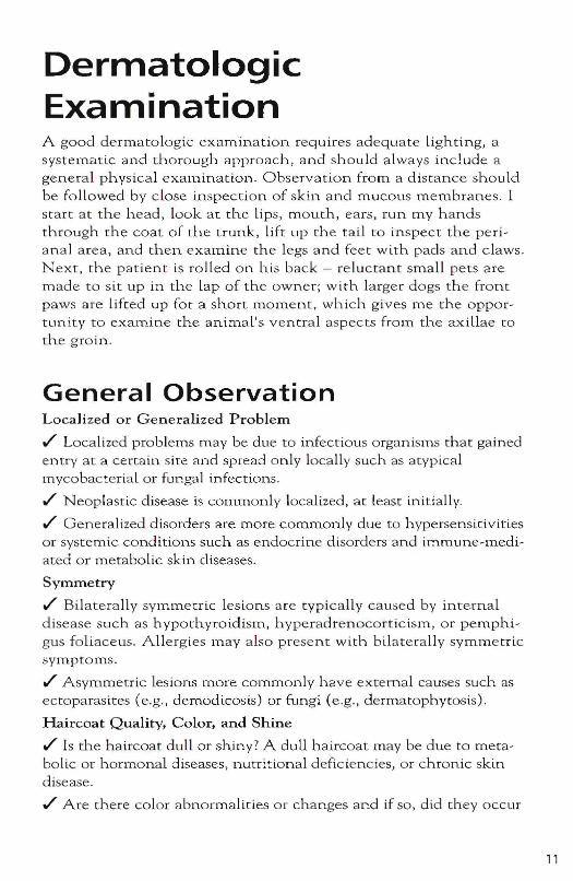
Dermatologic Examination A good dermatologtc examination requires adequate lighting, a systematic and thorough approach, and should a lways include a general physical exam.ination. Observation from a distance shou ld be followed by close inspection of skin and mucous membranes. I start at the head, look at the lips, mouth, ears, run my hands through the coat of the trunk, lift up the tail to inspect the periana l area, and then examine the legs and feet with pads and claws. Next, the patient is rolled on his back - reluctant small pets are made to sit up in the lap of the owner; with larger dogs the front paws are lifted up for a short moment, which gives me the opportunity to exaluine the anin"lal's ventral aspects from the axillae to the groin.
General Observation LocaJized or Generalized Problem
./' Localized problems may be due to infectious organisms that gained entry at a certain site and spread only locally such as atypical mycobacterial or fungal infections .
./' Neoplastic disease is commonly localized, at least initi a lly .
./' Generalized disorders are more commonly due to hypersensitivities or systemic conditions such as endocrine disorders and immune-mediated or metabolic skin diseases.
Symmetry
./' Bilaterally symmetric lesions are typically caused by internal disease such as hypothyroidism, hyperadrenocorticism, or pemphigus foliaceus . A llergies may a lso present with bilaterally symmetric syrnptoms .
./' Asymmetric lesions more commonly have external causes such as ectoparasites (e.g., demodicosis) or fungi (e.g., dermatophytosis).
Haircoat Quality, Color, and Shine
./' Is the haircoat dull or shiny? A dull haircoat may be due to metabolic or hormonal diseases, nutritional deficiencies, or chronic skin disease .
./' Are there color abnormalities or changes and if so, did they occur
11
before or concurrent with th e onset of skin disease or as a consequence of the disease. Hair color changes may be associated withhormonal disease or follicular dysplasia .
./ Changes in t h e hair quality (either to a coarse coat or to afine puppy coat) may again po in t to hormonal disease or follicular dysplasia.Close inspection of t h e skin and mucous membranes follows thegeneral observation. Pay special attention to any ind iv id uallesions. Primary lesions are initial eruptions tha t are causeddirectly by the underlying disease process. Secondary lesionsevolve from primary le s io n s 01- are caused by the patient (selftrauma) or environment (medications). It is important that theclinician be able both to differentiate between primary and secondary lesions and to understand the underlying pathornec.h-an ism because this helps in the formulation of a relevant list ofdifferential diagnoses. I next discuss the individual lesions andtheir implications and give the most common differential diagnoses for each lesion.
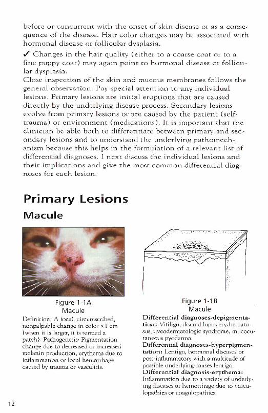
Primary LesionsMacule
12
Figurel-1AMacule
Definition: A focal, circumscribed,nonpalpable change in color <1 cm(when it is larger, it is termed apatch). Pathogenesis: Pigmentationchange due to decreased or increasedmelanin production, erythema d ue toinflammation or local hemonhagecaused by t rauma or vasculitis.
". : ::
.~:Pt~~~t~~~.:~::~\~~:;;~~
',;::
Figure l -1BMacule
Differential diagnoses-depigmentation: Vitiligo, discoid lupus erythematosus, uveodermarologic syndrome, mucocutaneous pyoderma.Differential diagnoses-hvperpigmentation: Lentigo, hormonal di seases orpost-inflarnmarory with a multitude ofpossible underlying-causes lentigo.Differential diagnosis-erythema:Inflammation due to a variety of underlying d iseases or hemorrhage due to vasculoparhies or coagulopathies,
Papule
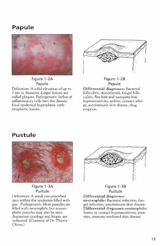
Figure 1-2A Papule
Definiti on : A so lid elevation of up to 1 em in diameter. Larger Lesions are called plaques. Pathogenesis: Influx of inflammatory ce lls imo the dermi~, focal epidermal hype rplasia, early neoplastic lesions.
Pustule
Figure 1-3A Pustule
Definition: A small circumscribed area within the epidennis filled with pus. Pathogenesis: Most pustules are filled with ll.eutrophils, but eosinophilic pustules may also be seen. Aspiration cytology and biopsy are indicated . (Courtesy of Dr. Thierry Olivry.)
Figure 1-28 Papule
Differential diagnoses; Bacterial fo lliculitis, democlicosis, funga l fa ll i culitis, fl ea-bite and mosquito-bite hypersensitivity, sC8hies, contact all ergy, autoimmune skin disease , drug etuption .
Figure 1-38 Pustule
Differential diagnosesneutrophils: Bacteria l infection, fungal infection, autOimmune skin disease . Differential diagnoses-eosinophils: Insect o r contact hypersensitivity, parasites, immune-mediated skin di sease
13
14
Vesicle
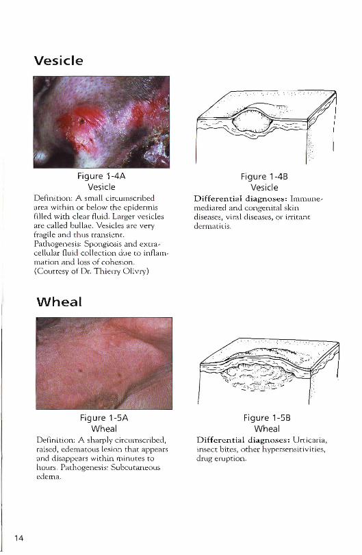
Figure 1-4AVesicle
Defini t ion: A sma ll circumscribeda rea with in o r be low m e epide r misfilled with clear fluid . Large r ves iclesare ca lled bullae . Ves icles are veryfrag ile and thus transient.Path ogenes is: Spo ng iosis and extrace llular fluid collec t ion du e to infl am mation and loss of cohesio n.(Courtesy of D r. T h ierry O livry)
Wheal
Figure 1-5AWhea l
Definition: A sharply circumscribed ,raised, ede matous lesion th at appea rsand disap pears with in minutes rohours. Pathogenesis: Subcutaneou sede ma.
"
Figure 1-48Vesicle
Differential diagnoses: Immunemediated and co nge nita l skindiseases, viral di seases, or irritantderma t it is.
Figure 1-5BWheal
Differen t ial diagnoses: U rticari a,insect bites, o the r h ypersen sit iv ities,drug erup t ion .
Nodule
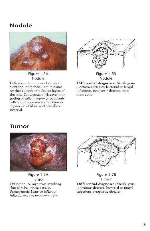
Figure 1-6A Nodule
Definition: A circumscribed, solid elevation more than 1 cm in diameter that extends into deeper layers of the skin. Pathogenesis: Massive infiltration of infl ammatory or neoplastic ce lls into the dennis and subcutis or deposition of fibr in and crysmlline materia l.
Tumor
Figure 1-7A Tumor
Definition: A large mass involving skin or subcutaneous tissue. Pathogenesis: Massive influx of inflammatory or neoplastic cells.
Figure 1-6B Nodule
Differential diagnoses! Sterile granulomatous diseases, bacteria l o r fungal infect ions. neoplastic diseases. calcinosis cut is
Figure 1-7B Tumor
Differential diagnoses! Sterile granulomatous diseases. bacterial or fungal infections, neoplastic diseases.
15
Primary or Secondary LesionsAlopecia
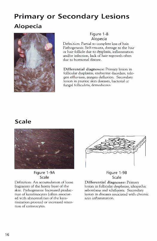
Figure 1-8Alopecia
Definition: Part ial to complete loss of hair.Pathogenesis: Se lf-trauma, damage to the h airo r h ai r foll icle due to dysplasia , in flam mat ionand/or infect ion , lack of h air regrowth oftendue to hormonal d isease.
Differential di agnose s: Primary lesion infol licular dvsp las ias, endocrine d iso rders, telo gen effluvium, anagen deflux ion . Secondarylesion in pr ur it ic skin diseases, bacter ial orfun gal folliculi tis, demodicosis.
Scale
- " j'/,/
16
Figure 1-9AScale
Definition: An acc umulat ion of loosefragments of the horny layer of theskin . Pathogenesis: In creased pro duct ion of kerar inocvtes (often assoc iated with abnormalit ies of the kerat in ization process) o r increased reten t ion of corneocy tes.
Figure 1-9BScale
Differential diagnoses: Primarylesion in follicu lar dysplasias, idiopathicseborrheas and ichthyosis. Seco n da rylesion in diseases assoc iated with chronicsk in inflammat ion .
Crust
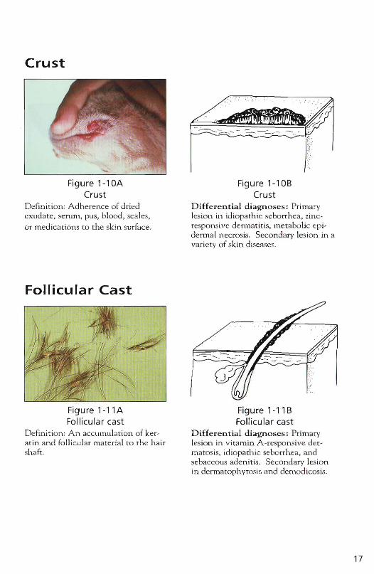
Figure1 -10ACrust
Defin it ion: Ad herence of driedex udate. serum , pus, blood. sca les .or medi cation s (0 the skin surface .
Follicular Cast
Figure1-11AFollicul ar cast
Defini tion : An. acc umu lat ion of ker at in and follicular mater ial (0 the hairshaft .
Figure 1-108Crust
Differential diagno ses : Primarylesion in id iopa th ic seborrhea. zincrespon sive dermatitis, metabolic epiderm al necrosis. Secondary lesion in avariety of skin d iseases .
Figure 1-118Follicular cast
Differential diagnoses : Pr imarylesion in vitamin A -responsive der mat osis, idiopathic seborrhea. andsebaceous adenitis. Secondary lesionin dermatophv tos is an d de rnod icosis.
17
18
Pigmentary Abnormalities
Hyperpigmentation
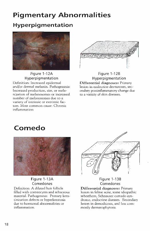
Figure 1-12A Hyperpigmentation
Defin ition: Inc reased epiderma l and/o r dermal melanin. Pa thogen esis: Increased produc tion, size , o r melanization of m elanosomes or increased n umber o f melanosomes due to a variety o f intrinsic o r extrinsic factors. Most commo n cause: C h ronic inflammation
Comedo
Figure 1-13A Comedones
Definition : A di lated h air fo llicle filled with com eocytes and sebaceous materia l. Pathogenes is: Primary kerat inization defects o r hyperkeratosis due to hormon al abnormalities o r in flammation .
Figure 1-12B Hyperpigmentation
Diffe r entia l diagn oses: Primary lesion in endocrine dermatoses, secondary postinflammatory ch ange due to a variety of skin diseases.
Figure 1-138 Comedones
Differential diagn oses : Primary lesion in feline acne, some idiopathic seborrheas, Schnauzer comedo syn drome, endocrine diseases. Secondary lesion in demodicosis, and less common ly dermacophytosis.
Secondary Lesions
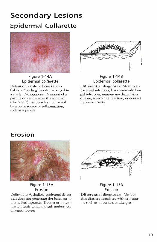
Epidermal Collarette
Figure 1-14A Epidermal collarette
Definition: Sca le of loose keratin flakes or "peeling" kera tin alTanged in a circle. Path ogen es is: Remnant of a pustule or vesicle after the top part (the "roof") h as been lost, or caused by a point source of inflammation, slIch as a papule.
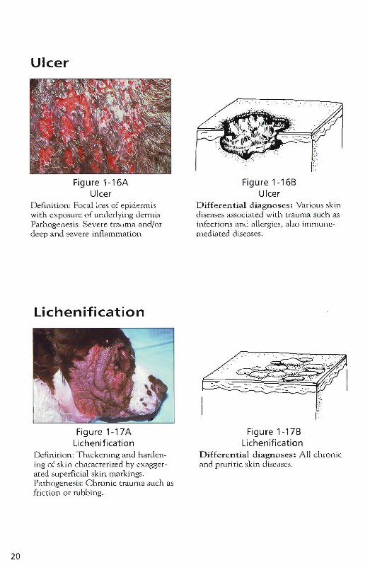
Erosion
Figure 1-15A Erosion
Definition: A shallow epidermal defect that does not penetrate the basal membrane. Pathogenesis: Trauma or inflammation leads to rapid death and/or loss of keratinocytes
Figure 1-14B Epidermal collarette
Differential diagnoses: Most likely bacterial infection , less commonly fungal infection, immune-medi ated skin disease , insect-bite reaction, or conmct hypersens iti vity.
Figure ' -15B Erosion
Differential diagnoses: Various skin diseases associated with se lf trauma such as infections or allergies.
19
20
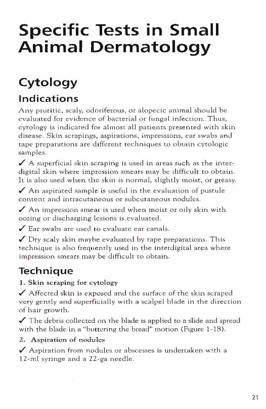
Ulcer
Figurel-16A Ulcer
Definition: Focal loss of epidermis with exposure of underlying dem1is Pathogenesis: Severe trauma and/or deep and severe inflammation
Lichenification
Figure 1-17A Lichenification
Definition: Thickening and hardening of skin characterized by exaggerated superficial skin markings. Pathogenesis: Chronic trauma such as friction or rubbing.
Figure 1-16B Ulcer
Differential diagnoses: Various skin diseases associated with trauma such as infections and 811ergies, also immunemediated diseases.
Figurel-17B Lichenification
Differential diagnoses: All chronic and pruritic skin diseases.
Specific Tests in Small Animal Dermatology
Cytology Indications Any pruritic, sca ly, odoriferous, or a lopecic animal should be evaluated for evidence of bacterial or fungal infection. Thus, cytology is indicated for almost all patients presented with skin disease . Skin scrapings, aspirations, impressions, ear swabs and tape preparations are different tecl"lOiques to obtain cytologic samples .
./ A superfic ial skin scraping is used in areas such as the interdigital skin where impression smears may be difficult to obtain. It is also used when the skin is normal, slightly moist, or greasy .
./ An aspirated sample is useful in the evaluation of pustule content and intracutaneous or subcutaneous nodules .
./ An impression smear is used when moist or oily skin with oozing or discharging lesions is evaluated .
./ Ear swabs are used to evaluate ear canals .
./ Dry scaly skin maybe evaluated by tape preparations. This technique is also frequently used in the interdigital area where impression smears may be difficult to obtain.
Technique 1. Skin scraping for cytology
./ Affected skin is exposed and the surface of the skin scraped very gently and superfic iaLLy with a scalpel blade in the direction of hair growth .
./ The debris collected on the blade is applied to a slide and spread with the blade in a "buttering the bread" motion (Figure I - IS).
2. Aspiration of nodules
./ Aspiration from nodules or a bscesses is undertaken with a lZ-ml syringe and a 22-ga n.eedle.
21
22
./ The nodule is firmly grasped and the needle is then inserted (Flgure 1-19), aspirated several ti mes (up (0 the 10-ml mark if possible), the pressure released, and the syringe with n eed le st ill a ttach ed is withdrawn .
./ It is important (0 release the pressure before withdrawaL of the needle or else the asp ira te can be sucked back into the barrel of the syringe - from which it may not be retrieved .
./ The needle is detached , the plunger pulled back, a nd th e needle reattached .
./ Ce lls are then blown onto a s lide. The smear is air dried.
3. Impression Smears
./ Cotton swabs a re used to obtain samples from ear canals by inserting them into the canal, rotating, and Withdrawing them. They are then ro ll ed gently onto a slide. I hold ear sl ides uniform.ly on the left side with my left hand, the cotton swab from the left ear is rolled onto the mid-section of the slide a n d the cotton swab from the right ear onto the right third of the same slide .
./ In patients with dry skin , a cotton swap may be moistened with saline solution and rubbed on the surface of affected skin before it is rolled onto a s lid e .
./ In patients w ith moist or g reasy sk in, the slide can be rubbed or impressed directly onto affected skin (Figure 1-20).
4. Tape Preparation
./ A direct imp ressi.on techni.que uses clear sticky tape to collect debris from the surface of the skin. Although quick, this m ethod does take practice to establish what is "normal"
./ The tape is pressed sticky side down onto the skin (Figure 1-21) .
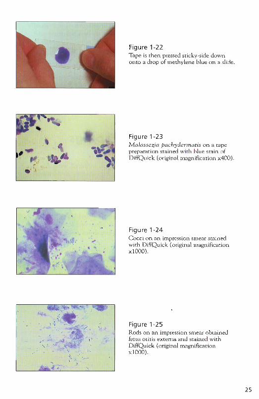
./ Next, it is pressed (also sticky side down) onto a drop of methylene blue or the b lue stain of DiffQuick on a slide (Figure 1-22) .
./ The tape serves as a cover slip : the sam.p le can be evaluated even under o il immersion (with a small droplet of o il placed directly on top of the tape) .
./ This technique is espec ia lly useful for Malassezia evaluation. Other items of interest that can be identified include inflammatory ce lls such as neurrophils (wh ich may have passed through the epiderm.is in response to a sup erficial infection) , nucleated epithel ia l cells (which are not normal and reflect a keratinization abnormallty)' cocci, rods, macrophages, short-bodied demodex mites, Cheyletie lla , and occasiona lly Smcoptes mites.
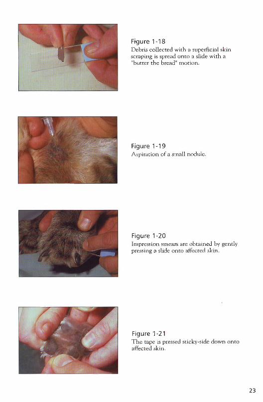
Figure 1-18 Debris collec ted with Cl superficial skin scrClping is spread OntO a slide with a "butter the bread" mo t ion .
Figure 1-19 A spiration of a small nodule.
Figure 1-20 Impression smears are obtained by gently press ing a slide onto affected skin .
Figure 1-21 The tape is pressed sticky-side down onto affected skin.
23
24
Stain ./ A modified Wright's stain (e .g., o iff Quick) can be used to stain the air-dried slides. It is much faster and easier than Gram's stain a nd sufficient to evaluate nearly all skin cytology samples. But Gram's stain is also suitable.
Interpretation ./ Yeast organisms are most often M. pachydermatis (Figure 1-23), although Candida spp. may occasionally be involved .
./ Cocci are most often Staphylococcus intermedius (Figure 1-24). S. aureus or Streptococci may be found in some patients .
./ Rod-shaped organisms are found mostly in the ear canal and are most often Pseudomonas aeruginosa or Proteus mirabilis (Figure 1-25).
h The number of organisms is important. Occasional cocci or yeast are probably not relevant. On the skin, I consider one or more yeast organisms per high-power field (HPF) relevant; cocci should be seen in high numbers. In the infected ear, yeast and cocci typically occur in high numbers; any rods present are abnormal. Don't mistake exogenous bacterlal contaminants for infection .
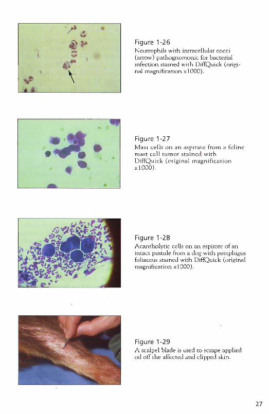
./ Inflammatory cells with intracellular organisms are pathognomonic for a clinically relevant infection (Figure 1-26) .
./ Eosinophils typically indicate allergic or parasitic skin disease.
If:' Neoplastic cells may be difficult to recognize so tha t the help of a clinical pathologist is typically needed. Even with high skill levels, neoplastic skin disease should not be diagnosed exclusively by cytology (with the exception of mast cel1 tumors); a biopsy should always confirm any suspicions raised clinically and cytologically .
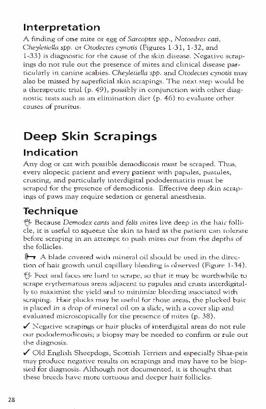
./ If mast cells are found cytologically (Figure 1-27), the diagnosis of mast cell tumo r is confirmed, but complete surgical excision should still be confirmed by histopathology. In some patients with mast cell tumors, mast cell granules do not stain with routine DiffQuick and thus a negative cytologic result can not rule out mastocytosis .
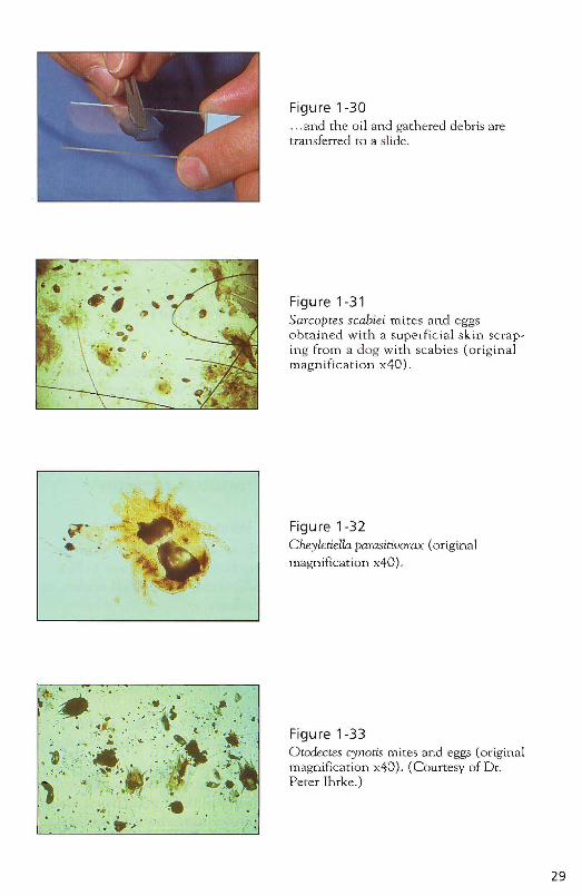
./ Acantholytic cells are keratinocytes that have lost their intercellular connections (desmosomes) and present as round cells with a purple cytoplasm and a central dark purple nucleus (Figure 1-28). These cells suggest pemphigus foliaceus or erythematosus but can also be seen on cytologic samples of severe pyodermas. A biopsy is indicated to confirm the diagIl.osis.
.....~
. , ll,
Figure 1-22Tape is then pressed sticky-side downonto a drop of methylene blue on a slide .
Figure 1-23Malassezia pachydermatis on a tapepreparat ion stained with blue stain ofD iffQ uick (original magnification x400) .
Figure 1-24Cocci on an impression smear stainedwith DiffQuick (original magnificationxlOOO).
..\\ '-
-.
Figure 1-25Rods on an impression smear obtainedfrom Otitis externa and stained w ithDiffQuick (original magnificationxlOOO) .
25
26
h Remember, that bacterial and yeast infections are usually secondary to other diseases, which need to be identified and treated to prevent recurrence of the infection.
h Cytologic re-evaluation at the end of antimicrobial therapy is crucial because organisms may change during treatment. For instance, a dog initially presented with bacterial infection may develo p a yeast infection during successful antibacterial treatment, preventing clinical improvement and vice versa.
Treatment of bacterial infections and antifungal therapy are discussed in Section 3.
Superficial Skin Scrapings Indications Any pruritic or scaly dog and cat may be infested with Cheyletiella spp., Otodectes cynotis, Scabies scabiei, or Notoedrescari and should be scraped.
Technique ,/ If scabies is suspected, preferred areas for scrapings are the elbows, hocks, and ventrum. Ear margins should be scraped thoroughly if any pruritus or scaling is observed in this area. Sometimes scaling is subtle and only becomes evident on close exatnination .
./' Sites are gently clipped with #40 clipper blades. Mites may be difficult to find (especially canine scabies mites), so that the bigger the surface area scraped, the greater will be the chance of a positive skin scraping.
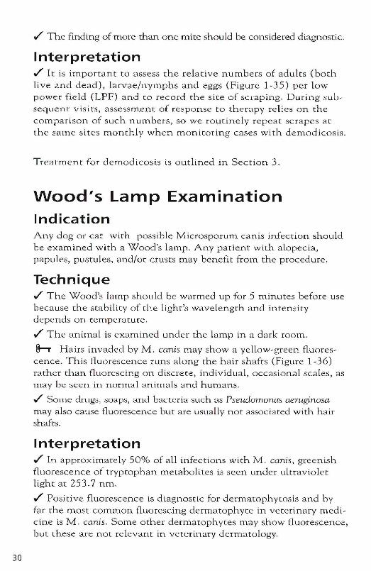
~ Several drops of mineral oil are applied directly to the clipped skin and distributed evenly in the area .
./' The oil is scraped off with a #11 scalpel blade (Figure 1-29) and transferred to one or more glass slide(s). Scrape 10 to 15 times especially when canine scabies is suspected .
./' A cover slip is used to aLLow rapid yet thorough evaluation of collected debris (Figure 1-30) and the slide(s) is (are) evaluated under low power (x40 or xlOO) systematically from the left upper corner co the right lower corner.
;.
\
Figure 1-26Neurrophils with intracellular cocci(arrow) pathognomonic for bacterialinfection stained with DiffQuick (original magnification xlOOO).
Figure 1-27Mast cells on an aspirate from a felinemast cell tumor stained withDiffQuick (original magnificationx 1000).
,.
Figure 1-28Acantholvtic cells on an. aspirate of anintact pustule from a dog with pemphigusfoliaceus stained with D if/Quick (origina lmagnification xlOOO).
Figure 1-29A scalpel blade is used to scrape appliedoil off the affected and clipped skin.
27
28
Interpretation A finding of one mite or egg of Sarcoptes spp., Notoedres cati, CheyletieUa spp. or Otodectes cynotis (Figures 1-31, 1-32, and 1-33) is diagn.ostic for the cause of the skin disease. Negative scrapings do not rule our the presence of mites and clinical disease particularly in canine scabies. Cheyletiella spp. and Otodectes cynotis may also be missed by superficial skin scrapings. The next step would be a therapeutic trial (p. 49), possibly in conjunction with other diagnostic tests such as an elimination diet (p. 46) to evaluate other causes of pruritus.
Deep Skin Scrapings Indication Any dog or cat with possible demodicosis must be scraped. Thus, every alopecic patient and every patient with papules, pustules, crusting, and particularly interdigital pododermatitis must be scraped for the presence of demodicosis. Effective deep skin scrapings of paws may require sedation or general anesthesia.
Technique 'f} Because Demodex canis and felis mites live deep in the hair follicle, it is useful to squeeze the skin as hard as the patient can tolerate before scraping in an attempt to push mites out from the depths of the follicles.
h A blade covered with mineral oil should be used in the direction of hair growth until capillary bleeding is observed (Figure 1-34).
'f} Feet and faces aTe hard to scrape, so that it may be worthwhile to scrape erythematous areas adjacent to papules and crusts interdigitally to maximize the yield and to minimize bleeding associated with scraping. Hair plucks may be useful for those areas, the plucked hair is placed in a drop of mineral oil on a slide, with a cover slip and evaluated microscopically for the presence of mites (p. 38) .
./' Negative scrapings or hair plucks of interdigital areas do not rule out pododemodicosis; a biopsy may be needed to confirm or rule out the diagnosis .
./' Old English Sheepdogs, Scottish Terriers and especially Shar-pels may produce negative results on scrapings and may have to be biopsied for diagnosis. Although not documented, it is thought that these breeds have more tortuous and deeper hair follicles.
. "~P
.~ .. .. ----'"
l4t ... ~
,(.
I~ - .':~
Figure 1-30... and the oil and gathered debris aretransferred [0 a slide .
Figure 1-31Sarcoptes scabiei tTl ites and eggsobtained with a superficial skin scraping from a dog with scabies (originalmagnification x40).
~~ . ~.. Figure 1-32Cheyletiella parasuivorax (original
magnification x40).
'f:1•.•' ··-:; . ~ '. ,., -_ . : ..• . • ., 's)..' . .s: ' .:' ,1~ . ,~ . ~:'t ',-, • of. • • •'t l '- , . . '_ . • • "" '.. .. J ' • . ' • • . , _
'* : .....•.. .: . .~.-:,;,: :.'I w. .;. '. ..r
o
• • ..
. ! ..;, . , - ,' - " .,. . ' .~, · ,· ·~ ·'·l· ' .>' - •.:, .,, ;0,. • , ' . \ •, . -. , ":, ' ~ ... '" • . .:: " Ie.~ '; ' .• ', .;,.
:..: ........ ,
Figure 1-33Otodectes cynotis mites and eggs (originalmagnification x40). (Courtesy of Dr.Peter Ihrke.)
29
30
./ The finding of more than one mite should be considered diagnostic.
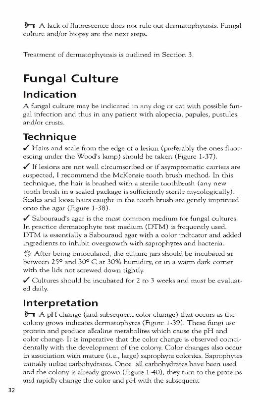
Interpretation ./ It is important to assess the relative numbers of adults (both live and dead), larvae/nymphs and eggs (Figure 1-35) per low power field (LPF) and to record the site of scraping. During subsequent visits, assessment of response to therapy relies on the comparison of slIch numbers, so we routinely repeat scrapes at the same sites monthly when monitoring cases wtth demodicosis.
T reatment for demodicosis is outlined in Section 3.
Wood's lamp Examination Indication Any dog or cat with possible Microsporum cants infection should be examined with a Wood's lamp. Any patient with alopecia, papules, pustules, and/or crusts may benefit from the procedure.
Technique ./ The Wood's la mp should be warmed up for 5 minutes before use because the stability of the light's wavelength and intensity depends on remperawre .
./ The animal is examined under the lamp in a dark room.
h Hairs invaded by M. canis may show a yellow-green fluorescence. This fluorescence runs along the hair shafts (Figure 1-36) rather than fluorescing on discrete, individual, occasional scales, as rnay be seen in normal animals and humans .
./ Some drugs , soaps, and bacteria such as Pseudomonas aeruginosa may also cause fluorescence but are usua lly not associated with hair shafts.
Interpretation ./ In approximately 50% of all infections with M. canis, greenish fluorescence of trypwphan metabolites is seen under ultraviolet light at 253.7 nm .
./ Positive fluorescence is diagnostic for dermatophywsis and by far the most common fluorescing dermatophyte in veterinary medicine is M. canis . Some other dermatophytes may show fluorescence, but these are not relevant in veteri.nary dermatology.
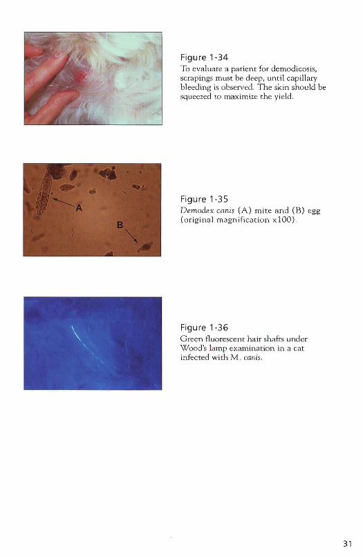
Figure 1-34 To evaluate a patient for demodicosis, scrapings must be deep, until capill ary bleeding is observed. The skin should be squeezed to maximize the yield.
Figure 1-35 Demodex canis (A) mite and (B) egg (o rigina l magnification xlOO).
Figure 1-36 Green Huorescem hair shafts under Wood's lamp examination in a cat infected with M . canis.
31
32
h A lack of fluorescence does noc rule out dermatophytosis. Fungal culture and/or biopsy are the next steps.
Treatment of dermatophytosis is outlined in Section 3.
Fungal Culture Indication A fungal culture may be indicated in any dog or cat with possible fun~ gal infection and rhus in any patient with alopecia, papules, pustules, and/or crusts.
Technique ./ Hairs and scale from the edge of a lesion (preferably the ones fluor~ escing under the Wood's lamp) should be taken (Figure 1~37) .
./ If lesions are not well circumscribed or if asymptomatic caniers are suspected, 1 recommend rhe McKenzie tooth brush merhod. In this technique, the hair is brushed with a sterile toothbrush (any new tooth brush in a sealed package is sufficiently sterile mycologically) . Scales and loose hairs caught: in the tooth brush are gently impdnted onto the agar (Figure 1~38) .
./ Sabouraud's agar is the most common medium. for fungaL cultures. In practice dermatophyte test medium (DTM) is frequently used. DTM is essentially a Sabouraud agar with a color indicator and added ingredients 1:0 inhibit overgrowth with saprophytes and bacteria.
~ After being innoculated, the culrure jars should be incubated ac between 25° and 30° C at 30% humidity, or in a warm dark corner with the lids not screwed down tightly.
./ Cultures should be incubated for 2 to 3 weeks and must be evaillat~ ed daily.
Interpretation h A pH change (and subsequent color change) that occurs as the
colony grows indicates dermatophyces (Figure 1~39). These fungi use protein and produce alkaline metabolites which cause the pH and color change. It is imperative that: the color change is observed coinci~ dentally '.vith the developm.ent of the colony. Color changes also occur in association with mature (i.e., large) saprophyte colonies. Saprophytes initially utilize carbohydrates. Once all carbohydrates have been Llsed and the colony is already grown (Figure 1-40), they turn to the proteins and rapidly change the color and pH with the subsequent
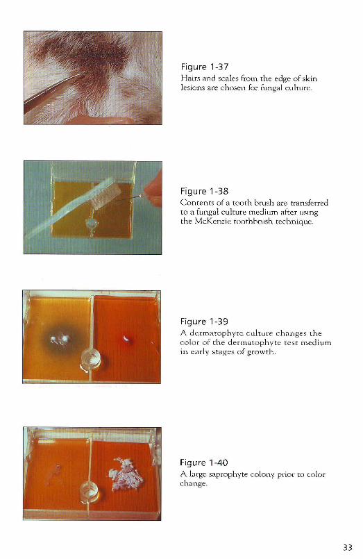
Figure 1-37 Hairs and scales from the edge of skin lesions are chosen for fungal culture.
Figure 1-38 Contents of a tooth brush are transferred to a fungal culture medium after usmg the McKenzie toothbrush technique.
Figure 1-39 A dermatophyte culture changes the color of the dermatophyte test medium in early stages of growth.
Figure 1-40 A large saprophyte colony prior to color change.
33
34
alkaline metabolites (Figure 1-41). It may be impossible to distinguish on gross appearance whether a mature colony with significant red pigmentation to the underlying and surrounding agar is a pathogenic or saprophytic fungus .
./ Always check the colony microscopically for characteristic macroconidia. Clear sticky tape is impressed gently onto the culture (sticky side down), then laid onto a drop of methylene blue or the blue stain of DiffQuick (also sticky side down) on a microscope slide and evaluated under the microscope. The sUlface of the sticky tape acts as its own cover slip. If required, microscope oil can be placed directly onto the sUlface of the tape .
./ Microsporum canis grows in a white, wooly colony with a yellowish reverse pigment (which maybe difficult to assess if grown on DTM). Abundant spindle-shaped macroconidia with knobs at the terminal ends and typically mnre than six internal compartm.ents are seen microscopically (Figure 1-42).
't} M. canis is a zoophilic fungus and patients typically were infected by another anilnal or human. Humans and other animals in contact with the patient are at risk to develop t he infection or may be asymptornatic carriers and need to be carefully evaluated and possibly treated as well.
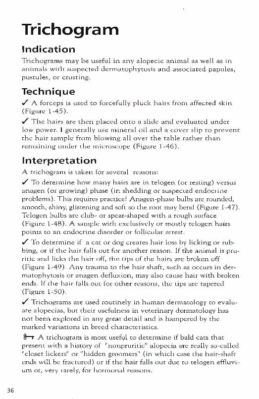
./ M. gypseum grows in a granular beige culture with yellowish reverse pigment and has thin-walled echinulate macroconidia wi th fewer than six internal compartments (Figure 1-43). M. gypseum is a geophilic fungus that is acquired by exposure to contaminated soil and thus has a limited zoonotic potencial.
./ Trichophyton mentagrophytes grow in colonies of variable texture and color that characteristically have a few cigar-shaped macroconidia and globous microconidia (Figure 1-44). Typical hosts fo r T. mentagrophyces are rodents and humans; infections are usuaL! y associated with exposure to these hosts or their ilnmediate environlnent.
See Section 3 for treatment of dermatophytosis
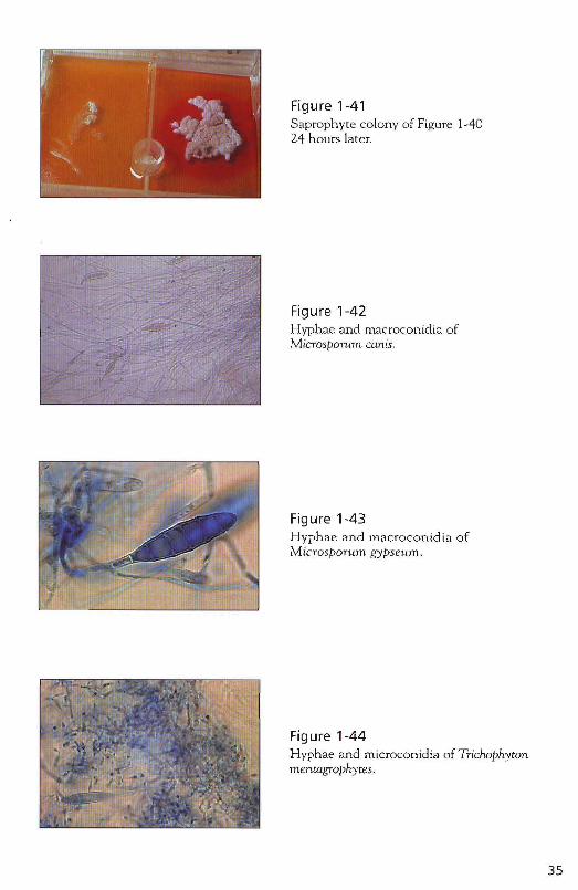
Figure 1-41 S<tprophyte colony of Figure 1-40 24 hours later.
Figure 1-42 Hyphae and rnacroconidia of Microsporum canis.
Figure 1-43 Hyphae and m<tcroconidia of Microsporum gypseum.
Figure 1-44 Hyphae and microconidia ofTTichophyton menwgrophytes.
35
36
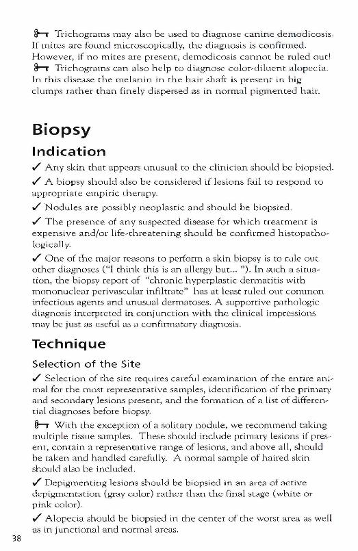
Trichogram Indication Trlchograms may be useful in any alopecic animal as well as in animals with suspected dermatophytosis and associated papules, pustules, or crusting.
Technique ./ A forceps is used to forcefully pluck hairs from affected skin (Figure 1-45) .
./ The hairs are then placed onto a slide and evaluated under low power. I generally use mineral oil and a cover slip to prevent the hair sample from blowing all over the table rather than remaining under the microscope (Figure 1-46).
Interpretation A trichogram is taken for several reasons:
./ To deterrn.ine how many hairs are in telogen (or resting) versus anagen (or growing) phase (in shedding or suspected endocrine problems). This requires practice! Anagen-phase bulbs are rounded, smooth, shiny, glistening and soft so the root may bend (Figure 1-47). Telogen bulbs are club- or spear-shaped with a rough surface (Figure 1-48). A sample with exclusively or mostly telogen hairs points co an endocrine disorder or follicular arrest .
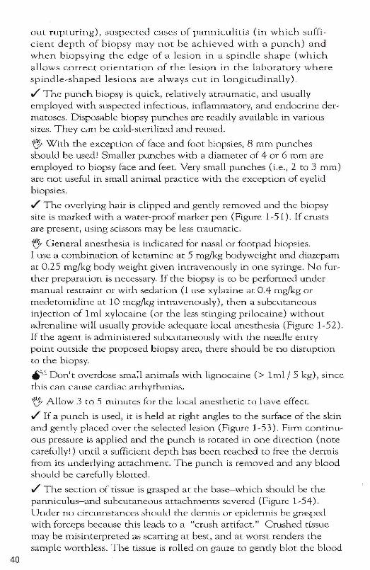
./ To determine if a cat or dog creates hair loss by licking or rubbing, or if the hair falls out for another reason. If the animal is pruritic and licks the hair off, the tips of the hairs are broken off (Figure 1-49). Any trauma to the hair shaft, such as occurs in dermatophytosis or anagen defluxion, may also cause hair with broken ends. If the hair falls out for other reasons, the tips are tapered (Figure 1-50) .
./ Trichograms are used routinely in human dermatology to evaluate alopecias, but their usefulness in veterinary dermatology has not been explored in any great detail and is hampered by the marked variations in breed characteristics.
h A trichogram is most useful to determine if bald cats that presen t with a history of "nonpruritic" alopecia are really so-called "closet lickers" or "hidden groomers" (in which case the hair-shaft ends will be fractured) or if the hair falls out due to telogen effluvium or, very rarely, for hormonal reasons .
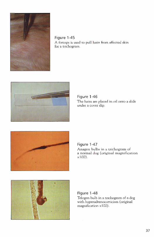
Figure 1-45A for ceps is used to pull hairs from affected sk infor a tri chogram.
L-, Figure 1-46The hair s are p laced in oil on to a slideunder a cov er slip.
Figure 1-47A n agen bu lbs in a t rich ogr am o fa n orma l dog (ori gi n al m agn ifica t ionx l OO)
Figure 1-48Telogen bulb in a trich ogram of a dogwith. h yperad renocortic ism (o riginalmagnificat ion xlO O).
37
38
h Trichograms may also be used to diagnose canine demodicosis. If mites are found microscopically, the diagnosis is confirmed. However, if no mites are present, demodicosis cannot be ruled out' h Trichograms can also help to diagnose color-diluent alopecta. In this disease the melanin in the hair shaft is present in big clumps rather than finely dispersed as in norma l pigmented hair.
Biopsy Indication ./' Any skin that appears Llnusual to the clinictan should be biopsied .
./' A biopsy should also be considered if lesions fail to respond to
appropriate empiric therapy .
./' Nodules are possibly neoplastic and should be biopsied
./' The presence of any suspected disease for which treatment is expensive and/m life-threatening should be confirmed histopathologically .
./' One of the lTlajor reasons to perfonn a skill. biopsy is to rule out other diagnoses (HI think this is an allergy but ... "). In such a situation, the biopsy report of "chronic hyperplastic dermatitis with mononuclear perivascular infiltrate" has at lea.<;t ruled our COlnrnon infectious agents and unusual dermatoses. A supportive pathologic diagnosis interpreted in conjunction with the clinical impressions may be just as useful as a confirmatory diagnosis.
Technique
Selection of the Site
./' Selection of the site requires careful examinarion of the entire animal for the most representative samples, identification of the primary and secondary lesions present, and the fonnation of a list of differential diagnoses before biopsy.
h With. the exception of a solitary nodule, we recommend taking multiple tissue sam.ples. These should include primary lesions if present, contain a representative range of lesions, and above all, should be taken and haJ.1.dled carefully. A normal sample of haired skin should also be included .
./' Depigmenting lesions should be biopsied in an area of active depigmentation (gray color) rather than the final stage (white or pink color) .
./' Alopecia should be biopsied in the center of the worst area as well as in junctional and normal areas.
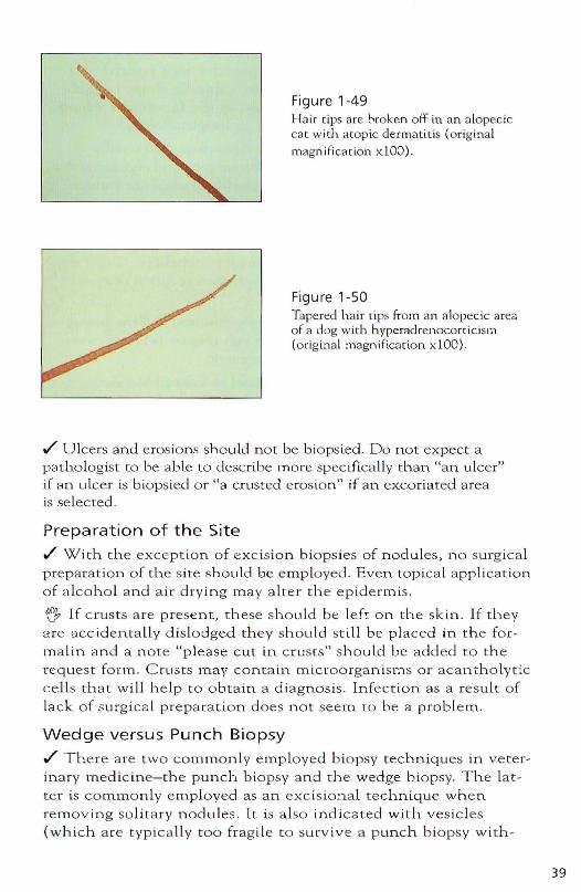
Figure 1-49 Hair tips are hroken off in an a lopecic cat with <Iwpic dermatltis (original magn i(icat.ion x lOO).
Figure 1-50 Tapered hair rips fTom an alopecic area of a dog with hyperadrenocorticism (original magniflcation x 100) .
./ Ulcers and eros ions should not be bi ops ied. Do not exp ect a pathologist [0 be able to describe m o re specifically than "an ulce r" if an ulcer is biopsied o r "a crusted eros io n" if an excoriated area is selec ted.
Preparation of the Site
./ With the exception of excision biops ies of nodules, n o surgical preparat ion of cil.e site should be employed. Even topica l applicatio n of a lcoho l a nd a ir drying ma y a lter the ep idermis.
fJ If crusts are presen t , these sh o uLd be left on the skin. If they are aCC identa Lly dislodged they should still be placed in t h e formalin an d a n ote "please cut in c rusts" should be added t o the reques t form. C rusts may contain microorganisms or acan th o lyt ic cells that will heLp to obtain a d iagn os is. Infection as a result of Lack of surg ica l pre p a ra tion does n o t seem to be a proble m .
Wedge versus Punch Biopsy
./ The re are two commonly empLoyed biopsy techniques in ve terinary medic ine-the punch biopsy a nd the wedge biopsy. The Latter is comrnonly e mployed as an exc is ional technique whe n removing solita ry nodu les . It is a lso indica ted with vesicles {which a re typically too frag ile to su rvive a p unch b iopsy with -
39
40
out rupturing), suspecte d cases of panniculitis (in which sufficient depth of biopsy may not be achieved with a punch) and when biopsying the edge of a lesion in a spindle shape (which allows correct orientation of the lesion in the laboratory where spindle-shaped lesions are always cut in longitudinal! y) .
./ The punch biopsy is quick, relatively atraumatic, and usually employed with suspected infectious, inflamm.atory, and endocrine dermatoses. Disposable biopsy punches are readily available in various sizes. They can be cold-sterilized and reused.
~ With tlLe exception of face and foot biopsies, 8 mm punches should be used I Sn1.aller punches with a diameter of 4 or 6 rnrn are employed to biopsy face and feet. Very small punches (i.e., 2 to 3 mm) are not useful in small animal practice with the exception of eyelid biopsies .
./ The overlying hair is clipped and gently removed and the biopsy site is marked with a water-proof marker pen (Figure 1-51). If crusts are present, using scissors may be less traumatic.
~ General anesthesia is indicated for nasal or footpad biopsies. I use a combination of keramine at 5 mg/kg bodyweight and diazepam at 0.25 mg/kg body weight given intravenously in one syringe. No further preparation is necessary. If the biopsy is to be perfomled under manual restraint or with sedation (I use xylazine at 0.4 mg(kg or medetomidine at 10 mcg(kg intravenously), then a subcLltaneoLls injection of Iml xylocaine (or the less stinging prilocaine) without adrenaline will usually proVide adequate local anesthesia (Figure 1-52). If the agent is administered subcutaneously with the needle entry point outside the proposed biopsy area, there should be no disruption to the biopsy.
ttji:' Don't overdose small animals with lignocaine (> Iml / 5 kg), since this can cause cardiac an-hythmias.
~ Allow 3 to 5 minutes for the local anesthetic to have effect .
./ If a punch is used, it is held at right angles to the surface of the skin and gently placed over the selected lesion (Figure 1~53). Firm continuOllS pressure is applied and the punch is rotated in one direction (note carefully!) until a sufficient depth has been reached to free the dennis from its underlying attachment. TILe plUlch is removed and any blood should be carefully blotted .
./ The section of tissue is grasped at the base- which should be the panniculus-and subcutaneous attachments severed (Figure 1-54). Under no circumstances should the dermis or epidermis be grasped with forceps because this leads to a "crush artifact." Crushed tissue may be misinterpreted as scan-ing at best, and at worst renders the sample worthless. The tissue is rolled on gauze to gently blot the blood
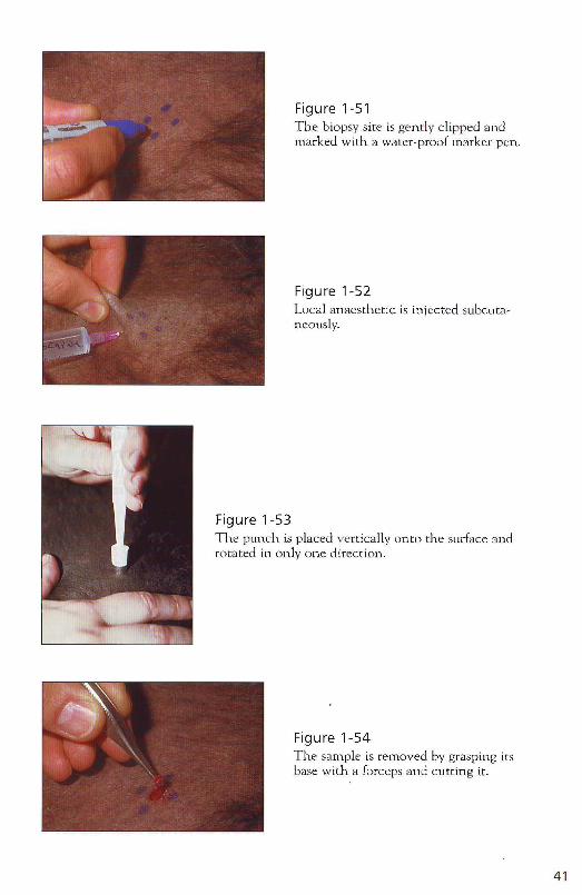
Figure 1-53
Figure 1-51 The biopsy site is gentl y clipped Clnd marked with a water-proof marker pen.
Figure 1-52 Local anaesthetic is injected subcutaneously.
The punch is placed vertically onw the surface and row ted in only one dirccrion .
Figure 1-54 The sample is removed by grasping its base with a forceps and cutting it.
41

42
from its surface. A thin sample should then be placed with the pan~ niculus face down-onto a rigid piece of cardboard or broken tongue depressor (Figure 1~55). This prevents the tissue from curling when placed in the formalin optimizing the interpretation by the patholo~ gist. Thick samples may be placed in formalin without such SUppOl-t .
./ The unit of tissue and cardboard is placed in 10% formalin (tissue side down) and allowed to fix for several hours before sec~ tioning. The volume of formalin required is at least 10 times the volume of the sample. Nodules should be sectioned into 1 cm thick pi.eces to a llow adequate penetration of the formalin into the center of the lesion.
Submission of Biopsy Samples
~ Careful completion of the appropr iate skin biopsy request form, including history and physical examination, will greatly improve the chances of a diagnostic report.
~ A list of differential diagnoses is important with any clinical case but is essential with dermatologic patients. Seborrhea or draining tracts can be the result of a wide range of d isease processes. This list is i.mportant for the clinician to ensure that he or she has considered all options and obtained as much information as possible frOln both pet and owner as necessary before taking the biopsy. It is also important for [he pathologist and may a id in choosing speCial stains to rule out or confirm unusual diseases.
Serum Testing for Allergen-specific IgE Indication Useful if the owner of an atopic dog or cat diagnosed by history, clinical examinat ion, and ruling out of differential diagnoses, is either curious about what causes the problem or is interested in allergen~specific immunotherapy.
Interpretation ./ Different serum tests are ava il able. Laboratory techniques have improved over the years and serum testing has become an alternative to intradermal skin testing for many small an imal practitioners. However, teStS vary in their sensitivity and speci~ ficity so that careful se lection of an appropriate test is important.
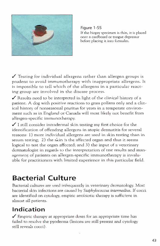
Figure 1-55 If the biopsy spec imen is thin, it is placed onto a cardboard or tongue depressor before placing it into formCllin .
./ Testing for individual allergens rather than a llergen groups is prudent to avoid immunotherapy with inappropriate a llergens . It is impossi.ble to te ll which of the a llergens in a particular reactin g group are involved in the disease process .
./ Results need [0 be interpreted in light of the clinical history of a patient. A dog with positive reactions to grass pollens only and a clinic a l history of nonseasonal pruritus for years in a temperate environment such as in England Or Canada will most likely not benefit from a llergen-spec ific immunotherapy .
./ I still consider intradermal skin testing my first choice for the identification of offending allergens in atopic dermatitis for several reasons: 1) more individual allergens are used in skin testing than in sewm test ing; 2) the skin is the affected o rgan and thus it seems logical to test the organ affected; and 3 ) the input of a veterinary dermatologist in regards to the interpretation of test resu Its and managemen~ of patients on allergen-specific immunotherapy is invaluable for practitioners with. limited experience in this particular field .
Bacterial Culture Bacterial cultures are used infrequently in veterinary dermatology. Most bacteria l skin. infections are caused by Staphylococcus intennedius. If cocci are identified on cytoIOb'Y, empiric antibiotic therapy is sufficient in almost a ll patients.
Indication ./ Empiric therapy at appropriate doses for an appropri:ate time h as failed to resolve the pyoderma (les ions are st ill present and cytOlogy still revea b cocci).
43
44
./ Numerous rod~shaped bacteria are identified on cytology samples from ear canals. These organisms may also rarely playa role in cuta~ neous infections of patients clinically not responding to empiric therapy.
Procedure ./ Swabs are taken from ear canals as described for cytology samples .
./ Aspirates from intact pustules are useful in patients with superficial pyoderma.
h Swabs from the skin surface to culture organi.sms from patients with deep pyoderma are not suitable. Samples are raken in a similar manner to that used in biopsies under aseptic condi~ tions (scrub the skin surface and use sterile instruments and gloves). The upper half of the tissue sample with the epidermis and hair is cut off and the lower half is submitted in a sterile container placed on a sterile gauze pad soaked in a sterile saline solution for maceration culture. This prevents overgrowth of the culture by surface bacteria not relevant to the deep infection.
~ Each sample for culture and sensitivity should be accompa~ nied by cytologic examination, and culture resul t s must be in[er~ preted in relationship to cytologic findings.
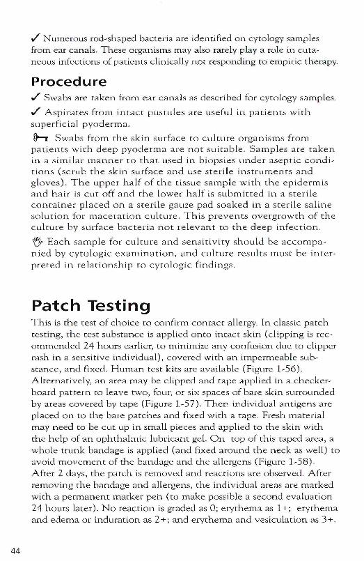
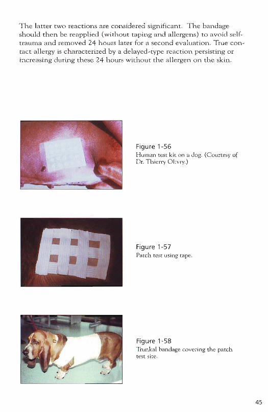
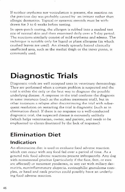
Patch Testing This is the test of choice to confirm con tan allergy. In classic patch testing, the test substance is applied onto intact skin (clipping is rec~ ommended 24 hours earlier, to minimize any confusion due to clipper rash in a sensitive individual), covered with an impermeable sub~ stance, and ([xed. Human test kits are available (Figure 1 ~ 56). Alternatively, an area may be clipped and tape applied in a checker~ board pattern to leave two, four, or six spaces of bare skin surrounded by areas covered by tape (Figure 1~57). Then individual antigens are placed on to the bare patches and fixed with a tape. Fresh material may need to be cut up in small pieces and applied to the skin with the help of an ophthalrn.ic lubricant geL On top of this taped area, a whole trunk bandage is applied (and fixed around the neck as well) to avoid movement of the bandage and the allergens (Figure 1~58). After 2 days, the patch is removed and reactions are observed. After removing the bandage and allergens, the indlvidual areas are marked with a permanent marker pen (to make possible a second evaluation 24 hours later). No reaction is graded as 0; erythema as 1+; erythema and edema or induration as 2+; and erythema and vesiculation as 3+.
T h e la t ter two re actions are con sidered sign ifican t . T h e ban dageshould th en be re app lied (wi thout tap in g and a llergens) to avoid sel ft ra uma an d re moved 24 h ours la ter for a second evaluat io n . T ru e contact a llerg y is characterized by a d e layed -typ e reac t ion pe rsistin g o rin cre asin g durin g these 24 h ours without th e a llergen o n the sk in.
Figure 1-56Human test kit on a dog. (C ourtesy ofDr, Thierry O livry.)
Figure 1-57Patch test using tape.
Figure 1-58Trunkal bandage covering th e pa tchtest site.
'-" '
45
46
If neither erythema nor vesiculation is present, the reaction on the previous day was probably caused by an irritant rather than allergic dermatitis. Topical or systemic steroids must be withdrawn for 3 to 6 weeks before testing.
rn open-patch testing, the allergen is rubbed into a marked test site of normal skin and then examined daily over a 5-day period. The reactions Similarly consis t of mild erythema and edema. The technique is suitable only for liquid or plant allergens (in which crushed leaves are used). An already sparsely haired clinically unaffected area, such as the medial thigh or the inner pinna, is commonly used.
Diagnostic Trials Diagnostic trials are well accepted tests in veterinary dermatology. They are performed when a cercain problem is suspected and the trial is either the only or the best way to diagnose (he possible underlying disease. A response to the trial confirms the diagnosis in some instances (such as the scabies treatment trial), but in other ins ranees a relapse afcer discontinuing the trial with subsequent resolution on restarting the trial is diagnostic (such as in elimination diets). If there is no response to a well-conducted diagnostic erial, the suspected disease is extremely unlikely (which helps veterinarian, owner, and patient, and needs to be emphasized to clients frustrated by the lack of response).
Elimination Diet Indication An elimination diet is used to evalLtate food adverse reaction which can occur with any food fed over a period of time. As a general rule food adverse reactions present infrequently. Any dog with nonseasonal pruritLis (particularly if the face, feet, or ears are affected) or recurrent pyoderma, or any cat with miliary dermatitis, noninflammatory alopecia, eosinophilic granuloma complex, or head and neck pruritus could possibly have an underlying food adverse reaction.
Procedure h An e limina tion diet for dogs con s ists of one protein source and one carbohydrate source previously not fed! This means that the e limination diet for a particular patient is d ete rmined by the diet fed so far to this a nima l. Cats a re fed only o n e pro~ tein without the carbohydrate source to enhance compliance .
./ Poss ible options for proteins a re c h ick e n, turkey, duck, veni~
son, mutton, beef, h o rse, buffa lo, rabb it, h a re , kangaroo, emu, v a rious sorts of fish, am.ong others. Carbohyd ra tes may consist of rice , potatoes, swee t potatoes, beans, or others .
./ T he diet chosen needs to be fed exclu sively I Concurrent h eart-w o rm prophylaxis or supplements must not contain food flavor extracts .
./ It may t ake 6 to 8 weeks befo re a response becomes evident.
h After initial improvement, a rech a lle nge with the n ormal diet previously fed is essential b ecause improvem.ent m ay result froln oth er factors such as seasonal or env iro nmenta l ch anges or concurrent medication. If a re lapse occurs w ithin 2 weeks and clinica l signs resolve again after reinstitution of the elimination diet , the diagnosis is confirmed.
Tips to increase compliance
./ Warming the food may improve pat ient compliance .
./ Spices such as garlic or salt (in sm a ll amounts) may a lso be beneficial to improve paLatibiLity.
'f!} If the anima l (and o wner) is used to treats, the h ab it should be continued in a m odified fashio n to prevent feeding of inappropr iate protein s. Little pieces of the selected m eat protein can be fried and kept in the fridge for llse as treats . The selected meat can be dried (in the oven or microwave ) and given. as treats. If an an ima l is rece iving potatoes in the diet, then fried pieces of potato may be used (so long as they are not fried in butter, but in a plant-derived oi l). If rice is chosen, rice cakes may be an addit io n a l option .
./ If bones are part o f the normal die t , bones of the m ea t se lected for the elirnination diet may be fed if ava ilab le .
./ Good client communication is essentia l. It must be made clear that an occasiona l sllp in feeding h abits (as little as o n ce or twice weekly of a very small amount of a different protein) may d estroy all th e effort .
./ It may be worthwhile to advise neighbors about the diet as well.
47
48
./ If a home-cooked diet is not an option, a commercial diet consisting exclusively of a protein source and a carbohydrate SOUTce not previously fed may be considered. The same principles apply to commercial as to home-cooked elimination diets. However, some animals with food adverse reactions may be missed when using commercial diets.
After a diagnosis of food adverse reaction is conflrmed, the client has two options: 1. To continue a commercial elimination diet forever-the more convenient option; 2. A home-cooked diet. It should be properly balanced (the help of a veterinary nutritionist may be indicated) .
./ Identifying of the offending allergen allows a more varied diet and is achieved through a sequential rechallenge with proteins formerly fed. Beef, lamb, chicken, or cheese and milk products are added to the elimination diet one at a time for 2 weeks each. If a relapse occurs within the first 2 weeks (many patients show symptoms within the first 2 days), the protein is discontinued until the patient's condition settles. That particular protein is avoided in the future. After 2 weeks of a given protein without clinical symptoms, a reaction to this protein is ruled out and it may be fed in the future. Some dogs will tolerate any homecooked diet, but relapse on commercial diets may be caused by a reaction to additives or preservatives.
Insect Control Trial Indication An insect cancro I trial may be used in any patient with suspected insect-bite hypersensitivities. Most animals with insect-bite hypersensitivities will be allergic to fleas. Clients generally accept these trials more readily when they are labeled "insect control trials" because many do not believe fleas cause the problem, whereas most will accept ants or mosquitoes as a possible cause. Any dog with pruritus, alopecia, and/or a papular or crusty rash in the tailbase or inguinal area, and any cat with miliary dermatitis, noninflammatory alopecia, or eosinophilic granuloma complex luay benefit from an insect control trial. Mosquito bite hypersensitivity in the cat is characterized by papules and crusts on the nose, pinnae, and foot pads. A trial using insect repellents may be beneficial co these animals.

Procedure ./' The patient should be treated regularly with an insecticide. In a diagnosti c trial, 1 often inc rease the frequency of administration above t h e m a nufacturer's recommendations. Fipronll spray, imida~ cloprid, permethrin, a nd selamectin spot~ons a re administered every 2 weeks. Pyrethroid sprays a re adm ini stered daily d epending on the product. N itenpyram tablets are given either daily o r every other d ay. Which products to use depends on the individua l cir~ cumstances . M o re details are provided o n page 138 .
./' At the start of the trial, treat the a nitnal's environment with an insect~deve lopment inhibi tor su ch as methoprene , fenoxycClrb, or pyriproxifen. M o re details are provided on page 138 .
./' Contact anima ls (ei ther Living in the same household or those that v is it on a regular basis) mus t be treated as w e ll, although the frequency between adulticide app Licatio n s may b e increased to the lua nufacturers' recommendat ions .
./' At the start of the trial, I often prescribe 5 to 7 d a ys of pred~ nisolone at 1 mg/kg bodywe ight dai ly to hasten clini.cal response.
If there is good respon se to the triaL, insect~bite hypersensitivity is present a nd insect contro L may be tape red to the minimum required.
~ R e m e mber th a t the req uired minimum. treatment typica lly varies season a lly, as does the insecc Load.
Scabies Treatment Trial Indication Any pruritic dog or ca t could poss ibLy be infested with Sarcopte.s scabiei o r Notoedres cati, respect iv e ly, part icuLa rly if the pruritus was of sudden o n set o r if pinnae, vencrum, and elbows a re pruri~ tic. With spot~ons used for flea contro l , I have seen patients with. pruritus and les ions limite d to ventrum and Lower legs. Negative superficial skin scrap ings do not rule o ut scabies (p. 26) so tria l treatment is indicated in any patient with s uspected scabies irre~ spective of negative skin. scraping results. In as much as C hey letiella spp. and Otodectes cynotis are sensitive to the sam e antiparasitic agents, a scabies treatment tria l will be useful for these parasites as well.
49
50
Procedure ./ Several treatments for scabies are available but many of them are not Labeled for thls use .
./ Topical treatments include ivermeccin, lime sulfur dlPS, amitraz, and other antiparasitic rinses . They are used weekly for 4 weeks. More details are given on page 133 .
./ Systemic therapy may be undertaken with selameccin, ivermectin or milbemycin. Treannenc details are outlined on page 133 .
./ All animals in contact with the patient need to be treated as well!
./ Initial deterioracion during the first days of treatment may occur. Treat with glucocorticoids daily for 3 co 4 days at 1 mg/kg body weight .
./ Remission should be achieved within 4 weeks although some patients may need extended treatment for up to 8 weeks.

Section 2
The Approach to Common
Dermatologic Presentations
51
52
In this section, I offer an approach to various COlnmon presenta~ tions in veterinary dermatology. I begin each topic in this sec~ tion with general comments followed by tables containing the most common differential diagnoses, their clinical features, diagnostic procedures of choice, treatment, and prognosis. I have attempted to list diseases in order of prevalence. Diseases marked with an (*) and a colored screen are potentially difficult to diagnose or their management often requires considerable experience to achieve the best possible outcolne. You lnay con~ sider offering your client a referral to a veterinary dermatologist if you do not feel comfortable diagnosing or treating this disease.
This is not a textbook of veterinary dermatology so these tables do not contain all possible details but rather a concise overview concentrating on the most important features. Similarly, the flow charts at the end of each topic are concise and simplified to maximize the benefit for the busy small animal practitioner. They will be useful in most instances, but remember that SOlne of your clients may not have read the textbooks. Even though this information is aimed at helping you as competent veterinarians to reach a diagnosis and formulate a treatment plan, your critical acumen, examination, and cOlumunication skills remain the most crucial instrulnents for Sllccess in YOUI daily practice.
The Pru ritic Dog
Key Questions
All questlons discussed in Section 1 (pages 2 ~ lO) may be r e le~
vant for a p ru r itic parieIn.
Differential Diagnoses If lesions are prese n t, see p age 58, The Dog with Papules, Pustu les and C ru sts. If n o les ions a re present, differential dlagn oses a re listed in Table 2~ 1 .
53
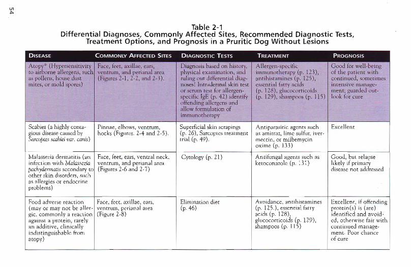
Table 2-1Differential Diagnoses, Commonly Affected Sites, Recommended Diagnostic Tests,
Treatment Options, and Prognosis in a Pruritic Dog Without Lesions
DISEASE COMMONLY AFFECTED SITES DIAGNOSTIC TESTS TREATMENT PROGNOSIS
Atopf (Hypersensitiv ity Face, feet , ax illae, ears , Diagnosis based on history, A llergen -spec ific Good for well-beingto air orne allergens, such ventru m, and perian al area physical exa mina tion, and immunotherapy (p . 123 ), of the patient withas po llens, house dust (Figures 2-1,2-2, and 2-3 ). ruling out differen tial diag- an tihistamines (p. 125), con tinued, some-ti mesmites, or mold spores) noses! Intradermal skin test essentia l fat ty acids inten sive m anage-
or serum test for allergen- (p . 128) , glucocorticoids merit ; guarded ou t-specific IgE (p, 42) identify (p , 129), sha mpoos (p. 115) look for cureoffending allerge ns andallow formulation ofimmunotherapy
Scabi es (a high ly conta- Pinnae, elbows, ven trum, Supe rficial skin scrapings A ntiparasitic agen ts such Excellent~iOUS disease ca used by hock s (Figures. 2-4 and 2-5 ). (p. 26), Sarcoptes treatment as arnit raz, lime sulfur, iver -arcoptes seabiei vaT. canis) trial (p. 49 ). rnect in , or milbem ycin
ox ime (p . 133)
Malassezia derm atitis (an Face , feet, ears, veneraI neck, Cy to logy (p . 21) Anti fungal agen ts such as Good, but relapseinfection with Malasseva ventru rn, and perian al area ketoconazole (p. 131) likely if primarypachyd.ermaris secondary to (Figures 2-6 and 2-7) d isease not addressedothe r skin d isord ers, suchas allergies or endocrinepro blems)
Food adverse react ion. Face, feet , axi llae , ears, Elimination diet Avoidance , an tihista mines ExceHen t, if offend ing(mayor may not be aller- ven trum, per ian al area (p . 46) (p. 125.), essen t ia] fatty prot e in (s) is (are)gic, co mm on ly a reac t ion (Figure 2-8) acids (p. 128) , identified and avoid -agains t a protein , rarely glucocorrico ids (p. 129), ed, ot herwise fair withan add it ive , clinically sha mpoos (p. 115) continued manage-indis tinguish ab le from mem oPOOt chancea to py) of cure
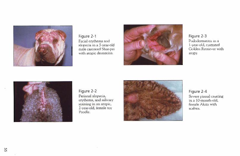
Vl V1
Figure 2-1 Facial erythema and alopecia in a 5-year-old male castrated Shar-pei with atopic dermatit is.
Figure 2-2 Perianal alopecia, erythema, and salivary staining in an atopic, 2-year-old, fema le toy Poodle.
Figure 2-3 Pododermatitis in a l -year-o ld, cclstrated Golden Retriever with atopy.
Figure 2-4 Severe pinnal crusting in a to-monch-old, female Akita with scab ies.
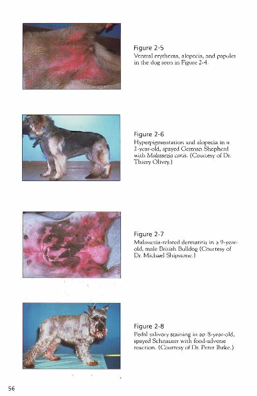
56
Figure 2-5 Ventral erythema, alopecia, and papules in the dog seen m Figure 2-4.
Figure 2-6 Hyperpigmentation and alopecia in a 2-year-old, spayed German Shepherd with Maia.ssezja canis. (Courtesy of Dr. Thiery Olivry.)
Figure 2-7 Malassezia-related dennatitis in a 9-yemold , male British Bulldog (Courtesy of Dr. Michael Shipswne .)
Figure 2-8 Pedal sa livary staining in an 8-year-old, spayed Schnauzer with food-adverse reaction. (Courtesy of Dr. Peter lhrke.)
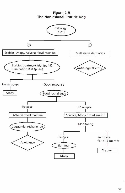
Figure 2-9The Nonlesional Pruritic Dog
Scabies, Atopy, Adverse food reactionI
Malassezia dermatitis
No response
~Good response
Relapse No relapse
IAdverse food reaction Scabies, Atopy out of season
,
Remissionfor> 12 months
~
Monitoring
II I
Relapse
Skin test
Sequential rechallenge
57
58
The Dog vvith Papules, Pustules, and Crusts
Key Questions
./ What is the breed of this patient? (p. 2)
./ How old was this patient when clinical signs were first recognized? (p. 3)
./ How long has the disease been present and how did it progress? (p. 3)
./ On which part of the body did the problem start? (p. 4)
./ Is the animal itchy? (p. 5)
./ Is the disease seasonal? (p. 6)
./ Are there other clinical signs, such as sneezing, coughing, or diarrhea? (p. 7)
./ What do you feed the animal? Was a special diet used in the past? (p. 7)
./ Are there any ocher animals in the household? (p. 8)
./ Does anybody in the h.ousehold have skin disease? (p. 8)
./ Was the disease treated before? If so, which drugs were used and how successful was treatment 7 (p. 8)
./ What is used for flea control currently 7 (p. 9)
./ When was the last medication given? (p. 9)
Differential Diagnoses Papules may develop into pustules and crusts, and any dog with an acute papular rash may eventually show pustules or crusts. Some diseases are characterized by papules that do not typically develop further into pustules (such as flea-bite hypersensitivity); other diseases typically show crusting as their predominant symptom (such as zincresponsive dermatitis). Tables 2-2, 2-3, and 2-4 list the major differential diagnoses for dogs with papules, pustules, and crusts. Lesions may be follicular or nonfollicular (Figure 2-10). Follicular papules and pustules indicate a pathologic process concentrating on the hair follicle, most commonly bacterial folliculitis, demodicosis , or derrnato-
phytosis. N onfollicular le sions m ay indica t e pathologic p rocessesconce n trat ing on. the ep iderm is, dermis, o r d erma -epidermal junction.such as superf ic ia l spreading pyoderma, flea-bite, con tact hypersensit iv ity, O r Immune-mediared ski n diseases. Be a ware that some n o rifollicular proces ses m ay occ as io nally involve h air follicles as well.
Nonfollicular papule and pustule
Foll icular papule and pustule
...• ./ .'---' .
~/
Figure 2-10
59
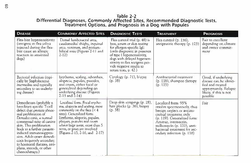
ma
Table 2-2Differential Diagnoses, Commonly Affected Sites, Recommended Diagnostic Tests,
Treatment Options, and Prognosis in a Dog with Papules
DISEASE COMMONLY AFFECTED SITES DIAGNOSTIC TESTS TREATMENT PROGNOSIS
Flea-bite h ypersensitivity Dorsal lumbosacral area , Flea control trial (p. 48) is Flea contro l (p. 136), Fair to excellent(antigens in flea saliva caudomedial thighs, inguinal best, serum or skin testing antipruritic therapy (p . 123) de pending on climateinjected during th e flea- area, ventrum, and perium- for allergen -specific IgE and owner commit-bite cause an allergic bilical area (Figures 2-11 and (only diagnostic in presence menrreaction in sensitized 2-12) of type I hypersensitivity,dogs) dogs with delayed hvpersen-
sitivity to flea anti gens pro-VIde negative results toserum tests, p. 42.)
Bacterial infection (rvpi- Erythema , scaling, seborrh ea, Cytology (p. 21) , biopsy Antibacterial treatment Good , if underlyingcally by Staphylococcus alopecia, papules, pustules, (p.38) (p . 118) ,shampoo therapy d isease can be idenri -intennedius and typically and crusts , either focal or (p.lIs) fied and treatedsecondary to an underlv- generalized depending on appropriately. Relapseing disease) underlying disease (Figures like ly, if this is not
2-13 and 2-14) possible
Demodicosis (probably a Localized form: Focal erythe- Deep skin scrapings (p. 28), Localized form : 95% Fairhereditary specific Tccll rna, alopecia and scaling, most hair plucks (p . 36), biopsy resolve spontaneously, thusdefect that permits abnor- commonly on the lace « 4 (p.38) benign neglect or antimi-mal proliferation of Sites). Generalized form: crobial treatment on lyDemodex canis, a normal Erythema, alopecia, papules, (p . 119). Genera lized form:commensal mite of canine plaques, pustules and crusts Amitraz, ivermecrin,skin. This proliferation where large areas, more than 5 milbemycin (p. 133) , anti-leads to a furthe r parasi te- areas, or paws are involved bacterial treatment for sec-induced imrnunosuppres- (Figures 2-IS, 2-I6, and 2-17) ondary infection (p . 118)sion. Adult-onset dernodi-cosis frequently secondaryto hormonal diseases, neo-plasia, steroids , or otherchem otherapy. )
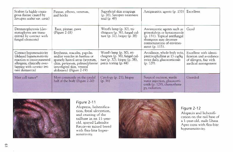
Scabies (a highly coma- Pinnae, elbo ws, ven trurn, Superfic ial skin scrapings Antiparasitic agent s (p. 133) Excellentgious disease caused by and hocks (p. 26) , Sarcoptes treatmentSarcoptes scabieivaT. canis) erial (p. 49)
Dermatophytosis (de r- Face, pinnae, paws Wood's lamp (p. 30), rri- Antimycot ic agen ts such as Goodrnat ophyres are trans- (Figure 2-18) chograiu (p. .36 ), fungal cui- griseofulvin or ketoconazoiemitred by contact with Me (p, 32), biopsy (p. 38) (p . 131) . Top ical antifungalfun gal elemen ts ) shampoos may decrease
con tam ination of en viro n-merit (p . 115 ),
Contact hvpersensitiviry Ery thema, macu les, papules Wood's lamp (p, 30) , tri- Avoidance , whole-body suits, Excellen t with identi-(delayed hypersensitivity and/or vesicles in hairless or chogram (p, 36), fungal cul- pen roxyphvlline at 15 mg/kg fication and avo idancereaction to environmental sparsely haired areas (scrotum, rure (p 32) , biopsy (p. 38 ), twice daily, glucocorticoids of allergen, fair withallergens, c1 in ically over- chi n, perineum, palmar/pla ntar patch resting (p. 44) (p. 129). medical managementlappi.ng with cont act irri- in terdigital skin , ven traltan t dermatitis) abdomen) (Figure 2-19)
Mast cell tumor" Most commonly on the caudal C ytology (p. 21), biopsy Surgical exc ision, sterile Gu ardedha lf of the body (Figure 2-20) (p. 38) water injection , glucocort i-
coids (p. 129) , chemoth era-py, radiation,
---; A
O'l
Figure 2-11A lopecia, lichenifica t io n , focal ulcera tion ,and c rusting o f therailbase in an Ll- yea ro ld, sp ayed La bra do rRetrie ver m ixed breedwith flea-b ite h ypersen sit iv ity.
Figu re 2-12Alopec ia and lichen ification on the ta il base ofa I -ve ar-old , male LhasaApso cross with flea-biteh ypersensit ivit y.
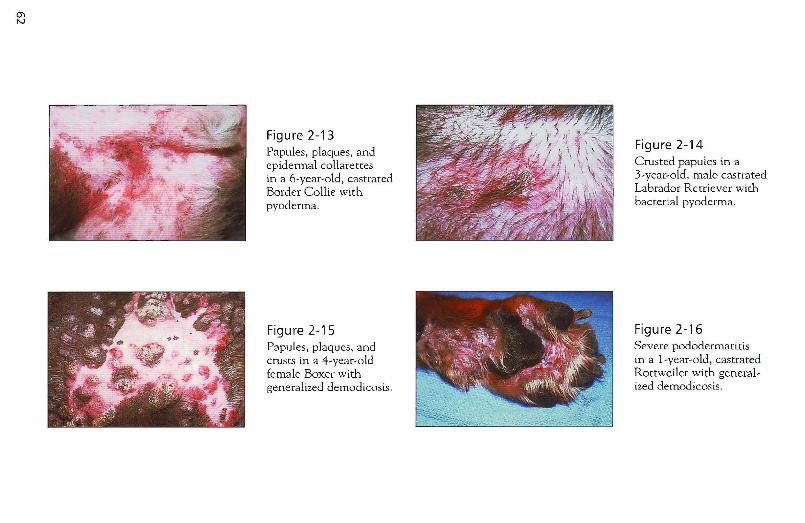
Figure 2-13 Papules, plaques. and epidermal collarettes in a 6-year-old. castrated Border Collie with pyoderma.
Figure 2-15 Papules, plaques, and crusts in a 4-year-old female Boxer with generalized demodicosis.
Figure 2-14 Crusted papules in a 3-year-old, male castrated Labrador Retriever with bacterial pyoderma.
Figure 2-16 Severe pododerm.atitis in a l-year-old, castrated Rottweiler with generalized demodicosis.
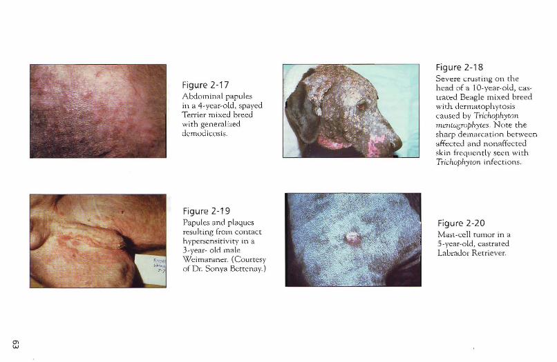
Figure 2-17 Abdominal papules in a 4-year-old, spayed Terrier mixed breed with generalized demodicosis.
Figure 2-19 Papules and plaques resulting from contact hypersensitivity in a 3-year- o ld male Weimaraner. (Courtesy of Dr. Sonya Bettenay.)
Figure 2-18 Severe crusting on the head of a lO-year-old, castrated Beagle mixed breed with dermarophytosis caused by Tricho/lhyton mentagTophyces. Note the sharp demarcation between affected and nonaffected skin frequently seen with TrichophytOn infections.
Figure 2-20 Mast-cell tumor in a 5-year-old, castrated Labrador Retriever.
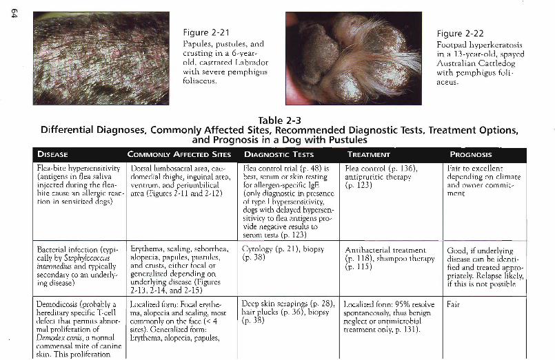
Figure 2-21Papules, pustules, andcrusting in a 6-yearold, castrated Labradorwith severe pemphigusfoliaceus.
Figure 2-22Footpad hyperkeratosisin a 13-year-old, spayedAustralian Carrledogwith pemphigus foliaceus.
Table 2-3Differential Diagnoses, Commonly Affected Sites, Recommended Diagnostic Tests, Treatment Options,
and Prognosis in a Dog with PustulesDISEASE COMMONLY AFFECTED SITES DIAGNOSTIC TESTS TREATMENT PROGNOSIS
Flea-bite hvpersensitivirv Dorsal lumbosacral area, cau- Flea control trial (p. 48) is Flea control (p. 136), Fair to excellent(antigens in flea saliva domedial thighs, inguinal area, best, serum or skin resting antipruritic therapy depending on climateinjected during the flea- ventrurn, and periumbilical for allergen-specific IgE (p. 123) and owner commit-bite cause an allergic rcac- area (Figures 2-11 and 2-12) (only diagnostic in.presence merittion in sensitized dogs) of type I hypersensitivity,
dogs with delayed hvperscn-sirivirv to flea antigens pro-vide negative results to
serum tests (p. 123)
Bacterial infection (rvpi- Erythema, scaling, seborrhea, C ytology (p, 2!), biopsy Antibacterial treatment Good, if underlyingcally by Staphylococcus alopecia, papules, pustules, (p.38) (p. 118), shampoo therapy disease can be identi-intermedius and rypically and crusts, either focal or (p.115) fied and treated appro-secondary ro an underlv- generalized depending on priate ly. Relapse likely,ing disease) underlying disease (Figures if th is is not possible
2-13,2-14, and 2-15)
Demodicosis (probably a Localizedform: Focal ervthe- Deep skin scrapings (p. 28) , Localized form: 95% resolve Fairhereditary specific Tccll rna, alopecia and scaling, most hair plucks (p. 36), biopsy spontaneously, thus benigndefect that pennits abnor- commonly on the face « 4 (p.38) neglect or antimicrobialmal proliferation of sites). Generalized form: treatment only, p. 131).Demodex canis, a norma! Erythema, alopecia, papules,commensal mite of canineskin. This proliferation
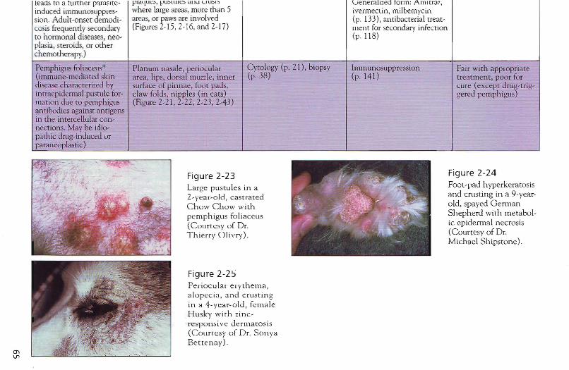
leads to a turtn er parasite- pl(lque~ , pustuies anu ,- ru~L') Lienernllzed torrn: Arrurraz,induced irnrnunosuppres- where large areas, more than 5 iverrnectin , milbemyci.n.sion. Adult-onset dernodi- areas, or pawsare involved (p. 133) , ant ibacterial treat-cosis frequently secondary (Figures 2-15, 2-16, and 2-17) rnen t for secondary infectionto hormonal diseases, neo- (p. 118)plasia, steroids, or otherchemotherapy.)
Pemphigus foliaceus* Planum nasale, periocular Cyto logy (p, 21) , biopsy Immunosuppression Fair with appropriate(immune-mediated skin area, lips, dorsal muzzle, inner (p.38) (p . 141) treatment, poor fordisease characterized by surface of pinnae, foot pads, cure (except drug-trig-imraepiderrnal pustule for- claw folds, nipples (in cats) gered pe mph igus)marion due to pemph igus (Figure 2-21, 2-22, 2-23,2-43)anti bodies against an tigensin the intercellular con-nections. May be idio-parhic drug-induced orparaneoplasric)
O'lV1
Figure 2-23Large pustu les in a2-year-old, ca stratedC how C h ow withpemphigus foli aceus(Courtesy of Dr.Thierry O livry) .
Figu re 2-2 5Per iocu la r e rythema ,a lo pec ia, and crustingin a 4- year-old , femal eHusk y with zincresponsi ve dermatosis(Courtesy of Dr. SonyaBettenay).
Figure 2-24Foot-pad hyperkeratosisand crusting in a 9-yearold , spayed Germa nShepherd with me tabolic epide rm al necrosis(Courtesy of Dr.Michael S h tpsto ne).
Table 2-4Differential Diagnoses, Commonly Affected Sites, Recommended Diagnostic Tests,
Treatment Options, and Prognosis in a Dog with Crusts
DISEASE COMMONLY AFFECTED SITES DIAGNOSTIC TESTS TREATMENT PROGNOSIS
Bacterial infection Dorsal lumbosacral area, cau- Insect control trial (p. 48), Flea control (p. 136), Fair to excellentFlea-bite hypersensitivity domedial thighs, inguinal serum or skin testing for antipruritic depending on climate(antigens in flea saliva area, venrrurn, periumbilical allergen-specific IgE (only therapy (p. 123) and owner commit-injected during me flea area (see Figures 2-11 and diagnostic in presence of menthite cause an allergic reac- 2-12.) rvpe I hypersensitivity, dogstion iJIsensitized dogs) with delayed hvpersensitivi-
ty to flea antigens are nega-rive on serum rests, p. 42)
Demodicosis (probably a Localizedform: Focal crythe- Deep skin scrapings (p. 28), Localized form: 95% resolve Fairhereditary specific Tcell rna, alopecia and scaling, most hair plucks (p. 36), biopsy spontaneously, thus benigndefect that permits abner- commonly on me face « 4 (p.38) neglect or antimicrobialmal proliferation of sites). Generalized form: treatment only, p. 131).Demodex canis, a normal Ervrhema, alopecia, papules, Generalized form: Arnirraz,commensal mite of canine plaques, pustules and crusts iverrnecrin, milbemycinskin. This proliferation where large areas, more than 5 (p. 133), antibacterial treat-leads to a further parasite- areas, or paws are involved ment for secondary infectioninduced irnmunosuppres- (Figures 2-15, 2-16, and 2-17) (p. 118)sian. Adult-onset demodi-cosis frequently secondaryto hormonal diseases, neo-plasia, steroids, or otherchemotherapy. )
Scabies (a highlv conta- Pinnae, elbows, venrrurn, Superficial skin scrapings Antiparasitic agents Excellentgious disease caused by and hocks (p. 26), Sarcoptes treatment (p.133)Sarcoptes scabiei var. canis) trial(p.49)
Pemphigus foliaceus* Planum nasale, periocular C ytology (p, 2 L), biopsy Immunosuppression Fair with appropr iate(immune-mediated skin area , lips, dorsal muzzle , inner (p. 38) (p, 141) treatment, poor fordisease characterized by surface of pinnae, foot pads, cure (except drug-tri g-intraepidermal pustule cla w folds, n ipples (in cats) gered pemphigus)formation due to pemphi- (Figure 2-21, 2-22, 2-23, 2-43)gus ant ibodies againstantigens in the Inrercell-ular connect ions, Maybeidiopath ic drug-induceder paraneoplastic ) ,-
en"-J
Metabolic ep idermal Muzzle, mucocutaneous june- Biopsy (p. 38) Antimicrobial treatment, Poornecrosis* t ions, distal limbs, foot pads, vita min and mineral supple-(pa thogenesis unclear) elbows, hocks, ventrum ment, h igh-quali ty prot ein,
(Figure 2-24) intravenous am ino acids
Dermatophytosis (de r- Face, pinnae, paws Wood's lamp (p, 30), tri- Antimycotic agents such as Goodrnarophyres are trans- (Figure 2-18) chogram (p. 36). fungal cul- griseofulvin or kerocon azolerni tted by co nt ac t with ture (p, 32 ), biopsy (p. 38) (p , 13 1) . Topical ant ifungalfunga l elements) sha mpoos may decrease
contaminati on of env iron-men t.
Zinc-responsive dermatitis Periocular, perioral , pinnae, Biopsy (p. 38) Zinc supp lement at ion, low- Fair(Zinc deficiency due to ch in, foot pads, planum dose glucocorrico ids [0
insufficient zinc in the nasale, pressure points increase zinc absorptiond iet or insufficient absorp- (Figure 2-25)tion of zinc, especially inarctic breeds)
Idiopath ic seborrhea* Otitis extern a, digital hyper- Biopsy (p. 38 ) Antiseborrheic shampoos G ood to guarded for(primary kerat in ization keratosis, dry flaky skin , or (p, 115), moisturizers, well-being, poor fordefect as autosomal reces- seborr heic dermati tis pre- retinoids, corticoste roidssive trait with decreased dominantly on face, feet, (p . 129 )
cure.
epidermal cell renewal ventral neck , and ventraltime and thus hyperprolif- abdomen (Figure 2-26)eration of epidermis, seba-ceous glands, and follicu-lar infundibulum.Secondary to inflamma-tion, endocrine disease, ornutritional deficiencies)
Dermato myositis (autoso- Erythema, scaling alopecia, Skin biopsy, muscle biopsy, Vitamin E (200-800 iu/day), Variesmal dominant in Collies mild crusting in face (particu- EMG pentoxyphylline (20 mg/kg Dogs typically will notand Shelties, first signs in lad y periocular area) eartips, q 12 h), for acute flares deteriorate furtherpuppies) carpal and tarsal regions, dig- predn isolon e (1-2 mg/kg q after 1 year of age.
its, tail tip, myositis, and in 2.4 h )severe cases, rn egasoph agus
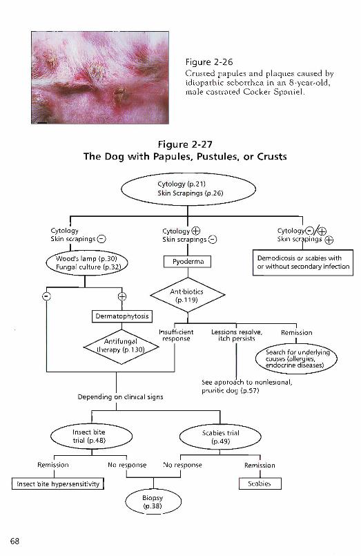
Figure 2-26Crusted papules and plaques ca used byidio path ic seborrhea in an 8-year-old,male castrated Cocker Spaniel.
Figure 2-27The Dog with Papules, Pustules, or Crusts
Cytology (p.21)Skin Scrapings (p.26)
Depending on clinical signs
CytologySkin scrapings <:)
Wood's lamp (p.30)Fungal culture (p.32
CytologyEBSkin scrapings <:)
Remission
Seeapproach to nonlesional,pruritic dog (p.57)
Insect bitetrial (p.48)
Scabies trial(p.49)
68
Remission No response No response Remission
II Scabies I
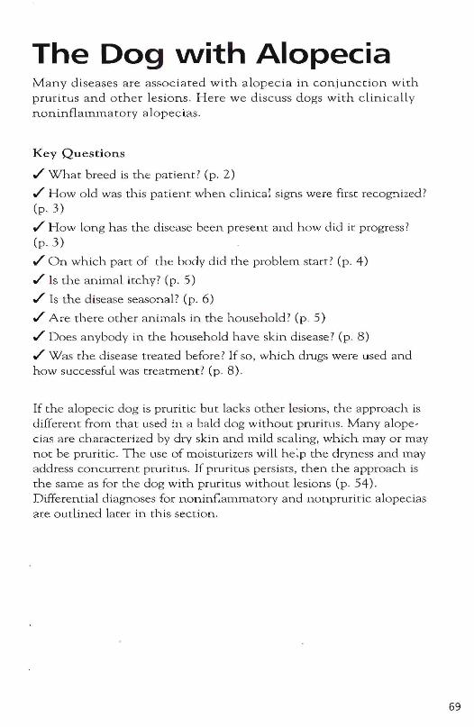
The Dog vvith Alopecia Many diseases are associated with alopecia in conjunction with pruritus and other lesions. Here we discuss dogs with clinicaLLy noninflamma tory 8 ]opecias.
K ey Questions
.I' What breed is the pat ient? (p. 2)
.I' How old was this pmient when clinic81 signs were first recogrtized? (p. 3)
.I' How long has the disease been present and how did it progress? (p.3)
.I' On which part of the body did the problem start? (p. 4)
.I' Is the animal itchy? (p. 5)
.I' Is the disease seasonal? (p. 6)
.I' Are there other animals in the household? (p. S)
.I' Does anybody in the household h8ve skin disease? (p. 8)
.I' Was the disease treated before? If so, whlch drugs were used and how successful was treatment? (p. 8).
If the alopecic dog is prurit ic but lacks othe r lesions, the approach is different from that lIsed in a bald dog without pruritus. Many alopec ias are characterized by dry skin and mild scaling, which mayor may not be pruritic. The LIse of moisturizers wil l help the dryness and may address concun-ent pruritus. If pnlritus persists, (hen the approach is the same as for the dog with pruritus without Lesions (p. S4). Differential diagnoses for noninflammatory and nonpruritic aLopecias are outlined Later in this section.
69
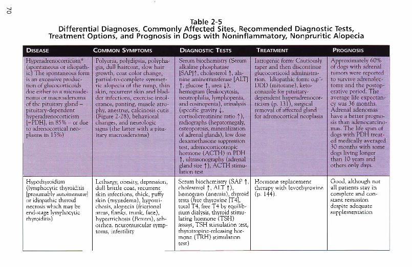
-.....Io
Table 2-5Differential Diagnoses, Commonly Affected Sites, Recommended Diagnostic Tests,
Treatment Options, and Prognosis in Dogs with Noninflammatory, Nonpruritic Alopecia
DISEASE COMMON SYMPTOMS DIAGNOSTIC TESTS TREATMENT PROGNOSIS
Hvperadrenocor ticism" Polyuria, polydipsia , polypha- Serum biochemistry (Serum Iatrogenic form: Cautiously Approximately 60%(spontaneous Of idiopath- gia, dull haircoat , slow hair alkaline phosphatase taper and then discontinue of dogs with adrenalic) The spontaneous form growth, coa t color change, [SAP] t ,cholesterol t , ala- glucocorticoid adrninisrra- tumors were reportedis an excessive produc- par tial-to-com plete svmme t- nine aminotransferase [ALT] tion. Idiopathic form: 0,1"-- to survive adrenalee-tion of glucccorricoids ric alopecia of the rump, thin t, glucose t, mea ~) , DDD (mitotane), keto- corny and the postop-due either to a microade- skin, recurrent skin and blad- hemogram (leukocytosis, conazole for pituitary- erarive period. Thenoma or ·macroadenoma de, infections, exercise in tol- neutrophilia, lymphopenia, d,ependem hYrradr.~nocor- average life expectan-..of the pituitary gland - erance, panting, muscle atro - and eosinopenia), urinalysis ncism (p. 131 ,surgical ey was 36 months.pituitary-dependent phv, anestrus, calcinosis cutis (specific gravity ~, removal of affected gland Adrenal adenomashyperadrenocorticism (Flgure2-28), behavioral cortisol.creatinine ratio t), fer adrenocortical neoplasia have a better progno-[=PDHl, in 8&% - or due changes, and neurologic radiographs (hepatomegaly, sis than adenocarcino-to adrenocortical neo- signs (the Iatter with a p itu- osteoporosis, mineralization mas. The life span ofplasmsin 15%) italiy. macroaderrorna) of adrenal glands), low dose dogs with PDB treat-
dexamethasone suppression ed medically averagedtest, adrenocorticotropic 30 mon ths with some .hormone (ACTH) in PDH dogs living longert, ultrasonography (adrenal than 10 years andgland size t), ACTH stimu- others onLydays.lation test
Hypothyroidism Lethargy, obesity, depression, Serum biochemistry (SAP t, Hormone replacernent Good, although not(lymphocytic thyroiditis dull brittle coat, recurrent cholesterol t, ALT t), therapy with levothyroxine all patients stay in[presumably autoimmune] skin infections, thick, puffy hemogram (anemia), thyroid (p.144). complete and con-or idiopathic thyroid skin (myxedema), hvpotri- tests (free thyroxine [T4], stant remissionnecrosis which may be chosis, alopecia (frictional total T4, free T4 by equilib- despite adequateend-stage lymphocytic areas, flanks, trunk, face), riurn dialysis, thyroid stimu- supplementationthyroiditis) hypertrichosis (Boxers), seb- laring hormone (TSH)
orrhca, neuromuscular svrnp- assays,TSH stimulation (est,toms, infertiliry rhvrotropine-releasing hor-
mone (TRH) stimulationtest)
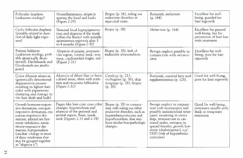
"
Follicu lar dysplasia Noninflamm atory alope cia Biopsy (p. 38), ruling ou t Rer inoids, melaton in Excellent fo r wel l-(unknown e t iology) sparing th e head and limb s endocrine disord ers in (p. 144) being, guarded for
(Figure 2-29 ) equ ivocal cases. hai r regrowth
Cycl ic follicular dysplasia Seasonal local hyperp igmenta- Biopsy (p. 38 ) Me lat oni n (p . 144) Excellent fo r gene ra l(possibly related (0 d ura- tion and alopecia of th e trunk well-be ing, fair fortion of da ily light expo- (often the flan ks) with in itia lly prevent ion of hai r losssure) spont aneous regrowth afte r 3 with treatment
to 4 months (Figure 2-30 )
Pat tern baldness A lopeci a of pinnae, posta ur ic- Biopsy (p. 38 ), lack of Be n ign neglect possibl y in Excell ent for we ll-(unknown e t iology, prob- ular region , ventral nec k, veri- endoc rine abnorma lit ies. co n jun ction wit h mo istu r- being, poor for ha irably gene tica lly deter- t rurn, caudorned ia l th ighs , rail izers . regro wthmin ed ). Dac hsh unds and (Figure 2-3 1)G reyhou nd s are pred is-posed
C olor di lution alopecia A lopecia of d ilut e blue or fawn Cvrology (p. 21 ), Reti noids, essent ial fat ty acid Good fa r we ll-b ei ng,(geneti ca lly de term ine d co lored areas, often with sca li- t richogram [p . 36 ) , skin supp lementatio n (p. 128). poo r for hair regrowthde generative process ness and recu rrent folliculitis scrapings (p. 26), b iopsyresu lt ing in ligh ter h air ( fi gure 2-3 2) (p . 38 )co lo r with p igme n taryclum ping and da mage to
th e hai r shaft and bulb)
G rowth hormone-respon - Pupp y-like ha ir COat , coat color Biopsy (p. 38 ) in co njunc- Benign neglect in co nj unc - G ood for we ll-b e ing ,sive derm atosis, estrogen- chan ges, hypotrichosis and [ion with ruling out other t ion with moi stur izers and treatment usua lly on lyrespon sive dermatosis, cas- alopec ia of the pe rineal and endoc rine disorders, such as possibl y an t imicrob ial trea t- leads to tempo rarytrarion-re sponsive de r- gen ital region, flank , trunk, hyperadrenocorticism and ruen r, neu ter ing in intact remissionmatosis, adrena l sex hor- neck (Figures 2-33 and 2-34) hypothyroidism, [ha t may clogs, testosterone in cas-mone imbalance, testes- have similar histopath ologic [rat ed males, est rogen interone-respon sive de r- changes. spaye d fem ales, grow th lior -rnarosis, hypogon ad ism mone (d iabe togen ic), o, p'-(unclear enology in most DO D (ri sk of hvpoadreno-of the se syndromes that ccrt icism)may be group ed toge the ras "alopec ia XU)
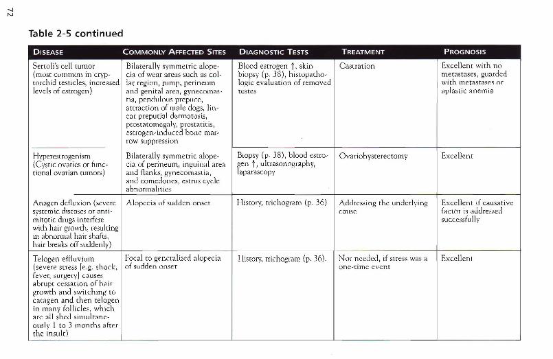
Table 2-5 continued
DISEASE COMMONLY AFFECTED SITES DIAGNOSTIC TESTS TREATMENT PROGNOSIS
Sertoli's cell tumor Bilaterally symmetric alope- Blood estrogen i , skin Ca stration Excellent with no(most common in cryp- cia of wear area s such as co l- biopsy (p. 38) , histopatho- met astases, guardedtorchid testicles, increased lar region, rump , perineum logic evaluation of removed with meta stases orlevels of estrogen) and genital area, gvnecornas- testes apla st ic anemia
ria, pendulous prepuce,attraction of male dogs, lin-ear preputial dermatosis,prostatomegalv, prostatitis,estrogen-induced bone mar-row suppression
Hyperestrogenism Bilarerally symmetr ic alope- Biopsy (p. 38), blood estro- Ovariohysterectomy Excellent(Cystic ovaries or tunc- cia of perineum, inguinal area gen i, ulrra sonographv ,tional ovarian rumors) and flanks , gynecomastia, laparascopy
and comedones, estrus cycleabnormalities
Anagen defluxion (severe Alopecia of sudden onset Hisrorv, trichograrn (p. 36) Addressing the underlying Excellent if causativesystemic diseases or anti- cnuse factor is addressedmitotic drugs interfere successfuIIywith hair growrh , resultingin abnormal hair shafts,hair breaks off suddenly)
Telogen effluvium Focal to generalized alopecia History, rrichogram (p. 36) . Not needed, if stress was a Excellent(severe stress [e.g. shock, of sudden onset one-time eventfever, surgery] caus esabrupt cessat ion of ha irgrowth and switching tocatagen and rhen telogenin many follicles, whichare all shed simulrane-ouslv 1 to 3 months afterrhe insult)

Post -clipping A lopecia Clipped areas Diagnosis based on signal- None. Hair will grow back Excell ent(arc tic or plushcoated ment, history, ami presence in 6-24 monthsbreeds fail to regrow hair of noninflammatory alope-in clipp ed areas; cause is cia in clipped areas only.unknown)
-...JW
Figure 2-28Papules and crusts in anS-year-old, male castrated Bull Terrier withc alc inosis cutis due topituitary-dependenthyperadrenocorticism
Figure 2-30A lope c ia and hyperpigmentation in the flankarea of a 6-year-old,m ale castrated Boxerwith c yclic folliculardysplasia .
Figure 2-29Alopecia due to foll iculardysplasia in a 2-year-old,female Curly-coatedRetriever
Figure 2-31Pattern alopecia.(Courtesy of Dr. PeterIhrke .)
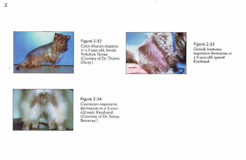
Figure 2-32 Color dilurion alopecia in a 3-year-old, female Yorksh.ire Terrier. (Courresy of Dr. Thieny Olivry.)
Figure 2-34 Cas rracion-respons ive dermacosis in a 5-ycarold male Keeshond. (Courcesy of Dr. Sonya Benenay.)
Figure 2-33 Growth honnoneresponsive dennamsis in a 9-year-old, spayed Keeshond .
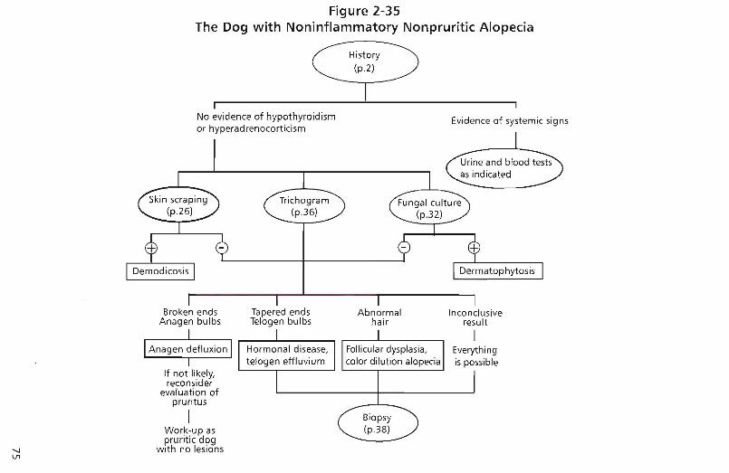
Figure 2-35The Dog with Noninflammatory Nonpruritic Alopecia
History(p.2)
rNo evidence of hypothyroidismor hyperadrenocorticism
Evidence of systemic signs
""-J\J1
Broken endsAnagen bulbs
If not likely,reconsider
evaluation ofpruritus
IWork-up aspruritic dog
with no lesions
Tapered endsTelogen bulbs
Abnormalhair
Inco nclusiveresult
Everythingis possible
76
The Dog vvith Nodules
Key Questions
./ How old was this patient when clinical signs were first recognized? (p.3)
./ How long has the disease been present and how did it progress? (p.3)
./ Are there any other animals in the household? (p. 8)
./ Does anybody in the household have skin disease? (p. 8)
./ Was the disease treated before? If so, which drugs were used and how sllccessful was treatment? (p. 8)
Differential Diagnoses The differential diagnoses are predicted primarLly based on two separate features: (1) Is there only one lesion (which increases the likelyhood of neoplasia or a kerion) or are there multiple lesions (which may be due to sterile inflammatory diseases, more aggressive neoplastic disease or severe infection); and (2) Are draining tracts absent or present (increasing the likelihood of foreign bodies, severe bacterial or fungal infection, or sterile inflammatory disease)?
The approach to the dog with nodules is straightforward. History and clinical exarnination are followed by microscopical evaluation of impression smears (if draining tracts are present) and aspirates in any dog with nodules (p. 21) . In some patients, cytology will reveal an infectious organism or classic neoplastic cells and thus a diagnosis. In most patients, cytologiC examination will further narrow the list of differential diagnoses, but a biopsy (p. 38) will be necessary to reach a diagnosis. With nodular lesions, complete excision of one or more nodules shouLd be performed. If draining tracts are present and/or cytology indicates possible infection, a culture may be llseful as well. Deep tissue should be submitted rather than a culture :;wab (p. 28).
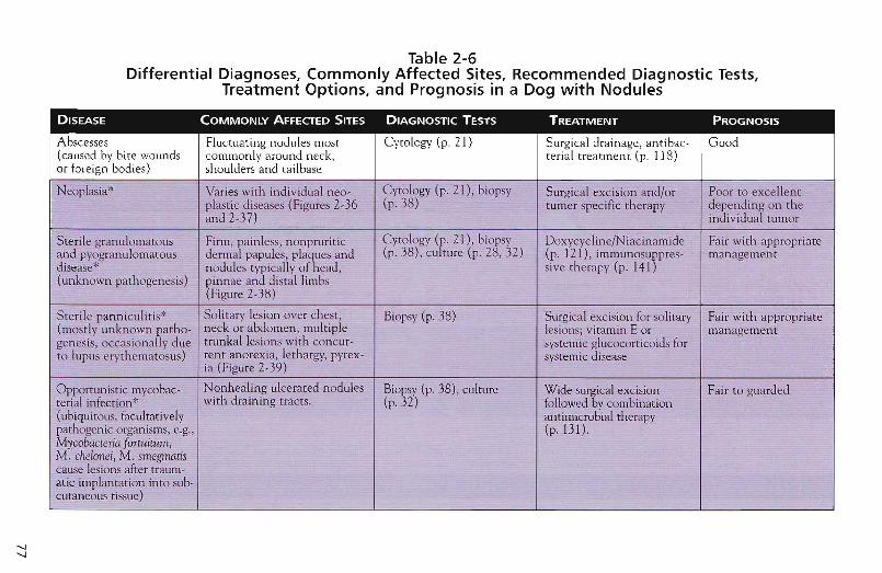
-...J-...J
Table 2-6Differential Diagnoses, Commonly Affected Sites, Recommended Diagnostic Tests,
Treatment Options, and Prognosis in a Dog with Nodules
DISEASE COMMONLY AFFECfED SITES DIAGNOSTIC TESTS TREATMENT PROGNOSIS
Abscesses Fluctuating nodules most Cytol ogy (p, ZI) Surgical drainage, antibac- Good(caused by bite wounds commonly around neck, rerial treatment (p. 118)or foreign bodies) sho ulders and railbase
INeoplasia" Varies with ind ividual neo- Cytology (p. 21), biopsy Surgical excision and/or Poor to excellentI plast ic diseases (Figures Z-36 (p. 38) tu mer spec ific therapy depend ing on th e
~ and 2-37) individ ual tum or
I Sterile gran ulomatous Firm, painless, nonpruri tic C ytology (p. 21) , biopsy Doxycycl ine/N iacinamide Fair with approp riateand pyogranulomatous dermal papules, plaques and (p, 38) , cult ure (p. 28,32) (p. 121), immunosuppres- managementdisease" nodules typically of head, sive therapy (p. 141)(un known pathogenesis) pinnae and distal limbs
(Figure 2-38 )
Sterile panniculitis* Solitary lesion over ches t, Biopsy (p. 38) Surgical exc ision for solitary Fair with app ropr iate(mostly un known patho- neck OJ abdomen, multiple lesions; vitamin E or managementgenesis, occasiona lly due trunkal leslons with concur- systemic glucocorticoids forto lupus erythe matos us) ren t anorexia , leth argy, pyrex- systemic disease
ia (Figure 2-39)
Opportu n istic rnycobac- Nonhealing ulcerated nodules Biopsy (p. 38) , culture Wide surt ical excision Fair to guardedrerial infection" with draining trac ts. (p. 32) followed v combination(ubiqui tous, facultatively antimicrobi~l therapypathogenic organ isms, e.g., (p, 13l).Mycobacteria fortuitum,M . chelonei, M . smegmatiscause lesions after traum-at ic implan tation into sub-cutaneous tissue)
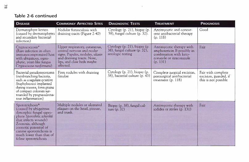
Table 2-6 continued
DISEASE COMMONLY AFFECTED SITES DIAGNOSTIC TESTS TREATMENT PROGNOSIS
Dermarophyre kerion Nodular furunculosis with Cvro logv (p. 21), biopsy (p. Antimycot ic and concur- Good(caused by derrnarophvtes draini ng [facts (Figure 2-40) 38), fungal culrure (p. 32). rent antibacterial th erapyand secondary bacterial (p, 118)infect ion )
Cryptococcosis* Upper respiratory, cut aneous, Cytology (p. 21), biopsy {p. Antimycot ic th erapy with Fair(Rare infection in often central nervous and ocular 38) , fungal culture (p. 32) , amphotericin B possibly inimmunocompromized host signs. Papu le , nodules, ulcers serologic test ing comb ination with keto-with ubiquitous, sapro- and draining trac ts. Nose, con azole or itraconazolephytic, yeast-like fungus lips, and claw beds maybe (p, 131)Cryptococcus neojormens) affected.
Bacterial pseudornycetorna Firm nodules with draining Cytology (p. 21) , biopsy {p. Co mplete surgical excision , Fair with com~le te(nonbranching bacteria, fistulae 38), bacterial culture (p. 43) postsurgical anri bacteriaI excision, guar ed, ifsuch as coagulase-positive treatment (p. 118) this is not possibleStaphylococci implantedduring trauma, form grainsof compact colonies sur-rounded by pvogranuloma-tous inflammation)
Sporotrichosis" Mul tiple nodules or ulcera ted Biopsy (p. 38), fungal cul- Antimycotic therapy with Fair{caused by ubiquitous plaques on th e head , pinnae, ture (p. 32) iodides or azoles (p. 131)dimorph ic fungal sapro- and trunk.phy te Sporothrix schenkiithat infects wounds)Zoonosis, althoughzoonot ic potential ofcan ine sporo trichosis ismuch Lower than that offeline sporotrichosis
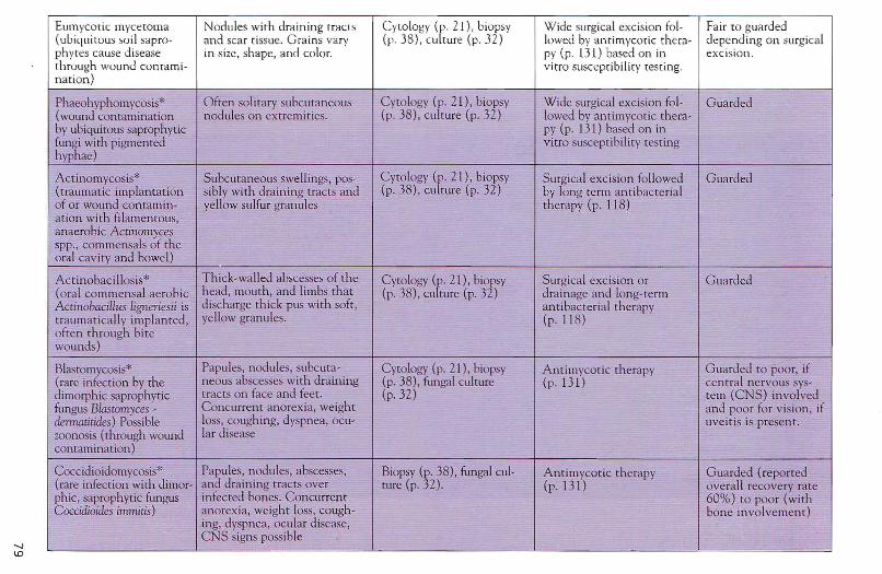
'-I1D
Eumycotic mycetoma Nodules with draining trac ts Cyto logy (p. 21), biopsy Wide surgical excision fol- Fair to guarded(ub iqui tous soil sapro- and scar tissue. G rains vary (p. 38), cu ltu re (p, 32) lowed by antirnvcotic the ta- depend ing on surgicalph ytes cause disease in size, shap e, and color. PI' (p . 131) based on in exci sion.throug h wound con rami- vitro susceptibility testing.nat ion )
Phaeohyphomycosis* Often solitary subcutaneous Cy tology (p. 2 \), biopsy W ide surgical excision fol- G uarded(wound contamination nodules on ex tremities, (p. 38), culture (p. 32) lowed by antimycotic rhera-hy ubiquitous saprophytic py (p. 131) based on infungi with pigmented vitro suscept ibility testinghyphae)
Actinomycosis* Subcutan eous swellings, pos- Cytology (p, 2\), biopsy Su rgical excision followed Guarded(t raumatic implantation sibly with draining tracts and (p. 38) , cult ure (p. 32 ) by long term antibacte rialof or wound contamin- yellow sulfur gran ules ther apy (p, 118)ation with filamentous,anaerobic Actinomycesspp., commensals of theoral cavity and bowel)
Actinobacillosis" Thick-walled abscesses of the Cytology (p. 21), biopsy Surgica l excision or Guarded(oral commen sal aerobic head , mouth , and limbs that (p. 38 ), culture (p. 32) drainage and lon g-termActinobadllus ligneriesii is discharge thick pus with soft, antibacterial therapytraum atically implanted , yellow gran ules. (p. 118)often through bitewounds)
Blastomycosis* Papules, nodules, subcuta- Cytology (p, 21), biopsy Antimycotic therapy Guarded to poor, if(rare infect ion by the neous abscesses with draining (p. 38) , fungal culture (p . 131) central nervous sys-dimorphic saprophytic tracts on face and feet . (p. 32) tem (eNS) involvedfungus BlastOmyces - C oncurrent anorexia, weight and poo r for vision , ifdemuuitides) Possible loss, cough ing, dyspnea , ocu- uveitis is present.zoonosis (through wound lar diseasecontamination )
Coccidioidomycosis" Papules, nodules, abscesses, Biopsy (p, 38), fungal cul- A ntimycotic therapy Guarded (reported(rare infection with dimor- and draining tracts over ture (p. 32) . (p.131) overall recovery ratephic, saprophytic fungus infected bones. Concurrent 60%) to poor (WithCoccidioides immitis) anorexia, weight loss, cough- bon e involvement)
ing, dyspne a, ocular disease,CNS signs possible
.. . ... - - ' - -'~ ....
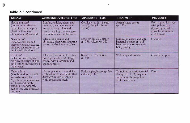
coo
Table 2-6 continued
DISEASE COMMONLY AFFECTED SITES DIAGNOSTIC TESTS TREATMENT PROGNOSIS
Histoplasmosis" Papules, nodules, ulcers, and Cytology (p. 21), biopsy Antimyootic agents Fair to good for dogs(uncommon infection draining tracts. Concurrent (p. 38), fungal culture (p. 131) with pulmonarywith dimorphic, sapro - anorexia, weight loss and (p.32) disease, guarded tophytic soil fungus fever, coughing, dyspnea, gas- grave for dissemin-Histoplnsma capsulatum) trointestinal and ocular disease ated disease
Nocardiosis" Ulcerated nodules and Cytology (p. 21), biopsy Surgical drainage and anti- Guarded(Nocardia. spp. are soil abscesses, often with draining (p. 38), culture (p. 32) bacterial th erapy (p. 118)-sapmphytes and cause res- tracts, on the limbs and feet based on in vitro suscepti-piratorv, cutaneous, or dis- bility testingseminated infect ions)
Pythiosis* Ulc erated nodules of the face Biopsy (p. 38), culture Wide surgical excision Guard ed to poor(infection with aquatic and legs develop into boggy (p. 32)fungi by exposure of dam- masses with ulcer ati on andaged skin to infected stag- draining tractsnant water)
Tuberculosis* Ulcers, plaques, and nodules Radiographs, biopsy (p. 38) , Combin ation antimicrobial Poor(rare infection in small on he ad, neck, and limbs th at culture (p. 32) therapy (p . 131), frequentan imals caused by discharge yellow-green pus euthanasia due to publicMycobacterium tubercu/o- with unpl easant smell health concernssis, bOllis and rarelvallium, predominantlyrespiratory and digestivelesions)
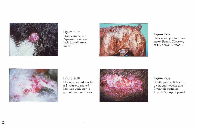
00
Figure 2-36 Histiocytoma in a 2-year-old castrated Jack RusseLlluixed breed.
Figure 2-38 Nodu les and ulcers in a 2-year-o ld spayed Maltese wi th ste rile granu lomatous disease.
Figure 2-37 Sebaceolls cysts in a castrated Boxer, . (Courtesy of Dr. Sonya Bettenay.)
Figure 2-39 Sterile panniculitis with ulcers and nodules in a 9-year-old castrated English Springer Spaniel.
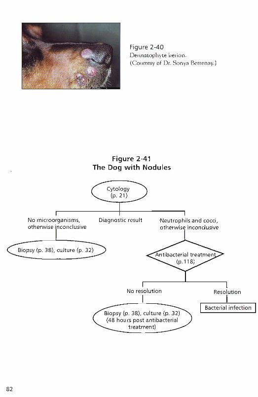
Figure 2-40Derma tophvte kerion.(Courtesy of Dr. Sonya Be tte nay.)
Figure 2-41The Dog with Nodules
~Cytol:
~~'I---- -------.1No microorganisms, Diagnos tic result Neutrophils and cocci,otherwise inconclusive otherwise inconclusive
Biopsy (p. 38), culture (p. 32)
82
No resolution
IResolution
II Bacterial infect ion I
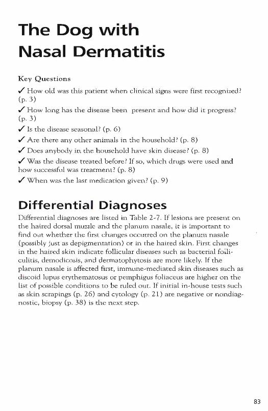
The Dog \Nith Nasal Dermatitis
Key Questions
~ Howald was this patient when clinical signs were first recognized? (p.3)
~ How long has the disease been present and how did it progress? (p . 3)
~ Is the disease seasonal? (p. 6)
~ Are there any other animals in the household? (p. 8)
~ Does anybody in the household have skin disease? (p. 8)
~ Was the disease treated before? If so, which drugs were used and how successful was treatment? (p. 8)
~ When was the last medication given? (p. 9)
Differential Diagnoses Diffe rential diagnoses are listed in Table 2-7 . If Lesions are present on the haired dorsal muzzle and the planum nasale, it is important to find out whether the first changes occurred on the planulTl nasale (possibly just as depigmentation) or in the haired skin. First changes in the haired skin indicate follicular diseases such as bacterial foniculitis, demodicosis, and dermatophytosis are more likely. If the planum nasale is affecred first, immune-mediated skin diseases such as discoid lupus erythematosus or pemphigus foliaceus are higher on the List of possible conditions to be ruled out. If initial in-house tests such as skin scrapings (p. 26) and cytology (p. 21) are negative or nondiagnostic, biopsy (p. 38) is ci1.e n ext step.
83
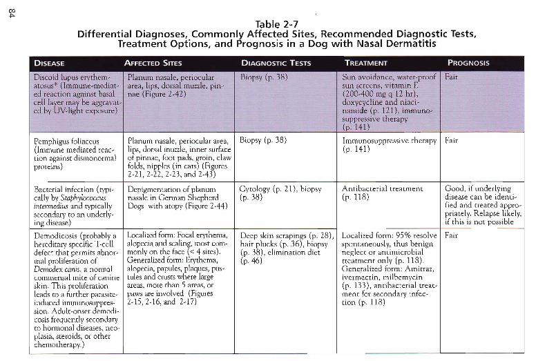
Table 2-7Differential Diagnoses, Commonly Affected Sites, Recommended Diagnostic Tests,
Treatment Options, and Prognosis in a Dog with Nasal Dermatitis
DISEASE AFFECTED SITES DIAGNOSTIC TESTS TREATMENT PROGNOSIS
Discoid lupus eryrhem - Planum nasa Ie, periocular Biopsy (p . 38) Sun avoid-ance, water-proo f Fairat osus* (Imrnune-mediar- are a, lips, do rsal muzzle, pin- sun scre ens, vitam in Eed react ion against basal nae (Figure 2-42 ) (200-400 mg q 12 h r) ,cell layer may be aggravat- doxycycline and ni aci-ed bv Uv-lighr exposure ) namide (p. 121), imrnuno -
suppressive therap y(p . 141)
Pemphigus foliaceus Planum nasal e, periocular area, Biopsy (p. 38) Immunosuppressive th erapy Fair{Immune mediated rcac - lips, dorsal muzzle, inn er surface (p. 141)tion again sr disrnonormal of pinnae, foor pads, groin, clawproteins) folds, nippl es (in cats) (Figures
2-21,2-22. 2·23 , and 2-43)
Bacterial infection (tvpr- Dep igmemation of planum C yto logy (p. 2 1) , biopsy Antibacterial treatment Good , if underlyingcallv by Swphylococcus nasale in German Shepherd (p. 38) (p.118) d isease can be identi-intennedius and typically Dogs with atopy (Figure 2-44) fied and rreated appro-secondary to an underlv- pria telv, Relapse likely,ing disease) if this is not possible
Demodicosis (probably a Localized form: Focal erythema, Deep skin scrapings (p. 28), Localized form: 95% resol ve Fairhereditary specific Tcell alopecia and scaling, most corn- hair plucks (p . 16), biopsy spontaneously, thus benigndefect that permits abnor- monly on the face « 4 sites). (p . 38) , elimination diet neglect or antimicrobialmal proliferation of Generalized form: Erythema, (p. 46) treatment only (p. 118) .Demodex canis, a normal alopecia, papules , plaques, pus- G eneralized form: Amirraz,commensal mite of canine rules and crusts where large ivcrrnectin, milbemycinskin . This proliferation areas, more than 5 areas, or (p. 133), antibac terial treat-leads to a further parasite- paws are involved (Figures menr for secondary infec -induced immunosuppres- 2-15,2-16, and 2-17) tion (p . 118)sian. Adul t-onset dem odi-cosis frequently secondaryto hormonal diseases, neo-plasia, steroids, or otherchemotherapy.)
-

Dermarophvrosis (der- Face, pinnae , paws Wood's lamp (p. 30), rri- Antimycotic agents such as Goodmatoph ytcs are transmit- (Figure 2-18) chogram (p. 36), fungal cut- griseofulvin or ketoconazoleted by contact with fun - ture (p. 32), biopsy (p. 38) (p. 131). Topical antifungalgal elements) shampoos may decrease
contamina t ion of environ-ment.
..
Sporotrichosis* Multiple nodules or ulcerated Biopsy (p, 38), fungal cul- Antimycotic therapy with Fair(caused by ubiquitous plaques on the head, pinnae, ture (p, 32) iodides or azolesdimorphic fungal sapro- and trun k. (p. 131)phvte Sporothrixschenkiithat infect wounds)Zoonosis, althoughzoono tic potential ofcan ine sporotrichosis ismuch lower than that offeline .spere trichosis
C ry.ptococcosis* . Upper respiratory, cutaneous, Cyto logy (p. 21) , biopsy Antimycot ic therapy with Fair(Rare infection in: often central nervous and ocular (p. 38 ), fungal culture amphoter icin B possibly inimmunocompremized signs. Papules, nodules, ulcers (p, 32), serologic testing combination with keto-
;least with ubiquitous, and draining tracts. Nose, conazole or itraconazolesaprophytic, yeast-like lips, and claw beds mavbe (p. 1.3 1)fungus Crypwwccus neo- affected.jormsns )
OJV1
Figure 2-42Depigmentation, erosions,and ulcers in a 3-year-oldspayed Au stralian Shepherdmixed breed with discoidlupus eryth ematosus.
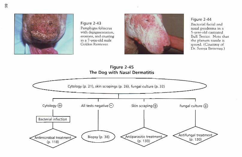
Figure 2-43Pem ph igus foliaceuswith depig mentation ,erosion s, and crustingin a 7-vear-old maleG olden Retriever.
Figure 2-44Bacte ria l faci a l andnasal pyoderma in a5 -year-o ld ca stratedBull Terrie r. N ote th atth e p lanum n asale isspared . (Courtesy ofDr. Sonya Betten av.)
Figure 2-45The Dog with Nasal Dermatitis
Cytology (p. 21), skin scrapings (p 26), fungal culture (p. 32)
Cytology EB All tests negative 8I
Skin scrapingEB Fungal culture EB
The Cat \Nith Miliary Dermatitis
Key Questions
./ How old was this patient when clinical signs were first recognized? (p.3)
./ How long has the disease been present and how did it progress? (p.3)
./ On which part of the body d id the problem start? (p. 4)
./ Is the disease seasonal? (p. 6)
./ Are there other clinical signs such as sneezing, coughing, or diarrhea? (p. 7)
./ \Vhat do you feed the anima l? Was a special diet used in the past? (p. 7)
./ Are there any other animals in the household? (p. 8)
./ Does anybody in the household have skin disease? (p. 8)
./ Was the disease treated before? If so, which d rugs were used and how successful was ueatmenr? (p. 8)
./ Wha t is used for flea control now? (p. 9)
./ When was the last medication g iven? (p. 9)
Differential Diagnoses Classic lesions of miliary dermatitis are focal or generalized smaLL papules and crusts (Figures 2-46 and 2-47). Miliary dermatitis is not a diagnosis but rather a descriptive term for a feline cutaneous reaction pattern with rn.any possible causes. Most cats suffer from an underlying flea-bite hypersensitivity. The d ifferential diagnoses for feline miliary dermatitis are listed in Table 2-8.
87
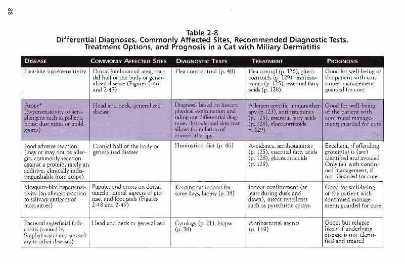
coco
Table 2-8Differential Diagnoses, Commonly Affected Sites, Recommended Diagnostic Tests,
Treatment Options, and Prognosis in a Cat with Miliary Dermatitis
DISEASE COMMONLY AFFECTED SITES DIAGNOSTIC TESTS TREATMENT PROGNOSIS
Flea-bite hypersensitivity Dorsal lumbosacral area, cau- Flea conrrol trial (p . 48) Flea control (p. 136). gluco- Good for well-being ofdal half of the body or genet- corticoids (p. 129), antihista- the patient with con -alized disease (Figures 2-46 mines [p, 125). essential fatty tinued management;and 2-47) acids (p. 128). guarded for cure
Atopy* Head and neck, generalized Diagnosis based on history, Allergen-s)ecific imrnunother- Good for well-being(hvpersensitivitv to aero- drsease. physical examinat ion and apy (p.l23 ,antihistamines of the patient withallergens such as pollens, ruling out differential diag- (p, 125), essential fatty acids continued manage-house dust mites or mold noses. Inrradermal skin test (p, 128), glucocorticoids ment: guarded for curespores) allows formulation of p.129).
immunotherapy
Food adverse reaction Cranial half of [he body or Elimination diet (p. 46) Avoidance, antihistamines Excellent, if offending(mayor may nor be aller- generalized disease (p. 125), essent ial fatty acids proteints) is (are)gic, commonly reaction (p. 128), glucocorricoids identified and avoided.against a protein, rarely an (p. 129). Only fair with contin-additive, clinically indis- ued management, iftinguishable from atopy) not. Guarded for cure
Mosquito-bite hvpersensi- Papules and crusts on dorsal Keeping cat indoors for Indoor confinement (at Good for well-beingtivirv (an allergic reaction muzzle, lateral aspects of pin- some days, biopsy (p. 38) least during dusk and of the patient withto salivary antigens of nae, and foot pads (Figures dawn), insect repellents conrinued manage-mosquitoes) 2-48 and 2-49) such as pyrethrine sprays. menr; guarded for cure
Bacterial superficial folli- Head and neck or generalized Cytology (p. 21), biopsy Antibacterial agents Good, but relapseculitis (caused by (p.38) (p.119) likely if underlyingStaphylococci and second- disease is not idenri-arv to other diseases) fied and treated
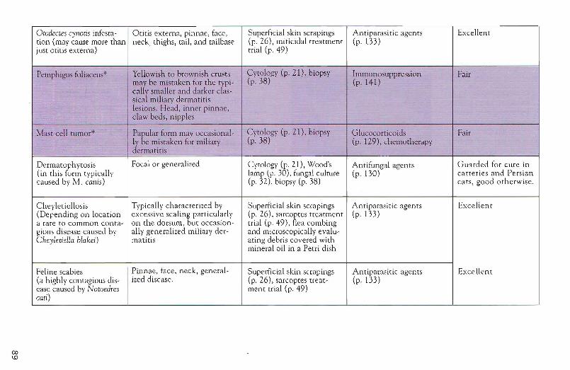
001.0
Orodectes cynotis infesra- Otitis cxterna, pinnae, face, Superficial skin scrapings Antiparasitic agents Excellenttion (may cause more than neck, thighs, tail, and tailbase (p, 26), miticidal treatment (p . t33)just otitis externa) trial (p . 49)
Pemphigus foliaceus* Yellowish to brownish crusts Cyto logy (p, 21) , biopsy Immunosuppression Fairmay be mista ken for the typi- (p. 38) (p. 141)callv smaller and darker clas-sical miliary dermati tislesions. H ead, inner pinnae,claw beds, n ipp les
M ast ce ll tumor" Papular form may occasiona l- Cyto logy (p. 21), biopsy G lucocorticoids FairIy be mistaken for miliary (p.38) (p. 129). chemotherapydermat itis
Dermatophytosis Focal or generali zed Cytolog y (p. 21) , Wood's Antifungal agents Guarded for cure in(in this form rypically lamp (p. 30). fungal culture (p . 130) catteries and Persiancaused by M . canis) (p. 32) , biopsy (p. 38) cats, good otherwise.
Cheyleticllosis Typically characterized by Superficial skin scrapings Antiparasitic agent s Excellent(Depending on location excessive scaling particularly (p . 26), sarcoptes treatment (p .133),Irare to common conta- on the dorsum , but occas ion- trial (p, 49), flea combinggious disease caused by ally gen eralized miliary der- and microscopically evalu -Clley!elie/la bla/(ei) matitis at ing debris covered with
mineral o il in a Petri dish
Feline scabies Pinnae. face, neck , general- Superficial skin scrapings Antiparasitic agents Excellent(3 highly cont agious dis- ized disease. (p, 26), sarcoptes treat- (p .133)ease caused by Notoedres ment trial (p. 49)emi)
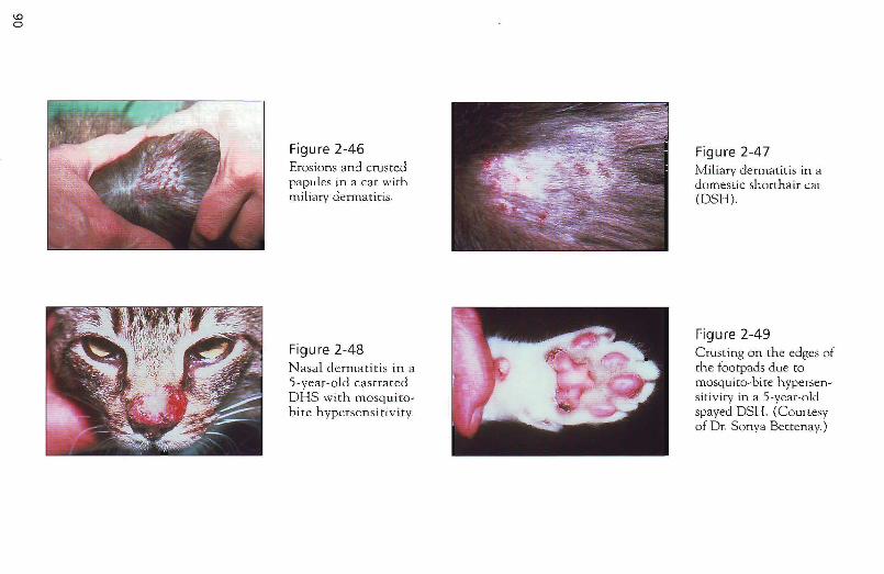
Figure 2-46 Erosions and crusted papules in a cat with miliary dermatitis.
Figure 2-48 Nasal derm:Hitis in 3
5-year-old castrated DHS with mosquirobite hypersensiti v ity.
Figure 2-47 Miliary dermatitis in a domestic shormair cat (DSH).
Figure 2-49 Crusting on the edges of the footpads due to mosquim-bite hypersensitivity in a 5-year-old spayed DSH. (Courtesy of Dr. Sonya Beccenay.)
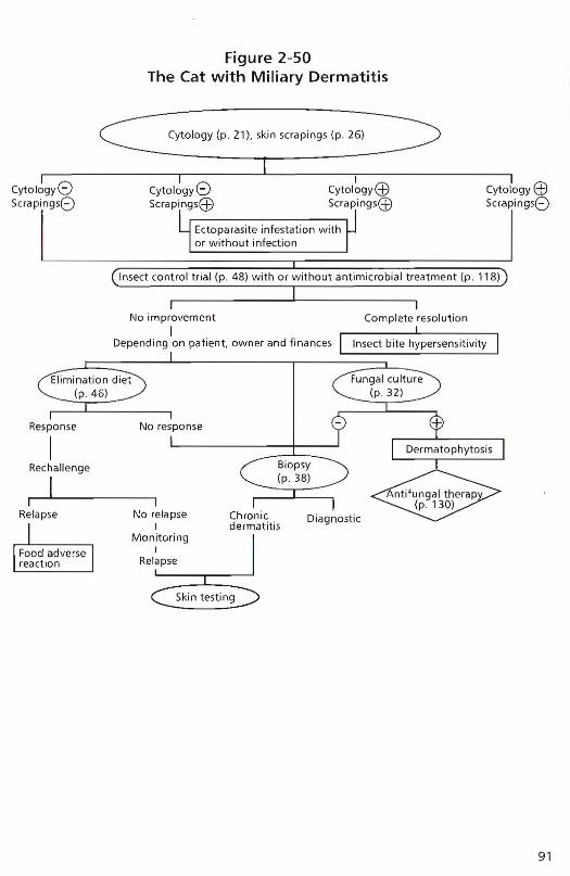
Figure 2-50The Cat with Miliary Dermatitis
Cytology (p. 2 1), skin scrapings (p. 26)
Ectopa rasite infestat ion withor w itho ut infection
cyto logy8Scrapings8
Cytology 8Scrapings@
ICytology@Scrapings(j;)
CytologyEf)Scrapings8
Insect control trial (p . 48) with or w ithout antimicro bial tr eatment (p . 118)I
Compl ete resoluti on
Fungal cul ture(p .32)
DiagnosticChronicdermatitis
No relapseI
MonitoringI
Relapse
No improvem ent
IDepending on patient, own er and fin ances
Elimination di et(p. 46 )
Response No respon se
IRechall eng e
~ I
Relapse
Skin testing
91
92
The Cat vvith Noninflammatory Alopecia
Key Questions:
tI' How old was this pacient when clinical signs were first recognized? (p. 3)
tI' How long has the disease been present and how did it progress? (p.3)
tI' On which part of the body did the problem start? (p. 4)
tl'Is the disease seasonal? (p. 6)
tI' Are there other clinical signs such as sneezing, coughing or diarrhea? (p. 7)
tI' What do you feed the animal? Was a speclal diet used in the past? (p. 7)
tI' Are there any other animals in the household? (p. 8)
tI' Does anybody in the household have skin disease? (p. 8)
tI' Was the disease treated before? If so, which drugs were used and how successful was treatment? (p . 8)
tI' What is used for flea control now? (p. 9)
tI' When was the last medication given? (p. 9)
Differential Diagnoses Noninflammatory alopecia is a feline cutaneous reaction pattern that may have various causes. H onnonal alopecia is extremely rare in the feline and typically affected cats show other severe signs. Psychogenic alopecia in the cat is greatly overdiagnosed. The disease usually affects pure-bred cats with a nervous disposition. Environmental changes such as a new partner, baby, pet, or a move to a new house precede clinical signs. Most cats with noninflammatory alopecia are pruritic as a result of allergies and may be closet groomers. Many cats previously diagnosed with hormonal alopecia and treated successfully with medroxyprogesteron acetate or megestrol acetate have responded because the anti-inflammatory action of these medications have controlled their pruritus caused by allergies, not because of a correction of their hormonal imbalance. The differential diagnoses for feline noninflammatory alopecia are listed in Table 2-9.
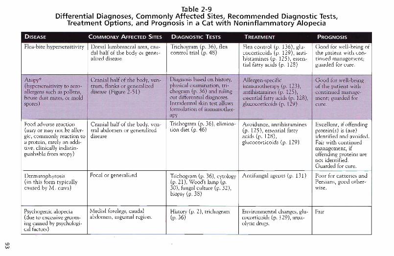
\DW
Table 2-9Differential Diagnoses, Commonly Affected Sites, Recommended Diagnostic Tests,
Treatment Options, and Prognosis in a Cat with Noninflammatory Alopecia
DISEASE COMMONLY AFFECTED SITES DIAGNOSTIC TESTS TREATMENT PROGNOSIS
Flea-bite hypersensiti vity Dorsal lumbosacral area, cau - Trichogram (I". 36), flea Flea con trol (I'" 136) , glu- Good for well -being ofdal half of the body or gener- control trial (I'" 48) cocorricoids (I'" 129), anti- the patien t with con-alized disease histamines (I'" 125), essen- tinued management;
tial fatty acids (I'" 128 ) guarded for cure.
Atopy" Cranial h alf of the body, ven- Diagnosis based on history, A llergen -specific Good for well-being(hyp ersensitivitv to aero- tru m, flanks or generalized physical examination, tr i- immunotherapy (I!>. 123 ), of the patient withallergens such as pollens, disease (Figure 2-51) chogram (I". 36) and ruling antih istamines (I'" 125), co nt inued manage-.house dust mites, or mold out differen t ial diagnoses. essential fatty acids (I". 128), menr, guarded for.spores) Intradermal skin test allows glucocortic oids (p . 129) cure.
formu lat ion of irnrnunothe r-apy
Food adverse reacti on Cranial half of th e body, ven- Trichogram (I". 36), elirnina- Avoidance , antihistamines Excellent. if offending(mayor may not be aller- rral abdomen or genera lized tion diet (I". 46) (I'" 125) , essential fatty proteints) is (are)gic, commonly reaction to disease acids (I'" 128) , identified and avo ided.a protein, rarelv an addi - glucocorticoids (I". 129) Fair with con t inuedtive , clinicall y indistin- management, ifguishable from atopy) offending proteins are
not identified.Guarded for cure.
Dermamph ytosis Focal or generalized Tuchograrn (I'" 36), cytology A ntifungal agen ts (I'" 13 1) Poor for catte ries and(in this form typically (p . 21), Wood's lamp (p, Pe rsians , good o ther-caused by M. canis) 30) , h.lI1gal culture (p, 32), wise.
biopsy (p . 38)
Psych ogenic alopecia Medi al forel egs, caudal History (p. 2), rrichogram Environmental changes, glu- Fair(due to exce ssive groom- abdomen, inguinal region. (p.36) cocorticoids (p . 129) , anxi -ing caused by psvchologi- olvtic drugs.cal factor s)
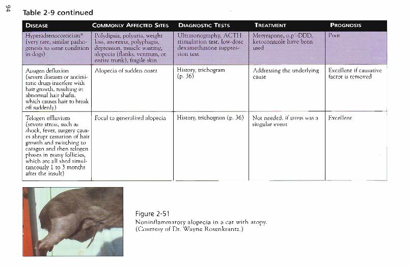
Table 2-9 continued
DISEASE COMMONLY AFFECTED SITES DIAGNOSTIC TESTS TREATMENT PROGNOSIS
Hvperadrenoccrticism" Polydipsia, polyuria, weight Ultrasonography, ACTH Metyrapone, o,p'-DDD, Poor(very rare, similar patho- loss, anore-xia, polyphagia, stimulation test, low-dose ketoconaaole have beengenesis to same condition depression , muscle wastin g, dexamethasone suppres- usedin dogs) alopecia (flanks, ventrum, or sian test
entire m ink), fragile skin
Anagen defluxion Alopecia of sudden onset History, rrichogram Addressing the underlying Excellent if causative(severe diseases or antim i- (p. 36) cause factor is removedtotic drugs interfere withhair growth, resulting inabnormal ha ir shafts,which causes hair to breakoff suddenly)
Telogen effluvium Focal to gene ralized alopecia History, trichogram (p. 36) Not needed, if stress was a Excellenc(severe stress, such as singular eventshock , fever, surgery caus-es abrupt cessation of hairgrowth and switch ing tocaragen and then telogcnphases in many follicles,which are all shed simul-tancously 1 to 3 monthsafrer rhe insult)
Figure 2-51Noninflammatory alopecia in a cat with atopy.(Courtesy of Dr. Wayne Rosenkrantz.)
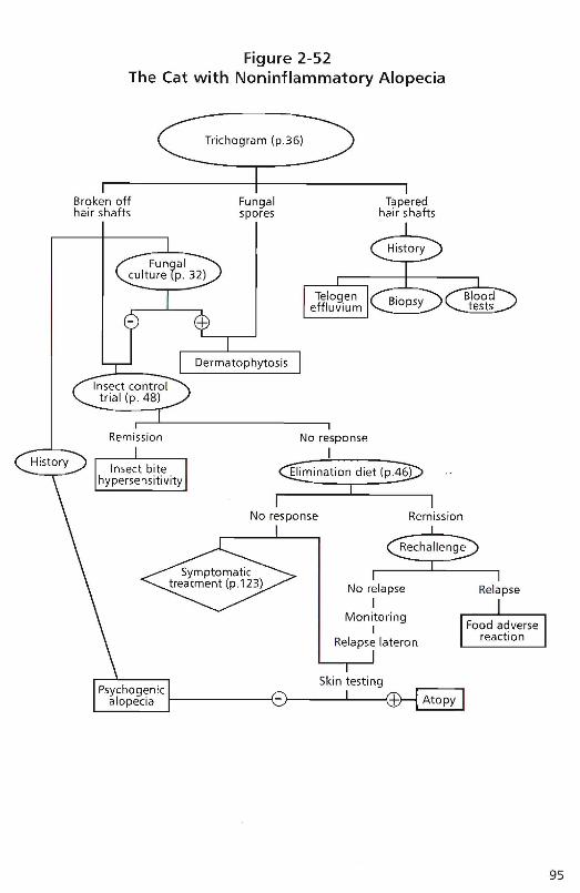
Figure 2-52The Cat with Noninflammatory Alopecia
Trichogram (p.36)
Broken offhair shafts
Fungalspores
Taperedhair shafts
Relapse
Remission
No relapseI
MonitoringI
Relapse lateran
~ination diet (p~I
No response
No response
IRemission
95
96
The Cat vvith Lesions of the Eosinophilic Granuloma Complex
Key Ques tions
v' How old was this patient when clinical signs were first recognized? (p.3)
v'Is the disease seasonal? (p. 6)
v' Are there other clinical signs such as sneezing, coughing, or diarrhea? (p. 7)
v' What do you feed the animal? Was a special diet used in the past? (p. 7)
v' Was the disease treated before? If so, which drugs were used and how successful was treatment? (p. 8)
v' What is used for flea control now? (p. 9)
v' When was the last medication given? (p. 9)
Differential Diagnoses All subsets of the eosinophilic granuloma are mucocutaneous reaction patterns in the cat.
v'Indolent (or eosinophilic or rodent) ulcer commonly affects the upper lip unilaterally or bilaterally (Flgure 2-53), but may occur in the oral cavity or elsewhere on the body (Figure 2-54). The well-circumscribed ulcers with raised borders are rarely painful or pruritic; frequently the owner is more bothered by the lesions than the cat. The differential diagnoses of the feline eosinophilic ulcer are neoplastic diseases such as squamous cell carcinoma and infectious ulcers (eosinophilic ulcers are often secondarily infected as well). Diagnosis is confirmed by biopsy (p.38). Prior antimicrobial treatment (p. 118) is recommended if cytology (p. 21) is indicative of infection.
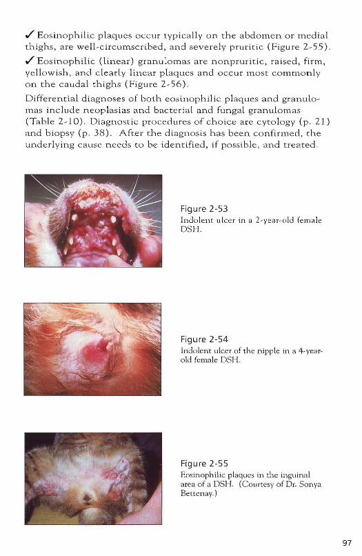
./ Eosinophilic plaques occur typically on the abdomen or medial thighs. are well-circumscribed. and severely pruritic (Figure 2-55) .
./ Eosinophilic (linear) granulomas are nonpruritic. raised. firm. yellowish. and clearly linear plaques and occur most commonly on the caudal thighs (Figure 2-56).
Differential diagnoses of both eosinophilic plaques and granulomas include neoplasias and bacteri a l and fungal granulomas (Tab le 2-10). Diagnostic procedures of choice a re cyto logy (p . 21) and biopsy (p. 38). After the diagnosis has been confirmed, the underly ing cause needs to be identified, if possible. and treated.
Figure 2-53 Indolent ulcer in a 2-yea r-old female DSH.
Figure 2-54 Indolent ulcer of th e njpple in a 4-yearold female DSH.
Figure 2-55 Eosinophilic plaque~ in the inguinal area of a DSH. (Courtesy of Dr. Sonya Bettenay.)
97
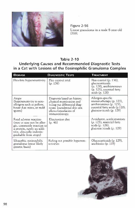
Figure 2-56linear granuloma in a male 8-year-oldDSH.
Table 2-10Underlying Causes and Recommended Diagnostic Tests
in a Cat with Lesions of the Eosinophilic Granuloma Complex
Flea-bite hypersensitivity
Atopy(hypersensinvicv to aeroalle rgens such as pollens,house dust mites, or moldspores)
Food adverse reaction(mayor may not be allergic, commonly reaction toa protein, rarely an addirive, cl inically indistinguishable from atopy)
Idiopathic eosinophilicgranuloma (most lik e lygen er ic basis)
Flea control trial(p.136)
Diagnosis based on hisrorv,physical examination andruling Out differen tial diagnoses. Intradermal skin testa llows formulation ofimmunotherapv.
Elimination diet(p.46)
Ruling out possible hypersensirivities
Flea control (p. 136),glucocorticoids(p. 129 ) , an tihistamines(p, 125), essential fattyacids(p. 128)
Allergen-speci ficimmunotherapy (p . 123),antihistamines (p. 125),essential fatty acids (p.l28),glucocorticoids (p. 129)
Avoidance, antilustarnines(p, 125), essential fattyacids (p. 128),glucocorticoids (p. 129 )
Glucocorricoids (p. 129),antibiotics (p. 119 )
98
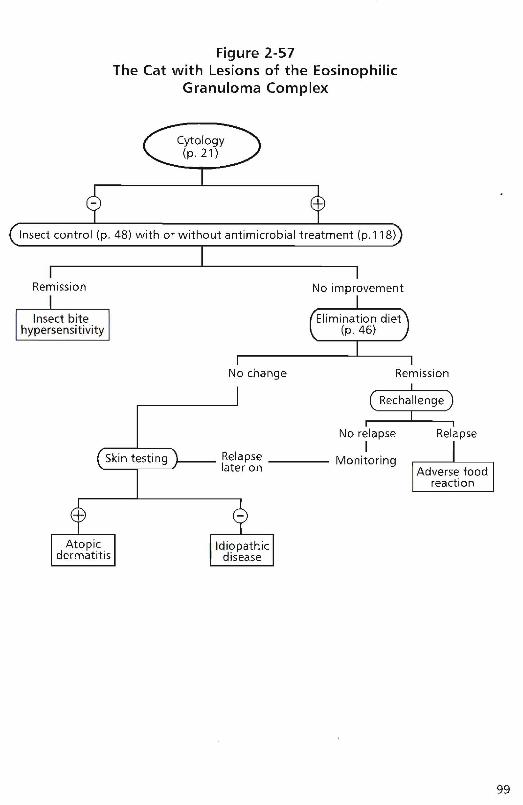
Figure 2-57The Cat with Lesions of the Eosinophilic
Granuloma Complex
Insect control (p. 48) with or without antimicrobial treatment (p.118), ..
Remission No improvement
No change Remission
No relapseI
l ,)"", Lt:::~L"'Sl r--- Relapse Monitoring~ later on
Relapse
99

100
The Cat vvith Nodules
Key Questions
./ How old was this patient when clinical signs were first recognized? (p.3)
./ How long has the disease been present and how did it progress? (p. 3)
./ Are there o cher clinical signs such as sneezing, coughing, or diarrhea? (p. 7)
./ Was the disease treated before? If so, which drugs were used and how successful was treatment? (p. 8)
Differential Diagnoses The differential diagnoses depend primarily on two features: the number of lesions and whether draining tracts are present or not. Is there only one lesion? This increases the likelihood of neoplasia or a kerion. Or are there multiple lesions? These may b e due to sterile inflammatory diseases, more aggressive neoplastic disease, or severe infection. The presence of draining tracts increases the likelihood of foreign bodies, severe bacterial or fungal infection, or sterile inflammatory disease.
In a cat with nodules, history taking and clinical exam ination are followed by microscopic eva luation of impression smears (if draining tracts are present) and aspirates (in any cat with nodules) (p . 21). In some patients, cytology will reveal an infectious organism or classic neoplastic ce lls and thus a diagnosis. In most patients, cytology will aid in further limiting the list of differential diagnoses, but a biopsy (p . 38) will be necessary to reach a diagnosis . With nodular lesions, a comp lete excision of one or more nodules shou ld be performed. If draining tracts are present and/or cytology indicates possible infection, a tissue culture may be useful as well (p . 43) .
The differential diagnoses for feline nodules are listed in Table 2-11 .
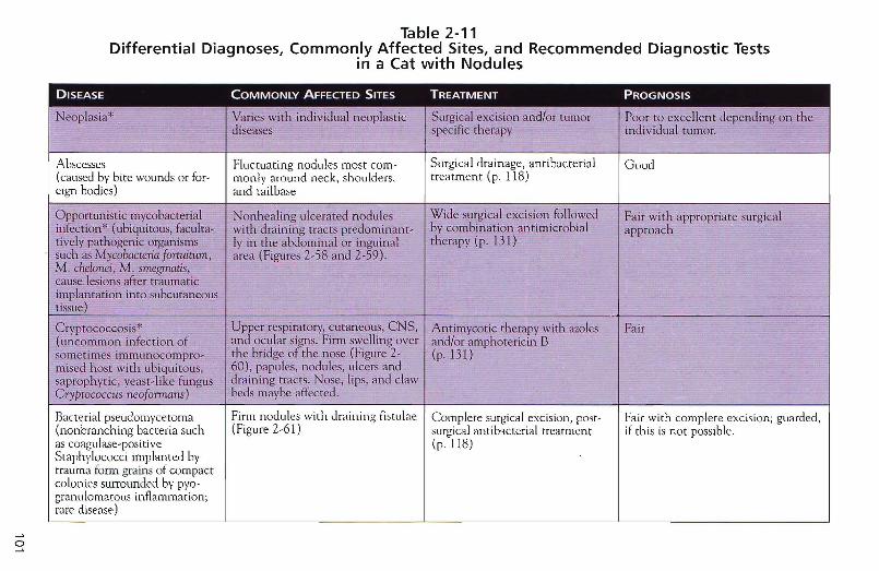
o
Table 2-11Differential Diagnoses, Commonly Affected Sites, and Recommended Diagnostic Tests
in a Cat with Nodules
DISEASE COMMONLY AFFECTED SITES TREATMENT PROGNOSIS. .
Neoplasia" "' Varies with individual neoplastic Surgical excision and/or tumor Poor to excellent depending on thediseases specific therapy individual tumor.
Abscesses Fluctuating nodules most com- Surgical drainage, antibacter ial Good(caused by bite wounds or for- manly around neck, shoulders, treatment (p. 118)eign bodies) and tailbase
Opportun istic mycobacterial Nonhealing ulcerated nodules Wide surgical excision followed Fair wi th appropriate surgica linfect ion" (ubiquitous, faculta- with draining tract s predominant- by combination antimicrobial approachtivelv pathogen ic organisms Iy in the abdom inal or inguinal therapy (p. 131)such as M?/cobaeteria fQ~t1~m , area (Figures 2-58 and 2-59 ).M. chelonei, M .~gmati$,
cause lesions after traumaticimplantation into subcutaneoustissue)
Crv ptococcosis" Upper respiratory, cutaneous, e NS, Ant imycot ic therapy with azoles Fair(uncommon infection of and ocular signs. Firm swelling over and/or amphotericin Bsomet imes immunocompro- the br idge of the nose (Figure 2- (p. 131)rnised host with ubiquitous, 60) , papules, nodules, ulcers andsaprophytic, yeast-like fungus draining tracts. Nose, lips, and clawGry/?tococcus neoformans) beds maybe affected.
--
Bacterial pseudomyceroma Finn nodules with draining fistulae Complete surgical excision, post- Fair with complete excision; guarded.(nonbranching bacteria such (Figure 2-61) surgical antibacterial treatmen t if this is not possible.as coagulase-positive (p.1 18)Staphylococci implanted bytrauma form grains of compactcolon ies surrouncled by pya-granu lomatous inflammation;rare disease)
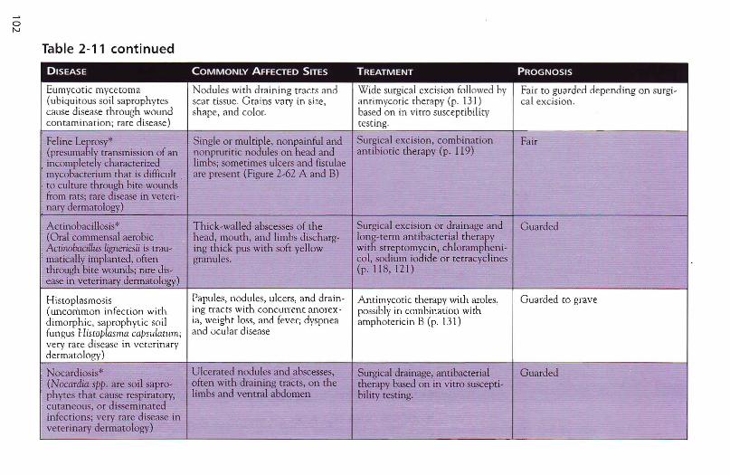
oN
Table 2-11 continued
DISEASE COMMONLY AFFECTED SITES TREATMENT PROGNOSIS
Eumycotic mycetoma Nodules with drainin g tracts and Wide surgical excision followed by Fair to guarded depend ing on surgi-(ubiquitous soil saprophytes scar tissue. G rains vary in size, anti mycotic therapy (p. 13J) cal excision .cause disease through wound shape. and color. based on in virro susceptibilitycontamination; rare disease) testing.
Feline Leprosy" Single or multiple, nonpainful and Surgical exc ision, combination Fair(presumably transmission of an nonprur itic nodules on head and antibiotic therapy (p. 119)
: incompletely characterized limbs; somet imes ulcers and fistulae· mycobacterium that is difficult . are present (Figure 2-62 A and B)· to culture thro ugh bite wounds· from rats; rare disease in veter i-: nary dermato logy)
• Actinobacillosis* Thick-walled abscesses of the Surgical excision or drainage and G uarded, (Ora l commensal aerobic head, mouth, and limbs discharg- long-term antibacterial therapy, ActinobaciUus ligneriesii is trau- ing thick pus with soft yellow with streptomycin, chlorarnphen i-• mat ically implanted, often gran ules. col, sod ium iod ide or tetracycl ines, through bite wounds; rare dis- (p. 118, 121)[ ease in veterinary dermatology)
Histoplasmosis Papules, nodu les, ulcers, and drain - Amimycotic therapy with moles, G uarded to grave(uncommon infect ion with ing tracts with concurren t anorex- possibly in combination withdimorphic, saprophyt ic soil ia, weight loss, and fever; dyspnea amphotericin B (p. 131)fungus /-liscoplasrna capsulaturn; and ocular diseasevery rare disease in vcrerinarvdermatology)
Nocardiosis" Ulcerated nodules and abscesses, Surgical drainage, antibacterial Guarded(Nocardia spp. am soil sapro- often with dra ining tracts, on the therapy based on in vitro suscepti-phvtes that cause respirarory, limbs and ventral abd omen bi lity testing.cutaneous, or d isseminatedinfection s; very rare disease inveterinary dermatology)
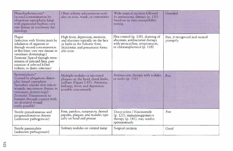
ow
Phaeohyphomycosis* Often solitary subcutaneo us nod- Wide surgical excision followed Guarded(wound conta minat ion by ules on nose, trunk, or extremit ies by ant imycotic therapy (p. 131)ubiqu itous saprophytic fungi based on in vitro suscept ibilitywith pigmented hyphae; very test ing.rare disease in vete rinary der-matology
Plague H igh fever, depression, anorexia, Flea contro l (p. 136) , draining of Fair, if recognized and treated(infection with Yersinia pestis by and abscesses typically on the face abscesses, ant ibacte rial therapy promptly.inha lation of organism or or limbs in the bubonic form. with tetracycline, strepto rnycin,through wound contaminat ion Septicemic and pneumonic form" or ch loramphen icol (p, 118)or flea bites; very rare disease in also exist.veterinary dermato logy)Zoonosis: Spread thro ugh trans-mission of infected fleas, pres-entation of infected killedrodents, or direct infect ion!
Sporotrichosis" Multip le nodules or ulcerated Antimycot ic therapy with iodides Fair(caused by ubiquitous dimor- plaques on the head, distal limbs, or azoles (p . 131)ph ic funga l saprophyte tailbase (Figure 2-63) . Anorexia,Sporothrix schenkii th at infects lethargy, fever, and depressionwounds; uncomm on disease in possible concurren tlyveter inary dermato logy)Zoonosis: Transmission tohumans th rough contact withan ulcerated woundeasily possible !
Ste rile granulomatous and Firm, painless, nonp ruritic dermal Doxycycline I N iacinamide Fairpyogranulomatous d isease papules, plaques, and nodules typ i- (p. 121), immunosuppressive(unknown path ogenesis) cally on head and pinnae the rapy (p. 141), may resolve
sponta neously
Sterile panniculitis Solitary nodules on vent ral rump Surgical excision Good(unknown pathogenesis)
Table 2-11 continued
DISEASE COMMONLY AFFECTED SITES TREATMENT PROGNOSIS
Tuberculosis*(very rare in small animal dermatology; predominant ly respiratory and digestive lesions)
Insidious ulcers, plaques, and nodules on head, neck, and limbs discharging yellow-green pus withunpleasant odor.
Combination antimicrobialtherapy, frequent euthanasia(public health concerns).
Poor
Figu re 2-58A 5-year-old cas tratedDSH with nod ules anddraining rracts result ingfrom atyp ical mycobacte ria. (Courtesy ofDr. Sonya Betrenay. )
Figure 2-59Dra ining tracts due to
atypical mycobacteriain a J-year-old castratedDSH.
Figure 2-60Nasal swelling cau sed bycryptococcosis in a 6- yearold female domestic longhair car. (Courtesy of Dr.T hierry Olivry.)
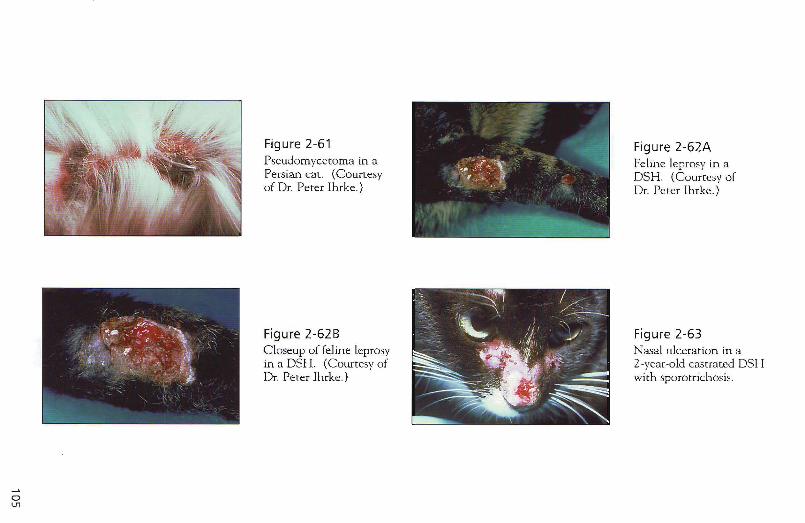
o IJl
Figure 2-61 Pseudo mycetoma in a Persian cat. (Courtesy of Dr. Peter Ihrke.)
Figure 2-62B Closeup of feline leprosy in a DSH. (Courtesy of Dr. Peter Ihrke.)
Figure 2-62A Feline leprosy in a DSH. (Courtesy of Dr. Peter Ihrke.)
Figure 2-63 Nasal ulceration in a 2-year-old castrated DSH with sporotrichosis .
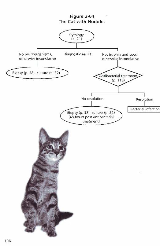
Figure 2-64The Cat with Nodules
~~~....1---- -------.1No microorganisms. Diagnostic result Neutrophils and cocci,otherwise inconclusive otherwise inconclusive
I- -Biopsy (p. 38), culture (p. 32)
106
No resolution
IResolution
II Bacterial infection
The Patient vvith Otitis Externa Otitis externa may be seen wi.th many diseases in conjunctioo with other clinical signs, which are helpful in the formulation of a list of differential diagnoses. This discussion is the approach to the dog with otiti.s externa and no other symptoms.
It is important to differentiate between predisposing, primary, and perpetuating factors in the pathogenesis of otitis externa. Predisposing factors are iodependent from the underlying disease and alone will not cause disease, but will facilitate the pathologic process . Conformation, including dense hair in the ear canal, a Lo ng and narrow ea r canal, pendulous ears, and climate-re18ted seasonal factors such as iocreased remperature and humidity are examples of predisposing factors. Complicating factors occur only after the primary pathologic process has begun, but continue to be a problem after the primary disease has been successfu lly identified and treated. Examples are otitis media, bacterial or fuogal iofections, and chronic proliferative changes due to inflammation . These complicating factors nee d to be treated independe ntly. The most common primary factors are listed in Tab le 2-12.
Key Questions
./ HowaLd was this patient when clinical signs were first recognized? (p.3)
./ Is the disease seasonal ? (p. 3)
./ What do yOll feed the aninl.al? Was a special diet used in the past? (p. 7)
./ Are there any other animals in the household? (p. 8)
./ Was the disease treated before? If so, which drugs were used and how successful was treatment! (p. 8)
./ When was the last medication given? (p . 9)
107
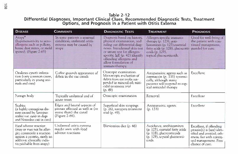
o00
Table 2-12Differential Diagnoses, Important Clinical Clues, Recommended Diagnostic Tests, Treatment
Options, and Prognosis in a Patient with Otitis Externa
DISEASE COMMENTS DIAGNOSTIC TESTS TREATMENT PROGNOSIS
A topy* In some patients a seasonal Diagn osis based on history, Allergen- specific immune- G ood for well-b eing of(hypersensi tivity to aero- co nd ition; unilateral otit is physical exam inat ion , and therapy (p. 123) , anti- the pa t ien t with con-allergens such as po llens , externa may be cau sed by ru ling out differen tial diag- histamines (p , 125).essent ial tinued management,house dust mites, or mold atopy noses. In tradermal skin test fat ty acids (p, 128), glucocorti guarded for cure .spores). (Figure 2-65) or serum test for allergen - co ids (p. 129),
specific IgE (p . 42 ) identify ropical glucocorr icoids.offending allergens andallow formul at ion ofimmunotherapy.
Otodectes cynoti.l infesta- Coffee grounds appearance of Otoscopic examination. Antiparasitic agents such as Excellenttion (very common cause, debris in the ear canals Microscopic evaluation of iverrnecrin (p. 133) systemi -particularly in young ani- debris from ear swabs sus- cally, although manyrnals and cats) pended in mineral oil; rniti- pat ients will respond to rap-
cidal treatment trial ical miticidal rherapy(p. 49)
foreign body Typically unilareral and of Otoscopic examination Removal Excellentacute onset
Scabies Edge s and lateral aspects of Superficial skin scrapings Antiparasiric agents Excellent(a highly contagious dis- pinnae affected as well as (or (p. 26), sarcoptes treatment (p. 133)ease caused by Sarcoptes worse than) the canal trial (p. 49).scobiei var. canis in dogs (Figure 2-66).and Notoedres cad in cars)
Food adverse reaction Unilateral otitis cxterna Elimination diet (p. 46) Avoidance, anuhisramines Excellent, if offending(rnav or may not be aller- maybe seen with rood (p. 125),essential fatty acids protein(s) is (are) iden-gic; commonly a reaction adverse reactions (p. 118), glucocorricoids rified and avoided; oth-against a protein, rarely an (p. 129), topical glucocorti- erwise (air with connn-add itive; clinically ind is- colds. Lied management. Poortinguishable from atopy) chance of cur e.
o\.0
Hyperadrenocorticism* Subrle clinical signs may be Serum biochemistry Iatrogenic form: discontinue Approx imately 60% of(spontaneous or idiopath- overlooked (see table 2-5). (SAP t t , cholesterol t . glucocorticoid adminisrra- dogs with adrenalic. The spontaneous form Complete response to therapy ALT t. glucose t, urea ~), tion. Idiopathic form: o,p' - tumors were reportedis an excessive production of seconda ry ear infect ion hemograms (leukocytos is, DOD (rnitotane), ketocona- to survive adrenalecto -of glucocorticoids either neutrophili a, lymphopenia zole for PDB my and the postopera-due to a microadenoma or and eosinopenia), urinalysis (p. 13I), surgical removal tive period. The aver-macroadenoma of the (specific gravity ~, corti- of affected gland for adreno- age life expectancy waspituitary gland (PDH, sol.creat inine ratio t), radi- cortical neop lasia 36 months. Adrenal85%) or due to adrenocor- ographs (hepatomegaly, adenocarcinomas havetical neoplasms in 15%) osteoporosis, mineralization a better prognosis than
of adrena l glands), low- adenomas. The lifedose dexamethasone sup- span of dogs with PDHpression test, ACTH assays, treated medically aver-ultrasonograph y (adrenal aged 30 months withgland size t), ACTH srim- some dogs livingulation test longer than 10 years
and others only days.
Pemphigus foliaceus* In ner surface of pinnae typi- Cytology (p. 21), biopsy Immunosuppression Fair with appropriate(immune-mediated skin cally worse than can als (p. 38) (p.141) treatment, poor fordisease characterized by (Figure 2-67) cure (except drug-intraep idermal pustule for- induced pemphigus)rnation due to pemphigusantibodies against antigensin the interce llular con-nections. Ma'( be dtll%"induced or paraneoplast ic )
Neoplasia* Unilateral, older an imals Otoscopic examination Surgical excision (vertica l G ood, if completely(ceruminous gland adeno- or complete ablation of the excised and nomas and adenoca rcino- ear canal) metastasesmas-both types in cats,the former more commonin dogs)
o
Table 2-12 continued
DISEASE COMMONLY A FFECTED S ITES DIAGNOSTIC TESTS TREATM ENT PROGNOSIS
Hypothyroidism Subtle other cIinical signs Serum biochemistry Hormone replacement Good , al though not(lymphocytic thyroiditis, (nay be overlooked (see Table (SAP i , cholesterol t , ALT therapy with levotlwroxine all patients stay inpresumablv auroirnmune, 2-5). Complete response to i ), hemograms (anemia) , (p. 144) complete and con-or idiopathic th yroid therapy of secondary ear th yroid tests (free T4, tot al sran t remissionnecrosis which may be infecti on T4 , free T 4 by equilibrium despite adequate sup-end-stage lymphocyric dialysis, TSH assays, TSH plernentation .th vroiditis) stimulation test, TRH stim-
ularion test)
Idiopath ic seborrhea" Excessive wax formation even Biopsy [p, 38 ) Ear cleaners, retin oids, Fair to guarded for(primary kerat inization wirh topical medication. cor ticoste roids well-being; poordefect as autosomal reces- for curesive trait with decreasedepidermal cell renewaltime and thus hyperprolif-erarion of epidermis. seba-ceous glands. and follicu-lar infundibulum second-ary to inflammation.endocr ine disease, ornutritional deficiencies) .. --
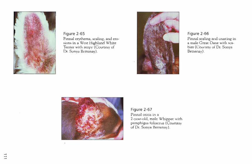
Figure 2-65 Pinnal erythema, scaling, and erosions in a West Highland White Terrier with atopy (Courtesy of Dr. Sonya Bettenay).
Figure 2-67 Pinnal otitis in a 2-year-old, male Whippet with pemphigus foliaceus (Courtesy of Dr. Son ya Benenay) .
Figure 2-66 Pinnal scaling and crusting in a male Great Dane with scabies (Courtesy of Dr. Sonya Bettenay).
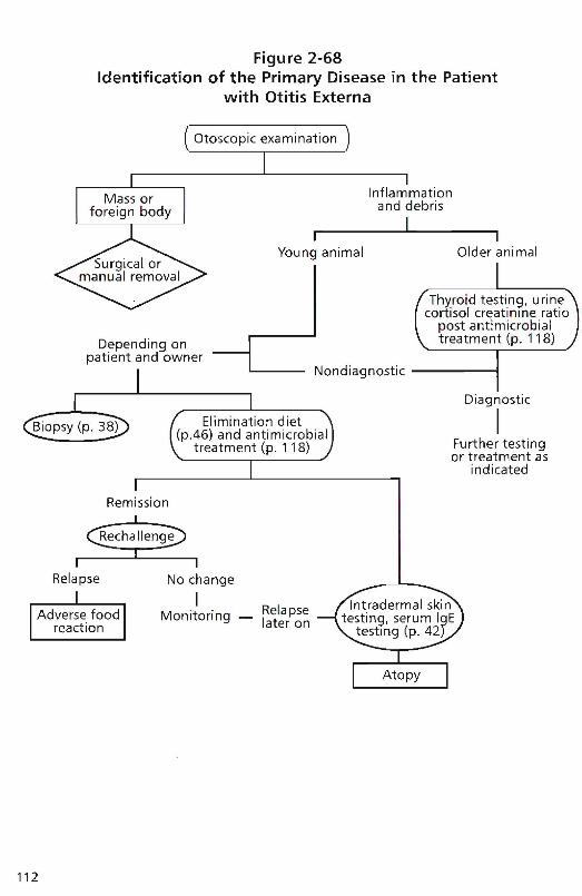
Figure 2-68Identification of the Primary Disease in the Patient
with Otitis Extema
Nondiagnostic ,
Diagnostic
IFurther testingor treatment as
indicated
Thyroid testing. uri necortisol creatinine ratio
post antimicrobialtreatment (p. 118)
Inflammationand debris
Ir 1
Young animal Older animal
Depending onpatient and owner
Remission
Relapse No change
I. RelapseMonitoring - later on
112
Cytology is essential in any dog or cat with otitis externa; examination must be separate in the left and right ear canals <1S infective microorganisms may be different from one ear to the other. In some animals, particularly in patients with chronic longstanding otitis externa and concurrent otitis media , organisms in the middle ear may differ from the those isolated in the externaL ear. Antimicrobial treatment in the ear canaL is most effective topicaLly and determined by cytology (p . 21) and in vitro susceptibility (p . 42 and 46). Repeat cytology examinations during treatment is essential and changes in the otic flora may necessitate changing medications.
Otitis media is best treated with systemic medication. Many dogs with chronic otitis externa and otitis media may not respond to treatment because of severe accumulation of purulent or waxy material in the ear canals . An ear flush under anesthesia may be needed followed by a new attempt of topical and concurrent systemic therapy. The tympanum may rupture prior to flushing or during flushing because in an inflamed ear the tympanum is much more fragile than normal. Frequently there are few alternatives for antimicrobial treatment in these patients. Sometimes they may have to receive potentially ototoxic topical medication. Be sure to discuss this possibility with owners before beginning therapy. Regular cytologic examinations are a precondition for successful management of patients with otitis externa. They are not specifically mentioned in Figure 2-68 . Therapeutic trials and tests for primary diseases may be influenced by concurrent topical medication and thus must be planned, executed, and interpreted with care. Chronic Long-standing otitis extern a is extremely frustrating for patients, clients, and veterinarians, and patients may benefit from referral to a veterinary dermatologist. "',
'"
113
Section 3
Treatments
114
In this section, I will summarize the most common treatment modalities, their formulations (which may vary in different parts of the world), indications, and doses. Given that detailed discus~ sion of individual drugs, their mechanisms of action, pharmacokinetics, and protocols is beyond the scope of this text, further reading may be required. See Recommended Readings.
Drugs marked with an (*) and a colored screen are potentially dangerous and the clinician inexperienced with these medications may consider offering referral to a veterinary specialist or seeking further advice from a colleague with more knowledge about that particular agent.
Shampoo Therapy of Various Skin Conditions
Shampoo therapy can provide effective management of dermatoses with both medical and cosmetic presenting complainrs (Table 3-1). There are few adverse effects associated with shampoo therapy, although they are recognized. However, shampoo therapy is symptomatic treatment; it rarely "cures" a dermatosis.
Prescribing a shampoo involves selecting the proper shampoo for both the dem1.atosis and the client. Shampoo manufacturers have undertaken considerable research and development in order to produce for~ mulations, which lather well, have an appealing smell, offer little irritation, and serve their intended purposes.
In addition to selecting the appropriate shampoo, the veterinarian's instructions will have a significant impact on the efficacy. The frequency of bathing and duration of skin contact time will influence the obtained result. A 10-minute contact time is generally recommended. This is a long time for the owner of a fidgety, shivering dog to wait and it will frequently be cut short! Techniques to improve contact tirne include:
./ Take a clock into the bathing area and time 10 minutes accurately .
./ Use the time for patting and bonding.
115
116
./ Massage the skin for the full 10 minutes; it will usually be enjoyable for the dog and occupy both pet and owner .
./ Take the dog outside to play ball or go for a walk with the soap still on (if climate permits) .
./ Rinse the shampoo off thoroughly for at least 5 to 10 minutes.
The frequency of shampooing will vary with severity and type of the disease process. In general, the more severe the disease, the more frequently bathing is indicated.
The major reasons for failure of shampoo therapy are
1. lack of client compliance (frequency and/or duration of application)
2. incorrect selection of shampoo for the disease process
3. shampoo irritation
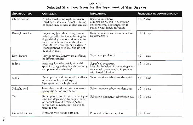
'"-J
Table 3-1Selected Shampoo Types for t he Treatment of Skin Disease
SHAMPOO TYPE COMMENTS INDICATIONS FREQUENCY OF ADMINISTRATION
Chlorhex id ine Ant ibacter ial, an tifungal, not inacri- Bacterial infection s. q 1-14 daysvated by organic matter, not irritat ing May also be helpful in dec reasingor drying, may be used in dogs and cats environ mental contamina tion in
pat ients with fungal infecti on
Benzoyl peroxide Degreasing (and th us drying), kera- Bacterial infections, sebaceous aden i- q 7-14 dayscolytic, possibly follicu lar flush ing . In tis, de modic osisdogs with dry or normal skin , a rnois-tur izer must be used after the sham -poo! Ma y be irritating, particularly inconcentrations over 3% . Should notbe used in cats!
Eth yl lactate May be drying. Controversial efficacy Superficial pyoderma q 7-14 daysi.n differen t studies
Iodine Antifungal, ant ibacterial, virucida l, Superficial pyoderma. q 7- 14 dayssporicida l, degreasing, but also sta in ing May also be helpful in decreasing en vi-and potenti ally irritating! ronrnental conta mination in patien ts
with fungal infection
Sulfur Keraroplasric and keratolytic, ant ibac- Sebo rrhea sicca, seborrheic dermatitis. q 3-14 daystcrial and mildly an tifungal.Synergistic with salicylic ac id
Salicylic ac id Kera tolytic , mildly an ti-in flammatory, Seborrhea sicca, seborrheic dermat it is q 3- ] 4 da yssynergistic actio n with sulfur
Tar Keratop last ic and keratolyt ic, anr ipru- Seborrheic denn atiu s, seborrhea oleosa q 3-14 daysririe and degreasing. In dogs with dryor norm al skin , it needs to be fol-lowed with a moisturi zer. No t to beused in ca ts!
Co lloidal oatmeal Hydrates th e strat um corneum Prurtric skin disease, dry skin q z- l-l davs
118
Treatment of Bacterial Infection
./ Antibiotics are frequently used in veterinary dermatology, because many conditions are associated with secondary bacterial infection. Dogs with chronic allergies, irmnune~mediated der~ matoses, or endocrinopathies frequently develop secondary pyo~ dennas that exacerbate these conditions and necessit3te antibacterial treatment (Table 3~Z) .
./ Not all available antibiotics are useful for skin infections so that spectrum of activity as well as pharmacology of the different antibacterial drugs has to be considered.
h The overwhelming majority of skin infections in the dog and cat is caused by Staphylococcus intermedius. Mixed infections can involve organisms such as Escherichia coli or Pseudomonas aeruginosa, which usually develop concurrently with most patients' primary agent, S. intermedius.
h Proper dosage and proper duration are important for the suc~ cess of antibacterial therapy. Antibiotics should be given for at least 3 weeks or longer or until at least 1 week after resolution of clinical signs. Relapses are common in patients on short courses of pharmacotherapy or those receiving medications at low dosages! Deep infections may take 6 to 1Z weeks to resolve .
./ Pyodermas can, at least initially, be treated empirically. If appropriate therapy does not resolve the condition, taking a culture is indicated (p. 32) .
./ Each sample for culture and sensitivity should be accompanied by cytologic examination and culture results interpreted in light of the cytology, as growth of different microorganisms does not indicate necessarily that they ate present in significant numbers in vivo.
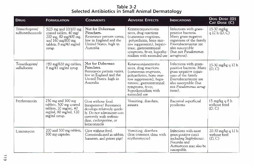
IV
Table 3-2Selected Antibiotics in Small Animal Dermatology
DRUG FORMULATION COMMENTS AOVERSE EFFECTS INDICATIONS DOG DOSE (D)CAT DOSE (C)
Trimcrhop rim/ 5/25 mg and 20/100 mg N ot for D ober man Keratoconjunctiviti s In fections with gram- L5-30 mg/kgsulfamerhoxazole coated tablets, 40 mg/ P inschers . sicca, drug reactions positive bacteria. q 12 h (D, C)
200 mg, 80 mg/400 mg, Resistance patt ern varies, (cutaneous eruptions, Many gram-negativeand 160 mg/800 mg low in Eng land and the pol yarthritis, bone mar- organ isms of the fam ilytablets , 8 mg/40 mg/ml Uni ted States, high in row suppression ), hepato- Enterobacteriaceae aresyrup. Australia toxic , gastroi ntestinal a lso susceptible
sympto ms, fever, hvporhy- (b ut not Pseudomonasroidism with extended use aeruginosa) .
Trimethoprtrn/ 180 mg/820 mg tablets, N ot for Doberm an Keratocon junctivitis In fection s with gram- 15-30 mg/kg q 12 hsulfadiazine 9 mg/4 1 mg/mt syrup P inschers. sicca , drug reac tions posit ive bacteria. Ma ny (D,C)
Resistance pat tern va ries, (cutaneous erupt ions, gra m-nega tive organ -low in England and the pol yarthritis. bone mar- isms of the familyUn ited Stares, high in row suppression ), hepa- Enrerobacteriaceae areAu stralia totoxic , gastrointestinal also suscep t ible (but
symptoms, fever , no t PseudomoTU.l.S aeTug-h ypothyroid ism with mosc).extended use
Erythromycin 250 mg and 500 mg Gi ve witho ut food. Vom iting , dia rrhea , Bacterial supe rfic ia l 15 mg/kg q 8 htab lets, 500 m~ coated Inexpensi ve1 Resistance n ausea pyode rma without foo dtablets, 20 mg ml, 40 develops relatively qu ick- (D,C)mg/ml , 80 mg/m l, L20 Iy. Do not admi nister co n-mg/ml syrup. currently with rerfena-
dine, cyclo spor ine , orketaconazo le
Lincomycin 200 and 500 mg tab lets, G ive without food. Vomi ti ng , diarrhea Infections with most 20-30 mg/kg q 12 h500 mg capsules. Contraind icated in rabbits, (less common than with gram-positive cocci without food
hamsters, and guinea pigs! er ythromycin ) including Swphylococci . (D,C)Nocardia andActinomyces may also besusceptible.
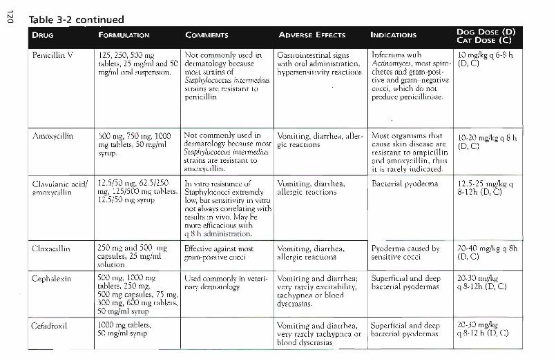
~ Table 3-2 continued
DRUG FORMULATION COMMENTS ADVERSE EFFECTS INDICATIONS DOG DOSE (D)CAT DOSE (C)
Peni cillin V 125, 250, 500 mg Not commonly used in Gast rointestinal signs Infections with 10 mg/kg q 6-8 Ittablets, 25 mg/ml and 50 dermatology because with oral administra tion , Actinomyces , most spiro- (D ,C)mg/ml ora l suspension . most stra ins of hype rsens itivity reactions chetes and gram-post-
Staphylococcus imennedius rive and gram- neg ativesrra ins ate resistant to cocc i, which do notpenicillin produce penicill inase.
Amoxycillin 500 rng, 750 mg, 1000 Not co mmo n ly used in Vomiting, di arrhea, <Iller- Mos t o rgan ism s that 10-20 mgfkg q 8 hmg tabl et s, 50 mg/ml dermatology becau se most gic reactions cause sk in d isease are (D,C)syrup. Staphylococcus inrelmediLts res istant to am picillin
stra ins are res istan t to a nd a moxycill in , th usarnoxvci Ilin . it is rarel y indicated.
C lav ula n ic acid/ 12.5 /50 mg, 62 .5/250 In vitro resistance of Vom iti ng, d iarrh ea, Bac ter ia l pyoderm a 12. 5- 25 mg/kg qamoxyci llin mg, 125/500 mg tablets, S taphylococci ex tremely all ergic reactions 8-12h (D , C )
12.5/ 50 mg syrup low, bur sensiri viry in vitronor always correlating withresults in vivo . May bemore efficacious withq 8 h admin istrat ion .
Cloxacillin 250 mg and 500 mg Effect ive against most Vomiting, diarrhea , Pyoderma caused by 20- 40 mgjkg q 8hca psules, 25 mg/ml gram-positive cocci all ergic reactions sensiti ve cocci (D ,C)so lut ion
C ephalexin 500 mg, 1000 mg Used commo nly in veteri- Vo m it ing and diarrh ea ; Supe rfic ial and deep 20-30 mg/kgtabl e ts, 250 mg, nary dermatology ve ry rar ely excitabili ty, bac terial pvoderrnas q 8- 12h (D , C)500 mg capsules, 75 mg, tachypnea or b lood300 mg, 600 mg tablets , dvscrasia s.50 mg/m l syrup
Cefadroxtl 1000 mg tablet s, Vomiting an d d iarrhea , Su perficial and deep 20-30 mg/kg50 mg/ml syrup ve ry rarely ta chypnea or bac te ria l pyodermas q 8-12 h (D , C)
blood dvscr asias-
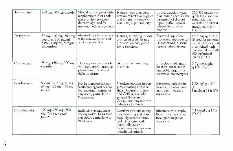
N
Tetracycline 250 mg, 500 mg capsules Should not be given with Nausea, vomiting, discol- In combination with 250-500 mg/animalamphotericin B or eryth- oration of teeth in puppies niacinamid used for the q 8 h (in combina-rornycin. In veterinary and kittens, phototoxic treatment of discoid uon with niaci-dermatology used for reactions, hepatotoxicity lupus erythematosus, namide at 250-500immunomodulatory effects idiopathic onvcho- mg/animal q 8 h)
rnadesis. (D,C)
Doxycyline SO mg, 100 mg, 200 mg Also used for effects on cells Nausea, vomiting, discol- Bacterial superficial 2.5-5 mg/kg q 24 hcapsules, 100 mg/ml of the immune system and oration of teeth in pup- pyoderma, mycobacter- (if used for immune-paste, I mg/ml, 5 mg/ml cyrokine production pies and kittens; photo- ial infections, discoid mediated diseases, itsuspension toxic reactions lupus er ythematosus. is combined with
niacmamide at 250-500 rug/animalq8h) (D, C)
Clindamycin 75 mg, ISO mg, 300 mg Do nor give concurrently Skin rashes, vomiting, Infections with gram- 5-10 mg mg/kgcapsules with cvclosporin, iron sup- diarrhea. positive cocci, mo st q 12h (D, C)
plementation , and oral anaerobic organisms,diabetic agen ts. Nocardia , Actinomyces
Enrofloxacin 5.7 mg, 22.7 mg, 50 mg, Not in irrunarure animals' Cartilage erosions in pup- Infections with staphv- 5-20 mg/kg q 24 h68 mg, 136 mg, 150 mg Ineffective against anaero- pies. vomiting and diar - lococci, mycobacteria, (D)tablets bic organisms. Resistance rhea. Hypersensitivities most gram-negative 5 mg/kg q 24 h (C)
may occur, particularly ro and CNS signs could organ ismsPseu.domonas . potentially occur.
Crystalluria may occur indehydrated ani mals
Ciprotloxacin 100 mg, 250 mg , 500 Ineffective againsr anaer- Cartilage erosions in pup- Infections with staphy- 5-15 mg/kgq 12 hmg, 750 mg coated obe organism s. Resistance pies; vomit ing and diar- lococci , mycobacteria. (D,C)tablets may occur, particularly TO rhea. Hypersensitivities most gram-negative
Psewiomonas. and CNS signs could organismspotentially occur.Crystalluria may occur indehydrated animals
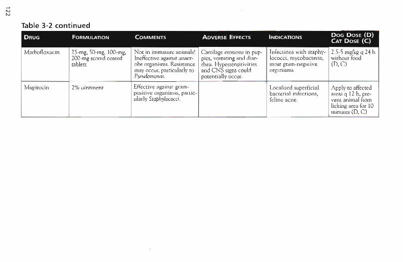
Table 3-2 continued
DRUG FORMULATION COMMENTS ADVERSE EFFECTS INDICATIONS DOG DOSE (D)CAT DOSE (C)
Marbofloxacin 25-mg, 50-m g, l OO.mg, NO[ in immature an ima lsI C arti lage erosio ns in pup ' Infec t ions with sraphv- 2.5·5 mg/kg q 24 h200 -mg scored coated Jneffective against anaer- pies, vomiting and d iar- locoeci , mycob acteria, witho ut foodtable ts obe organ isms. Resistan ce rhea . Hypersensit ivit ies most gra m-negat ive (D,C)
may occ ur, particularly to and C NS signs could organ ismsPseudomonas. potentially OCCUt.
Mupirocin 2% o in tment Effect ive <1gain st gram · Loca lized superfici a l Apply to affec tedpos it ive organ isms, part ie- bact er ial infecti ons, areas q 12 h, pre-ularl y Staphylococci. feli ne acn e. ven t an imal from
licking area for 10min utes (D, C )
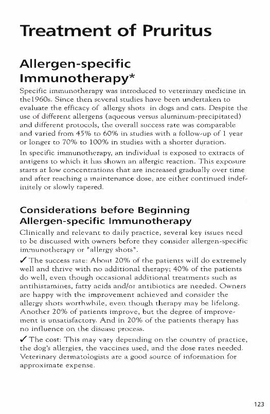
Treatment of Pruritus
Allergen-specific Immunotherapy* Specific immunothera py was introduced to veterinary medicine in the 1960s. S ince then several studies have been undertaken to
evaluate the efficacy of allergy shots in dogs and cats . Oespi te the use of different a llergens (aqueous versus aluminum-precipitated) and different protocols , the overall success rate was comparable and varied from 45% to 60% in stud ies with a fo llow-up of 1 year o r longe r to 70% to 100% in stud ies with a shorter d uration.
In specific immunotherapy, an ind ividual is exposed to extracts of antigens to which it has shown an a llergiC reaction. This exposure starts at low concentrations that a re increased gradually over t ime and after reaching a main ten an ce dose, are either continued indefinitely or slowly tapered.
Considerations before Beginning Allergen-specific Immunotherapy C lin ica lly and relevant to daily practice, several key issues need to be discussed w ith owners before they cons ider allergen-specific imlnunotherapy or "a lle rgy sh o ts" .
./ The success rate: Abo u t 20% of the patients will do extremely we ll and thrive with n o add itional therapy; 40% of the patients do we ll . even though occas iona l additional treatments such as antihistamines, fatty acids and/or antibiotics are needed . Owners are happy with the improvement achieved and consider the allergy shors worthwh il e , even though therapy may be lifelong. Another 20% of patients improve , but the degree of improvement is unsat isfactory. And in 20% of the patients therapy has no influence on the d isease process .
./ The cost: This may vary depend ing on the country of pract ice, the dog's allergies, the vacc ines used, and the dose rates needed. Veterinary dermatologists are a good source of information for approxitnate expense.
123
124
./ The time to improvement: First im.provement may be seen as early as 4 weeks into therapy and as late as 12 months after starting the allergy shots. On average, improvement is expected after between 4 and 6 months .
./ The duration of treatment: A minority of owners may discontinue their dogs' immunotherapy after 2 years, with theif pets being perm.anently cured and syrnptolTI fTee. Other dogs, however, will require lifelong therapy!
./ The involvement: Atopic dogs are "high maintenance" and as such need constant care, most likely at least initially regular rechecks and concurrent medication. Allergy shots are not an easy way out but at this point the best of many available treatments, all of which involve long-term administration of medications of SOITIe sort.
Points to Remember if You Have Patients on Immunotherapy ~ Glucocorticoids may be given on all. occasional basis at antiinflammatory dosage levels without interfering with therapy .
./ Antihistamines, farty acids, and antimicrobials do not interfere with immunotherapy so that they may be, and often are, used concurrently.
h If the condition of patients receiving allergy shots suddenly deteriorates, always evaluate for infection. Yeast and bacteria are common complicating factors secondary to these allergies, which can cause dramatic increases in pruritlls. Cytology Cp. 21) wilL identify cocci, rods, yeasts and inflammatory cells and thereby guide antimicrobial therapy .
./ If no infeC(ion is detected, antihistamines or glucocorticoids can be used to regulate the padent's pruritus .
./ If there is a regular increase in pruritus after the injection, the dose and frequency may need to be adjusted. Decreased doses may be helpful.
./ If there is a regular increase in pruritus before the injection that is greatly improved by the injection of allergen extract, the time interval between the injections is probably too long and needs to be shortened. The dose may also be decreased in some of these patients without decreased efficacy.
./ If there is no response to allergen~specific immunotherapy after 4 to 6 lnonths, 1 recommend that you contact your n earest ve terinary dermatologist for advice while the re is sti ll sufficient vacc ine left t o change the dose a nd frequency of the injections by adjusting them to the needs of tha t particular patient. Many patients need an approach suiting their particular requirements and the h e lp o f a veterinarian experienced in im.munotherapy m.ay be of great benefit.
1 s incere ly believe that allergen-specific immunoth e rapy is currently the best ava ilable treatment for canine atopic dermat itis , but it will o nl y be successful in most atopic a nima ls if owners a nd veterinarians have realistic expectations and a re prepared to put in significant e ffort over the period that som et imes extends over many months. Only then will maxima l benefit be achieved ! In as much as the first tnonths on immunotherapy m ay be draining for owner and veterinarian, con s ider offering referra l to a veterinary d ermatologist, particula rly if you are not experienced in this therapy.
Antihistamines ./ Antihistamines are useful adj unctive agents in the manage~
ment of pruritic patients. The classica l a ntihista mines ac t by b locking Hl~receptors. First~generati on antihistamines a[so h a ve an antic h o linerg ic, sedative, and loca l a naesthetic effec t. Second ~generation antihistamines penetrate less through the blood bra in barri e r or have a low affinity fo r the brain compared with the action on peripheral Hl~receptors. Thus t hey a re effective yet produce less sedation (Table 3~3) .
./ The a dvantage of antihistamines is th e rare occurrence of adve rse effects. Dro wsiness is the m ost commo n finding a nd may decrease after 2 to 3 days of thera py. Thus , it m ay be worthwhile continuing treatment for seve ral days before fina l evalu a tio n . Less COlnmon a re gastrointestinal signs. A c ute poisoning following an overdose is characterized by eNS hyperexcitab ility. Due t o the anticholinergic properties of t erfe nacline a nd cyprohepta~ dine, these drugs should not be used in patients with severe car~ diovascula r disease, since they m ay cause hypertensio n .
./ The necessity of frequent administration (two to three times dally) a n d the high cost of some a ntihistamines limit their longterm use in m a ny patients, especia lly larger dogs.
125
126
./ A further shortcomlng in dogs is the relative ly lo w success rate , which varies between 5% and 30%, depending on dosage and drug used .
h Cats are much more like ly to respond to antihistam lnes than dogs. However, admlnlstertng ora l medication o n a lo ngterm basis may be challenging in this species .
./ If a patient responds to a ntihist a mine therapy and the o wner lS willing to maintain the anim a l on it, antihistamines represent safe lo n g- term treatment th at is preferable to glucocorticoids!
./ Antihistamines in human s a re no t used to treat present symptoms but to prevent onset of symptoms. Thus, adminlstration sh o uld not be intermitten t ass uming the same h o lds true i.n a nim.a ls .
~ The potentlal success ra te can be increased by trying severa l different anti histamines seq uen tia lly because patients m ay be respons i ve to one antihistamlne but not to another.
~ Antihistamines h ave been reported to be effective in lowe ring the corticosteroid dose, even if they did n o t help the animal as a s lng le therapeutiC agent .
./ Because the withdra w a l time o f antihistamines before an in trade rma l skin test is rrlUch sh orter than tha t of g lucocorticoids, they can be used to r e li eve pruritus during the preparation time where the latter a re contra indicated .
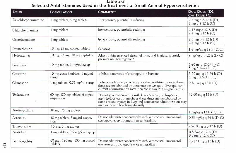
N'-J
Table 3-3Selected Antihistamines Used in the Treatment of Small Animal Hypersensitivities
DRUG FORMULATION COMMENTS DOG DOSE (D),CAT DOSE (C)
Dexch lorphenirarnine 2 mg tablets, 6 mg tablets in expensive , po ten t ially seda ting 2-6 rug q 8-12 h (D),2 mg q8-12 h (C)
C hlorpheni rami ne 4 mg tablets Ine xpe nsive, poten tially seda ti ng 2- 12 mg q 12 h (D )2-4 mgq I2 h (C)
C yproheptad ine 4 mg tablets Inexpensive, potentially sedat ing 2-8 mg q 8 -12 h (D)2-4 mg q 12 h (C)
Prom et hazine 10 rug, 25 mg coa ted tablets Sedating 1-2 mg/kg q 12 h (D ,C)
Hydroxyzine 10 mg, 25 rng, 50 mg capsules A lso inhi bits mast cell degran ulat ion , and is tr icyclic an t ide - 2 mg/kg q 8-12 h (D , C )pressant and teratogeni c!'
Loraudi ne 10 mg tablet , 1 mg/m l syrup 5-20 In q 12-24 h (0)5 mg q 12-24 h (C )
Cet irizine 10 mg coated tablets, 1 mg/ml Inhibits exocvtosis of eosmophils in hu mans 5-20 mg q 12-24 h (D)syrup 5 mg q 12-24 h (C )
Clernasrine I mg ta blets, 0.05 mg/rnl syrup Enh ances cho linergic activity of other an rihisrarn ines as these 0.5- 1 mg q 12 h (D )drugs are metabolized by same enzyme system in liver and con-current admin ist ration may increase serum levels significan tly.
Terfenadinc 60 rng, 120 mg tablets, 6 rng/rnl Do not give concurrently with ketoconazole, cyclosporine, 30-60 mg q 12 h (D)suspension asteruizo l, o r ervrhrornycin as the se drugs are metabolized by
same enzyme sysrem in liver and conc urrent ad ministration mayincrease serum levels sign ificantly.
A mitrypti lline 10 mg, 25 mg tabletsI mg/kg q 12 h (0 , C )
Aste rni zo l 10 mg tablets, 2 mg/m l suspen- Donot admin ister concurrently with ketoconazol, itraconazol, 0.25 mg/kg q 24 h (D, C)sion cyclosporine, erythromycin , or terfenadine .
Tr imeprazine 2.5 mg, 5 mg tablets 2.5-10 mg q 8- 12 h (D )
Azatid ine 1 mg tab lets, 0 .5 mg/5 ml syrup 0 .5-2mg q 12 h (0 )0 .5 mg q 12 h (C )
Foxofenac hi n 60 mg , 120 mg, 180 mg coa ted Do not administer conc urrently with keroconazol, itracon azol, 30-120 rng q 12 h (0 )tablets erythro mycin , cvclosporine, or tertenadine
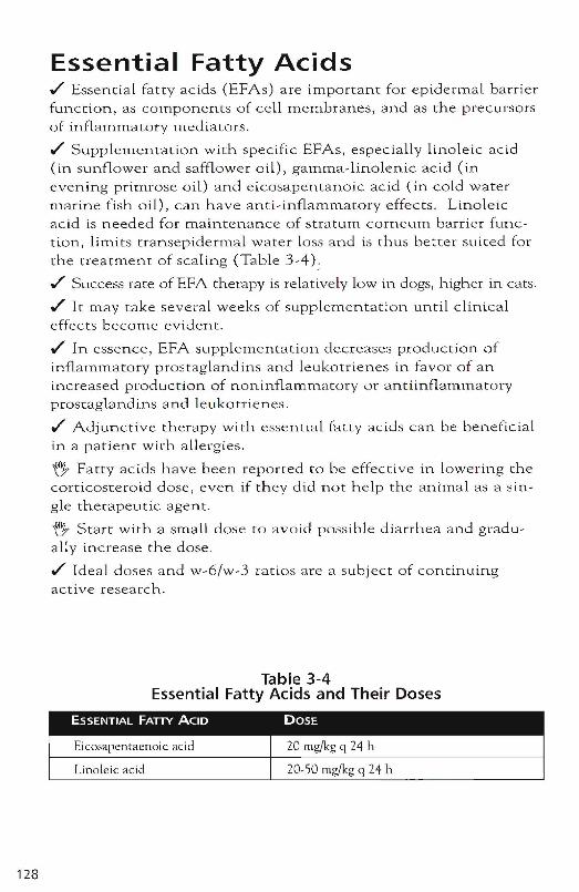
128
Essential Fatty Acids./ Essential fatty acids (EFAs) are important for epidermal barrierfunction, as cornporierits of cell membranes, and as the precursorsof inflammatory mecliators .
./ Supplementation with specific EFAs, especially linoleic acid(in sunflower and safflower oil), gamma-linolenic acid (inevening primrose oil) and elcosapentanoic acid (in cold watermaririe fish oil), can have anti-inflammatory effects. Linoleicacid is needed for maintenance of stratum corneum barrier function, limits transepidermal water loss and is thus better suited forthe treatment of scaling (Table 3-4):
./ Success rate of EFA therapy is relatively low in dogs, higher in cats .
./ It may take several weeks of supplementation until clinicaleffects become evident .
./ In essence, EFA supplementation decreases production ofinflammatory prostaglandins and leukotrienes in favor of anincreased production of noninflammatory or ancttnflammaroryprostaglandins and leukotrienes .
./ Adjunctive therapy with essential fatty acids can be beneficialin a patient with allergies.
~ Fatty acids have been re p o n ed to be effective in lowering thecorticosteroid dose, even if they did not help the animal as a single therapeutic agent.
~ Start with a small dose to avoid possible diarrhea and gradually increase the dose.
./ Ideal doses and w-6/w-3 ratios are a subject of continuingactive research.
Table 3-4Essential Fatty Acids and Their Doses
Linoleic acid
Glucocorticoids .,/' Glucocorticoids are very commonly used in the treatment of skin conditions (Table 3-5) .
.,/' At anti-inflammatory dosage, they decrease inflamma tory cell activity and migratio n .
.,/' Corticosteroids are effective in most patients with atopic disease and resolve the symptoms at least initially on reasonably low dosages. Flea-allergic animals will also often respond to "these drugs, although typically at slightly higher dosages .
.,/' G lucocorticosteroids can be considered the treatment of choice in animals with a l1.l.ild seasonal pruritus of 1 to 2 months duration that is conn-oIled with anti-inflammatory dosages « 1 mg/kg ) of prednisolo ne every other day .
.,/' Every other day therapy is definitively preferred over daily drug administration because as it is tho ught to Lower the chances of iatrogenic hyperadrenocorticism .
.,/' I llse prednisolone at anti-inflammatory doses for severely affected dogs a fter skin testing and short term to break the itch-scratch cycle. However, the need to inc rease the dosage over time to control the clinical signs in most of these patients, combined with the potentially severe long-term side effects m 8ke glucocorticoids a poor long-term c hoice for 8topic patients .
.,/' Adverse effects include poLyuria, poLydipsia, polyphagia, increased susceptibility to infec tion, and other well-known symptoms of iatrogeniC hyperadrenocorticism. The most commonly encountered infec tions affect the urinary tract, skin, and lungs .
.,/' Drugs should a lways be tapered to the lowest effective dose.
h Frequently, the dose necessary to control clinical signs cah be decreased when adjunctive therapy is used . Fatty acids and antihistamines have been reported to be e ffective in lowering the cor ticosteroid dose, even if they did not h e lp the animal as a single therapeutic agent. Regular topica l therapy (e .g. , shampoos) may be another m eans of decreasing the n eed for system.ic glucocorticoids.
h I recommend to the owners of my atopic patients trea ted with glucocorticoids the lowest possible dose, on which the animal is mildly itchy but not uncomfortable. If n o pruritLls is present, the dose used is too high.
h The g lucocorticOid dose needed with individual patients often varies seasonally.
129
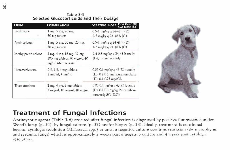
wo
Table 3-5Selected Glucocorticoids and Their Dosage
DRUG FORMULATION STARTING DOSE DOG DOSE (D)CAT DOSE (C)
Prednison e 1 mg, 5 mg, 20 rng, 0.5-1 IUg/kg q 24-4 8 h (D)
50 mg tablets 1-2 lng/kg q 24-48 h (C)
Prednisolone 1 mg, 5 mg, 20 mg, 25 mg, 0.5-1 mg/kg q 24-48 h (D)
50 mg tablets 1-2 mg/kg q 24-48 h (C)
Methylprednisolone 2 mg, 4 mg, 16 mg, 32 mg, 0.4-0.8 mg/kg q 24-48 h orally
100 mg tablets, 20 mg/ml , 40 (D), intramuscularly
mg/ml Met. acetate
Dexamethasone 0.5, 1.5, 4 mg tablets, 0.05-0.1 mg/kg q 48-72 h orally
2 mg/mI, 4 mg/m] (D) , 0.2-0.5 mg/ intramuscularly
(D) ; 0.1-0 .25 Ing/(e),
Triamcinolone 2 mg, 4 mg, 8 mg tablets , O.OS-O.! mg/kg q 48-72 h orally
3 mg/ml, 10 mg/ml, 40 mg/ml (D); 0.1-0.2 mg/kg 1M or subcu-
taneouslv SC (D,C)
Treatment of Fungal InfectionsAntimycotic agents (Table 3-6) are used after fungal infection is diagnosed by positive fluorescence underWood's lamp (p. 30), by fungal culture (p. 32) and/or biopsy (p. 38). Ideally, treatment is continuedbeyond cytologic resolution (Malassezia spp .) or until a negative culture confirms remission (dermatophytesand systemic fungi) which is approximately 2 weeks past a negative cultu re and 4 weeks p ast cytologicresolution .
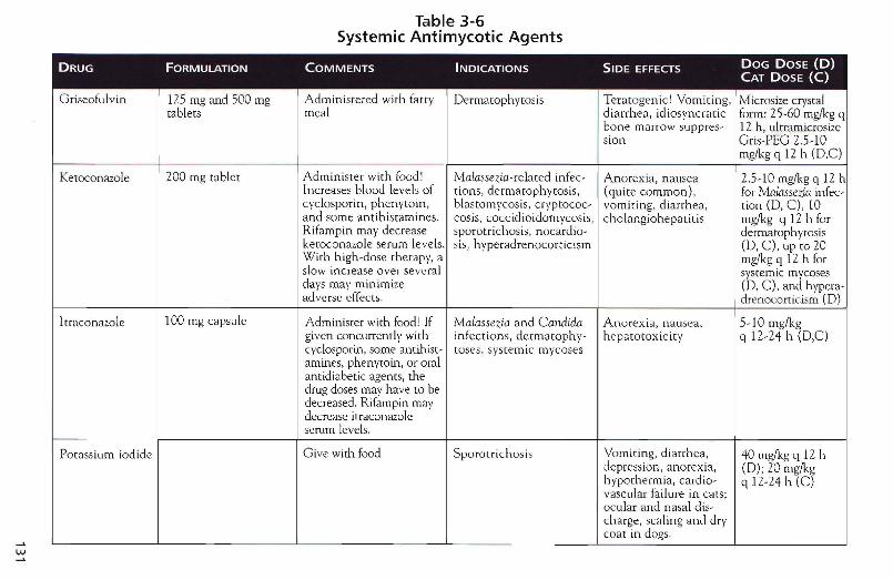
w....
Table 3-6Systemic Antimycotic Agents
DRUG FORMULATION COMMENTS INDICATIONS SIDE EFFECTS DOG DOSE (D)CAT DOSE (C)
Griseofulvin 125 mg and 500 mg Administered with fatty Dermatophytosis Teratogenic' Vomiting, Microsize crystaltablets meal diarrhea, idiosyncratic form: 25-60 mg/kg q
bone marrow suppres- 12 h, ultrarnicrosizesion Gri s-PEG 2.5-10
mg/kg q 12 h (D,C)
Keroconazole 200 mg tablet Administer with food' Malassezia-relmed infec- Anorex ia, nausea 2.5-10 mg/kg q 12 hIncreases blood levels of tions, dermatophytosis, (quite common) , for Malassezia infec-cyclosporin, phenytoin, blastomycosis, cryptococ- vomiting, diarrhea , tion (D, C) , 10and some antihistamines. cosis, coccidioidomycosis, ch olangiohepatitis mg/kg q 12 h forRifampin may decrease sporotrichosis, nocardio- de rmatophytosiskeroconazole serum levels. sis, hyperadrenocorticism (0, C), up to 20WIth high-dose therapy, a mg/kg q 12 h forslow increase over several systemic mycosesdays may minimize (0, C), and hvpera-adverse cffects. drenccorticisrn (D)
1traconazole 100 mg capsule Administer with food! If Malassezia and Candida Anorexia, nau sea, 5-10 mg/kggiven concurrently with in fect ions, derrnatophy- hepatotoxicity q 12-24 h (D,C)cyclosporin, some anrih ist- loses, systemic mycosesaruines, phenytoin, or oralantidiabetic agents , thedrug doses may have to bedecreased. Rifarnpin maydecrease itraconazoleserum levels.
Potassium iodide Give with food Sporotrichosis Vomiting, diarrhea, 40 mg/kg q 12 hdepression, anorexia, (D) ; 20 mg/kghypothermia, cardio- q 12-24 h (C)vascular failure in cats;ocular and nasal dis-charge, scaling and drycoat in dogs.
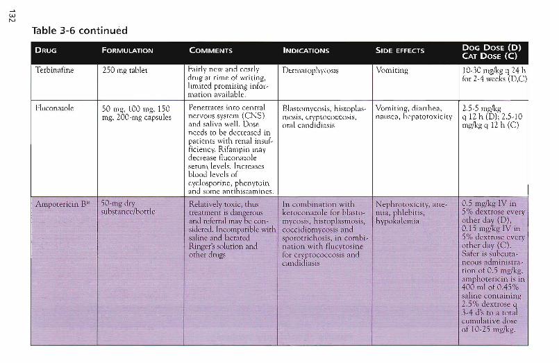
WN
Table 3-6 continued
DRUG FORMULATION COMMENTS INDICATIONS SIDE EFFECTS DOG DOSE (D)CAT DOSE (C)
Terbinafine 250 mg tablet Fairly new an d costly De rma tophytos is Vomit ing 10-30mg/kg q 7.4 hdrug at time of writ ing, (or 2-4 weeks (D,C)limited promising infer-rnarion availab le.
Flucon azole 50 rng, 100 mg, 150 Penetrates in to ce ntral Blasrom vcosis, histopl as- Vomiting , d iarrhea , 2.5-5 mg/kgmg, WO-mg capsules ner vous system (CN S) mosis, c rvpto co ccos is. nau sea, hepat ot oxicity q 12 h (D); 2.5-10
and saliva well. Dos e ora I cand id iasis mg/kg q 12h (C)need s to be decreased inpati ents with renal insuf-ficiencv, Rifampin maydecrease fluco nazoleserum level s. In creasesblood levels ofcvclospo n ne, phe nyto inand some antihistamines.
A mpotericin B* 5Q-mg dry Relatively toxic, thus In co mbination with Nephrotox icity, ane- 0.5 mg/kg IV in
;substance/bottle t reatment is dangero us ketoconazo le for blasto- mia, phleb itis, 5% dextrose event
and referral may be con- mycosis , histopl asm osis, h ypokalemia other daft (0),sidered. Incom patible with coccidiomycosis and 0.15 mg kg IV insaline and lactated sporotrichosis, in combi- 5% dextrose everyRinger's solurion and n ation with flucvtoslne other day (C ).orher drugs for cryptococcosis and Safe r IS subcuta-
candid iasis neous adrninistra-tion of 0.5 mg/kg,ampho tericin is in400 ml of 0.45%saline co ntain ing2.5% dextrose q3-4 d 's co a totalcu mulative doseof 10-25 mg/kg.
Ectoparasiticidal AgentsW h e n treating p a tie n t s wi th ec.toparasires, env ironment and contac t animals h a v e to be considered as well.Environmental contamination is significant with fleas a nd chiggers (Trombicula, N eocrombicula, Walchia) andpossibly with Cheyleti ella . Contact animals must b e treated for all ectoparasites ex cept Demodex a n d chiggers, but, if dogs are affected w ith sca b ies, cats may not have to be t reated an d v ice v e rs a (Table 3 - 7 )
Table 3-7Selected Ectoparasiticidal Agents in Small Animal Dermatology
DRUG FORMULATION COMMENTS INDICATIONS SIDE EFFECTS DOG DOSE (D)CAT DOSE (C)
ww
lvermecrin 10 mg/kg bovinein jectable so lut ion(often given ora lly tosma ll anima ls) , 10lng/m l equine oralso lut ion
Id iosyncrat ic toxic iti esmost commo n inColl ies and O ldEngl ish Sheepdogs.but also possible ino ther breeds. G rad ua ldose increasefro m 50 fAg/kg on d ayI to LOa ~lg on da y Z,150 !Jog on day 3, ZOOIJ.g on day 4 and 300IJ.g o n da y 5 to identi fvsensit ive pa tien tsbefore se vere ad ve rseeffects occu r.
Heartworm prevention.Oral and subcutaneousadministrati on : Canineand feline scabie s.Orodcctes cynotis infesta tions, demodicosis,cbeyleriellosis, liceTopical adminis tra tion:Canine scabies
Lethargy, at axia,tremors, mydr iasis,coma, respiratory arres t
Heartworm preventionat 6 lAg/kg monthly. Oraland subcu ta neo usadm in ist ration (Withgradual increase from 50to 300 IA with in 4 days):300 lAg/kg daily fordemodi cosis until 8weeks after rhe first negative skin scraping, fouradmin istrations 1 weekapart for all othe rec to parasites. (D, C)Topical admin istrat ion :Iverrnectin pour-on forcattle administered at500 mg/kg (0 .1 ml/k g)applied along the dorsalmidline twice Z weeksapa rt was reported to beeffecti ve for can inescabies.
Table 3-7 continued
DRUG FORMULATION COMMENTS INDICATIONS SIDE EFFECTS DOG DOSE (D)CAT DOSE (C)
Milbemycin 2.3 mg, 5.75 mg, Fairl y costl y Heartworm preventi on , Very safe drug Heartworm preventi on:ox ime ii.5 mg and 23 mg scabies, demodicosis ] mg/kg monthly. Scabies:
rabl et s 2 mg/kg twice weekly for4 weeks. Oemodicosis: 2mglkg daily unci! 8 weeksafter the first negativeskin scraping for dernod-icosis. (0, C )
Lufenuron 45 mg, 90 mg, A ll househ old animals Flea infestation Very safe drug 10-15 mg/kg once204.9 mg, 409 .8 mg need to be on monrh lv orally withtablets (dogs), 135 mg, lufenuron for effect ive food (0, C) or injectable270 mg suspension trea tment. Breaks life every 6 months (C) .(cars ). cycle, no sign ifican t
adultic idal activ ity.
Amitraz 10.6 rnl, 50 m l Animal must nor ger Canine demodicos is, Sedation , pruritus, Mix 10.6 ml in 2 gal ofconcen trate wet in between rinses. chevletiellosis, scabies hypothermia , hyper- water (D) and rinse q 7-
An ima ls sho uld no t and Owdectes cynotis glycemia, hypotension 14 days. Continue for 8be rinsed whe n wet. infestatio n weeks (D) after the first-Asthma tic people nega tive scrapin g.should not performthe rinse.
Lyme sulfur 97.8 % sulfurated lime Safe for even puppies Cheyletiellosis, sea- Very safe dru g Di lute 1 part ro 32solution and kittens bies, Otodectes cynotis parts water, apply as
in festa t ion . Feline rinse or dip wee kly fordemod icosis 4 weeks (0, C )
W\Jl
Fipro ni l 0 .29% spray in 100 and Spray more effective Flea-bite hypcrscnsit ivi- Temporary irritat ion Spray: 4-6 ml/kg q 2-12250 ml bo tt les. 9 .7% than spot-on. formu la- ry, ticks, possibly scabies <J[ site of app licat ion , weeks (8- 12 pumps usingsolut ion (0 .5 ml, tiou , but strong smell and chey let iellosis rare hypersensi rivit ies. the 100 ml bott le, 2-40.67 ml , 1.34 rnl, unavoidable during Contrai nd icated in pumps using the 2S0-ml2.68 ml ) applicat ion. For highest rabbits bottle ). (D, C )
efficacy, an ima ls may be Spa r-on: Applv onceshampooed 48 hours ruonrhlvbefore applicat ion , butnot any soone r; expo-sure to water does notinterfere with efficacy.
Selamec rin 15 mg, 30 mg, 45 mg, C osme tically ap pealing, Flea- b ite hypersensitiv- Focal alopecia 6-12 mg/kg monthly as60 mg, 120 mg, and disperses quickly. Too itv, O todectes cynotis (reversible) a spot-on. (D, C )240 mg tubes. ear ly to comment fur- infestat ion, scab ies ,
th er a t time of writ ing heartworm prevention,roundworm, and hook -wor m infes ta t ions
Imidacloprid 9 . 1% so lut ion (0 .4 rnl , Freq uent sha mpoo Flea -bi te h ypersensi- Focal alopec ia 0 .4 mi (C)< 4 kg,0 .8 rnl, 1 rnl, 2.5 rul , therap y or water expo- t ivit y 0.8 ml (C ) > 4 kg.4 rnl rubes) su re will sign ifica n tly 0 .4 ml (0) > 5 kg,
decrease efficacy. 1 rnl (0) 5-10 kg,So lvent ma y d isco lor 25 ml (D) 10 - 25 kg,lacqu er on furn itu re. 4 rnl (D ) > 25 kg
Pyrerh rin Sprays and pow de rs at Repe llent as well as Flea-bi te h vpersensi- P tya lism , t re mors, Use q 24-72 h (D , C )0.05-0.2% ad ulric ide . Lo w toxic i- t iv it y atax ia, vo mit ing,
ty poten tia l, sa livat ion depression , hype raes-in cats thesia , se izures,
dyspnea
Perrneth rin Sprays, spot-e ns and Repellent as well as C a n ine flea-b ite Pt ya lism, trem ors, Use q 3-] 0 da ys (D)sha mpoos at 0.2-2% ad ult ic ide. Low to xicity hype rsensitivit y atax ia, vo mit ing,
in dogs. Not to be used depressio n, hyperaes-in cats' rh esia, seizures,
dyspnea

136
Insect Control Trials and Individual Management of Patients with Flea-bite Hypersensitivity
Flea control trials ./ Treatment recommendations will vary significantly with indi~ vidual situations. Confirmed flea~bite hypersensitivity, suspected f1ea~bite h ypersensitivity, or pets that show no sign of discomfort, but have some fleas, are treated very differently .
./ Reasonably safe and effective products are available (Table 3~7). As veterinarians, we are in the best position to advise clients on a flea-control program tailored to their specific needs that considers their personalit y and life style as well as their pet's little peculiarities.
h A major reason for failure of flea control programs is owner compliance. They are either unwilling, not educated pwperly, too careless, or simply not physically able to do what we ask them to do for whatever reason. Choosing th.e right protocol and educating owners properly, taking th.e time and possibly using nursing staff, brochures, and message boards will greatly increase your chance of success.
~ With all topical products, the first application should be administered in the clinic by the veterinarian or the veterinary technician/nurse to demonstrate the correct procedure to the owner.
./ Another reason fo r failure may be resistance of the organism to the products used. Resistance will always develop to any prod~ uct, the question is thus not if, but rather when. In essence, we speed up evolution and create resistant fleas by putting pressure on the population when using products for flea control. However, there are ways to delay this development of resistance. The first possibility is to combine different flea products, as it: is much less likely that an individual flea gets resistant to two drugs at the same time. This approach is called integrated flea contwl and is becoming more popular all over the world . The second possib ility is to switch products quickly when signs of resistance occur and kill the resistant flea with another effective product before it has time to multiply in big numbers.
./ Suspected flea-bite hypersensitivity: A ggressive flea control is needed for 4 to 6 weeks. If there is n o i.mprovement, we most like ly do n ot deal with flea-bite hypersensitivity. Wi th s ignificant improvement or remission, we es tablished a diagnos is and need to discuss long-term strategies with that particular owne r. In such a trial, w e usually recommend the freque nt use of an adulticide in combination with an insect growth regulator in the env ironment to quickly lower the flea pressure (Tab les 3-8 and 3-9) .
./ Confinned flea-bite hypersensitivity: Ideally, we recommend a n insect g rowth regulator/insect development inhibito r o n a pennanent basis (systemically, to pically or in the e nvironme nt) and a n ad ultic ide as n eeded (Tables 3-8 a nd 3-9 ) . The sec o nd op t ion is a n adulticide only, in which case we n eed to switch products very quickly a t the first s ign of res istance. However, as ad ulticides a re tapered slowly to identify t h e lo n ges t possible inte rval in be tween applications, recurrences m ay indicate insufficient frequency of application rather than resistance .
./ No flea-bite hypersensitivity present: In these cases, w e do n ot recommend flea control because permanent fl ea exp os ure m.ay b e less likel y to induce flea-bite h yp e rse n s it ivity th a n offand -on flea concro l by an o wner who is not pressed into complia nce by a n itc hy pe r. If the client does want to start som e so rt of fl ea control, insect g rowth regul a tors o r d evelopme nt inhibitors are recomm ended.
Mosquito-bite trial The safest a nd m ost thorough mosqui to-bite tria l in cats with papules a nd crusted papules o n n ose, pinnae o r foot pad s is to keep the patient indoors fo r 2 weeks. When there is n o exposure to mosquitoes, the disease regresses rap idly_ However, in cats u sed to o utdoo rs thi~ option m ay n o t be viable . A lte rnatively, exposure is decrease d when o utdoor act ivi t ies a re limited and cats are trained to c o m e in before dawn by feeding them in th e la te afternoon. In add ition, a mosquito repe lle nt safe for u se in ca ts such as pyre thrin spray m ay be app lie d by wetting a clo th a nd wiping the feet and h ead d a ily before t h e cat goes o u t.
137
wco
Table 3-8Administration, Advantages, and Disadvantages of Selected Flea Control Products
DRUG ADMINISTRATION ADVANTAGES DISADVANTAGES COMMENTS
Insect growth regulators/development inhibitors
Fenoxvcarb Indoors: spray q 12 m. Comparatively safe and Work-intensive Foggers are more convenient but do notOutdoors: Spar treat- effective, rapid onset of cover more than 2 rooms/can. The insecti-menr of allergic pet 's act ion cide stays on shelves and furniture, but area sfavorite spars q 6-12 m underneath furniture ate not covered 'in dry environments. Sprays 3re less convenient and more work-
intensive, but insecticide is deposited onlywhere needed. Use in all rooms with petacce ss' Carpeted area s, crev ices, and comersas well as areas underneath furniture aremost important. Indi cated with frequent vis-itin g animals not on flea control as well asat the start of an insect -control tria l.
Lufenuron 10-15 mg/kg q 30 d orally Convenient, Expensive in multi-pet Indi cated in house -holds with few pers and(D) , 25-50 mgjkg SC safest environmentally hou seholds, lag period of no visits from an imals without thorough fleaq6m(C). several weeks to months. control.
Meth oprene Indoor spray q 6 m Comparatively safe and Work -intensive Foggers are more convenient, but do notor effective, rapid onset of cover more than 2 room s/can. The insecti-Pvriproxifcn action. cide stays on shelves and furniture, but areas
underneath furniture are nor covered !Spra ys are less convenient and more work -intensive, but insecticide is deposued onlywhere needed . Use in all rooms with petaccess ' Carpeted areas, crevices, and cor-ners, as well as areas underneath furnituremost important. Indicated with frequent vis-iting anim als not on flea control as well asar the start of an insect control trial.
w~
Adulticides
Fipron il 0 .29 g/ lOO ml, 10-1 5 Water-proof (but not No repelling ac tion , Spray needs to be app lied ca refully, coveringmg/kg as a spray d isrrih- sh ampoo proof") . ex pens ive in large an i- the ani mal's whole body. Spot-on formula-uted over the who le co nven ien t , because used mals, viab le egg produc - tion easier to apply, but less effect ive .body q 2-8 week, spo t-on on ly every 2-4 weeks tio n pos sible . Ad min iste r o utdoors or in well-vent ilatedq 2-8 week area due to Strong smell du ring first minu tes.
Sha mpoo on ly 2 da ys before new app licat ion .
Imidaclc prid 100 mg/m l Conven ient , because N ot wate r proof, no Sh ampoo onl y 2 days before ne w app lica -used on ly every 2-4 repellingviab le egg t ion . Sw imm ing or roaming in ra in y wea therwee ks, easy ro app ly prod uct ion possible . not recommend ed .
N iten pyram Tablets q 1-2 d or as C onv e n ien t , rapid onse t Flea needs to b ite May be given da ily with no adverse effects.needed of act ion , safe an imal to d ie, on ly Part icularly useful for pro ph ylactic adrnin is-
effec t ive for less than tra t ion in an ima ls on lufenuro n di rec tlyone day befor e anticipated exposure in shows or visits.
Pvrethr in Repels insec ts, q uick Wo rk-intensive, Soak an imal with sprays (pressure pump spraysknock -out rare but possible toxici- may be useful for bigger ur long-haired dogs).
ti es, de pend ing on prod -uc t and patient; daily-to -monthl y applica t ion
Table 3-8 continued
DRUG ADMINISTRATION ADVANTAGES DISADVANTAGES COMMENTS
Permethrin 744 mg in I ml as a spot- Repelling action, qui ck Work-intensive, Soak animal with sprays (pressure pumpon for dogs <15 kg, 1488 knock-out. rare but possible toxici- sprays may be useful for b igger or long-mg in 2 ml for dogs> 15 ties, depending on prod- haired dogs) .kg. Do not use in cats. uct and patient; daily-to-
monthly application
Selamectin 6- 12 mg/kg monthly as a Easy to use, cosmetically Too new to comment at Too new to comment at time of writing.spot-on appealing, safe time of writing
Table 3-9Application of Selected Flea Products in Patients with Confirmed versus
Suspected Flea-bite Hypersensitivity
DRUG CONFIRMED FLEA-BITE HVPERSENSITIVlTY* SUSPECTED FLEA-BITE HYPERSENSITIVITY
Fipronil spot-on q 2-4 weeks Nor used for insect control trials
Fipronil spray q 4 - 12 weeks q 7-14 d for 4-6 weeks"
Imidacloprid spot-on q 4 weeks q 7-14 d for 4-6 weeks"
N itenpyram In addition to lufenuron, when pat ient shows clini- q 1-2 d for 4-6 weeks"cal signs or before suspected exp osure
Pennerhrin spar-on q 4 weeks Nor used for insect control trials
Permerhrin spray Varying depending on individual product lip to Varying depending on indiv idual product up to qq 7-14 d 2-3 d for 4-6 weeks"
Pyrerhrin spray Varying depend ing on indiv idual product up to Varying depending on individual product up to qq 7-14 d 2-3 d for 4-6 weeks"
Selamecrin q 4 week q 14 d for 4-6 weels"
* In an y flea control t rial adulricides are combined wirh an insecr growrh regulator used in rhe environment ar rhe beginningor the trial.
Immunosuppressive Therapy
fi7" Before YOLl think abollt immunosuppressive therapy you must be sure about your diagnosis. It can be very dangerous for your pati.ent to start imm.unosuppressive drugs based only on history and clinical examination as a confirmation of the diagnosis of immune~mediated skin disease. If the animal has an infectious disease (fungal, bacterial, or parasitic), it can rapidly deteriorate and even die. There is no place for trial therapy in immunemediated disease (except in the case of a patient facing euthanasia otherwise).
t} Patients with immune~mediated skin disease commonly have secondary infections that need to be identified and treated. In patients with mild-to-moderate disease, 1 start ant imicrobia l therapy 3 weeks before immunosuppressive therapy to eva luate how many of the clinical signs are due to the infection and how many are due to the immune~mediated disease . In cases of seve re clinical disease, however, treatment of infection and of the immune-mediated disease should be started concurrently.
h It is impossible to give YOll a good general purpose recipe for immunosuppression. Every dog or cat reacts differently to each of the drugs mentioned later in this section and you have to individualize treatment for each patient. Immunosuppression is a technique requiring instinct, sensitivity, and experience as well as theoretic knowledge that is beyond the scope of this text. There are, however, certain generalizations as well as certain starting dosages and ranges .
./ Probably the best way is to start using one preferred drug, then, if your approach fails, refer the patient and learn from the way the specialist treats it. After you are familiar with that new drug, YOll add anothe r one to your repertoire and use both of them and so on .
./ The doses mentioned in Table 3-10 are starting doses that are tapered as soon as possible to the smallest effective dose.
141
142
./ Taper the drug once the patient is in clinical remission or if adverse effects are intolerable. In a patient with severe adverse effects and concurrent clinical signs of ac tive disease, new drugs need to be added at the same time.
fi:',< Monitoring, as described in Table 3-10, is essential. 1 only cOlnprolnise on Inonitoring standards because of financial considerations in patients facing euthanasia otherwise!
./ Some dogs will have seasonal relapses. This mechanism is currently not understood. If a well-controlled patient suddenly seems to relapse, always check for demodicosis and fungal or bactertal infections first. Rather than a flare-up of the immune-mediated disease you may be encountering a problem secondary to your treatme nt. These patients are immunosuppressed and thus easily may be affected by infectious diseases l Increasing the dose of the immunosuppressive drug may not always be a good idea.
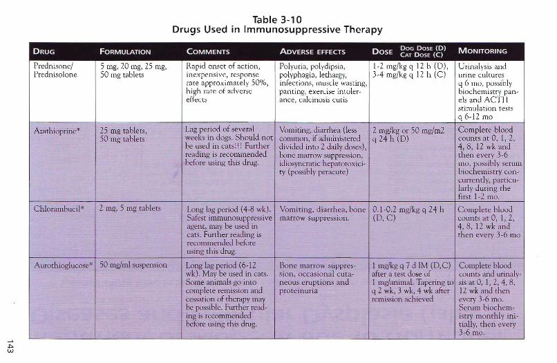
+:>w
Table 3-10Drugs Used in Immunosuppressive Therapy
DRUG FORMULATION COMMENTS ADVERSE EFFECTS DOSEDOG DOSE (D) MONITORINGCAT DOSE (C)
Prednisone/ 5 mg. 20 mg, 25 mg. Rapid onset of action. Polyuria, polydipsia, 1-2 mg/kg q 12 h (0), Urinal ysis andPredniso lone 50 mg tablets inexpens ive. response polyphagia, lerhargy. } -4 mg/kg q 12 h (C) urin e cultures
rare approximate ly 50% , infections, muscle wasting, q 6 mo. possiblyhigh rate of adverse panti ng, exercise inroler- biochemistry pan-effects ance, calcinosis cutis els and ACTI-J
st imulat ion testsq 6- 12 mo
Azarhioprine* 25 rng tablets, Lag period of several Vomiting, diarrhea (less 2 mg/kg or 50 mg/m2 Complete blood50 mg tablets weeks in dogs. Should not common . if administered q 24 h (D) counts at 0 , 1, 2,
be used in catsl!l Further divided into 2 daily doses), 4, 8, 12 wk andreading is recommended bone marrow suppression, the n every 3-6before using thi s drug. idiosyncratic hepatotoxici- rno, hOSSibly serum
tv (possibly peracute) bioc emistry con-currently, parricu-larly during thefirst 1-2 mo.
Chlorambucil* 2 rug, 5 mg tablets Long lag period (4-8 wk). Vomiting, diar rhea, bon e 0.1 -0 .2 mg/kg q 24 h Co mplete bloodSafest imrnunosuppressive marrow suppre ssion . (D, C) counts at 0, I, 2,agent, may be used in 4, 8, 12 wk andcats. Further reading is then every 3-6 morecommended beforeusing this drug.
Aurothioglucose* 50 mg/ml suspension Long lag period (6-12 Bon e marrow suppres- 1 mg/kg q 7 d 1M (D,C) Complete bloodwk). May be used in cars. sion , occas ional cuta- after a test dose of counts and urinalv-Some an imals go into neo us eruptions and 1 mg/animal. Tapering to sis at a, 1, 2, 4. 8,complete remission and proteinuria q 2 wk, 3 wk, 4 wk after 12 wk and thencessation of therapy may remission achieved every 3-6 mo.be possible. Further read- Serum bioche rn-ing is recommended istrv month ly ini-before using th is drug. rially, then every
3-6 mo.

Treatment of Alopecia due to HormonalDiseases and Follicular Dysplasia (Table 3-11) .
Table 3-11Selected Drugs Used in the Treatment of Endocrine Disorders with Cutaneous Symptoms
DRUG DOSE DOG DOSE (D) INDICATIONS ADVERSE EFFECTSCAT DOSE (e)
o,p '-DDD* (mirotane) 25 mg/kg q 12 h dur ing induction Idiopathic hyperadren ocorticism, Le thargy, ataxia, vomiting, d iarrhea,(5-14 d), same dose q 12 hon 2 COR- adrenal sex hormone imbalance (I anorexia.secutive days of each week as m ain - do not recommend the drug fortenance. Length of induction deter- th is latter d isease) . Further read-mine d bywater int ake, food intake, ing is recom mended prior to usingand AcrH stimulat ion test . (D, C) th is drug.
Levothvroxine 20 I-tg/kg q 12 h . If patient condi- Hypothyroidism Polydipsia, polyuria , nervousness,tion is well controlled, medication aggressiveness, panting, diarrhea ,may be changed to once daily at tachycardia, pyrexia, pruritus, heartdouble dose. (D, C) failure in dogs with cardiac disease,
exacerbation of adrenal crisis in dogswith hypoadrenocorticism
Testosterone 0.5-1 mg/kg (up to a maximal dose Aggressive behavior, greasy haircoat,of 30 rng) q 24 h orally (D, C) prostatic hypertrophy, hepatotoxicity
Estrogen 0.02 mgjkg q 48 h for 6-12 wk oral- Estrogen-responsive dermatosis Estrus induction, bone marrow sup-Iy or q 24 h for 3 wk, then 1 wk off, pression, hepatoroxicirv, pyometra,then repeat cycle (D). spontaneous abortion
Melatonin 3-6 mg q 12-24 h for 2-3 rna (D) Cyclic follicular dysplasia, follicu- Abscess formation with injection oflar dysplasia, alopecia. repository capsules.
Growth hormone" 0.1 IU/kg q 56 h for 6 wk (D) Adrenal sex hormone imbalance, Anaphylaxis, acromegaly, diabetesgrowth-hormone responsive dis- me llitusease (I do not recommend treat-menr with this drug).
Appendices A. Breed Predilections Abyssinian cat
Airedale
AkLta
Basse t H ound
Beagle
Belgian Te rvure n
Bo rder Collie
Borzoi
Boston Terrier
Boxer
Bullmastiff
Bullte rrier
Cairn Terri e r
Ceruminous otitis extenla Psychogenic a lopecia
Adult-on set demodicosis
Pemphigus fo liaceus Sebaceous adenitis Uveodenn ato log ic s)'ll.drome
Atopy Intertrigo Malassezia dennatiti s Sebo rrhea
Atopy Demodicos is IgA defic ien cy
Vitiligo
Systemic lupus erythematosus
Hypo thyroidism
Atopy Demodicosis Intertrigo
Atopy Cyclic follicular dysplasia Demodicosis Hyperadrenoco rticism Muzzle and/o r pedal bacterial funm culosis
Bacteria l furunculosis
Atopy Acrodermatitis Bacteria l furunculosis Solar d erm atitis
Atopy
Ches';peake Bay Retriever Atopy
Ch ihuahua Demodicosis
Chow C h ow
Collie
Adrena l sex hormone abnorrnalities Pemphigus foli aceus Demodicosis Hyposomatotfopism Hypothyroidism
Dermatomyosit is
145
146
Curly-coated Retriever
Dachshund
Dalmatian
Dobennan
English Bulldog
German Shepherd
Golden Retriever
Lupus erythematosus Pemphigus erythematosus
Follicular dysplasia
Bacterial pyoderma Color dilution. alopecia Hyperadrenocorticism Hypothyroidism Juvenile ce lluli t is Malassezia dermatitis Pattern alopecia Pinnal vasculitis Sterile pyogranulomatous dermatitis Sterile nodular panniculitis
Atopy Demodicosis Solar dermatitis
Acral lick dermatitis Bacterial pyodenna Color dilution alopecia Demodicosis Drug reaction (particularly against sulfonarn.ides) Follicula r d ysplasia Hyporhyroidism Vitiligo
Atopy Bacterial pyoderma Cyclic follicular dysplasia Demodicosis Intertrigo Hypothyroidism Malassezia dermatitis Sterile pyogranuloma syndrome
Atopy Bacte ria l pyoderma Ear tip fly dermatitis Eosinophilic funmculosis Flea-bite hypersensitivity Food adverse reaction Idiopathic onychomadesis Mucocutaneous bacterial pyoderma Pemphigus erythematosus Pituitary dwarfism Systetn ic lupus eryth ematosus Tarsal fistulae Vitiligo
Acral lick dennatitis Atopy Bacterial pyoderma Hypothyroidism ) uvenile cellulitis Nasal hypopigmentation ("Dudley nose") Pyotraumatic dermatitis
Gordon Setter
Great Dan e
Great PYl"enees
Irish Setter
Irish W ater Spaniel
Jac k Russel T~rrier
Keesh ond
Labrador Retriever
Lhasa Apso
Malamute
N ewfo undland
Old English Sheepdog
Pekingese
Persian Cat
Po inter
Pomeranian
Poodle
Porwguese Water Dog
Atopy Hypothyroidism
Acra l lick dermatitis Bacte ria l pyoderma Callus fonn a tion Demodicos is Hypothyroidism
Demodicos is Pyorraumatic dermatitis
Atopy Colo r dilution alopecia H ypo thyroidism
Follicula r dysplasia
Atopy Demodicosis
Alopecia X due to sex h o rmon e imbaLan ces Hyposomatotropism Hypothyroidism
Acral lick dermatitis Atopy Bacteria l pyodenna Food adverse reactio n Pyotraumatic clennatitis Seborrhea
Atopy Malassezia d ermatit is
Zinc-responsive dennatitis
Bacterial pyoderma Pyotraumatic derm.atitis
Atopy Demodicosis
Intertrigo
CheyletielLosis Dermatoph ytos is Intertrigo Seborrhea
AcraL mutil ation Demodicosis Hereditary lupo id dermatosis
Adrenal sex h ormon e a bnormalities H yposoma totropism
Hyperadren ocortic ism Hypothyro idism Injection reactions Sebaceous ad enitis (Standard)
Fo llicula r dysplasia
147
148
Pug
Rhodesian Ridgeback
Rortweiler
Samoyed
Schipperke
Scottish Terrier
Shar-pei
Schnauzer
Shetland Sheepdog
Shi-Tzu
Siamese Car
Siberian Husky
Spanieb
St. Bernard
Viszla
Weimarcmer
Atopy Intertrigo
Dermoid sinus
Bacterial pyodenna Vasculitis Vitiligo
Sebaceous adenitis
Pemphigus foliaceus
Atopy
Atopy Bacterial pyoderma Demodicosis Food adverse reaction Hyporhyroidism IgA defICiency Intertrigo Mucinosis
Atopy Aurotrichia Hyporhyroidism Schnauzer comedo syndrome Superficial suppurative necrolytic Den-narieis
Dermatomyositis Lupus erythematosus
AtOpy
Food adverse reaction H yporrichos is Periocular leukotrichia Vitiligo
EosinophilLc furunculosis Follicular dysplasia Zinc-responsive dermatitis Atopy
Food adverse reaction Hypothyroid ism Idiopathic onychomadesis Intertrigo Malassezia dermatitis Psoriasifonn-lichenoid dermatosis
(English Springer Spaniel) Seborrhea
Acral lick dermatitis Bacterial pyoderma
Sebaceous adenitis
Sterile pyogranulomatous syndrome
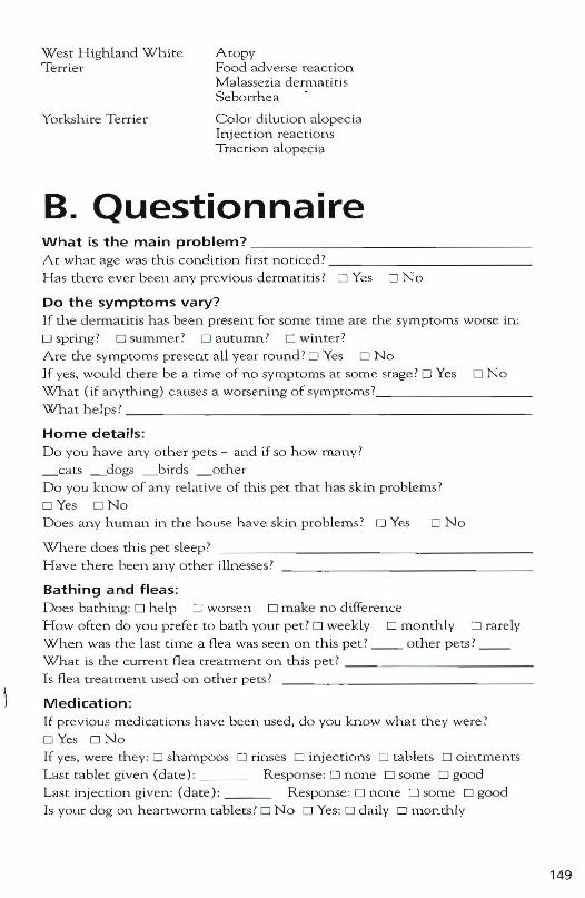
West High land WhiteTerrier
Yorkshire Terrier
A topyFo od adverse react ionM al assezia d errua ri t isSeborrh ea
Colo r dilution a lo pec iaInjec tion reac t io n sTrac tio n a lopec ia
B. QuestionnaireWhat is the main problem? _
A t what age wa s this condition first noti ced ? _
Has ther e ever b een an y previous de rmatitis? 0 Yes 0 N o
Do the symptoms vary?If the derm a t it is h as b een present fo r so me time are the sympto ms worse in:
o spring? 0 sum mer ? 0 autumn? 0 wi nter?
Are the sym pto ms p resent all year round ? 0 Yes 0 No
If yes, wou ld there b e a ti m e o f no sy mp tom s a t so me stage? 0 Yes 0 N oWhat ( if an yth in g) causes a worsening o f sym p to ms 7 _
W h a t h el ps ? _
Home details:D o you h av e an y o th e r pets - and if so h ow m any?
_ ca ts _dogs _ b irds _ o th.e r
Do you know of an y relat ive of this pe t th at has skin problems?
D Yes 0 N oDoes any human in the h ouse h ave skin problems? 0 Yes 0 N o
W h.ere do es th is pet sleep ?
H av e there been an y other illn esses ?
Bathing and fleas:Does b a thing: 0 h el p G worsen 0 make n o diffe rence
H ow often do you prefer to ba th your pet? 0 weekly 0 month ly 0 rarelyWh.en was the last t ime a flea was seen on this pet? __ other p e ts? __What is the cu rren t fle a treatm ent on this pet ? _
Is fl ea treatment used on o ther pets ?
Medication:If previous med ications h ave b een used, do you know what th ey were ?
D Yes 0 N oIf yes, were they : 0 sham po os 0 rin ses C in jec ti on s 0 tab lets 0 ointments
Last tablet g iven (date) : R esponse: 0 none 0 some 0 go od
Last in jec t io n given: (d ate ) : Response : 0 none 0 some 0 good
Is your d og on h eartworm tablets ?0 No 0 Yes : 0 d a ily 0 monthly
149
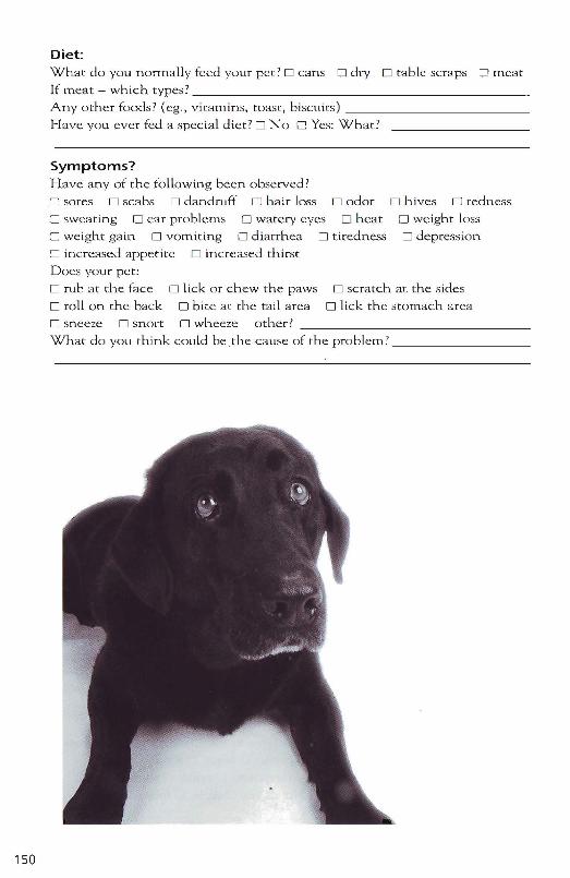
o scratch at the sideso lick the stomach area
D h ives D redn ess
o we ight loss
D depression.
150
Diet:Wnat do you nonna lly feed your p et? 0 cans 0 dry 0 tab le scraps Q m ea tIf meat - wh ich types? _
Any other foods? (eg ., vitamins, toast , b isc uits) _
Have you ever fed a special d iet? D N a 0 Yes: What?
Symptoms?H ave any of the following been observed ?
D so res D scabs D dandruff D hair loss D odor
o sweat ing 0 ear problems 0 watery eyes 0 heat
D we ight gain 0 vomit ing 0 dia rrhea 0 tiredness
D in creased appeti te D increased th irst
Does your per:
D rub at the face 0 l ick or chew the paws
D ro ll o n the back 0 bi te at the tail areaD sneeze D snort 0 wheeze o ther? _
What do you think could be the cause of the problem? _
Recommended Readings Bonagura JD. Ed. Kirk's Current Veterinary Therapy (XII and XIII ). Philadelphia, WB Saunders, 1995 and 2000.
Campbell KL. Ed. The Veterinary Clinics of North America~Small Animal Practice: Dermatology, Vol. 29 (6). Philadelphia, WB Saunders, 1995 .
Feldman EC, Nelson RW. Canine and Feline Endocrinology and Reproduction . 2nd Ed. Philadelphia, WB Saunders, 1996.
Greene CEo Infectious Diseases of the Dog and Cat. 2nd Ed. Philadelphia, WB Saunders, 1998.
Griffen CE, Kwochka KW, MacDonald JM. Eds. Current Veterinary Dermatology. St. Louis, Mosby, 1993.
Kunkle G. Ed. The Veterinary Clinics of North America~Small Animal Practice: Feline Dermatology, Vol. 25 (4) . Philadelphia, WB Saunders, 1995.
Kwochka KW, Willemse T, von Tscharnel' C. Eds. Advances in Veterinary Dermatology, Volume 3. Oxford, Butterworth Heinemann, 1998.
Locke PH, Harvey RG, Mason IS. Handbook of Small Animal Dermatology. Oxford, Pergamon, 1995.
Moriello KA, Mason IS. Handbook of Small Animal Dermatology. Oxford, Pergamon, 1995.
Ogilvie GK, Moore AS. Managing the Veterinary Cancer Patient: A Practice Manual. Trenton, Veterinary Learning Systems, 1995.
Plumb DC. Veterinary Drug Handbook. 3rd Ed. White Bear Lake, Pharma Vet Publishing, 1999.
Reedy RM, Miller WH, Willemse T. Allergic Skin Diseases of Dogs and Cats . 2nd Ed. Philadelphia, WB Saunders, 1997 .
Scott DW, Miller WH, Griffin CEo Small Animal Dermatology . 5th Ed. Philadelphia, WB Saunders, 1995 .
151
Dermatologyfor the Small Animal Practitioner
The quick reference manualyou need for Dermatology- Practical guidelines for diagnosis and
treatment of the skin diseases that you seeevery day in practice.
- Over 115 Color illustrations integrated withRalf S. Mueller the text for easy identification of lesions.Dr. mecl.vet., _ C f 11 desi d bl £ . kMACVSc Di ACVD are u y esrgrie ta es lor qU1C accessFACVSc ' P ' t o important diagnostic and treatment
information.
- Formulary of dermatologic drugs andpreparations.
- Provides quick identification of rare andcomplicated dermatologic tests.
- Clear, step by step descriptions of commonlyperformed diagnostic tests.
- User friendly, consistent design which helpsthe reader focus on the really importantinformation.
- Companion CD~ROM provided whichcontains the book plus an expanded libraryof color illustrations for instant search andretrieval of information.
Forthcoming titles in the Made Easy Seriesfor the Small Animal Practitioner:Thoracic Radiology· Transfusion Medicine· Endocrinology
• Echocardiography
~TNM