design, synthesis, application of biodegradable polymers
TRANSCRIPT

University of South FloridaScholar Commons
Graduate Theses and Dissertations Graduate School
March 2018
Design, Synthesis, Application of BiodegradablePolymersMussie GideUniversity of South Florida, [email protected]
Follow this and additional works at: https://scholarcommons.usf.edu/etd
Part of the Biochemistry Commons, and the Chemistry Commons
This Thesis is brought to you for free and open access by the Graduate School at Scholar Commons. It has been accepted for inclusion in GraduateTheses and Dissertations by an authorized administrator of Scholar Commons. For more information, please contact [email protected].
Scholar Commons CitationGide, Mussie, "Design, Synthesis, Application of Biodegradable Polymers" (2018). Graduate Theses and Dissertations.https://scholarcommons.usf.edu/etd/7625

Design, Synthesis, Application of Biodegradable Polymers
by
Mussie Gide
A thesis submitted in partial fulfillment
of the requirements for the degree of
Master of Science
Department of Chemistry
College of Arts and Sciences
University of South Florida
Major Professor: Jianfeng Cai, Ph.D.
James Leahy, Ph.D.
Shengqian Ma, Ph.D.
Chen Yu, Ph.D.
Date of Approval:
March 20, 2018
Keywords: Antimicrobial polymers, Dendrimers, Amphiphilic, Unimolecular micelles, Host defense
Peptide
Copyright © 2018, Mussie Gide

DEDICATION
To my parents, for their enormous support. To Selam Hagos for her amazing heart. To my
brother Samuel Gide and my uncle George Ghide for their everlasting encouragement. To Efrem
Moconnen and Sami Abdelwahab, for their indispensable friendship. To everyone who was ever
there for me when I needed them.

ACKNOWLEDGEMENTS
I would like to thank my research advisor, Dr. Jianfeng Cai, for the support and guidance
he has given during my academic study at USF. I would like to thank him for his guidance, endless
support and patience. It is very rare to find an advisor who always have time for their students to
listen to their little problems, I feel I am fortunate and I thank my advisor for always finding time
and encouraging to overcome the barriers during my research work.
I would like to thank my thesis committee members, Dr. James Leahy, Dr. Shengqian Ma,
Dr. Chen Yu, for their extended support, advice and encouragement in accomplishing my Master’s
degree at USF.
I want to express my honest thanks to every member in the Cai’s group. I enjoyed every
moment in my lab. Dr. Alekhya Nimmagadda mentored me to understand and apply all the basic
principles of polymer synthesis and all related staffs of my research. Especial thanks to Dr. Yan
Shi and Dr. Teng Peng for their motivations and help in all my research progress. I am thankful to
Tim, Annemarie and Sylvia for their efforts during my thesis writing. I also enjoyed time with
other lab members, Olapeju, Lulu, Ma Su, Fengyu, Dr. Peng Sang, Chunpu, Dr. Mengmeng, Dr.
Mi and Minghui, Songyi Xue, Cong Pan and Ruixuan Gao. I would specially thank my friend and
lab mate Sami for bearing brotherly advice and making every moment in the lab lively.
The time I spent with my room mates Selam, Sami and Efrem was cherished. Furthermore,
I would like to thank my family for their endless love and support. Finally, I would like to remark
this is thesis is dedicated to Selam Hagos and all my family and friends, who encouraged and
motivated me all the time during my study at USF.

i
TABLE OF CONTENTS
LIST OF TABLES ........................................................................................................................ iii
LIST OF FIGURES ....................................................................................................................... iv
LIST OF ABBREVIATIONS ........................................................................................................ vi
ABSTRACT .................................................................................................................................... x
CHAPTER 1: INTRODUCTION ................................................................................................... 1
1.1 Introduction ........................................................................................................................... 1
1.2 Outline of the Thesis ............................................................................................................. 2
1.3 Reference .............................................................................................................................. 3
CHAPTER 2: PEPTIDE BASED POLYCARBONATE POLYMESRS WITH POTENT AND
SELECTIVE ANTIMICROBIAL ACTIVITY .............................................................................. 5
2.1 Introduction ........................................................................................................................... 5
2.2 General Overview of Polymers ............................................................................................. 6
2.3 Statement of purpose of our work ......................................................................................... 7
2.4 Experimental: Materials and Methods .................................................................................. 7
2.4.1 Synthesis of Monomer 1 (M1) (Figure 2.1) ................................................................... 7
2.4.2 Synthesis of Monomer 2 (M2) - (Figure 2.2) ................................................................ 8
2.4.3 Synthesis of Monomer 3 (M3) - (Figure 2.3) ................................................................ 9
2.4.4 Synthesis of Polycarbonate Copolymers ..................................................................... 10
2.4.4.1 Random copolymerization of M1 and M2 (Figure 2.4) ............................................ 10
2.4.4.2 Random copolymerization of M1 and M3 (Figure 2.6) ............................................ 12
2.4.5 Antimicrobial Assay .................................................................................................... 13
2.4.6 MTT Assay .................................................................................................................. 13
2.5 Results and Discussion ....................................................................................................... 14
2.6 Conclusion .......................................................................................................................... 18
2.7 References ...................................................................................................................... 19
CHAPTER 3: LIPIDATED DENDRIMERS AS POTENT AND BROAD SPECTRUM
ANTIBACTERIAL AGENTS ...................................................................................................... 22

ii
3.1 Introduction ......................................................................................................................... 22
3.2 Dendrimer Overview .......................................................................................................... 24
3.3 Result and Discussion ......................................................................................................... 25
3.3.1 Solid phase Synthesis of lipidated dendrimers: ........................................................... 25
3.3.2 Antimicrobial and Hemolytic activity ......................................................................... 25
3.3.2 Mechanism of Action ................................................................................................... 29
3.3.3 Bacterial Kinetic Study ................................................................................................ 30
3.3.4 Bacterial Biofilm Assay ............................................................................................... 31
3.4 Materials and methods ........................................................................................................ 32
3.4.1 Antimicrobial Assay .................................................................................................... 32
3.4.2 Hemolysis Assay .......................................................................................................... 33
3.4.3 Fluorescence Microscopy ............................................................................................ 33
3.4.4 Time kill study ............................................................................................................. 34
3.4.5 TEM Study ................................................................................................................... 34
3.4.6 Biofilm assay ............................................................................................................... 35
3.5 Conclusion .......................................................................................................................... 35
3.6 References ........................................................................................................................... 36
CHAPTER 4: UNIMOLECULAR NANOPARTICLES AS POTENT ANTIMICROBIAL
AGENTS ....................................................................................................................................... 44
4.1 Introduction ......................................................................................................................... 44
4.2 Overview of Unimolecular Micelle Hyperbranched Polymers .......................................... 46
4.3 Experimental Section .......................................................................................................... 47
4.3.1 Synthesis of Hyperbranched Polyester Polyacid (HBPP) (Figure4.1)......................... 47
4.3.2 Synthesis of hydroxyl terminated hyperbranched polyester (Figure 4.3) .................... 48
4.3.3 Synthesis of Hydrophilic monomer (M1) (Figure 4.5) ................................................ 49
4.3.4 Synthesis of Hydrophobic monomer (M2) (Figure 4.6) .............................................. 50
4.3.5 Synthesis of Unimolecular micelle hyperbranched polymers (Figure 4.7 & Figure 4.8)
............................................................................................................................................... 50
4.3.6 Antimicrobial Assay .................................................................................................... 52
4.4 Results and Discussion ....................................................................................................... 53
4.5 Conclusion .......................................................................................................................... 56
4.6 References ........................................................................................................................... 57
CHAPTER 5: CONCLUSIONS AND FUTURE DIRECTIONS ................................................ 61

iii
LIST OF TABLES
Table 2.1 Copolymers synthesized from M 1 and M 2 monomers .................................................... 12
Table 2.2 Antibacterial activity of polycarbonates ..................................................................................... 17
Table 3.1 The Antibacterial and Hemolytic Activity of Lipidated Dendrimers ......................................... 27
Table 4.1 Unimolecular micelle hyperbranched polymers ............................................................52
Table 4.2 The Antibacterial Activity of Unimolecular micelle hyperbranched polymers ............55

iv
LIST OF FIGURES
Figure 2.1 Synthesis of the Monomer 1 ....................................................................................8
Figure 2.2 Synthesis of Monomer 2 ..........................................................................................9
Figure 2.3 Synthesis of the Monomer 3 ..................................................................................10
Figure 2.4 Synthesis of Random Amphiphilic Polycarbonates ...............................................11
Figure 2.5 1HNMR of Polymer MG-P7 .................................................................................. 11
Figure 2.6 Synthesis of MG-P9 amphiphilic polycarbonate ...................................................13
Figure 2.7 IC50 of Polymer MG-P9 against HepG-2 and HEK-293T cell lines ..................... 18
Figure 2.8 IC50 of Polymer MG-P7 against HepG-2 and HEK-293T cell lines ..................... 18
Figure 3.1 Solid phase Synthesis of D-A dendrimers .............................................................26
Figure 3.2 Solid phase Synthesis of D-B dendrimers .............................................................28
Figure 3.3 Fluorescence micrographs of MRSA and E. Coli that were treated or not treated with
25 μg/mL of D-A-2 for 2 h ....................................................................................29
Figure 3.4 TEM micrographs of MRSA and E. coli treated with 10 µg/mL of D-A-2.……...30
Figure 3.5 Time kill study of D-A-2 against MRSA E. coli.....................................................31
Figure 3.6 Inhibition of biofilms of MRSA and E. coli by D-A-2 .........................................32
Figure 4.1 Synthesis of HBPP .................................................................................................48
Figure 4.2 1H NMR of HBPP ..................................................................................................48
Figure 4.3 Synthesis of hydroxyl terminated hyperbranched polyester ..........................................49
Figure 4.4 1H NMR of hydroxyl terminated hyperbranched polyester ...................................49
Figure 4.5 Monomer 1 (M1) synthesis ....................................................................................50

v
Figure 4.6 Monomer 2 (M2) synthesis ....................................................................................50
Figure 4.7 Synthesis of unimolecular nanoparticles with random chains of polycarbonates…51
Figure 4.8 Synthesis of unimolecular nanoparticles with random chains of polycarbonates...52

vi
LIST OF ABBREVIATIONS
WHO World Health Organization
HDPs Host-defense peptides
MRSA Methicillin-Resistant Staphylococcus Aureus
TEM Transmission Electron Microscopy
LPS Lipopolysaccharides
DCM Dichloro methane
TEA Triethylamine
HCl Hydrochloric Acid
TFA Trifluoracetic acid
NMR Nuclear Magnetic Resonance
MIC Minimum inhibitory concentration
CFU Colony forming unit
MRSE Methicillin-resistant Staphylococcus epidermidis
VREF Vancomycin-resistant E. faecalis

vii
PBS Phosphate Buffered Saline
DAPI 4, 6-Diamidino-2-phenylindole dihydrochloride
PI Propidium iodide
PAMAM Polyamidoamine
HOBT 1-hydroxybenzotriazole monohydrate
DIC Diisopropylcarbodiimide
Boc Tert-Butyloxycarbonyl
Fmoc Fluorenylmethyloxycarbonyl chloride
DMF Dimethylformamide
HPLC High-performance liquid chromatography
MALDI-TOF Matrix-assisted laser desorption/ionization time-of-flight
TIS Triisopropylsilane
NMP N-Methyl-2-Pyrrolidone
E.Coli Escherichia Coli
P. aeruginosa Pseudomonas aeruginosa
FM Florescence Microscopy
TSB Tryptic soy broth

viii
OD Optical Density
DIPEA Diisopropylethylamine
TFE Tetrafluoroethylene
MeOH Methanol
MTT (3-(4,5-Dimethylthiazol -2-yl)-2,5-Diphenyl tetrazolium Bromide)
PA Pseudomonas aeruginosa
NaHCO3 Sodium bicarbonate
THF Tetrahydrofuran
Na2SO4 Sodium sulfate
E. faecalis Enterococcus faecalis
M Molarity
PEG Polyethylene glycol
Dde Dichlorodiphenyldichloroethylene
MDC 5-methyl-2-oxo-1,3-dioxane-5-carbonyl chloride
TU 1-(3,5-bis(trifluoromethyl)-phenyl)-3-cyclohexyl-2- thiourea catalyst
DBU 1,8-diazabicyclo [5.4.0] undec-7- ene

ix
MWCO Molecular Weight Cut-Off's
CH3CN Acetonitrile
DIC Diisopropylcarbodiimide
ROP Ring opening Polymerization
SnCl2
HepG-2
Stannous chloride
liver hepatocellular cells
HEK-293T Human embryonic kidney Cells
C. diff UK6 Clostridium difficile
HBPP Hyperbranched Polyester Poly acid

x
ABSTRACT
Bacterial infections have posed a serious threat to the public health due to the significant
rise of the infections caused by antibiotic resistant bacteria. There has been considerable interest
in the development of antimicrobial agents which mimic the natural HDPs, and among them
biodegradable polymers are newly discovered drug candidates with ease of synthesis and low
manufacture cost compared to synthetic host defense peptides. Herein, we present the synthesis of
biocompatible and biodegradable polymers including polycarbonate polymers, unimolecular
micelle hyperbranched polymers and dendrimers that mimic the antibacterial mechanism of HDPs
by compromising bacterial cell membranes. The developed amphiphilic polycarbonates are highly
selective to Gram-positive bacteria, including multidrug resistant pathogens and the unimolecular
micelle hyperbranched polymers showed promising broad-spectrum activity. However, lipidated
amphiphilic dendrimers with low molecular weight display potent and selective antimicrobial
activity against both Gram-positive and Gram-negative bacteria, including multidrug-resistant
strains. In addition to antibacterial activity against planktonic bacteria, these dendrimers were also
shown to inhibit bacterial biofilms effectively. These class of polymers may lead to a useful
generation of antibiotic agents with practical applications.

1
CHAPTER 1: INTRODUCTION
1.1 Introduction
Antibiotics are agents that can be produced by microorganisms and have the capacity to
inhibit the growth of similar microorganisms. In the history of medicine, antimicrobials are one of
the most powerful forms of chemotherapy. With the discovery of new antibiotics, the death rate
caused by infectious diseases was decreased from 797 per hundred thousand in 1900 to 36 per
hundred thousand in 1980.1 However, although the number of discovered antibacterial medications
continues to grow, bacterial resistance to many of these medications become a threat to the current
prevention and treatment of infections. Indeed, within the past twenty years, bacterial resistance
has been considered the greatest challenge to the antibiotic era; 30% of all deaths in America was
due to tuberculosis, pneumonia and gastrointestinal infections caused by resistant bacteria.2
Antibiotic resistance can occur via three general mechanisms.3 First, bacterial destruction
to the antibiotics by specific enzymes for instance some bacteria produce beta lactamase enzyme
which degrades beta lactam drugs. Another approach to drug resistance is when bacteria undergo
mutational changes in their structural and functional makeup by acquiring new genes from other
strains. This results in changes in the bacterial receptor conformation which leads to less
susceptibility of the bacteria towards the drug. An example of this type of bacterial resistance is
mostly shown by Vancomycin-resistant Enterococcus. The third major course of bacterial
resistance is through overexpression of several efflux pumps which leads to banishing of the drugs
from the bacterial cell which results in a decrease in concentration of the drug to a level which is

2
below the toxic threshold. An example of this mechanism is shown by gram-positive and gram-
negative bacteria but mainly by Pseudomonas bacterial strains.2,3,4
One of the promising ongoing research topics to address the challenges created by the
resistant bacteria is to mimic host defense peptides (HDP) by unnatural peptidomimetics including
β-peptides,5 peptoids,6 oligoureas,7 AApeptides.8 Small peptidomimetics can mimic the function
and mechanism of natural HDPs and have shown promising potent, broad spectrum antimicrobial
activity. HDPs, however, are generally not cost effective, as it is difficult to scale up the reaction
to produce in extensive quantities.9 Recently, polymers such as methacrylates,10 norborenene,11
amidoamines,12 polystyrenes, polycarbonates13, Dendrimers14 and hyperbranched unimolecular
nanoparticles15 are getting more attention as novel anti-infective candidates.
Polymers are macromolecular compounds composed of repeating units of monomers
which are linked to each other with a chemical bond. Polymers are currently becoming main
interest of the biomedical era.16 With the current innovations in polymer chemistry, polymers are
functionalized in various architectural designs, including dendrimers and unimolecular micelles.
Dendrimers are highly branched globular structures consisting of a central core from which
identical fragments are built up to make star-like macromolecular structures.17
1.2 Outline of the Thesis
In this thesis, we discuss the design, synthesis, and antibacterial application of biodegradable
polymers.
In chapter 2, we studied the design and antibacterial activity of amphiphilic peptide-based
polycarbonate polymers.
In chapter 3, we discuss the design, solid phase synthesis and the antibacterial activity of
lipidated dendrimers

3
In chapter 4, we describe the design, development and antibacterial activity of unimolecular
micelle nanoparticle.
In chapter 5, we recapitulate the research findings and conclude the future directions of our
research study.
1.3 Reference
1. Walsh & Wright. Introduction: Antibiotic Resistance. Chem. Rev. 105, 391–394 (2005).
2. Wright, G. D. Bacterial resistance to antibiotics: Enzymatic degradation and
modification. Mech. Antimicrob. Resist. Oppor. New Target. Ther. 57, 1451–1470 (2005).
3. Fair, R. J. & Tor, Y. Antibiotics and Bacterial Resistance in the 21st Century. Perspect.
Med. Chem. 6, PMC.S14459 (2014).
4. Guilhelmelli, F. et al. Antibiotic development challenges: the various mechanisms of
action of antimicrobial peptides and of bacterial resistance. Front. Microbiol. 4, (2013).
5. Karlsson, A. J., Pomerantz, W. C., Weisblum, B., Gellman, S. H. & Palecek, S. P.
Antifungal Activity from 14-Helical β-Peptides. J. Am. Chem. Soc. 128, 12630–12631 (2006).
6. Patch, J. A. & Barron, A. E. Helical Peptoid Mimics of Magainin-2 Amide. J. Am. Chem.
Soc. 125, 12092–12093 (2003).
7. Violette, A. et al. Mimicking Helical Antibacterial Peptides with Nonpeptidic Folding
Oligomers. Chem. Biol. 13, 531–538 (2006).
8. Li, Y. et al. Helical Antimicrobial Sulfono-γ-AApeptides. J. Med. Chem. 58, 4802–4811
(2015).

4
9. Costanza, F. et al. Investigation of antimicrobial PEG-poly(amino acid)s. RSC Adv 4,
2089–2095 (2014).
10. Chen, Y. et al. Amphipathic antibacterial agents using cationic methacrylic polymers
with natural rosin as pendant group. RSC Adv. 2, 10275–10282 (2012).
11. AL-Badri, Z. M. et al. Investigating the Effect of Increasing Charge Density on the
Hemolytic Activity of Synthetic Antimicrobial Polymers. Biomacromolecules 9, 2805–2810
(2008).
12. Calabretta, M. K., Kumar, A., McDermott, A. M. & Cai, C. Antibacterial Activities of
Poly(amidoamine) Dendrimers Terminated with Amino and Poly(ethylene glycol) Groups.
Biomacromolecules 8, 1807–1811 (2007).
13. Engler, A. C. et al. Polycarbonate-Based Brush Polymers with Detachable Disulfide-
Linked Side Chains. ACS Macro Lett. 2, 332–336 (2013).
14. Chen, C. Z. & Cooper, S. L. Recent Advances in Antimicrobial Dendrimers. Adv. Mater.
12, 843–846 (2000).
15. Zhou, Y., Huang, W., Liu, J., Zhu, X. & Yan, D. Self-Assembly of Hyperbranched
Polymers and Its Biomedical Applications. Adv. Mater. 22, 4567–4590 (2010).
16. Paul, D. R. & Robeson, L. M. Polymer nanotechnology: Nanocomposites. Polymer 49,
3187–3204 (2008).
17. Tülü, M. & Ertürk, A. S. Dendrimers as Antibacterial Agents. Search Antibact. Agents
(2012). doi:10.5772/46051

5
CHAPTER 2: PEPTIDE BASED POLYCARBONATE POLYMERS WITH POTENT AND
SELECTIVE ANTIMICROBIAL ACTIVITY
2.1 Introduction
Natural antimicrobial peptides (HDPs) are one of the first lines of defense within our body
when bacteria or other microbes attack the body. They have broad-spectrum antimicrobial
activities.1 HDPs are found in many species, including humans, animals, plants, and invertebrates.
The mechanism of action for these peptides are not receptor-based interaction, but through direct
action on the bacteria’s membranes.2 The slowly progressing development of replacements for the
current ineffective antibiotic treatment results in a major risk to public health through multidrug
resistant bacteria. Different studies have revealed that bacteria can develop resistance to drugs in
numerous ways including effluxing the drug from inside the cell through membrane bounded
pumping proteins, undergoing mutational changes within the main receptors of the drug, and
developing enzymes which can selectively degrade the antibiotics.3 Presently, to face the
challenges caused by the multi-drug resistant bacteria various novel antimicrobial agents have
been developed. Compounds having cationic groups have arisen as auspicious candidates to
replace the current antibacterial drugs which already faced resistance. This is due to the short
amphiphilic positively charged peptides have the tendency to mimic HDPs and are selective
towards bacteria. Presently researchers are mainly working on synthetic peptides, polymers and
lipids as potential cationic compounds.4 Herein we report the synthesis and antimicrobial

6
application of peptide based polycarbonate polymers, in continuation to projects previously done
in our lab.5
2.2 General Overview of Polymers.
Polymers are large macromolecules consisting of repeating units, named monomers, linked
together through a covalent bond. Polymers can be obtained from natural sources called natural
polymers or artificially synthesized like that of synthetic polymers. Polymers have diverse
structural arrangements, ranging from linear to branched and cross-linked arrangement. Linear
polymers have long and straight chained arrangements like polyethylene and polyvinyl chloride.
Branched polymers contain some branches on the linear backbone, furthermore cross-linked
polymers are formed from conjugated and covalently bonded linear polymers.6 Generally
polymers are synthesized by a process called polymerization, where several types of
polymerization methods are applied today. In a polymerization reaction, the monomers used
should have a functional group that enables bond formation. Homo-polymerization and
copolymerization are the two most common practiced polymerization methods. In homo-
polymerization, one type of monomer is linked together, while in copolymerization two or more
distinct types of monomers are used.
Based on the sequential arrangement of the monomers several types of copolymers can be
synthesized including block copolymers, graft copolymers and random copolymers.7 Natural and
synthetic polymers are a necessity in our daily life. Growing biomedical applications of polymers
continue to be discovered and researched from time to time. It has been more than half a century
that drugs conjugated with polymers have been applied in biomedical fields for therapeutic
applications. As part of the therapeutic applications of polymers, polycarbonates are developed,
and their application is being studied. The mechanism of action is similar to the antimicrobial HDP

7
through disrupting the microbial membrane by the interaction of the cationic hydrophilic unit with
the negatively charged membranes.8,9
2.3 Statement of purpose of our work
In our lab, previous group members designed and synthesized amphiphilic polycarbonate
polymers which have primary amino groups. According to their findings, the polymers exhibited
potent antimicrobial activity and selectivity against gram-positive bacteria, including multidrug
resistant strains. Encouraged by the promising results obtained, we have planned certain
modifications in the composition of the monomers that may lead to the development of new
polymers which could potentially display broad spectrum antibacterial activity. Phenylalanine,
lysine and 4-Bromo benzoic acid were used as components of the backbone of 5-methyl-2-oxo-
1,3-dioxane-5-carbonyl chloride (MDC) and polymerization of the monomers was done
following the ring opening polymerization mechanism (ROP) as reported by Yang and Hedrick
et al.10 Lysine derived carbonate monomer was designated to afford the hydrophilic feature of
the polymer as it has a free primary amino groups, and both phenylalanine and 4- Bromo benzoic
acid derived monomers will offer hydrophobic characteristics to the polymer.11,10
2.4 Experimental: Materials and Methods
2.4.1 Synthesis of Monomer 1 (M1) (Figure 2.1)
To synthesize Monomer 1, First Di-tert-butyl (6-((2-hydroxyethyl)amino)-6-oxohexane-
1,5-diyl) (S)-dicarbamate was prepared by dissolving 1 equiv N2,N6-bis(tert-butoxycarbonyl)-L-
lysine and 1.5 equiv ethanolamine in Dimethylformamide (DMF), to this solution 1.5 equiv 1-
ethyl-3-(3-dimethylaminopropyl) carbodiimide hydrochloride (EDC), 1.5 equiv
Hydroxybenzotriazole (HOBT) coupling agents were added with a 1.5 equiv N,N-
Diisopropylethylamine (DIPEA) base. The mixture stirred for 6 h and after the reaction was

8
completed, work up was done using ethyl acetate and the organic layer was washed with 1N HCl
(100 mL×3) and brine (50 mL×1). Then product was dried with sodium sulfate and the solvent
was removed with a rotavapor and it was purified using flash chromatography and we got 80%
yield. As part of the monomer constituent, 5-methyl-2-oxo-1,3-dioxane-5-carbonyl chloride
(MDC) was also synthesized following the procedures given by Yang and Hedrick.10 Lastly
Monomer 1 was synthesized by dissolving 1.5 equiv MDC and 1 equiv di-tert-butyl (6-((2-
hydroxyethyl)amino)-6-oxohexane-1,5-diyl)(S)-dicarbamate in 30 mL Dichloromethane (DCM)
in a 100 mL round bottom flask, to which 1.5 equiv triethylamine (TEA) base was added. The
reaction was left for 4 h in an ice bath and then the product was extracted by DCM and was washed
with 1 N HCl (100 mL×3), brine (50 mL×1) and then dried over sodium sulfate. The solvent was
removed in vacuo to give oily product, which was further purified by flash chromatography (ethyl
acetate: hexane 1:1) to give the final white sticky solid product.
Figure 2.1 Synthesis of the Monomer 1.
2.4.2 Synthesis of Monomer 2 (M2) - (Figure 2.2)
To synthesize Monomer 2, first tert-butyl (S)-(1-((2-hydroxyethyl) amino)-1-oxo-3-
phenylpropan-2-yl) carbamate was prepared by dissolving 1 equiv tert-butoxycarbonyl-L-
phenylalanine and 1.5 equiv Ethanolamine in DMF with coupling agents 1.5 equiv EDC, 1.5 equiv
HOBT and 1.5 equiv DIPEA base. The reaction was left for 6 h, after the reaction was completed

9
work up was done by ethyl acetate and the product was washed with 1N HCl (100 mL×3) and
Brine (50 mL×1). The organic solvent was then dried with sodium sulfate and removed by
rotavapor to get white solid product. From that Monomer 2, was prepared by the adding the above
product (1 equiv tert-butyl (S)-(1-((2-hydroxyethyl)amino)-1-oxo-3-phenylpropan-2-yl)
carbamate) with 1.5 equiv MDC which is synthesized according to Yang and Hedrick,10 and the
mixture was dissolved in 30 mL DCM with 1.5equiv TEA in a 100 mL round bottom flask. The
reaction was left for 4 h in an ice bath and then the product was extracted by DCM and was washed
with 1 N HCl (100 mL×3), brine (50 mL×1) and then dried over sodium sulfate. The solvent was
removed by rotavapor to give solid product, which was further purified by flash chromatography.
Figure 2.2 Synthesis of the Monomer 2.
2.4.3 Synthesis of Monomer 3 (M3) - (Figure 2.3)
First 4-bromo-N-(2-hydroxyethyl) benzamide was prepared by dissolving 1 equiv 4-
bromobenzoic acid and 1.5 equiv Ethanolamine in DMF with coupling agents, 1.5 equiv EDC, 1.5
equiv HOBT and 1.5 equiv DIPEA base. The reaction was left for 6 h, after the reaction was completed
work up was done by ethyl acetate and the product was washed with 1N HCl (100 mL×3) and Brine
(50 mL×1). Then the organic solvent was dried with sodium sulfate and vacuo to give a yellowish
solid product. From that Monomer 3 was synthesized by dissolving 1equiv 4-bromo-N-(2-
hydroxyethyl) benzamide, 1.5 equiv MDC and 1.5 equiv TEA in 30 mL DCM in a 100mL round

10
bottom flask. The reaction was completed left for 4 h and then the product was extracted by DCM and
was washed with 1 N HCl (100 mL×3), brine (50 mL×1) and then dried over sodium sulfate. The
solvent was removed in vacuo to give solid product, which was further purified by flash
chromatography.
Figure 2.3 Synthesis of the Monomer 3.
2.4.4 Synthesis of Polycarbonate Copolymers
2.4.4.1 Random copolymerization of M1 and M2 (Figure 2.4)
Batches of polymers were synthesized from M1 and M2 monomers (Table 2.1). All polymers
were synthesized via ring opening polymerization (ROP) method. The detailed synthesis of random
copolymer MG-P7 will be given as an example and all further polymers will follow the reaction steps
and conditions. First 1 equiv initiator benzyl alcohol was dissolved in 10 mL of DCM in a round
bottom flask after purging with nitrogen gas. Then the two monomers, 30 equiv M1 (hydrophilic
monomer) and 20 equiv M2 (hydrophobic monomer) were added to the flask together, to this mixture
1 equiv (3,5-bis(trifluoromethyl)-phenyl)-3-cyclohexyl-2-thiourea catalyst (TU) and 1 equiv 1,8-
diazabicyclo [5.4.0] undec-7-ene (DBU) base were added. The reaction was left stirring for 6 h, and
then 1.3 equiv benzoic acid was added to quench the reaction. The polymers were then purified by
dialysis against methanol and distilled water for 3 days using dialysis tubing MWCO 3500 with
methanol and water being replaced twice a day. The polymer solvent was then vacuumed and then the
polymers were treated with 15 ml of a 1:1 TFA:DCM mixture for 3 h to deprotect the Boc protecting

11
group in order to obtain the free amine functional groups.12 Again, the polymers were purified in a
dialyzing tube against methanol and water for 4 days and then freeze dried to get the final product and
the product was characterized by 1HNMR (Figure 2.5).13 As such, a series of polycarbonate polymers
containing varying numbers of hydrophobic and hydrophilic groups were prepared in the same manner
(Table 2.1).
Figure 2.4 Synthesis of Random Amphiphilic Polycarbonates.
Figure 2.5 1HNMR of Polymer MG-P7

12
Table 2.1 Copolymers synthesized from M 1 and M 2 monomers.
Compound
Type of Co-
Polymer
Hydrophilic Units
M1
Hydrophobic units:
M2
Ideal Molecular
Weight
MG-P1 Random 10 30 13932.96
MG-P2 Random 15 25 13837.95
MG-P3 Random 15 30 15589.81
MG-P4 Random 20 25 15494.80
MG-P5 Random 20 40 20750.36
MG-P6 Random 30 10 13552.92
MG-P7 Random 30 20 17056.63
MG-P8 Random 30 30 20560.34
MG-P9 Random 20 20 14459.48
2.4.4.2 Random copolymerization of M1 and M3 (Figure 2.6)
Polymer MG-P9 was prepared in similar fashion as above procedure, but the hydrophobic
monomer M3 was used instead of M2.

13
Figure 2.6 Synthesis of MG-P9 amphiphilic polycarbonate.
2.4.5 Antimicrobial Assay
Minimum Inhibitory Concentration (MIC) of the polycarbonate polymers was determined
using a broth micro dilution method in 96-well plates against Methicillin-resistant S. aureus (MRSA,
ATCC 33591), Clostridium difficile (C. diff UK6) and Gram-Negative bacteria E. coli (ATCC 25922).
Bacterial cells were grown overnight at 37 °C in 4 mL TSB. Approximately 106 CFU/mL bacterial
suspension in TSB was prepared. Aliquots of 50 µL bacterial suspension were added to 50 µL of the
polycarbonate polymers to prepare serial diluted concentrations (50 to 0.25 µg/mL) in each well. The
plates were then incubated at 37 °C for 20h. Optical density (OD) at 600 nm was measured after 18h
using Biotek microplate reader and the MIC was determined accordingly.14
2.4.6 MTT Assay
The cytotoxicity of the polymers was determined by a cell viability assay called MTT assay.
MTT is a tetrazolium salt which is very soluble in water and turns to an insoluble purple formazan
through the cleavage of the tetrazolium ring by the dehydrogenase enzyme once it is inserted into the
cell. Formazan will be accumulated in the cell as it becomes impermeable through the cell
membrane.15,16 MTT is expressed in terms of IC50 ,which is defined as the concentration of a drug that
inhibits the growth of 50% of the viable body cells. In this assay, an MTT (3-[4,5-dimethylthiazol-2-
yl]-2,5-diphenyltetrazolium bromide) reagent was used. To determine the IC50 of the synthesized
polymers, first 100μL of HepG-2 (human liver carcinoma cells) and HEK-293T (Human embryonic

14
kidney cells) cells with a consistent density (5,000-10,000 cells per well) were added to the well plates
and incubated overnight. And different polymer concentrations ranging from 128 μg/mL to 0.125
μg/mL were prepared and added to the cell culture medium and incubated for 16-48h. MTT solution
was prepared by dissolving MTT in PBS, and then 10μl of the MTT stock solution was added to each
well and allowed to incubate for 4 h at 37 °C. Solubilizing of formazan was done by carefully removing
the media from each well without disturbing the cells and 100μl of DMSO was added to each well
with continuous mixing by pipetting up and down. After incubating the mixture at 37°C for 15
minutes, absorbance was measured at 540 nm immediately.17
2.5 Results and Discussion
Recently, the synthesis of polycarbonate polymers from monomers with primary amino groups
using benzyl alcohol initiators through ring-opening polymerization (ROP) of MDC was reported as
an effective way of synthesizing polycarbonate polymers in our lab.12,10 These polymers, containing
primary amino groups with appropriate hydrophobic group showed optimal amphiphilicity and were
found to be potent and highly selective antimicrobial polymers, but they were only active against
gram-negative bacteria.12 To extend this work, we sought to make a change in the previously used
monomers by some other peptide scaffolds as peptides are potential biological molecules that can be
modified and mimic the antimicrobial mechanism of natural host defense peptides. Studies revealed
that cationic peptide Epsilon-poly-L-lysine has antimicrobial activity and is used as a food
preservative.18 Considering the application of lysine as an antibacterial residue, we designed our
monomer where lysine could be a main chain and linked to MDC backbone by ethanolamine to
produce hydrophilic M1 monomer. Hydrophobic groups are necessary for a polymer to produce
antimicrobial effect, so we selected another peptide with hydrophobic characteristics to be
incorporated in to the MDC backbone to make M2 monomer. Phenylalanine was used to synthesize

15
the hydrophobic monomer as it has been proven for its antibacterial applications mixed with cationic
peptides.19 Furthermore, to see the effect of non-peptide hydrophobic residue, 4-bromobenzoic acid
was used in synthesizing another hydrophobic monomer which is M3. This monomer was selected
because many antimicrobial analogs with halogen group in their structures like chlorine, fluorine and
bromine have been discovered as potent antibacterial residues and we decided to take advantage of
those groups.20
The synthesis of the polymers was direct and straight forward. Copolymerization was done
through ROP following the published protocols given by Yang and Hedrick et al.10 Compared to
diblock copolymers random copolymers have proven potent antimicrobial activity according to the
previous study done in our lab.12 As Random core structure ease the interaction of the polymer with
the membrane of the bacteria via the cationic and hydrophobic interactions,21we decided to focus our
synthesis mainly on random copolymers.
Amphiphilicity is the most crucial factor to determine antimicrobial potency of a polymer. To
see the effect, we synthesized nine polymers by intermixing M1 and M2 monomers as well as M1 and
M3 monomers (Table 2.1). The synthetic strategy employed to make random copolymers is shown in
Fig 2.4 and Fig 2.6. To explore our proposal, the polymers were tested against clinically relevant
threatening strains of Gram-positive bacteria Methicillin-resistant Staphylococcus aureus (MRSA,
ATCC 33591), and a Gram-negative bacteria Escherichia coli (E. coli, ATCC 25922) for their
antimicrobial activity (Table 2.2).
As shown in Table 2.2, Polymers MG-P1:MG-P8 showed similar antimicrobial activity
against MRSA at 3µg/mL except for MG-P4 which does not show any activity. Polymer MG-P4 had
almost equal ratios of hydrophilic and hydrophobic residues 20 equiv and 25 equiv respectively. The
remaining seven polymers had unequal ratios of the residue and showed moderate antibacterial activity

16
against MRSA. The obtained data reveals that the cationic group in the polymer is vital for the
interaction of the polymers with the negatively charged membrane of the bacterial cell. Though this
aids in the activity of cell death, the component of hydrophobicity is necessary for the eradication of
bacterial cells.22 Furthermore, in order to study more about the importance of hydrophobic residue in
a polymer we synthesized polymer MG-P9 from M1 and M3 monomers where M3 is non-peptide
analog with bromophenyl group in its structure. We hypothesized the activity of the polymers will be
enhanced, as halogen encompassing analogs have already been proven to have potent antibacterial
activities.22 To synthesize polymer MG-P9, the same type of hydrophilic monomer M1 was used and
a new hydrophobic monomer M3 was used instead of M2. MG-P9 had 20 equiv hydrophobic and 20
equiv hydrophilic residues. As expected, the obtained MIC result was encouraging, and it was 0.75 µg
/mL. This value clearly reveals the presence of halogens in the hydrophobic residue increases the
hydrophobicity and ensures potent antimicrobial activity. Unfortunately, the desired activity against
gram negative bacteria was not obtained.
To further confirm the antimicrobial application of the polymers, we tested the polymers
against other gram-positive C. diff UK6 bacteria and the result obtained by all polymers was
encouraging. As shown in Table 2.2, polymers MG-P1, MG-P4, MG-P7 and MG-P9 were found to
be the most potent polymers against C. diff bacteria as their MIC result is below 1µg /mL. Polymer
MG-P4, which showed no activity against MRSA, displayed potent activity towards C. diff UK6 at
0.25 µg /mL. Similarly, polymer MG-P9 showed potent activity of 0.25 µg /mL, as we expected. From
the MIC results of all the polymers and especially that of polymer MG-P9, we can suggest that the
presence of halogens, especially bromine, has a significant role in the antimicrobial potency.

17
Table 2.2 Antibacterial activity of polycarbonate Polymers.
Compound
Hydrophilic
units
Hydrophobic
units
MIC- Gram Positive bacteria
(µg/mL)
MTT
(IC50) (µg/mL)
MRSA C.diff UK6 HepG-2 HEK-293T
MG-P1 10 30 6-3 0.5 54.74 49.01
MG-P2 15 25 6-3 4 - -
MG-P3 15 30 6-3 4 - -
MG-P4 20 25 NA 0.25 47.13 74.19
MG-P5 20 40 6-3 2 - -
MG-P6 30 10 6-3 1 65.98 75.15
MG-P7 30 20 < 3 0.5 129.8 126.1
MG-P8 30 30 6-3 2 - -
MG-P9 20 20 0.75 0.25 86.26 82.5
The cytotoxic activity of the polymers was also analyzed by an MTT assay against HepG-
2 (human liver carcinoma cells) and HEK-293T (Human embryonic kidney cells). The obtained
results reveal that the most potent polymers, (MG-P1, MG-P4, MG-P6, MG-P7 and MG-P9)
which had MIC less than 1μg/mL against C. diff UK6, showed better selectivity. Notably, Polymer
MG-P9 had IC50 of 86.26 μg/mL and 82.5 μg/mL against HepG-2 and HEK-293T respectively
which is > 300-fold of selectivity for C. diff UK6 (Figure 2.7). Similarly, polymer MG-P7 had an
IC50 of 129.8 μg/mL and 126.1 μg/mL against HepG-2 and HEK-293T respectively (Figure 2.8)
which is > 300-fold of selectivity for C. diff UK6. The results from the MTT assay confirmed that

18
the polymers were highly selective towards bacteria and we decided to do further animal study for
polymers MG-P9 and MG-P7 to research their cytotoxic property in animals.
Figure 2.7 IC50 of Polymer MG-P9 against HepG-2 and HEK-293T cell lines.
Figure 2.8. IC50 of Polymer MG-P7 against HepG-2 and HEK-293T cell lines.
2.6 Conclusion
To summarize, we have developed a series of polycarbonate antimicrobial polymers based on
ring opening polymerization method. These polymers display good potency against multidrug-
resistant Gram-positive bacteria. Both peptide and nonpeptide analogues were used as hydrophilic
and hydrophobic monomers to synthesize the polycarbonate polymers. The intermixed polymer from
lysine derived hydrophilic monomer and nonpeptide bromophenyl derived hydrophobic monomer

19
showed the most potent activity towards gram – positive bacteria and the polymer from peptide derived
lysine and phenyl alanine monomers showed the highest selectivity. Two polymers are being selected
for further in vivo study and the study is currently ongoing.
2.7 References
1. Arnt, L., Nüsslein, K. & Tew, G. N. Nonhemolytic abiogenic polymers as antimicrobial
peptide mimics. J. Polym. Sci. Part Polym. Chem. 42, 3860–3864 (2004).
2. Lienkamp, K. et al. Antimicrobial Polymers Prepared by ROMP with Unprecedented
Selectivity: A Molecular Construction Kit Approach. J. Am. Chem. Soc. 130, 9836–9843 (2008).
3. Wright, G. D. Bacterial resistance to antibiotics: Enzymatic degradation and modification.
Mech. Antimicrob. Resist. Oppor. New Target. Ther. 57, 1451–1470 (2005).
4. Carmona-Ribeiro, A. M. & de Melo Carrasco, L. D. Cationic Antimicrobial Polymers and
Their Assemblies. Int. J. Mol. Sci. 14, 9906–9946 (2013).
5. Li, Y. et al. Helical Antimicrobial Sulfono-γ-AApeptides. J. Med. Chem. 58, 4802–4811
(2015).
6. Paul, D. R. & Robeson, L. M. Polymer nanotechnology: Nanocomposites. Polymer 49,
3187–3204 (2008).
7. Young, R. J. & Lovell, P. A. Introduction to Polymers, Third Edition. (CRC Press, 2011).
8. Santos, M. R. E. et al. Recent Developments in Antimicrobial Polymers: A Review.
Materials 9, (2016).
9. Lee, J. & Lee, D. G. Antimicrobial Peptides (AMPs) with Dual Mechanisms: Membrane
Disruption and Apoptosis. J. Microbiol. Biotechnol. 25, 759–764 (2015).

20
10. Chin, W. et al. Biodegradable Broad-Spectrum Antimicrobial Polycarbonates:
Investigating the Role of Chemical Structure on Activity and Selectivity. Macromolecules 46,
8797–8807 (2013).
11. Nimmagadda, A. et al. Polycarbonates with Potent and Selective Antimicrobial Activity
toward Gram-Positive Bacteria. Biomacromolecules 18, 87–95 (2017).
12. Nimmagadda, A. et al. Polycarbonates with Potent and Selective Antimicrobial Activity
toward Gram-Positive Bacteria. Biomacromolecules 18, 87–95 (2017).
13. Costanza, F. et al. Investigation of antimicrobial PEG-poly(amino acid)s. RSC Adv 4,
2089–2095 (2014).
14. Teng, P. et al. Small Antimicrobial Agents Based on Acylated Reduced Amide Scaffold.
J. Med. Chem. 59, 7877–7887 (2016).
15. Fotakis, G. & Timbrell, J. A. In vitro cytotoxicity assays: Comparison of LDH, neutral red,
MTT and protein assay in hepatoma cell lines following exposure to cadmium chloride. Toxicol.
Lett. 160, 171–177 (2006).
16. Al-Sheddi, E. S. et al. Novel All Trans-Retinoic Acid Derivatives: Cytotoxicity, Inhibition
of Cell Cycle Progression and Induction of Apoptosis in Human Cancer Cell Lines. Molecules 20,
8181–8197 (2015).
17. Vybrant® MTT Cell Proliferation Assay Kit. Available at:
https://www.thermofisher.com/us/en/home/references/protocols/cell-culture/mtt-assay-
protocol/vybrant-mtt-cell-proliferation-assay-kit.html. (Accessed: 6th February 2018)

21
18. Hyldgaard, M. et al. The Antimicrobial Mechanism of Action of Epsilon-Poly-l-Lysine.
Appl. Environ. Microbiol. 80, 7758–7770 (2014).
19. Vaara, M. & Porro, M. Group of peptides that act synergistically with hydrophobic
antibiotics against gram-negative enteric bacteria. Antimicrob. Agents Chemother. 40, 1801–1805
(1996).
20. Garrison, A. T. et al. Structure–Activity Relationships of a Diverse Class of Halogenated
Phenazines That Targets Persistent, Antibiotic-Tolerant Bacterial Biofilms and Mycobacterium
tuberculosis. J. Med. Chem. 59, 3808–3825 (2016).
21. Zhou, C. et al. High Potency and Broad-Spectrum Antimicrobial Peptides Synthesized via
Ring-Opening Polymerization of α-Aminoacid- N -carboxyanhydrides. Biomacromolecules 11,
60–67 (2010).
22. Garrison, A. T. et al. Structure–Activity Relationships of a Diverse Class of Halogenated
Phenazines That Targets Persistent, Antibiotic-Tolerant Bacterial Biofilms and Mycobacterium
tuberculosis. J. Med. Chem. 59, 3808–3825 (2016).

22
CHAPTER 3: LIPIDATED DENDRIMERS AS POTENT AND BROAD SPECTRUM
ANTIBACTERIAL AGENTS
3.1 Introduction
The resistance developed by bacteria against conventional antibiotics have contributed to
the sharp rise in illness and deaths caused by bacterial infections that were once curable.1
Conventional antibiotics are generally small molecules that exert their activity by targeting specific
cellular nucleic acids and proteins, or cell wall enzymes of bacteria. Bacteria are likely to develop
mutations rapidly on the targets upon prolonged antibiotic treatment, leading to the development
of drug resistant bacterial strains.1 In order to combat the emerging resistance, research efforts
have been focused on developing host-defense peptides (HDPs) as bacteria are believed to have
less probability to develop resistance to HDPs due to their distinct antimicrobial mechanisms.2
It is known that HDPs are naturally occurring peptides that are rich in cationic and
hydrophobic residues. Despite the diversity in three-dimensional structures, upon association with
the bacterial membranes, HDPs generally obtain globally amphipathic structures which are critical
for membrane action on bacteria.3 The interaction occurs considerably selectively for bacteria as
the outer leaflet of membranes of bacteria is predominantly rich in negatively charged
phospholipids.4 In addition, in Gram-positive bacteria, teichoic acids or lipo-teichoic acids are
frequently identified on the peptidoglycan layer, whereas lipopolysaccharides are common
components on the outer membranes of Gram-negative bacteria.5 These negative charges greatly

23
contribute to the initial attraction of the catatonically charged HDPs onto the bacterial membranes.
Subsequently, the hydrophobic patches of HDPs help to penetrate the bacterial membranes through
hydrophobic interactions with phospholipids. In contrast, the outer surface of mammalian cell
membranes is largely zwitterionic as they are dominated by neutral phospholipids such as
cholesterol, phosphatidylcholine, and sphingomyelin, whereas their negatively charged
phospholipids are essentially located in the inner leaflet of membranes. As such, HDPs have less
probability to interact with mammalian cells compared with bacteria, which is believed to account
for at least a significant part of the selectivity of HDPs.6 Since membrane action lacks specific
molecular targets, it is generally believed that HDPs are less prone to development of antibiotic
resistance.7
Owing to the abovementioned advantages, HDPs have received notable attention as a new
generation of antimicrobial agents combating antibiotic resistance. However, there are noticeable
limitations associated with HDPs, including low-to-moderate activity, potential high cost of
manufacturing, susceptibility to proteolytic degradation etc.8 It is conceivable that the
antimicrobial agents which can mimic the mechanism of HDPs but with enhanced selectivity and
antimicrobial activity will be one viable strategy for antibiotic development to combat resistance.
To date, unnatural peptidomimetics such as β-peptides,9 peptoids,10a-10b oligoureas,11
AApeptides,12a-12f have been developed to mimic HDPs and target a wide range of bacterial strains.
These peptidomimetics are generally more active and more metabolically stable than natural
HDPs, however, they still suffer from potentially high manufacturing cost and difficulty in scale-
up.13 Another alternative approach is to develop cationic antimicrobial polymers including poly
(α-amino acid)s,14 nylon-3 polymers,15 polyacrylates,16,17 polycarbonates,18 and dendrimers such
as PAMAM,19 poly(propylene imine),20 etc. Herein we are exploring to design and synthesize

24
efficient and cost effective lipidated poly lysine dendrimer which has biomedical applications
especially as antibacterial agent.
3.2 Dendrimer Overview
Dendrimers are highly branched globular structures consisting of a central core from which
identical fragments are built up to make star-like macromolecular structures.21 Compared with
linear polymers, dendrimers are normally in the nano size scale and have narrow polydispersity,
uniform nanomorphology, and tunable surface entities.22 The terminal groups of the arms of the
dendrimer determine its solubility and reactivity,21 as well as capability for further modification.
Dendrimers have attracted significant interest in potential application in the biomedical and
materials sciences.23 For instance, they have been widely studied in targeted drug delivery systems
for the treatment of cancer,24 as antimicrobial agents,25 enzyme catalysis and surface engineering
techniques.26
Indeed, different attempts have been made to synthesize dendrimer compounds by
introducing functional groups pertaining to antimicrobial activity, e. g., poly(amidoamine)
dendrimers with quaternary ammonium salts.27 Lysine dendrimers were synthesized by coupling
with other peptides and confer activity against bacteria.28 Herein, we are presenting a new class of
lipidated dendrimers that encompass lysine amino acids to present a multicharged cationic surface
and bear a hydrophobic domain which is composed of different lengths of lipid tails. We also
evaluated the dendrimers for their antibacterial activity and mechanism of action. Intriguingly,
these dendrimers showed potent and broad-spectrum activity against both Gram-positive and
Gram-negative bacteria, including multidrug-resistant strains, in addition, the simple design of the
dendrimers allows easy scale-up and optimization using the solid phase peptide synthesis.29

25
3.3 Result and Discussion
3.3.1 Solid phase Synthesis of lipidated dendrimers
Lipidated dendrimers with one lipid tail were initially synthesized on Rink amide resin
following the standard Fmoc chemistry protocol used for the solid phase peptide synthesis. Briefly,
20% piperidine in DMF was used to remove the Fmoc protecting group in every coupling cycle
which was followed by the coupling of 2 equiv of the desired amino acid, 4 equiv of HOBT (1-
hydroxybenzotriazole monohydrate) and DIC (diisopropylcarbodiimide) in DMF for 4 h.30 The
coupling reaction time was prolonged to 6-8h upon increasing the number of generations of the
dendrimers. In order to introduce hydrophobic lipid tails, initially 4-Methyltrityl-Lys(Fmoc)-OH
was attached onto the rink amide resin. Then the 4-Methyltrityl protecting group was selectively
removed under 2% TFA. The lipid tail was attached, followed by coupling of the Fmoc-
Lys(Fmoc)-OH monomer (Scheme 1) until the desired dendrimers were obtained. The lipidated
dendrimers were cleaved from the solid support and purified by HPLC and then tested for their
antimicrobial activity against a panel of Gram-positive and Gram-negative bacteria (Table 2.1),
including three clinically threatening Gram-positive bacterial strains, methicillin-resistant S.
epidermidis (MRSE, RP62A), vancomycin-resistant E. faecalis (VREF, ATCC 700802), and
methicillin-resistant S. aureus (MRSA, ATCC 33591), and two Gram-negative bacterial strains,
E. coli (ATCC 25922), P. aeruginosa (ATCC 27853).
3.3.2 Antimicrobial and Hemolytic activity
As shown in Figure 3.1 and Table 3.1, the dendrimer D-A-1 which has only two positively
charged amino groups and one C16 tail already exhibited good antibacterial activity against Gram-
positive bacterial strains, with MICs of 3 µg/mL against MRSA and Enterococcus faecalis and 1.5
µg/mL against MRSE bacterial strains, respectively. Encouraged by the positive results, we

26
synthesized D-A-2 which bears twice the cationic groups and the same hydrophobic tail as D-A-
1. To our delight, the dendrimer exhibited potent and broad-spectrum activity against both Gram-
positive and Gram-negative bacterial strains with activity of 0.75 µg/mL against Gram-positive
strains and 3 µg/mL against Gram-negative strains. It is exciting that this dendrimer is highly
selective for bacteria because it only has very limited hemolytic activity (Table 3.1).
Figure 3.1 Solid phase Synthesis of D-A dendrimers.
To further understand the structure function relationship of this type of dendrimers in terms
of cationic charges, D-A-3 and D-A-4 were synthesized by increasing the terminal cationic amine
groups to 8 and 16, whereas the C16 hydrophobic tail was retained. Interestingly, bearing double
cationic charges, the activity of D-A-3 slightly reduced against all bacterial strains when compared
to D-A-2. D-A-4, containing 16 cationic charges, was found to be inactive against both Gram-
positive and Gram-negative bacterial strains. The results suggested that cationic charges by
themselves are not solely responsible for antimicrobial activity; optimized cationic charges and

27
hydrophobicity are necessary for both antibacterial activity and selectivity. Bearing balanced
cationic groups and hydrophobic tail, we hypothesized D-A-2 was able to selectively bind to
negatively charged bacterial membranes and insert its lipid tail into bacterial lipid layers. However,
with more cationic amino groups on the dendrimers, the hydrophobic tail was shielded in their
core, reducing its hydrophobic interactions with bacterial lipids, leading weaker or even abolished
capability to disrupt bacterial membranes.
Table 3. 1 The Antibacterial and Hemolytic Activity of Lipidated Dendrimers.
Next, we set out to evaluate the impact of hydrophobicity of the dendrimers on the
antimicrobial activity by synthesizing a series of dendrimers (D-A-2a to D-A-2d) which have the
same cationic charges as the most active dendrimer D-A-2 but varying hydrophobicity due to the
change in the length of the lipid tail. As shown in Figure 3.1 and Table 3.1, D-A-2a, D-A-2b, D-
A-2c, D-A-2d had a C14, C12, C10, C8 lipid tail, respectively. Interestingly, compared to D-A-2,
the dendrimers D-A-2a, D-A-2b exhibited reduced activity against Gram-positive bacteria,
whereas they lost activity against Gram-negative bacteria at the tested conditions. Furthermore,
Dendrimer
MW
Number
of positive
charges
Length
of
lipid tail
Gram-positive
bacteria (µg/mL)
Gram-negative
bacteria (µg/mL)
Hemolysis
(HC50 µg/mL)
Selectivity
indexof MRSA
(HC50/ MIC)
MRSA MRSE VREF E. coli P. A
D-A-1 511.80 2 16 3.0 1.5 3.0 >25 >25 125 42
D-A-2 768.15 4 16 0.75 0.75 0.75 3.0 3.0 >250 >333
D-A-3 1280.85 8 16 1.5 1.5 1.5 6.0 6.0 >250 >166
D-A-4 2306.25 16 16 >25 >25 >25 >25 >25 - -
D-A-2a 740.09 4 14 1.5 1.5 1.5 >25 >25 125 83
D-A-2b 712.06 4 12 3.0 1.5 3.0 >25 >25 125 42
D-A-2c 683.98 4 10 >25 >25 >25 >25 >25 - -
D-A-2d 655.93 4 8 >25 >25 >25 >25 >25 - -
D-B-1 878.39 2
Two C16
tails
>25 >25 >25 >25 >25 - -
D-B-2 1134.74 4 >25 >25 >25 >25 >25 - -
D-B-3 1647.44 8 >25 >25 >25 >25 >25 - -

28
D-A-2c and D-A-2d dendrimers completely abolished their activity (Table 3.1) toward both
Gram-positive and Gram-negative strains. This is consistent to previous findings,31,32 that C16
lipid tail is necessary to penetrate bacterial membranes. Ones with short lipid tails have less
probability to interact and lack capability to penetrate bacterial membranes.
Figure 3.2: Solid phase Synthesis of a series of D-B dendrimers.
To further evaluate the effect of hydrophobic lipid tails on the activity, we next synthesized
dendrimers containing two C16 lipid tails based on the most potent compound D-A-2. In order to
synthesize this type of dendrimer, after removal of the 4-Methyltrityl protecting group, Dde-
Lys(Dde)-OH monomer was coupled on to the first monomer on the solid support (Figure 3.2),
followed by the deprotection of the Dde group.33,34 Then to each of the two unprotected amines,
one C16 tail was attached. Their antimicrobial activity was also examined and shown in Table 3.1.
Interestingly, those sequences, D-B-1, D-B-2 to D-B-3, did not show any activity towards bacteria
29
under the tested conditions. Indeed, the results are consistent to our previous findings,35 and we
hypothesized that those dendrimers may self-assemble into micelles due to strong hydrophobic
interaction among each other, which deteriorates their ability to penetrate bacterial membranes.
3.3.2 Mechanism of Action
The antimicrobial activity of these dendrimers suggested that both cationic and
hydrophobic groups are required to be present in balance for the dendrimers to exhibit broad-
spectrum antibacterial activity, a structural motif analogous to that of HDPs. To confirm that these
dendrimers exhibit antibacterial activity by acting on bacterial membranes, Florescence
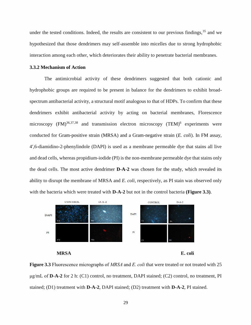
microscopy (FM)36,37,38 and transmission electron microscopy (TEM)6 experiments were
conducted for Gram-positive strain (MRSA) and a Gram-negative strain (E. coli). In FM assay,
4′,6-diamidino-2-phenylindole (DAPI) is used as a membrane permeable dye that stains all live
and dead cells, whereas propidium-iodide (PI) is the non-membrane permeable dye that stains only
the dead cells. The most active dendrimer D-A-2 was chosen for the study, which revealed its
ability to disrupt the membrane of MRSA and E. coli, respectively, as PI stain was observed only
with the bacteria which were treated with D-A-2 but not in the control bacteria (Figure 3.3).
MRSA E. coli
Figure 3.3 Fluorescence micrographs of MRSA and E. coli that were treated or not treated with 25
μg/mL of D-A-2 for 2 h: (C1) control, no treatment, DAPI stained; (C2) control, no treatment, PI
stained; (D1) treatment with D-A-2, DAPI stained; (D2) treatment with D-A-2, PI stained.

30
To further prove our hypothesis that lipidated dendrimers mimic the mechanism of action
of HDPs, TEM microscopy images were used to analyze the morphology of the drug treated and
non-drug treated bacteria. As shown in Figure 3.4 the membranes of both MRSA and E. coli were
compromised when treated with the dendrimer D-A-2.
Figure 3.4 TEM micrographs of MRSA and E. coli treated with 10 µg/mL of D-A-2 for 2 h: (a)
control, no treatment; (b) treatment with D-A-2, initial rupture of the bacterial membrane; (c)
treatment with D-A-2, complete cell rupture leading to bacterial cell death.
3.3.3 Bacterial Kinetic Study
To future investigate the time of bactericidal action of the dendrimers, bacterial kinetics
assay was conducted. The most active dendrimer D-A-2 was investigated and the time required to
show its bactericidal action was analyzed for Gram-positive and Gram-negative bacteria. Colony
forming units of the Gram-positive and Gram-negative bacteria on the agar plate were counted for
three different concentrations (50 µg/mL, 25 µg/mL, 12.5 µg/mL) of the D-A-2 treated bacteria
and the control bacteria at regular intervals of 10 min, 30 min, 1 h, 2 h. As shown in Figure 3.5, it
is evident that at the concentration of 50 µg/mL, 25 µg/mL 12.5 µg/mL, the Gram-positive bacteria
MRSA were eradicated completely after 1 h, whereas the Gram-negative bacteria, E. coli was also
eradicated after 2 h at the concentration of 50 µg/mL, 25 µg/mL, 12.5 µg/ml. This delay in
bactericidal action on the Gram-negative bacteria might be due to the presence of the extra outer
membrane layer, which is not present in the Gram-positive bacterial strains. Overall, the Time kill

31
studies deomonstrated that the dendrimer could rapidly kill bacteria.
MRSA E. coli
Figure 3.5 Time kill study of D-A-2 against MRSA and E. coli.
3.3.4 Bacterial Biofilm Assay
The infections caused by biofilms are a great threat to human life. Recent reports have
shown that 80% of the bacterial infections are caused due to biofilms.39 Bacterial biofilms have
become a severe problem specially in the cases of patients who suffer from infections that occur
from the antibiotic resistant bacteria.40 Biofilms formed by MRSA and E. coli are currently the
major concern in hospitals as they contaminate the surgical tools and organ catheters.41 Therefore,
development of new antibiotic agents that act against biofilm formation has become a major
strategy to treat bacterial infections. We thus analyzed the biofilm inhibiting efficiency of the most
active dendrimer D-A-2 against MRSA and E. coli. As shown in Figure 3.6, even at 0.17 μg/mL,
D-A-2 was able to inhibit 85% of biofilm formation of E. coli and was able to completely eradicate
the biofilm formation of MRSA. At the concentration of 0.8 μg/mL, D-A-2 was able to eradicate
biofilm formation of E. coli completely. The above stated results confirm that the dendrimers can
act as biofilm inhibitors.

32
0.0 0.3 0.6 0.9
0
50
100
MRSA
E. Coli
Rela
tiv
e B
iofi
lm B
iom
ass
(%
)
Compound D-A-2 (g/mL)
Figure 3.6. Inhibition of biofilms of MRSA and E. coli by D-A-2
3.4 Materials and methods
3.4.1 Antimicrobial Assay
Lipidated dendrimers were tested against different strains of bacteria to determine their
Minimum inhibitory concentration (MIC). Three clinically threatening Gram-positive bacterial
strains including methicillin-resistant S. epidermidis (MRSE, RP62A), vancomycin-resistant E.
faecalis (VREF, ATCC 700802), and methicillin-resistant Staphylococcus aureus (MRSA, ATCC
33591) and two Gram-negative bacterial strains, E. coli (ATCC 25922), P. aeruginosa (ATCC
27853) were used for testing. A serial dilution method was performed to determine the
antimicrobial activities. In 4 mL of TSB solution a single colony of bacteria was grown and
incubated at 37 °C overnight after which the cultured bacteria were diluted by 100-fold and were
shaken until their mid logarithmic phase was obtained. 50 μL of medium containing different
concentrations of the lipidated dendrimers was made in each vial of the 96 well plate. 50 μL
aliquots of bacterial suspension was added to the medium with different drug concentrations. The
96 well plate was incubated at 37 °C overnight and the cell growth was monitored using Biotek
synergy HT microtiter plate reader under the 600 nm wavelength absorbance. The assay was
repeated three times.42,42b,43

33
3.4.2 Hemolysis Assay
The selectivity of the lipidated dendrimers was determined through hemolysis assay. To
perform this assay fresh K2EDTA treated human red blood cells (hRBCs) were centrifuged at
1000g for 10 minutes. The step was done three times until the supernatant was clear, then the
desired suspended erythrocytes were collected and washed with PBS buffer a couple of times. The
collected RBCs were then diluted into 5% v/v suspension. From the diluted solution 50 μL was
taken and added to the already prepared serially- diluted lipidated dendrimer solutions and were
left for incubation at 37 °C for 1 hr. The incubated solutions were centrifuged at 3500 rpm for 10
minutes. To measure the absorbance, 30 μL of supernatant was diluted with 100 μL of PBS and
absorbance at a wavelength of 540 nm was recorded. Selectivity of the lipidated dendrimers (%
Hemolysis) was then calculated by applying the formula % hemolysis = (Abs sample − Abs PBS)/
(Abs Triton − Abs PBS) × 100%, where PBS was used as a negative control and 0.1% Triton x-
100 was used as a positive control. Results were repeated two times with duplicates each time.44
3.4.3 Fluorescence Microscopy
To assess whether the lipidated dendrimers act on the bacterial membrane or not, a double
staining Florescence microscopy assay was used. Both 6-diamidino-2-phenylindole
dihydrochloride (DAPI) and propidium iodide (PI) dyes were selected as fluorophores to
differentiate between the dead bacterial cells and viable cells. DAPI is capable of staining both
live and dead cells but PI can only stain dead cells due to its impermeable nature through the
membrane. The bacteria were grown to mid logarithmic phase and were incubated with lipidated
dendrimers at 37 °C for 2 h, the mixture was centrifuged at 1000g for 10 min. Once the bacterial
pellets were collected and washed with PBS three to four times. They were subsequently incubated
with PI (5 μg/mL) in the dark for 15 min at 0 °C, the excessive PI was rinsed using PBS several

34
times. Lastly, the cells were incubated with DAPI (10 μg/mL) for 15 min on ice and the excess
DAPI was removed by washing with PBS buffer. After the samples were ready 10 μL of the
bacteria were used for testing under Zeiss Axio Image Zloptical microscope using 100× oil-
immersion objective.44
3.4.4 Time kill study
The kinetic assay of the lipidated dendrimers was tested against MRSA and E. coli. The
bacteria were grown to midlogarithmic phase in TSB medium and then diluted into 106 CFUmL-1
suspensions. Different concentrations of the lipidated dendrimers were then added to the diluted
suspensions and incubated for 10 min, 30 min, 1 h, and 2 h, respectively. After incubation, the
mixture was diluted into 102 to 104 times, then dispersed on TSB agar plates and incubated
overnight at 37 °C. The colonies on the plates were counted and graphed against colony forming
unit of bacteria and incubation time.45
3.4.5 TEM Study
Similar procedure was used to grow the bacteria to mid logarithmic phase as in the case of
MIC study. Two samples were made, one batch has only bacteria and the other batch was the
bacteria treated with D-A-2. Samples were incubated at 37 °C for 2 h and the solution was
centrifuged at 1000g for 10 min. Bacterial pellets were collected at the bottom of the tube. Pellets
were washed with PBS three to four times before dissolving in water to a 10-6 M.32 A drop of the
solution was added on the carbon-coated Cu grid and the excess sample was wiped off with the
filter paper to avoid any aggregation. The grid was then left for 30 min on the bench top so that
the sample can be absorbed onto the grid. The samples on the grids were analyzed and images
were taken with FEI Morgagni 268D TEM instrument.46,47

35
3.4.6 Biofilm assay
The bacteria were grown and the treated with the dendrimers in 96 well plate in a similar
fashion as MIC study. The 96 well plates were left for incubation at 37 °C for 48 hours. The plate
was shaken over an empty tray to remove all the planktonic bacteria. The 96-well plate was then
rinsed in a large tray of water and shaken. Rinsing of the plate in water was done a couple of times.
The plate was placed on the paper towel to get rid of the water from the wells and was laid on
another paper towel and left to dry overnight. The wells were stained with 125 μL 0.1% crystal
violet solution for 10 minutes. The plate was shaken over the empty tray to remove the solution
and the plate was rinsed with water a couple of times until the wells are free of any liquid crystal
violet and left to dry overnight. 200 μL of 30% acetic acid were added to the wells to solubilize
the stained crystal violet and left for 10 min. 125 μL of the solution was transferred from each well
into a flat-bottom 96-well plate. The plate was read at OD 595 nm with a plate reader.43 The
average of the blank wells was subtracted from the OD of each sample that contained sample and
the average of the wells with samples were calculated. The averages of the sample wells were
normalized to the average of the control wells.4
3.5 Conclusion
We have developed a new class of antimicrobial lipidated dendrimers that can mimic the
HDPs. The design was straightforward which allows further development and optimization at ease.
Our findings suggest that amphiphilicity is required for the dendrimer to display potent and broad-
spectrum activity against a range of multidrug-resistant Gram-negative and Gram-positive
bacteria. These lipidated dendrimers were also proven to act as good biofilm inhibitors, further
augmenting their potential of practical applications. Furthermore, these dendrimers could be

36
developed for other biological applications to treat various fungal, viral infections and their activity
is yet to be explored.
3.6 References
1. Park, S. C.; Kim, N. H.; Yang, W.; Nah, J. W.; Jang, M. K.; Lee, D., Polymeric micellar
nanoplatforms for Fenton reaction as a new class of antibacterial agents. J Control Release 2016,
221, 37-47.
2. Savini, F.; Luca, V.; Bocedi, A.; Massoud, R.; Park, Y.; Mangoni, M. L.; Stella, L., Cell-
Density Dependence of Host-Defense Peptide Activity and Selectivity in the Presence of Host
Cells. ACS Chem Biol 2017, 12 (1), 52-56.
3. Thaker, H. D.; Som, A.; Ayaz, F.; Lui, D.; Pan, W.; Scott, R. W.; Anguita, J.; Tew, G. N.,
Synthetic mimics of antimicrobial peptides with immunomodulatory responses. J Am Chem Soc
2012, 134 (27), 11088-91.
4. Teng, P.; Huo, D.; Nimmagadda, A.; Wu, J.; She, F.; Su, M.; Lin, X.; Yan, J.; Cao, A.; Xi,
C.; Hu, Y.; Cai, J., Small Antimicrobial Agents Based on Acylated Reduced Amide Scaffold.
Journal of Medicinal Chemistry 2016, 59 (17), 7877-7887.
5. Hubbard, A. T. M.; Barker, R.; Rehal, R.; Vandera, K.-K. A.; Harvey, R. D.; Coates, A. R.
M., Mechanism of Action of a Membrane-Active Quinoline-Based Antimicrobial on Natural and
Model Bacterial Membranes. Biochemistry 2017, 56 (8), 1163-1174.
6. Nimmagadda, A.; Liu, X.; Teng, P.; Su, M.; Li, Y.; Qiao, Q.; Khadka, N. K.; Sun, X.; Pan,
J.; Xu, H.; Li, Q.; Cai, J., Polycarbonates with Potent and Selective Antimicrobial Activity toward
Gram-Positive Bacteria. Biomacromolecules 2017, 18 (1), 87-95.
7. Holthausen, D. J.; Lee, S. H.; Kumar, V. T.; Bouvier, N. M.; Krammer, F.; Ellebedy, A.
H.; Wrammert, J.; Lowen, A. C.; George, S.; Pillai, M. R.; Jacob, J., An Amphibian Host Defense

37
Peptide Is Virucidal for Human H1 Hemagglutinin-Bearing Influenza Viruses. Immunity 2017, 46
(4), 587-595.
8. Liu, R.; Chen, X.; Falk, S. P.; Mowery, B. P.; Karlsson, A. J.; Weisblum, B.; Palecek, S.
P.; Masters, K. S.; Gellman, S. H., Structure-activity relationships among antifungal nylon-3
polymers: identification of materials active against drug-resistant strains of Candida albicans. J
Am Chem Soc 2014, 136 (11), 4333-42.
9. (a) Karlsson, A. J.; Pomerantz, W. C.; Weisblum, B.; Gellman, S. H.; Palecek, S. P.,
Antifungal activity from 14-helical beta-peptides. Journal of the American Chemical Society 2006,
128 (39), 12630-1; (b) Porter, E. A.; Weisblum, B.; Gellman, S. H., Mimicry of host-defense
peptides by unnatural oligomers: antimicrobial beta-peptides. Journal of the American Chemical
Society 2002, 124 (25), 7324-30; (c) Porter, E. A.; Wang, X.; Lee, H. S.; Weisblum, B.; Gellman,
S. H., Non-haemolytic beta-amino-acid oligomers. Nature 2000, 404 (6778), 565.
10. (a) Patch, J. A.; Barron, A. E., Helical peptoid mimics of magainin-2 amide. Journal of the
American Chemical Society 2003, 125 (40), 12092-12093; (b) Ghosh, C.; Manjunath, G. B.;
Akkapeddi, P.; Yarlagadda, V.; Hoque, J.; Uppu, D. S. S. M.; Konai, M. M.; Haldar, J., Small
Molecular Antibacterial Peptoid Mimics: The Simpler the Better! Journal of Medicinal Chemistry
2014, 57 (4), 1428-1436.
11. Violette, A.; Fournel, S.; Lamour, K.; Chaloin, O.; Frisch, B.; Briand, J. P.; Monteil, H.;
Guichard, G., Mimicking helical antibacterial peptides with nonpeptidic folding oligomers.
Chemistry & biology 2006, 13 (5), 531-8.
12. (a) Li, Y. Q.; Wu, H. F.; Teng, P.; Bai, G.; Lin, X. Y.; Zuo, X. B.; Cao, C. H.; Cai, J. F.,
Helical Antimicrobial Sulfono-gamma-AApeptides. Journal of medicinal chemistry 2015, 58 (11),
4802-4811; (b) Padhee, S.; Smith, C.; Wu, H.; Li, Y.; Manoj, N.; Qiao, Q.; Khan, Z.; Cao, C.; Yin,

38
H.; Cai, J., The Development of Antimicrobial alpha-AApeptides that Suppress Proinflammatory
Immune Responses. Chembiochem : a European journal of chemical biology 2014, 15, 688-694;
(c) Li, Y.; Smith, C.; Wu, H.; Teng, P.; Shi, Y.; Padhee, S.; Jones, T.; Nguyen, A. M.; Cao, C.;
Yin, H.; Cai, J., Short antimicrobial lipo-alpha/gamma-AA hybrid peptides. Chembiochem 2014,
15, 2275-2280; (d) Li, Y.; Smith, C.; Wu, H.; Padhee, S.; Manoj, N.; Cardiello, J.; Qiao, Q.; Cao,
C.; Yin, H.; Cai, J., Lipidated cyclic gamma-AApeptides display both antimicrobial and anti-
inflammatory activity. ACS Chem Biol 2014, 9 (1), 211-7; (e) Niu, Y.; Wu, H.; Li, Y.; Hu, Y.;
Padhee, S.; Li, Q.; Cao, C.; Cai, J., AApeptides as a new class of antimicrobial agents. Org.
Biomol. Chem. 2013, 11, 4283-4290; (f) Shi, Y.; Teng, P.; Sang, P.; She, F.; Wei, L.; Cai, J., γ-
AApeptides: Design, Structure, and Applications. Accounts of Chemical Research 2016, 49 (3),
428-441.
13. (a) Niu, Y.; Padhee, S.; Wu, H.; Bai, G.; Qiao, Q.; Hu, Y.; Harrington, L.; Burda, W. N.;
Shaw, L. N.; Cao, C.; Cai, J., Lipo-gamma-AApeptides as a new class of potent and broad-
spectrum antimicrobial agents. Journal of medicinal chemistry 2012, 55 (8), 4003-9; (b) Padhee,
S.; Hu, Y.; Niu, Y.; Bai, G.; Wu, H.; Costanza, F.; West, L.; Harrington, L.; Shaw, L. N.; Cao, C.;
Cai, J., Non-hemolytic alpha-AApeptides as antimicrobial peptidomimetics. Chem Commun
(Camb) 2011, 47 (34), 9729-31; (c) Niu, Y. H.; Padhee, S.; Wu, H. F.; Bai, G.; Harrington, L.;
Burda, W. N.; Shaw, L. N.; Cao, C. H.; Cai, J. F., Identification of gamma-AApeptides with potent
and broad-spectrum antimicrobial activity. Chemical Communications 2011, 47 (44), 12197-
12199.
14. Costanza, F.; Padhee, S.; Wu, H.; Wang, Y.; Revenis, J.; Cao, C.; Li, Q.; Cai, J.,
Investigation of antimicrobial PEG-poly(amino acid)s. Rsc Adv 2014, 4 (4), 2089-2095.

39
15. (a) Liu, R. H.; Suarez, J. M.; Weisblum, B.; Gellman, S. H.; McBride, S. M., Synthetic
Polymers Active against Clostridium difficile Vegetative Cell Growth and Spore Outgrowth.
Journal of the American Chemical Society 2014, 136 (41), 14498-14504; (b) Chakraborty, S.; Liu,
R. H.; Hayouka, Z.; Chen, X. Y.; Ehrhardt, J.; Lu, Q.; Burke, E.; Yan, Y. Q.; Weisblum, B.; Wong,
G. C. L.; Masters, K. S.; Gellman, S. H., Ternary Nylon-3 Copolymers as Host-Defense Peptide
Mimics: Beyond Hydrophobic and Cationic Subunits. Journal of the American Chemical Society
2014, 136 (41), 14530-14535; (c) Liu, R. H.; Masters, K. S.; Gellman, S. H., Polymer Chain Length
Effects on Fibroblast Attachment on Nylon-3-Modified Surfaces. Biomacromolecules 2012, 13
(4), 1100-1105.
16. (a) Kuroda, K.; Caputo, G. A., Antimicrobial polymers as synthetic mimics of host-defense
peptides. Wires Nanomed Nanobi 2013, 5 (1), 49-66; (b) Oda, Y.; Kanaoka, S.; Sato, T.; Aoshima,
S.; Kuroda, K., Block versus Random Amphiphilic Copolymers as Antibacterial Agents.
Biomacromolecules 2011, 12 (10), 3581-3591.
17. Wang, J.; Chen, Y. P.; Yao, K.; Wilbon, P. A.; Zhang, W.; Ren, L.; Zhou, J.; Nagarkatti,
M.; Wang, C.; Chu, F.; He, X.; Decho, A. W.; Tang, C., Robust antimicrobial compounds and
polymers derived from natural resin acids. Chemical Communications 2012, 48 (6), 916-918.
18. (a) Engler, A. C.; Tan, J. P. K.; Ong, Z. Y.; Coady, D. J.; Ng, V. W. L.; Yang, Y. Y.;
Hedrick, J. L., Antimicrobial Polycarbonates: Investigating the Impact of Balancing Charge and
Hydrophobicity Using a Same-Centered Polymer Approach. Biomacromolecules 2013, 14 (12),
4331-4339; (b) Nederberg, F.; Zhang, Y.; Tan, J. P. K.; Xu, K.; Wang, H.; Yang, C.; Gao, S.; Guo,
X. D.; Fukushima, K.; Li, L.; Hedrick, J. L.; Yang, Y.-Y., Biodegradable nanostructures with
selective lysis of microbial membranes. Nature chemistry 2011, 3 (5), 409-414.

40
19. Vergara-Jaque, A.; Comer, J.; Sepúlveda-Boza, S.; Santos, L. S.; Mascayano, C.;
Sandoval-Yáñez, C., Study of specific interactions in inclusion complexes of amine-terminated
PAMAM dendrimer/flavonoids by experimental and computational methods. International
Journal of Polymeric Materials and Polymeric Biomaterials 2017, 66 (10), 485-494.
20. Zhisheng Chen, C.; L. Cooper, S.; C. Beck Tan, N., Incorporation of
dimethyldodecylammonium chloride functionalities onto poly(propylene imine) dendrimers
significantly enhances their antibacterial properties. Chemical Communications 1999, (16), 1585-
1586.
21. Niu, Y.; Wu, H.; Huang, R.; Qiao, Q.; Constanza, F.; Wang, X.; Hu, Y.; Amin, M. N.;
Naguyen, A.; Zhang, J.; Haller, E.; Ma, S.; Li, X.; Cai, J., Nanorods Formed from a New Class of
Peptidomimetics. Macromolecules 2012, 10.1021/ma3015992.
22. Xue, X.; Chen, X.; Mao, X.; Hou, Z.; Zhou, Y.; Bai, H.; Meng, J.; Da, F.; Sang, G.; Wang,
Y.; Luo, X., Amino-terminated generation 2 poly(amidoamine) dendrimer as a potential broad-
spectrum, nonresistance-inducing antibacterial agent. AAPS J 2013, 15 (1), 132-42.
23. Walter, M. V.; Malkoch, M., Simplifying the synthesis of dendrimers: accelerated
approaches. Chem Soc Rev 2012, 41 (13), 4593-609.
24. Wang, B.; Navath, R. S.; Menjoge, A. R.; Balakrishnan, B.; Bellair, R.; Dai, H.; Romero,
R.; Kannan, S.; Kannan, R. M., Inhibition of bacterial growth and intramniotic infection in a guinea
pig model of chorioamnionitis using PAMAM dendrimers. Int J Pharm 2010, 395 (1-2), 298-308.
25. Lopez, A. I.; Reins, R. Y.; McDermott, A. M.; Trautner, B. W.; Cai, C., Antibacterial
activity and cytotoxicity of PEGylated poly(amidoamine) dendrimers. Mol Biosyst 2009, 5 (10),
1148-56.

41
26. Shi, S.; Chen, F.; Ehlerding, E. B.; Cai, W., Surface Engineering of Graphene-Based
Nanomaterials for Biomedical Applications. Bioconjugate Chemistry 2014, 25 (9), 1609-1619.
27. Worley, B. V.; Slomberg, D. L.; Schoenfisch, M. H., Nitric oxide-releasing quaternary
ammonium-modified poly(amidoamine) dendrimers as dual action antibacterial agents. Bioconjug
Chem 2014, 25 (5), 918-27.
28. Janiszewska, J.; Swieton, J.; Lipkowski, A. W.; Urbanczyk-Lipkowska, Z., Low molecular
mass peptide dendrimers that express antimicrobial properties. Bioorganic & Medicinal Chemistry
Letters 2003, 13 (21), 3711-3713.
29. Siriwardena, T. N.; Stach, M.; He, R.; Gan, B.-H.; Javor, S.; Heitz, M.; Ma, L.; Cai, X.;
Chen, P.; Wei, D.; Li, H.; Ma, J.; Köhler, T.; van Delden, C.; Darbre, T.; Reymond, J.-L., Lipidated
Peptide Dendrimers Killing Multidrug-Resistant Bacteria. Journal of the American Chemical
Society 2018, 140 (1), 423-432.
30. She, F.; Nimmagadda, A.; Teng, P.; Su, M.; Zuo, X.; Cai, J., Helical 1:1 alpha/Sulfono-
gamma-AA Heterogeneous Peptides with Antibacterial Activity. Biomacromolecules 2016, 17 (5),
1854-9.
31. Niu, Y.; Padhee, S.; Wu, H.; Bai, G.; Qiao, Q.; Hu, Y.; Harrington, L.; Burda, W. N.; Shaw,
L. N.; Cao, C.; Cai, J., Lipo-γ-AApeptides as a New Class of Potent and Broad-Spectrum
Antimicrobial Agents. Journal of Medicinal Chemistry 2012, 55 (8), 4003-4009.
32. Li, Y.; Smith, C.; Wu, H.; Padhee, S.; Manoj, N.; Cardiello, J.; Qiao, Q.; Cao, C.; Yin, H.;
Cai, J., Lipidated Cyclic γ-AApeptides Display Both Antimicrobial and Anti-inflammatory
Activity. ACS Chemical Biology 2014, 9 (1), 211-217.
33. Díaz-Mochón, J. J.; Bialy, L.; Bradley, M., Full Orthogonality between Dde and Fmoc:
The Direct Synthesis of PNA−Peptide Conjugates. Organic Letters 2004, 6 (7), 1127-1129.

42
34. Shi, Y.; Challa, S.; Sang, P.; She, F.; Li, C.; Gray, G. M.; Nimmagadda, A.; Teng, P.;
Odom, T.; Wang, Y.; van der Vaart, A.; Li, Q.; Cai, J., One-Bead–Two-Compound Thioether
Bridged Macrocyclic γ-AApeptide Screening Library against EphA2. Journal of Medicinal
Chemistry 2017, 60 (22), 9290-9298.
35. Li, Y.; Smith, C.; Wu, H.; Teng, P.; Shi, Y.; Padhee, S.; Jones, T.; Nguyen, A.-M.; Cao,
C.; Yin, H.; Cai, J., Short Antimicrobial Lipo-α/γ-AA Hybrid Peptides. ChemBioChem 2014, 15
(15), 2275-2280.
36. Su, M.; Xia, D.; Teng, P.; Nimmagadda, A.; Zhang, C.; Odom, T.; Cao, A.; Hu, Y.; Cai,
J., Membrane-Active Hydantoin Derivatives as Antibiotic Agents. Journal of Medicinal Chemistry
2017, 60 (20), 8456-8465.
37. She, F.; Nimmagadda, A.; Teng, P.; Su, M.; Zuo, X.; Cai, J., Helical 1:1 α/Sulfono-γ-AA
Heterogeneous Peptides with Antibacterial Activity. Biomacromolecules 2016, 17 (5), 1854-1859.
38. Teng, P.; Nimmagadda, A.; Su, M.; Hong, Y.; Shen, N.; Li, C.; Tsai, L.-Y.; Cao, J.; Li, Q.;
Cai, J., Novel bis-cyclic guanidines as potent membrane-active antibacterial agents with
therapeutic potential. Chemical Communications 2017, 53 (87), 11948-11951.
39. Zhang, P.; Li, S.; Chen, H.; Wang, X.; Liu, L.; Lv, F.; Wang, S., Biofilm Inhibition and
Elimination Regulated by Cationic Conjugated Polymers. ACS Appl Mater Interfaces 2017, 9 (20),
16933-16938.
40. Bottcher, T.; Kolodkin-Gal, I.; Kolter, R.; Losick, R.; Clardy, J., Synthesis and activity of
biomimetic biofilm disruptors. J Am Chem Soc 2013, 135 (8), 2927-30.
41. Bjarnsholt, T.; Ciofu, O.; Molin, S.; Givskov, M.; Hoiby, N., Applying insights from
biofilm biology to drug development - can a new approach be developed? Nature reviews. Drug
discovery 2013, 12 (10), 791-808.

43
42. (a) Lin, S.; Koh, J. J.; Aung, T. T.; Lim, F.; Li, J.; Zou, H.; Wang, L.; Lakshminarayanan,
R.; Verma, C.; Wang, Y.; Tan, D. T.; Cao, D.; Beuerman, R. W.; Ren, L.; Liu, S., Symmetrically
Substituted Xanthone Amphiphiles Combat Gram-Positive Bacterial Resistance with Enhanced
Membrane Selectivity. J Med Chem 2017, 60 (4), 1362-1378; (b) Porel, M.; Thornlow, D. N.;
Artim, C. M.; Alabi, C. A., Sequence-Defined Backbone Modifications Regulate Antibacterial
Activity of OligoTEAs. ACS Chem Biol 2017, 12 (3), 715-723.
43. Joseph, R.; Naugolny, A.; Feldman, M.; Herzog, I. M.; Fridman, M.; Cohen, Y., Cationic
Pillararenes Potently Inhibit Biofilm Formation without Affecting Bacterial Growth and Viability.
J Am Chem Soc 2016, 138 (3), 754-7.
44. Hoque, J.; Konai, M. M.; Sequeira, S. S.; Samaddar, S.; Haldar, J., Antibacterial and
Antibiofilm Activity of Cationic Small Molecules with Spatial Positioning of Hydrophobicity: An
in Vitro and in Vivo Evaluation. J Med Chem 2016, 59 (23), 10750-10762.
45. Hoque, J.; Haldar, J., Direct Synthesis of Dextran-Based Antibacterial Hydrogels for
Extended Release of Biocides and Eradication of Topical Biofilms. ACS Appl Mater Interfaces
2017, 9 (19), 15975-15985.
46. Wu, H.; Li, Y.; Bai, G.; Niu, Y.; Qiao, Q.; Tipton, J. D.; Cao, C.; Cai, J., gamma-
AApeptide-based small-molecule ligands that inhibit Abeta aggregation. Chem Commun (Camb)
2014, 50 (40), 5206-8.
47. Song, B.; Zhang, Z.; Wang, K.; Hsu, C. H.; Bolarinwa, O.; Wang, J.; Li, Y.; Yin, G. Q.;
Rivera, E.; Yang, H. B.; Liu, C.; Xu, B.; Li, X., Direct Self-Assembly of a 2D and 3D Star of
David. Angew Chem Int Ed Engl 2017, 56 (19), 5258-5262.

44
CHAPTER 4: UNIMOLECULAR NANOPARTICLES AS POTENT ANTIMICROBIAL
AGENTS
4.1 Introduction
Antibiotic resistance is emerging to be one of the three greatest threats to global public
health in the 21st century.1,2 As a result, significant interest has been devoted to the development
of the new strategies combating drug resistance. Among them, natural cationic host-defense
peptides (HDPs) are believed to be most promising due to their minimum risk of resistance
development.3,4 It is known that conventional antibiotics generally target proteins involved in
bacterial cell wall biosynthesis, or intracellular targets such as nucleic acids.3,5 Such mechanisms
involving defined targets can frequently lead to mutations in bacteria, thereby inducing significant
antibiotic resistance.6 However, HDPs have existed universally in vertebrates for thousands of
years, and they still possess broad-spectrum antibacterial activity. It is intriguing that HDPs have
diverse three dimensional structures, such as α-helices and β-sheets,3 however they are all rich in
cationic and hydrophobic residues. The cyclic and linear forms of the HDP are due to the presence
of disulfide bonds and presence of multiple arginine and lysine amino acids in the sequences. This
also gives them a basic property and amphiphilic nature.7
Bacterial cell membranes are composed mainly of proteins and phospholipids. The
phospholipid layer has a polar hydrophobic glycerol and hydrophobic fatty acid tails which gives
the amphiphilic nature of the membrane. HDPs directly acts on the bacterial membrane. The
positively charged nature of HDPs allows its electrostatic interaction with the negatively charged

45
membrane as the glycoprotein present in the membrane is the binding sites for the HDPs.8, 9 HDPs
are believed to use their hydrophobic domain to penetrate and disrupt membrane lipid layers,
which subsequently leads to leakage of cellular contents and cell death.7,10 This explains why
HDPs have diverse three dimensional structures, because this mechanism of membrane disruption
is based on non-specific biophysical force of hydrophobic and hydrophilic interactions, and
therefore no defined secondary or tertiary structures are necessary.11
Although any therapeutic agents would inevitably develop resistance in their targets,
including cells and organs of the organisms, HDPs may be less prone to the trend because the
membrane disruption by HDPs is non-specific and HDPs lack defined membrane targets.9 It should
also be noted that the action of HDPs may also involve other mechanisms, such as targeting
intracellular and cell wall targets, much as traditional antibiotics do, however, the electrostatic
interaction and/or disruption of bacterial membranes still generally exist in these HDPs, which are
critical for their permeation into bacterial cells.8
One of the successful examples of HDPs is magainin-2,12 which shows moderate
antimicrobial activity against bacteria. Synthetic peptide analogs such as Pexiganan (also known
as MSI-78) have shown much improved activity and entered Phase III clinical trials for the
treatment of diabetic foot ulcers. However, it eventually failed in the clinical trial, largely due to
its moderate antimicrobial activity and high production cost.13 Furthermore, although the
selectivity of HDPs is good, it is still not ideal yet, as they display toxicity such as hemolytic
activity.
The scientific and clinical expansions in the antibiotic development is to advance agents
which can mimic the mechanism of HDP and with relatively low bacterial resistance and
maximum efficacy as well as minimum toxicity. Recently many research studies are launched to

46
develop synthetic peptidomimetics including β-peptides,14-16 peptoids,17 oligoureas,18
AApeptides.19-23 Though the ongoing development in the area of peptidomimetics is promising,
the selectivity problem, manufacturing cost and the efficiency of the synthesis method are
challenging.24-26
Another innovative approach to developing antimicrobial agents is mimicking the HDPs
through biodegradable polymers. Like HDPs, they contain both hydrophobic and hydrophilic
cationic groups in their sequences, which are synthesized by amphiphilic building blocks through
homo-polymerization, or by both hydrophobic monomers and cationic monomers through block-
by-block or random copolymerization. The antibacterial mechanism of action of polymers is
similar to that of HDPs. Some polymers function through interaction with bacterial membranes by
single random sequences,10,27-31 while others are designed to self-assemble into nanoparticles, then
act on the surface of bacterial membranes.27,32,33 It is suggested that through the formation of
nanoparticles, there is an increase of local mass and cationic charges on the bacterial surface which
facilitates disintegration of bacterial membranes.33 However, this limits the mechanism of action
and activity of self-assembled nanoparticles to be higher than their critical micelle concentration
(CMC).33
4.2 Overview of Unimolecular Micelle Hyperbranched Polymers
Unimolecular micelles are a class of small entities of micelles with a discrete core
surrounded by shells which are formed by a covalent bond chain. With the current innovations in
polymer chemistry, unimolecular micelles are functionalized in various architectural designs,
including amphiphilic dendrimers, amphiphilic star polymers and hyperbranched polymers called
polymeric micelles.34 Unimolecular micelle hyperbranched polymers are built from a dendrimer
core where the arms of the core are composed of both hydrophobic and cationic polymers. Bearing

47
special structural and functional properties, biodegradable-unimolecular polymeric micelles are
becoming imperative candidates for drug delivery applications, encapsulating and solubilizing a
hydrophobic guest molecule and antimicrobial applications in the biomedical era.35
The antimicrobial application of the unimolecular micelle is hypothesized to be more
potent and selective compared with the regular antimicrobial polymers, because of the spatial
architectural design, which improves their interaction with the bacterial membrane. Each
unimicelle is acting as a nanoparticle. Such a design is devoid of potential de-assembly of
assembled nanoparticles under low concentrations, ensuring constant high local mass and cationic
charges which is crucial for antimicrobial application mechanism. In addition, the hydrophobic
groups present in the structure may be sequestered in the interior of the micelles, which decreases
the probability of non-specific interaction of nanoparticles with mammalian cells, thereby
enhancing the selectivity towards bacteria. Indeed, these unimicelle nanoparticles can also be
scaled-up to make large quantities, and they are biodegradable, augmenting their potential in vitro
and in vivo applications.33,35 Herein, we present the design and application of unimolecular micelle
hyperbranched polymers as antimicrobial agents.
4.3 Experimental Section
4.3.1 Synthesis of Hyperbranched Polyester Polyacid (HBPP) (Figure4.1)
A weighed portion of 1 equiv of Boltorn H311 was heated to 140 OC for 2h under nitrogen
blowing to break the hydrogen bonds and make it anhydrous. Then it was cooled to 50 OC and
dissolved in 1,4-dioxane. To the heated solution of the Boltorn H311, a stoichiometric amount 5
equiv of maleic anhydride to each dendrimer arm (5 equiv*23) and catalytic amount of stannous
chloride (SnCl2) (1% with respect to the Boltorn weight) were added simultaneously. The mixture
was stirred at a temperature of 100 OC for 36 h. After cooling, the solvent was removed in vacuo

48
and the product was purified by dialysis against methanol for 2 days and distilled water for 2 days
using dialysis tubing MWCO 3500 where methanol and water being replaced twice a day.36 The
product was then freeze dried and characterized by 1HNMR (Figure 4.2).
Figure 4.1 Synthesis of HBPP
Figure 4.2 1H NMR of HBPP
4.3.2 Synthesis of hydroxyl terminated hyperbranched polyester (Figure 4.3)
To synthesize hydroxyl terminated hyperbranched polyester, 1 equiv of hyperbranched
polyester polyacid (HBPP, Figure 4.1) was dissolved in 20 ml DMF and then (5 equiv*23) of
ethanolamine (2-aminoethan-1-ol), (5 equiv*23) EDC, (2 equiv*23) HOBT and (2 equiv*23) DIPEA
base were added. The solution mixture was stirred at room temperature overnight. After the reaction
was completed, work up was done using 100 ml of ethyl acetate and the product was washed with 1N

49
HCl (100 mL×3) and Brine (50 mL×1). The product was then dried over anhydrous sodium sulfate
and the solvent was removed in vacuo to give yellowish oily product, it was then purified in a dialysis
tubing for 4 days using methanol and water.36 The product was then freeze dried and characterized by
1HNMR (Figure 4.4).
Figure 4.3 Synthesis of hydroxyl terminated hyperbranched polyester
Figure 4.4 1H NMR of hydroxyl terminated hyperbranched polyester
4.3.3 Synthesis of Hydrophilic monomer (M1) (Figure 4.5)
Monomer 1 (2-((tert-butoxycarbonyl) amino) ethyl 5-methyl-2-oxo-1,3-dioxane-5-
carboxylate) was synthesized following the published paper in our lab by Jianfeng Cai and Alekhya
Nimmagadda.37

50
Figure 4.5 Monomer 1 (M1) synthesis.
4.3.4 Synthesis of Hydrophobic monomer (M2) (Figure 4.6)
Monomer 2 was prepared according to the previously reported protocol by Yang and
Hedrick.38
Figure 4.6 Monomer 2 (M2) synthesis
4.3.5 Synthesis of Unimolecular micelle hyperbranched polymers (Figure 4.7 & Figure 4.8)
We propose to synthesize both random and diblock unimolecular micelles hyperbranched
polymers (Figure 4.7 & Figure 4.8). As shown in Figure 4.5 & Figure 4.6 first both hydrophilic and
hydrophobic monomers were synthesized following the protocols reported in the published paper of
our lab and a paper from Yang and Hedrick et al.37,38 Hyperbranched polymers were then synthesized
through ring-opening polymerization in the presence of the two complementary monomers and the
unimolecular micelle core.39 To synthesize random unimicelle polycarbonate P6, 1 equiv of hydroxyl
terminated hyperbranched polyesters as a macro initiator and 20 equiv of hydrophilic monomer (M1)
and 20 equiv of hydrophobic monomer (M2) were mixed together and dissolved in 10ml of anhydrous

51
DCM in a N2 purged round bottom flask. To this mixture 1 equiv of 1,8-diazabicyclo undec-7-ene
(DBU) and 1 equiv of 1-(3,5-bis(trifluoromethyl)-phenyl)-3-cyclohexyl-2- thiourea (TU) catalyst
were added and the reaction was left overnight and the next day the reaction was quenched by 1.1
equiv of benzoic acid and purification of the polymer was done by dialyzing against methanol in a
MWCO 3500 dialysis tubing for 3 days, after that the polymers were then treated with 15 ml of a 1:1
TFA:DCM mixture for 3 h to deprotect the Boc protecting group in order to obtain the free amine
functional groups. And then the polymer was purified again in a dialysis tubing (MWCO 3500) for 2
days in methanol and 2 days in water and it was then freeze dried and a brownish semisolid product
was attained. As such, a series of hyperbranched polymers containing varying numbers of hydrophobic
and hydrophilic groups were prepared in the same manner (Table 4.1).
Figure 4.7 Synthesis of unimolecular nanoparticles with random chains of polycarbonates.
Similarly, Di-block hyperbranched polycarbonate armed unimicelle polymers (Table 4.1)
were synthesized in the same way as the random block polymers. However, the two monomers were
added in two batches, first monomer 1 was added along with the macro initiator, TU catalyst and DBU
base and the reaction runs for 6 h and then monomer 2 was added and the reaction was left for
polymerization for 6 h (Figure 4.8).37

52
Figure 4.8 Synthesis of unimolecular nanoparticles with diblock chains of polycarbonates.
Table 4.1 Unimolecular micelle hyperbranched polymers.
Compound
Type of
Unimicelle polymer
Hydrophobic arms
Hydrophilic arms
P1 Di-block 10 10
P2 Random 10 10
P3 Di-block
15 15
P4 Random
15 15
P5 Di-block
20 20
P6 Random
20 20
P7 Di-block
25 25
P8 Random
25 25
P9 Di-block
30 30
P10 Random
30 30
P11 Di-block
40 40
P12 Random
40 40
4.3.6 Antimicrobial Assay
Antimicrobial assay of the synthesized unimicelle polymers was carried out following similar
protocols as reported in the published papers of our lab.40, 41Two clinically relevant bacterial strains,
Gram-positive Methicillin-resistant Staphylococcus aureus (MRSA, ATCC 33591) and Gram-
negative E. coli (ATCC 25922) bacteria were used to perform the MIC assay. The minimum inhibitory

53
concentration (MIC) is the lowest concentration that completely inhibits the growth of bacteria in 20
h. The MIC was made in sterile 96 well plate, first a single colony of bacteria was isolated and allowed
to grow in 4 ml TSb solution for 12 h at 37 °C in a shaking incubator. Next day the grown culture was
diluted by 100-fold and was shaken for 6 h to attain mild-logarithm phase. To the 96-wellplate 50 µL
of the unimicelle polymer solution in 2-fold serial dilution of TSB were added, then 50 µL diluted
bacterial in TSB medium (1 × 10⁶ CFU/mL) was added to each well. The well-plate was incubated for
20 h at 37 °C and MIC was determined from the Biotek Synergy HT microtiter plate reader absorption
at 600 nm wavelength.
4.4 Results and Discussion
Dendritic polymers, including HBPs in which polar amines, hydroxyl or amide groups
presented in their terminal ends were found to be a highly selective and potent antimicrobial
compounds in addition to the straight forward synthesis method and high yield production.42 We are
here presenting a contemporary design of unimolecular micelle hyperbranched polymers by
incorporating polycarbonate monomers in the arms of a dendrimer core (Boltorn™ H311) to function
as antimicrobial agents in biomedical era. We chose polycarbonates to be incorporated in the arms of
unimolecular micelle as polycarbonates are biocompatible, biodegradable, and can be polymerized
easily through ring-opening polymerization (ROP). Moreover, Yang and Hedrick et al have already
demonstrated the polycarbonates potential for antimicrobial applications.38
Polyol Boltorn™ (aliphatic hyperbranched polyester) are dendritic polymers which have a
highly branched flexible backbone that can embrace more than 20 terminal hydroxyl groups. In our
study, Boltorn™ H311 (MW 5300 g/mole) which has 23 terminal hydroxyl branches of functional
groups was used for the synthesis of the unimolecular nanoparticle as it can be adapted easily to
dendrimers as well as unimicelles.43,44 As per our design the polycarbonate monomers have to be

54
complexed to the arms of the Boltorn, so first the hydroxyl terminal of the Boltran has to be lengthened.
To achieve this approach then, first the hydroxyl terminal of Boltran was transformed into carboxyl
terminal to attain a high reactivity for the next reactions by reacting the Boltran H311-OH with maleic
anhydride in the presence of tin(II) chloride in dioxane (Figure 4.1). Once the reactive carboxyl group
was obtained ethanolamine was coupled to it to produce an elongated hydroxyl terminal arm of Boltorn
(Figure 4.3).36
Polymerization was done through ROP method, and 12 unimolecular micelle hyperbranched
polymers were synthesized by incorporating the two monomers M1 and M2 in to the arms of the
hydroxyl terminated hyperbranched polyester H311 macro initiator (Figure 4.7 and Figure 4.8). To
see the disparity in the antimicrobial activity among the 12 polymers, 6 were diblock polymers and
the rest were random block polymers (Table 4.1).
Earlier study in our lab discloses, polycarbonate polymer which has 20 equiv hydrophilic and
20 equiv hydrophobic monomers with benzyl alcohol initiator was the most potent towards gram
positive bacteria but were not active against gram negative bacteria.37 Bearing that in our mind we
hypothesized by manipulating the same polycarbonate polymers in unimolecular micelle core could
enhance the antibacterial activity of the polymers towards broad spectrum.
The antibacterial activity of the synthesized polymers was studied against Gram-positive
Methicillin-resistant Staphylococcus epidermidis (MRSE, RP62A) and Gram-negative E. coli (ATCC
25922) bacteria. As the antimicrobial activity is shown in Table 4.2 both diblock and random polymers
displayed significant antibacterial activity against Gram positive bacteria and to our delight diblock
polymer P5 and random block polymers P6 and P12 presented broad spectrum activity against both
gram positive and gram-negative bacteria.

55
Polymers P1and P2 which have the same number of hydrophilic and hydrophobic units (10
equiv:10 equiv) displayed potent antimicrobial activity (0.75µg /ml) against MRSA but they were not
active to gram negative bacteria, this could be due to the desired amphiphilicity was not obtained to
break the extra outer membrane of the gram-negative bacteria.
Table 4.2 The Antibacterial Activity of Unimolecular micelle hyperbranched polymers.
Polymer
Type of
Copolymer
Hydrophilic
units
Hydrophobic
units
MIC- MRSA
(µg/mL)
MIC- E. coli
(µg/mL)
P1 Di-block 10 10 0.75 NA
P2 Random 10 10 0.75 NA
P3 Di-block 15 15 6~12.5 NA
P4 Random 15 15 3~6 NA
P5 Di-block
20 20 6~12.5 12.5~25
P6 Random
20 20 1.5~3 12.5~25
P7 Di-block
25 25 6~12.5 NA
P8 Random
25 25 6~12.5 NA
P9 Di-block
30 30 6~12.5 NA
P10 Random
30 30 6~12.5 NA
P11 Di-block
40 40 12.5~25 NA
P12 Random
40 40 6~12.5 12.5~25
Next, we synthesized a series of polymers (P3, P4, P5 and P6) by increasing the number of
equivalents of hydrophilic and hydrophobic monomers and they were tested against both strains of
bacteria, among them polymers P6 (random block polymer) displayed potent activity against MRSA
(1.5 µg/mL) and moderate activity (12.5µg/mL) against gram negative E. coli, similarly the diblock

56
polymers P5 displayed broad spectrum activity against MRSA (6 µg/mL) and E. coli (12.5 µg/mL).
This result matched with our hypothesis that we propose the unimolecular micelle hyperbranched
polymer with 20 equiv of hydrophilic and hydrophobic monomers might show broad spectrum
activity. The broad-spectrum activity is probably obtained due to more interaction that is created
between the hyperbranched polymers and the outer membrane of the Gram-negative bacteria with the
increase of both hydrophilic and hydrophobic monomers in the arms of the Boltorn.
Encouraged by the MIC result of P5 and P6, we synthesized 6 more polymers by increasing
the hydrophilic and hydrophobic residues. Diblock polymer P11 and random polymer P12 which had
two times more hydrophilic and hydrophobic groups than the potent polymer P6 displayed lower
antimicrobial activity. Diblock polymer P11 displayed 12 µg/mL MIC towards Gram positive bacteria
and its activity totally lost against gram negative E. coli, similarly the random block polymer P12
displayed lower activity towards Gram positive bacteria 6 µg/mL but the activity against Gram
negative bacteria remains the same as polymer P6. The decreased activity of P11 and P12 might be
caused by the reduced interaction of the cationic region of the polymer with the negatively charged
bacterial membrane. From our findings we can propose that obtaining the desired amphiphilicity of
the polymer is crucial for its antimicrobial potency and from the obtained data we were able to identify
the number of equivalence of the hydrophobic and hydrophilic monomers required for potent and
broad-spectrum activity. Consistent with the previous study in our lab,37 polymers developed from 20
equiv of each hydrophilic and hydrophobic monomer showed potent and broad-spectrum activity.
4.5 Conclusion
In conclusion, we reported the design and development of potent antimicrobial unimolecular
micelle hyperbranched polymers. A series of polymers were synthesized and tested for their
antibacterial activity, and the MIC results disclose that the unimolecular unimicelle hyperbranched

57
polymers have broad-spectrum activity against both Gram-positive and Gram-negative bacteria
especially the random block polymers. Furthermore, from our finding we can conclude the potency
of the polymers is determined by its amphiphilicity. This is because the interaction of the bacteria with
a polymer depends on the nature of the amphiphilicity. Supplementary studies including hemolytic
assay, biofilm assay and TEM will be done for the potent polymers to determine the mechanism of
action of the polymers. Moreover, the design of unimolecular micelle is significant for synthesizing
advanced polymers by incorporating distinct kinds of monomers and it may lead to the advance of a
new class of antimicrobial polymers.
4.6 References
1. Niu, Y.; Wang, R. E.; Wu, H.; Cai, J. Recent development of small antimicrobial
peptidomimetics Future Med Chem 2012, 4, 1853.
2. Sabtu, N., Enoch, D. A. & Brown, N. M. Antibiotic resistance: what, why, where, when
and how? Br. Med. Bull. 116, 105–113 (2015). (3) Hancock, R. E.; Sahl, H. G. Nature
biotechnology 2006, 24, 1551.
4. Mercer, D. K.; O'Neil, D. A. Future Medicinal Chemistry 2013, 5, 315.
5. Hancock, R. E. W.; Brown, K. L.; Mookherjee, N. Immunobiology 2006, 211, 315.
6. Brown, K. L.; Hancock, R. E. W. Current opinion in immunology 2006, 18, 24.
7. Marr, A. K.; Gooderham, W. J.; Hancock, R. E. Current opinion in pharmacology 2006, 6,
468.
8. Matsuzaki, K. Bba-Biomembranes 2009, 1788, 1687.
9. Verkleij, A. J.; Zwaal, R. F. A.; Roelofse.B; Comfuriu.P; Kastelij.D; Vandeene.Ll
Biochimica et biophysica acta 1973, 323, 178.
10. Kuroda, K.; Caputo, G. A. Wires Nanomed Nanobi 2013, 5, 49.

58
11. McPhee, J. B.; Scott, M. G.; Hancock, R. E. W. Comb Chem High T Scr 2005, 8, 257.
12. Matsuzaki, K. Biochimica et biophysica acta 1998, 1376, 391.
13. Gottler, L. M.; Ramamoorthy, A. Bba-Biomembranes 2009, 1788, 1680.
14. Karlsson, A. J.; Pomerantz, W. C.; Weisblum, B.; Gellman, S. H.; Palecek, S. P. Journal
of the American Chemical Society 2006, 128, 12630.
15. Porter, E. A.; Weisblum, B.; Gellman, S. H. Journal of the American Chemical Society
2002, 124, 7324.
16. Porter, E. A.; Wang, X.; Lee, H. S.; Weisblum, B.; Gellman, S. H. Nature 2000, 404, 565.
17. Patch, J. A.; Barron, A. E. Journal of the American Chemical Society 2003, 125, 12092.
18. Violette, A.; Fournel, S.; Lamour, K.; Chaloin, O.; Frisch, B.; Briand, J. P.; Monteil, H.;
Guichard, G. Chemistry & biology 2006, 13, 531.
19. Li, Y. Q.; Wu, H. F.; Teng, P.; Bai, G.; Lin, X. Y.; Zuo, X. B.; Cao, C. H.; Cai, J. F. Journal
of medicinal chemistry 2015, 58, 4802.
20. Padhee, S.; Smith, C.; Wu, H.; Li, Y.; Manoj, N.; Qiao, Q.; Khan, Z.; Cao, C.; Yin, H.;
Cai, J. Chembiochem 2014, 15, 688.
21. Li, Y.; Smith, C.; Wu, H.; Teng, P.; Shi, Y.; Padhee, S.; Jones, T.; Nguyen, A. M.; Cao,
C.; Yin, H.; Cai, J. Chembiochem 2014, 15, 2275.
22. Li, Y.; Smith, C.; Wu, H.; Padhee, S.; Manoj, N.; Cardiello, J.; Qiao, Q.; Cao, C.; Yin, H.;
Cai, J. ACS Chem Biol 2014, 9, 211.
23. Niu, Y.; Wu, H.; Li, Y.; Hu, Y.; Padhee, S.; Li, Q.; Cao, C.; Cai, J. Org. Biomol. Chem.
2013, 11, 4283.
24. Niu, Y.; Padhee, S.; Wu, H.; Bai, G.; Qiao, Q.; Hu, Y.; Harrington, L.; Burda, W. N.; Shaw,
L. N.; Cao, C.; Cai, J. Journal of medicinal chemistry 2012, 55, 4003.

59
25. Padhee, S.; Hu, Y.; Niu, Y.; Bai, G.; Wu, H.; Costanza, F.; West, L.; Harrington, L.;
Shaw, L. N.; Cao, C.; Cai, J. Chemical communications (Cambridge, England) 2011, 47,9729.
26. Niu, Y. H.; Padhee, S.; Wu, H. F.; Bai, G.; Harrington, L.; Burda, W. N.; Shaw, L. N.; Cao,
C. H.; Cai, J. F. Chemical Communications 2011, 47, 12197.
27. Costanza, F.; Padhee, S.; Wu, H.; Wang, Y.; Revenis, J.; Cao, C.; Li, Q.; Cai, J. Rsc Adv
2014, 4, 2089.
28. Liu, R. H.; Suarez, J. M.; Weisblum, B.; Gellman, S. H.; McBride, S. M. Journal of the
American Chemical Society 2014, 136, 14498.
29. Chakraborty, S.; Liu, R. H.; Hayouka, Z.; Chen, X. Y.; Ehrhardt, J.; Lu, Q.; Burke, E.; Yan,
Y. Q.; Weisblum, B.; Wong, G. C. L.; Masters, K. S.; Gellman, S. H. Journal of the American
Chemical Society 2014, 136, 14530.
30. Liu, R. H.; Masters, K. S.; Gellman, S. H. Biomacromolecules 2012, 13, 1100.
31. Oda, Y.; Kanaoka, S.; Sato, T.; Aoshima, S.; Kuroda, K. Biomacromolecules 2011, 12,
3581.
32. Engler, A. C.; Tan, J. P. K.; Ong, Z. Y.; Coady, D. J.; Ng, V. W. L.; Yang, Y. Y.; Hedrick,
J. L. Biomacromolecules 2013, 14, 4331.
33. Nederberg, F.; Zhang, Y.; Tan, J. P. K.; Xu, K.; Wang, H.; Yang, C.; Gao, S.; Guo, X. D.;
Fukushima, K.; Li, L.; Hedrick, J. L.; Yang, Y.-Y. Nature chemistry 2011, 3, 409.
34. Fan, X., Li, Z. & Loh, X. J. Recent development of unimolecular micelles as functional
materials and applications. Polym. Chem. 7, 5898–5919 (2016).
35. Elsabahy, M. & Wooley, K. L. Design of polymeric nanoparticles for biomedical delivery
applications. Chem. Soc. Rev. 41, 2545 (2012).

60
36. Khannanov, A. A. et al. Hyperbranched polyester polyacids and their binary systems with
surfactants for doxorubicin encapsulation. Fluid Phase Equilibria 411, 93–100 (2016).
37. Nimmagadda, A. et al. Polycarbonates with Potent and Selective Antimicrobial Activity
toward Gram-Positive Bacteria. Biomacromolecules 18, 87–95 (2017).
38. Qiao, Y.; Yang, C.; Coady, D. J.; Ong, Z. Y.; Hedrick, J. L.; Yang, Y. Y., Highly dynamic
biodegradable micelles capable of lysing Gram-positive and Gram-negative bacterial membrane.
Biomaterials 2012, 33 (4), 1146-53.
39. Suzuki, M., Ii, A. & Saegusa, T. Multibranching polymerization: palladium-catalyzed ring-
opening polymerization of cyclic carbamate to produce hyperbranched dendritic polyamine.
Macromolecules 25, 7071–7072 (1992).
40. Teng, P. et al. Small Antimicrobial Agents Based on Acylated Reduced Amide Scaffold. J.
Med. Chem. 59, 7877–7887 (2016).
41. Su, M. et al. Membrane-Active Hydantoin Derivatives as Antibiotic Agents. J. Med.
Chem. 60, 8456–8465 (2017).
42. Zhou, Y., Huang, W., Liu, J., Zhu, X. & Yan, D. Self-Assembly of Hyperbranched Polymers
and Its Biomedical Applications. Adv. Mater. 22, 4567–4590 (2010).
43. Boltorn™ Advancing performance & comfort
44. Nabid, M. R. & Bide, Y. H40-PCL-PEG unimolecular micelles both as anchoring sites for
palladium nanoparticle

61
CHAPTER 5: CONCLUSION AND FUTURE DIRECTIONS
Here, in Chapter 5 we sum up the research findings of the three projects presented in the
previous chapters. We also propose some important ideas which could be helpful to further update
those projects or related project. The three projects compiled in this thesis are all aimed to study and
develop broad spectrum antibacterial biodegradable polymers including polycarbonate polymers,
dendrimers and unimolecular nano particle micelles.
We have developed biodegradable polycarbonates from both peptide and non-peptide
analog monomers through a ring opening polymerization technique. Though the activity was not
broad spectrum, the antibacterial activity and selectivity against clinically relevant and drug
resistant Gram-positive bacteria was promising. As our finding reveals, the incorporation of
halogens in the polymer enhances the antibacterial activity, the present polymers could be further
modified and designed so that broad spectrum could be achieved. The contemporary design can
be obtained through incorporating a halogen group into similar bioactive peptides that encompass
cationic and hydrophobic functional groups to make the monomer by itself amphiphilic. This
design could potentially enhance the amphiphilicity of the whole polymer so that broad spectrum
activity can be achieved.
We have also designed and synthesized both lipidated dendrimers and hyperbranched
unimolecular nanoparticle polymers as part of developing broad spectrum acting antimicrobial
polymers. The dendrimers showed potent antibacterial activity towards Gram negative and Gram-
positive bacteria. Furthermore, these dendrimers could be developed for other biological

62
applications to treat various fungal and viral infections. It can also be manipulated as a drug
delivery agent due to the presence of the internal voids in its structure.
Lastly, our research study was focused in developing hyperbranched unimolecular
nanoparticle polymers for antibacterial applications. The spatial architectural design of those micellar
structures allows us to build more branches in the arms of the core and this allowed more interaction
of the polymer and the bacterial membrane. A preliminary study has been done and the obtained result
is promising; the polymers displayed broad spectrum activity.