detection of enterobacter sakazakii from commercial...
TRANSCRIPT
Journal of Environmental Protection, 2015, 6, 1170-1175 Published Online October 2015 in SciRes. http://www.scirp.org/journal/jep http://dx.doi.org/10.4236/jep.2015.610104
How to cite this paper: Jaffaar, M.M., Shebli, M.K., Mussa, A.K.A., Hadi, B.H., Aenab, A.M. and Singh, S.K. (2015) Detection of Enterobacter sakazakii from Commercial Children Dry Milk. Journal of Environmental Protection, 6, 1170-1175. http://dx.doi.org/10.4236/jep.2015.610104
Detection of Enterobacter sakazakii from Commercial Children Dry Milk Mohammed Mosa Jaffaar1, Mohammed K. Shebli1, Abdul Khaliq Abbas Mussa2, Bayan Hassan Hadi2, Allaa M. Aenab3, S. K. Singh3 1Agricultural Research Directorate, Ministry of Science and Technology, Baghdad, Iraq 2Central Public Health Laboratory, Ministry of Health, Baghdad, Iraq 3Environmental Engineering Department, Delhi Technological University, Delhi, India Email: [email protected], [email protected] Received 19 August 2015; accepted 25 October 2015; published 28 October 2015
Copyright © 2015 by authors and Scientific Research Publishing Inc. This work is licensed under the Creative Commons Attribution International License (CC BY). http://creativecommons.org/licenses/by/4.0/
Abstract This study included isolation and identification of E. sakazakii from 51 different samples of pow-dered infant formula milk involved (Dialak 1 & 2, Celia 1 & 2, Primalac, Biomil 1 & 2, Similac, Nic-talia 1 & 2, MAMi, Novolac (AD), Novolac (AR), Novolac (Allernova), S-26 AR, Nursoy, S-26 PDF gold. The results showed that one batch from three of the batches identified of Novolac and Dialak were contaminated and all the types of other infants were non-contaminated. The strains code given as (E1, E2, E3, E4); the bacteria showed resistance to antibiotics used was cephalosporin batch; the third generation showed sensitive to antibiotics life results through inhibition processes such as (Cefotaxime, Sifutetan and Siftadizim), where the diameters of inhibition zone for Siftadizim (18 mm), Sifutetan (22 mm) & Cefotaxime (25 mm) confirmed the bacteria by API and Vitek Compact- 2 (biomero).
Keywords Enterobacter sakazakii, Infant Dry Milk, Antibiotic & Vitek Compact-2
1. Introduction Enterobacter sakazakii is a gram-negative bacteria, motile, non-spore forming, facultatively anaerobic, bacillus forming yellow pigmented colonies after 24 - 48 hours at 37˚C incubation on a non-selective medium rod that was formerly known as “yellow-pigmented Enterobacter cloacae” until 1980 [1]. This bacterium is an emerging opportunistic pathogen predominantly associated with bacterial meningitis in immune compromised neonates [2]. Other clinical presentations of infection include bacteremia and necrotizing enterocolitis [3]. It appears that
M. M. Jaffaar et al.
1171
the frequency of E. sakazakii infections is low. Enterobacter sakazakii has been associated with life-threatening infections in premature low-birth-weight infants. Contaminated infant milk formula (IMF) has been implicated in cases of E. sakazakii meningitis. Sensitive and quick methods to reveal low level of pollution sporadically present in IMF preparations would positively contribute towards risk reduction across the infant formula food chain. The bacterium has been cultured from an assortment of food matrices, including meat, grain, cheese, veg-etables, spices, bread and herbs [4]. Though the normal habitat of E. sakazakii has yet to be specified, infant milk formula (IMF) has been epidemiologically linked to cases of neonatal meningitis. As an oral pathogen rea-son for systemic infection, E. sakazakii must be in possession the instrumentarium to interfere the epithelial cell obstacle in the bowel in order to access the blood circulation and diffusion. Availability of an in vitro cell cul-ture model is essential to study the primary steps of entry of E. sakazakii into eukaryotic host cells and to iden-tify possibility virulence factors involved in such procedures. We used gentamicin protection examinations and confocal imaging of fluorescently tagged E. sakazakii cells to presenting that this bacterium actively invades human epithelial Caco-2 cells. Both f-actin and microtubule structures are needs for infestation. Disruption of cellular tight intersection increased the primary assembly of E. sakazakii with Caco-2 cells and significantly en-hanced the ability to pervade. Showed that E. sakazakii is able to penetrate rat brain endothelial cells and to sur-vive inside macrophages. The locative evidence also implies that E. sakazakii has to be able to translocate through the intestinal barrier and establish a systemic infection with symptoms such as bacteremia and meningi-tis [5]. Invasive bacteria are able to manipulate the host cell cytoskeleton. Doing this either directly by active secretion of bacterium while actin microfilaments are frequently associated with the bacterial invasion process, microtubules can be also involved in breakthrough by microbial pathogens [6].
2. Objectives and Aim of the Study Study aimed to detection and identification Enterobacter sakazakii from different type of infant dry milk by new methods and confirmatory API 20 E and Vitek compact-2.
3. Material and Methods 3.1. Milk Samples Fifty one samples have been received from public health laboratory of the Central. Samples were collected over a period of nine months between February and December of 2014.
3.2. Isolation of Enterobacter sakazakii Enrichment step dilution in peptone water and transfer (1 gm, 10 gm & 100 gm into (9 ml, 90 ml & 900 ml)) triple cat in EE broth (Enrichment Enterobacter broth) subsequent isolation of pure colonies on violet red bile glucose agar. Several isolated colonies were selected and streaked onto tryptone soy agar (TSA). Typical yel-low-pigmented colonies are detected after an overnight incubation for 48 to 72 h at 25˚C.
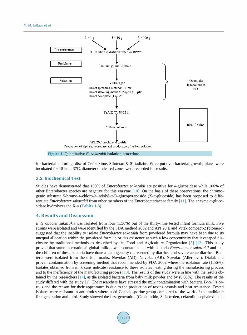
3.3. The Identification These presumptive colonies are identified biochemically [7]. This approach provides only a generic test for En-terobacteriaceae and lacks comparison of the effectiveness of antibiotics for the first and third generation ce-phalosporins within the group against bacteria the necessary capability to specifically identify E. sakazakii. Re-cently, a number of eclectic have become available to assist identification. One of these is the API 20 E bio-chemical test and vitek confirmatory system. Sakazakii were time and enriched for 6 h at 42˚C. summarizes the findings of these experiments. This method dependably revealed between 1 and 5 CFU E. sakazakii in 100 g with higher inocula producing a higher recovery of E. sakazakii. Tests were conducted according to the instruc-tions and approach Food and Drug Administration [7]. The limit of detection was determined to be 10 CFU/ml (equivalent to 2.5 × 10³ CFU in (250 ml)).Biochemical profiles are frequently used following primary isolation (Figure 1), but contradictions in identification may occur in different biochemical kits for the same strain [8].
3.4. Antibiotic Susceptibility Test Antibiotic was done for each isolate by standard Kirby Bauer method [9]. Muller Hinton agar plates were used
M. M. Jaffaar et al.
1172
Figure 1. Quantitative E. sakazakii isolation procedure.
for bacterial culturing, disc of Cefotaxime, Sifutetan & Siftadizim. Were put over bacterial growth, plates were incubated for 18 hr at 37C, diameter of cleared zones were recorded for results.
3.5. Biochemical Test Studies have demonstrated that 100% of Enterobacter sakazakii are positive for α-glucosidase while 100% of other Enterobacter species are negative for this enzyme [10]. On the basis of these observations, the chromo-genic substrate 5-bromo-4-chloro-3-indolyl-α-D-glucopyranoside (X-α-glucoside) has been proposed to diffe-rentiate Enterobacter sakazakii from other members of the Enterobacteriaceae family [11]. The enzyme α-gluco- sidase hydrolyzes the X-α (Tables 1-3).
4. Results and Discussion Enterobacter sakazakii was isolated from four (1.56%) out of the thirty-nine tested infant formula milk. Five strains were isolated and were identified by the FDA method 2002 and API 20 E and Vitek compact-2 (biomero) suggested that the inability to isolate Enterobacter sakazakii from powdered formula may have been due to its unequal allocation within the powdered formula or “its existence at such a low concentricity that it escaped dis-closure by traditional methods as described by the Food and Agriculture Organization [5] [12]. This study proved that some international global milk powder contaminated with bacteria Enterobacter sakazakii and that the children of these bacteria have done a pathogencity represented by diarrhea and severe acute diarrhea. Bac-teria were isolated from these four marks: Novolac (AD), Novolac (AR), Novolac (Allernova), Dialak and proven contamination by screening method that recommended by FDA 2002 where the isolation rate (1.56%). Isolates obtained from milk cans indicate resistance to these isolates heating during the manufacturing process and to the inefficiency of the manufacturing process [13]. The results of this study were in line with the results ob-tained by the researchers [14], as the isolated bacteria from baby milk powder and by (6.80%). The results of the study differed with the study [1]. The researchers have stressed the milk contamination with bacteria Bacillus ce-reus and the reason for their appearance is due to the production of toxins casuals and heat resistance. Tested isolates were resistant to antibiotics where used Cephalosporine group compared to the work of the antibiotic first generation and third. Study showed the first generation (Cephalothin, Safaberden, cefazolin, cephalexin and

M. M. Jaffaar et al.
1173
Table 1. Biochemical test of Enterobacter sakazakii and different bacteria.
Citrate Positive Positive Positive
Glucose K/A Positive A/A Positive A/A Positive
Sucrose Positive Positive Positive
Lactose Positive Positive Positive
α-glucosidase Positive Negative Negative
Urease Positive Negative Negative
Table 2. The proportion of bacteria in milk samples.
Type of samples Total of samples Isolated Percentage
Dialak 1, 2 51 1 1.49
Novolac AD 51 1 1.49
Novolac AR 51 1 1.49
Novolac Allernova 51 1 1.49
Table 3. Isolated bacteria from different types of children dry milk.
Type of Bacteria Batch No. Production time Country of origin Duplicates Type of milk
E. sakazakii 24 - 25 15 - 19 19 - 32
07.2013 03.2013 10.2013
Vietnam 3 Dialak 1, 2
19 - 27 11 - 15 6 - 64
11.2013 08.2013 10.2013
USA 3 Celia 1, 2
15 - 20 22 - 30 12 - 45
04.2013 08.2013 12.2013
Switzerland 3 Primalac
8500 5500 2000
10.2013 07.2013 04.2013
Belgium 3 Biomil 1, 2
09 - 42 03 - 26 02 - 17
11.2013 07.2013 03.2013
Ireland 3 Nursoy
41306 41308 41900
03.2013 03.2013 11.2013
Poland 3 Mami
36420 64222 92391
03.2013 06.2013 09.2013
France 3 Nicalia 1, 2
02 - 20 06 - 44 02 - 57
03.2013 07.2013 09.3013
Ireland 3 Similac
E. sakazakii 20 - 57 16 - 32 05 - 46
02.2013 07.2013 10.2013
Germany 3 Novolaclernova
E. sakazakii 08 - 52 10 - 58 03 - 36
02.2013 07.2013 10.2013
Germany 3 Novolac AD
E. sakazakii 19 - 44 08 - 30 16 - 16
02.2013 03.2013 07.2013
Germany 3 Novolac AR
2A02 2A27 2A34
10.2012 03.2013 10.2013
Ireland 3 S-26 AR
2L14 2L22 2L28
07.2012 03.2013 11.2013
USA 3 S-26 LF
M. M. Jaffaar et al.
1174
Table 4. Sensitivity of test Enterobacter sakazakii for third generation measured in Millimeter (mm) zone of inhibition ac-cording to (NCCL).
Antibiotic E1 E2 E3 E4
Cefotaxime 25 25 25 25
Sifutetan 22 22 22 22
Siftadizim 18 18 18 18
cephradine) did not have any result zone of inhibition. The third generation of the same batch of antibiotic which shows sensitive results through inhibition processes such as (cefotaxime (25 mm), Sifutetan (22 mm), Siftadizim (18 mm)), where the diameters of inhibition zone for Cefotaxime, Sifutetan and Siftadizim. The use of minimum inhibitory concentration Cephalosporine batch shows cefotaxime, Sifutetan and Siftadizim inhibition zone. The major cause is due to the group operates stop the work of the gyrase enzyme is present in prokaryotes and some eukaryotes work by competitive suppression of energy transduction of DNA gyrase by binding to the ATPase active site located on the GyrB subunit. Quinolones bind to these enzymes and prevent them from decatenation replicating DNA. Quinolone-resistant bacteria repeatedly harbor convert topoisomerases that resist quinolone binding 2009 Eukaryotic Cell, 11(8), 1759-1769. Gore J., Bryant Z., Stone M.D., Nollmann M., Cozzarelli N.R. 2006. Used in the study [5], this was identical to the present study in the case reported cephalosporins, such as cefepime, in collection with a second agent. Two studies in [15] and [2], supported the use of cefotaxime for childhood bacterial meningitis. In each of these studies, Enterobacter sakazakii meningitis was identified in at least 1 child, and the infection was treated successfully. In another study, [16] described E. sakazakii infection in 4 adults that occurred in 1995 and 1996 at the University of Massachusetts Medical Center. Of the 4 adults, 2 presented with pneumonia and 2 with bacteremia. The isolates were uniformly resistant to ampicillin, cefazolin, and extended-spectrum penicillins and were not uniformly susceptible to third-generation cephalosporins or to quinolones. This study proved that the four isolates were sensitive to the third generation cephalosporin group 9, Table 4.
References [1] Forsythe, S. and Iversen, C. (2008) Isolation of Enterobacter sakazakii and Other Enterobacteriaceae from Powdered
Infant Formula Milk and Related Products. Food Microbiology, 21, 771-773. [2] Lecour, H., Seara, A., Cordeiro, J. and Miranda, M. (1989) Treatment of Childhood Bacterial Meningitis. Infection, 17,
343-346. http://dx.doi.org/10.1007/BF01650726 [3] Van Acker, J., DeSmet, F., Muyldermans, G., Bougatef, A., Naessens, A. and Lauwers, S. (2001) Outbreak of Necro-
tizing Enterocolitis Associated with Enterobacter sakazakii in Powdered Milk Formula. Journal of Clinical Microbi-ology, 39, 293-297. http://dx.doi.org/10.1128/JCM.39.1.293-297.2001
[4] Kandhai, M., Reij, W., Gorris, L. and Guillaume, O. (2009) Occurrence of Enterobacter sakazakii in Food Production Environments and Households. Lancet, 363-369.
[5] Muytjens, H.L. and van der Ros-van de Repe, J. (1986) Comparative in Vitro Susceptibilities of Eight Enterobacter Species, with Special Reference to Enterobacter sakazakii. Antimicrobial Agents and Chemotherapy, 29, 367-370. http://dx.doi.org/10.1128/AAC.29.2.367
[6] Hoekstra, R.M., Kuehnert, M. and McDonald, L.C. (2007) Invasive Enterobacter sakazakii Disease in Infants. Nation-al Center for Infections Disease, 5, 885-895.
[7] FDA (2002) Isoilation and Enumeration of Enterobacter sakazakii from Dehydrated Infant Formula. [8] Iversen, C., Druggan, P. and Forsythe, S.J. (2004) A Selective Differential Medium for Enterobacter sakazakii. Inter-
national Journal of Food Microbiology, 96, 133-139. http://dx.doi.org/10.1016/j.ijfoodmicro.2004.01.024 [9] (2002) Performance Standards for Antimicrobial Disk Susceptibility Testing. NCCLSS, Wayne. [10] Muytjens, H. (1985) Enterobacter sakazakii: Identification and Clinical Significance. Antonie van Leeuwenhoek, 51,
618. http://dx.doi.org/10.1007/BF00404602 [11] Iversen, C., Druggan, P. and Forsythe, S. (2004) A Selective Medium for Enterobacter sakazakii, a Preliminary Study.
International Journal of Food Microbiology, 96, 133-139. http://dx.doi.org/10.1016/j.ijfoodmicro.2004.01.024 [12] FAO (1994) Codex Alimenterius Code of Hygienic Practice for Foods for Infants and Children. CAC/RCP 21-1979.
M. M. Jaffaar et al.
1175
Food and Agriculture Organization of the United Nations, Rome. [13] Pagotto, F. (2009) Pathogenesis of Cronobacter Enterotoxin Production, Adherence and Invasion of the Blood-Brain
Barrier, Burenu of Microbiol Hazards, Ireland. [14] Joshua, B., Jeffery, L., Kornacki, B. and Larry, R. (2005) Enterobacter sakazakii: Acoliform of Increased Concern to
Infant Heath. Center for Food Safety and Technology, Georgia. [15] Naqvi, S.H., Maxwell, M.A. and Dunkle, L.M. (1985) Cefotaxime Therapy of Neonatal Gram-Negative Bacillary Me-
ningitis. The Pediatric Infectious Disease Journal, 4, 499-502. http://dx.doi.org/10.1097/00006454-198509000-00012 [16] Lai, K.K. (2001) Enterobacter sakazakii Infections among Neonates, Infants, Children, and Adults. Case Reports and a
Review of the Literature. Medicine, 80, 113-122. http://dx.doi.org/10.1097/00005792-200103000-00004