detection of melanocytes in skin histopathological images
TRANSCRIPT
Pattern Recognition 46 (2013) 509–518
Contents lists available at SciVerse ScienceDirect
Pattern Recognition
0031-32
http://d
n Corr
E-m
Naushe
Naresh.
journal homepage: www.elsevier.com/locate/pr
Detection of melanocytes in skin histopathological images usingradial line scanning
Cheng Lu a,n, Muhammad Mahmood b, Naresh Jha c, Mrinal Mandal a
a Department of Electrical and Computer Engineering, University of Alberta, Edmonton, Alberta, Canada T6G 2V4b Division of Anatomical Pathology, Walter Mackenzie Health Sciences Centre, Edmonton, Alberta, Canada T6G 2B7c Department of Oncology, University of Alberta, Cross Cancer Institute, Edmonton, Alberta, Canada T6G 1Z2
a r t i c l e i n f o
Article history:
Received 11 November 2011
Received in revised form
29 May 2012
Accepted 29 July 2012Available online 8 August 2012
Keywords:
Histopathological image analysis
Object detection
Image analysis
Melanocytes
03/$ - see front matter & 2012 Elsevier Ltd. A
x.doi.org/10.1016/j.patcog.2012.07.020
esponding author. Tel.: þ1 780 492 0294; fax
ail addresses: [email protected] (C. Lu),
[email protected] (N. Jha), mmand
a b s t r a c t
In the diagnosis of skin melanoma by analyzing histopathological images, the detection of the
melanocytes in the epidermis area is an important step. However, the detection of the melanocytes
from the epidermis area is difficult because other keratinocytes that are very similar to the melanocytes
are also present. This paper proposes a novel computer-aided technique for detection of the
melanocytes in the epidermis area of skin histopathological images. An adaptive threshold technique
is first applied to segment all the keratinocytes in the image. In order to distinguish the melanocytes
from other keratinocytes, a novel technique based on radial line scanning is proposed to estimate the
halo region of the melanocytes. Based on the estimated halo region of all the nuclei, an area ratio of
estimated halo region and the nuclei is used to detect the melanocytes from all the keratinocytes.
Experimental results on 40 different histopathological images of skin tissue containing 341 melano-
cytes show that the proposed technique provides a superior performance.
& 2012 Elsevier Ltd. All rights reserved.
1. Introduction
Skin cancer is the most frequent and malignant types of cancer[14] and melanoma is the most aggressive type of skin cancer.According to a recent article, approximately 70,000 people arediagnosed with melanoma skin cancer, and about 9000 die from itin the United States every year [22]. The early detection ofmalignant melanoma will help to lower the mortality from thiscancer. Approaches to melanoma diagnosis have dynamicallyevolved during the last 25 years [18]. Although there are manynew emerging techniques, e.g., confocal microscopy [16], whichcould provide satisfactory performance, pathological examinationremains the gold standard for diagnosis as the histopathologyslides provide a cellular level view of the disease [10].
Traditionally, the histopathology slides are examined under amicroscope by pathologists. The diagnosis decisions are thenmade based on their personal experience. However, this judge-ment is subjective and often leads to intra-observer and inter-observer variability [7,19,8,1,4]. For example, it has been reportedthat in the diagnosing of follicular variant of papillary carcinoma(FVPC), the inter-observer agreement on benign and malignantdiagnoses is only 27% from 6 experts on 15 cases, and the intra-
ll rights reserved.
: þ 1 780 492 1811.
Mahmood),
[email protected] (M. Mandal).
observer agreement range from 17% to 100% [4]. Ruijter et al. [19]stated that at least 17% of all the grading errors result from themisinterpretation of the pathologists. To address this problem,automated computational tools are needed which can providereliable and reproducible objective results.
In melanoma diagnosis, the segmentation and detection of themelanocytes in the epidermis area is an important step before thediagnosis is made. If the melanocytes can be found correctly,architectural and cellular features (e.g. size, distribution, location)can then be used to grade or determine the malignancy of themelanotic skin tissue.
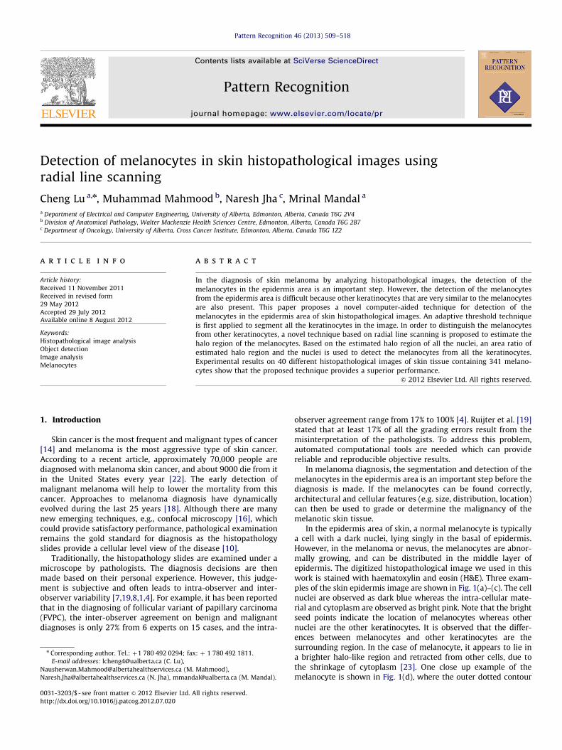
In the epidermis area of skin, a normal melanocyte is typicallya cell with a dark nuclei, lying singly in the basal of epidermis.However, in the melanoma or nevus, the melanocytes are abnor-mally growing, and can be distributed in the middle layer ofepidermis. The digitized histopathological image we used in thiswork is stained with haematoxylin and eosin (H&E). Three exam-ples of the skin epidermis image are shown in Fig. 1(a)–(c). The cellnuclei are observed as dark blue whereas the intra-cellular mate-rial and cytoplasm are observed as bright pink. Note that the brightseed points indicate the location of melanocytes whereas othernuclei are the other keratinocytes. It is observed that the differ-ences between melanocytes and other keratinocytes are thesurrounding region. In the case of melanocyte, it appears to lie ina brighter halo-like region and retracted from other cells, due tothe shrinkage of cytoplasm [23]. One close up example of themelanocyte is shown in Fig. 1(d), where the outer dotted contour

Fig. 1. Melanocytes in epidermis area from different skin tissues. Inter- and intra-image variations are observed in terms of the color. These images are sampled from the
skin digitized slide under 30�magnification. In (a)–(c), the bright seed points indicate the location of melanocytes whereas other nuclei are keratinocytes. (d) A close up
image of a melanocyte. (For interpretation of the references to color in this figure caption, the reader is referred to the web version of this article.)
C. Lu et al. / Pattern Recognition 46 (2013) 509–518510
represents the halo region and the inner solid contour representsthe nuclei. In contrast, the other keratinocytes are closely contactwith the cytoplasm and it has no or little brighter area. Thebrighter halo-like region of the melanocyte is an important patternfor differentiation of the melanocytes and other keratinocytes. Inthis work, we refer this pattern as ‘‘MPattern’’ and the bright regionaround the melanocytes as halo regions.
Several works have been conducted on the segmentation ordetection of various biological components in the histopathologicalimage using image processing techniques such as thresholding[6,17,12] and watershed [3]. These techniques typically fail whenconsiderable intensity variations are present in the images. Byincorporating the image color, texture and shape information, Naiket al. [15] proposed to segment the nuclei using the Bayesianclassifier. Sertel et al. [20] computed the probability map of karyor-rhexis cells based on the estimated likelihood function, and the cellnuclei are then segmented using thresholding. Although thesetechniques have been reported to provide good performance, theperformance is sensitive to the training samples.
In the melanocytes detection problem, the main difficulty ishow to differentiate the melanocytes and other keratinocytes inthe skin epidermis area. Similar problem has been addressed byBasavanhally et al. [2] in breast cancer diagnosis where thelymphocyte nuclei are differentiated from the cancer cell nucleiin H&E stained histopathological images. In their work, the basicassumption to differentiate two kinds of cells is based on thedomain knowledge regarding the nuclei size, intensity of thenuclei and spatial proximity. However, in skin histopathologicalimages, the size of melanocytes are very similar to that of otherkeratinocytes. In addition, the intensity value of the melanocytesand other keratinocytes are very close to each other. Therefore,the domain knowledge used in breast cancer [2] does not workwell in the case of melanocytes detection.
There is another closely related work in the literature wherethe keratinocytes nuclei are segmented in the skin epidermisarea [9]. In this work, a threshold is decided based on theassumption that cell nuclei covers approximately the darkest20% pixels in the image. The pixels whose values are less thanthe threshold are labeled as nuclei regions. Morphological opera-tions are then used to do the refinement. However, this globalthreshold based technique only works under the assumption thatthere are no intensity variations in the image and usuallygenerate the under-segmentation results (many of the nucleiare grouped together). Also, there is no attempt to differentiatethe melanocytes and other keratinocytes.
Template matching technique is a popular technique in com-puter vision for pattern detection. Naik et al. [15] have used fourbinary elliptical templates with different major and minor axesto detect the nuclei in breast cancer histopathological images.It is observed in Fig. 1 that the melanocytes typically have lowintensity value while its spatial surrounding space presents abrighter intensity value. It may be possible to detect the melano-cytes using a template matching technique with templates thathave similar appearance of the melanocyte. However, severaldifficulties need to be addressed. First, the size of the template ishard to decide due to the size variations of the melanocytes evenunder the same magnification level. In the case of cancer skin, themelanocytes are abnormal and larger than that in the case ofnormal skin or nevus skin. Second, the intensity level of thetemplate is hard to determine. Therefore, it is difficult to decide a‘good’ template to match the melanocyte patterns.
In this paper, we propose a novel technique to detect themelanocytes in the skin epidermis area. To our best knowledge,this is the first automated technique for the detection of themelanocytes in histopathological image of skin tissue. Thistechnique operates on reliable quantitative measures and provides

C. Lu et al. / Pattern Recognition 46 (2013) 509–518 511
objective and reproducible information complementary to that of apathologist. Such quantitative analysis of melanocytes is importantfor clinical applications, as well as for research purpose.
The organization of this paper is as follows. The proposedtechnique is described in Section 2, followed by the performanceevaluations in Section 3. The conclusion is presented inSection 4.
2. The proposed technique
The schematic of the proposed technique for melanocytesdetection is shown in Fig. 2. It is observed that there are threemodules. At first, the nuclei in the image are segmented using anadaptive thresholding technique which incorporates the domainknowledge. In the second module, the halo region is estimated foreach pre-segmented nuclei region by using the radial line scanning(RLS) algorithm. Finally, the melanocytes are detected based on theestimated halo region and the pre-segmented nuclei region. Detailsof these three modules are presented in the following.
2.1. Segmentation of the nuclei
Before performing the melanocytes detection, we first segmentall the nuclei in the image. Considering the intensity variations, anadaptive threshold technique is applied to segment the nucleiregions [13]. In order to reduce the influence from undesirablevariations in the image and to make the nuclei region homo-genous, a hybrid gray-scale morphological reconstruction methodis first applied. The hybrid gray-scale morphological reconstruc-tion method mainly consists of the Opening-by-Reconstruction andClosing-by-Reconstruction [5]. Next, a local region adaptive
Fig. 2. The schematic of the proposed
Fig. 3. Illustration of the RLS algorithm. (a) The original image with three nuclei regi
regions. (c) The radial lines for each nuclei region (nuclei regions are shown with thi
regions without resolving the overlap. (f) The overlap resolved results of (e). The valu
as well.
threshold selection method is used to achieve the segmentation.In this method, the local threshold is determined by minimizing apre-defined cost function which incorporates two domain specificknowledge: the size and the shape of the nuclei. Fig. 3(a) showsan original image containing three nuclei, and the melanocyte islabel with letter ‘M’. In Fig. 3(b), the segmented nuclei regions areshown as the white regions in a binary image.
2.2. Estimation of the halo regions using RLS
After the segmentation of nuclei, we are now ready to detectthe melanocytes from the pre-segmented nuclei. In order tocapture the ‘Mpattern’, we propose the RLS algorithm, whichmakes use of the radial lines originating from the centroid of anuclei. The RLS algorithm consists of four steps. The first step is toinitialize the radial center and radial lines for each nuclei regions.These radial lines will be used for measuring the information fromthe image in order to estimate the halo region. The second stepgenerates the gradient map of the image, which provides the edgeinformation for the radial lines. In the third step, the halo regionof each nuclei is estimated based on the gradient informationmeasured by the radial lines. In the final step, the overlappingregion problem is resolved. These four steps are described in thefollowing.
2.2.1. Initialization of radial center and radial lines
In this step, we first calculate the centroid of each pre-segmented nuclei. Let the centroid coordinate be denoted byðCx,CyÞ. Using the centroid as a radial center, a set of radial lines
melanocyte detection technique.
ons where the melanocyte is indicated with letter ‘M’. (b) The segmented nuclei
ck contours). (d) The suppressed smoothed gradient map. (e) The estimated halo
e of the parameter RHN discussed in Section 2.3 is shown inside the nuclei region

C. Lu et al. / Pattern Recognition 46 (2013) 509–518512
with different radial angles are calculated as follows:
yi ¼2pkði�1Þ, i¼ 1 . . . k, ð1Þ
where k indicates the number of radial lines. An illustration of theradial center ðCx,CyÞ and its radial lines yi, i¼ 1 . . . k is shown inFig. 4(a). Note that the nuclei contour is shown as the thick solidcontour whereas the dotted circle indicates the maximum rangeof the radial line (denoted as Rmax). The solid point in the centerindicates the radial center ðCx,CyÞ and a set of 16 radial lines areshown as the thick solid lines. In this paper, the radial lines aredenoted by its radial angle y. For example, the first and secondradial lines y1 and y2 are shown in Fig. 4(a) and the radial anglesfor these two radial lines are 0 and p=8, respectively. Denote thenuclei boundary point at given radial angle yi as bðyiÞ. In Fig. 4(a),bðy1Þ and bðy2Þ indicate the nuclei boundary point on given radiallines y1 and y2, respectively.
Note that Rmax specifies the maximum range of the radial lineand it is identical for all the radial lines, i.e., RmaxðyiÞ ¼ Rmax. InFig. 4(a), the maximum range Rmax of a radial line is labeled.
A radial line consists of Rmax points and is index from 1 to Rmax. Wedefine a valid scanning range for each radial line in order to reducethe computational time. We define RminðyiÞ as the distance from theradial center to the nuclei boundary point bðyiÞ. Note that theminimum valid scanning range maybe different for each radial line.Now, let m be the index of the point on the radial line, and the validscanning range of the index should range from RminðyiÞ to Rmax, i.e.,RminðyiÞrmrRmax. An illustration of the above mentioned notationis shown in Fig. 4(b). Note that one of the radial lines is shown as athick solid line. The hollow point on the radial line represents the mthvalid scanning point in the range of [RminðyiÞ,Rmax]. The shadow regionshown in Fig. 4(b) indicates the valid scanning range. The radialcenters and radial lines for Fig. 3(a) is shown in Fig. 3(c). These radiallines will be used later to measure the information from the image.
2.2.2. Gradient map generation
In the proposed technique, we estimate the halo region foreach nuclei by analyzing the image features on the pre-initializedradial lines. The gradient is a very useful feature in the image thatcan help to estimate the halo region, and hence in this step, wegenerate the gradient map for the latter analysis. Denote theimage as I and its 2-D coordinates as ðx,yÞ. In order to reduce thenoise, a smooth image Is is obtained by convolving the image witha Gaussian kernel Gsðx,yÞ ¼ s�1=2e�ðx
2þy2Þ=4s. In other words,Isðx,yÞ ¼ Gsðx,yÞnIðx,yÞ. The gradient magnitude map Gm and thegradient direction map Gd are then generated using the followingequations:
Gmðx,yÞ ¼ JrIsðx,yÞJ¼
ffiffiffiffiffiffiffiffiffiffiffiffiffiffiffiffiffiffiffiffiffiffiffiffiffiffiffiffiffiffiffiffiffiffiffiffiffiffiffiffiffiffiffiffiffiffiffiffiffiffiffiffiffiffidIsðx,yÞ
dx
� �2
þdIsðx,yÞ
dy
� �2s
, ð2Þ
Fig. 4. Illustration of the notation o
Gdðx,yÞ ¼ arctandIsðx,yÞ
dy
�dIsðx,yÞ
dx
� �: ð3Þ
To suppress the weak gradients, the following equations areused:
G0mðx,yÞ ¼Gmðx,yÞ if Gmðx,yÞZmeanðGmðx,yÞÞ,
0 otherwise,
(ð4Þ
G0dðx,yÞ ¼Gdðx,yÞ if Gmðx,yÞZmeanðGmðx,yÞÞ,
0 otherwise:
(ð5Þ
Eqs. (4) and (5) suppress the gradients whose magnitude areless than the mean gradient magnitude. The gradient map G0ðx,yÞis shown in Fig. 3(d) where the gradient (shown as arrows) issuper imposed on the image.
2.2.3. Estimation of the halo regions
The melanocytes can be distinguished from other keratino-cytes based on their bright halo-like regions. In this step, we usethe radial lines and the pre-computed gradient map to detect theboundary points for the halo regions estimation. On each radialline with radial angle yi, there are two kinds of boundary point:the inner boundary point and outer boundary point. We assumethat the inner boundary points are located at the boundary of thepre-segmented nuclei. Therefore, in this section, we aim to detectthe outer boundary point, denoted as BPðyiÞ, for the halo region.All the outer boundary points fBPðyiÞ, i¼ 1, . . . ,kg will then formthe outer halo contour which can constrain a halo region. InFig. 1(d), the dotted contour corresponds to the outer boundarycontour that consists of outer boundary points whereas the solidcontour corresponds to the inner boundary contour that consists ofinner boundary points. Ideally, for the melanocytes, the halo-likeregions surrounding the nuclei are segmented by the detected outerboundary points; whereas for the other keratinocytes, the detectedouter boundary points only encompass the nuclei themselves, i.e.,the outer boundary points are equal to the inner boundary pointssince there is no halo region.
Before introducing how to estimate the boundary points of thehalo region, let us first analyze the gradient information aroundthe halo region of a typical melanocyte (see Fig. 5). It is observedthat the gradient map (shown as small arrows) is superimposedonto the image where the gradient magnitude is represented bythe length of the arrow and the gradient direction is representedby the direction of the arrow. Because the intensity of the haloregion is higher than the neighboring regions, the gradientdirections point towards the halo region both from the nucleiand from the outer region. The dotted contour indicates theground truth halo region’s outer contour which consists of severalouter boundary points. It is noted that the outer boundary pointsshould have gradient direction that opposite to the radial line. For
f radial center and radial line.

Fig. 5. A close up example of a melanocyte with gradient map. The gradient map
(shown as small arrows) is superimposed onto the image. The dotted contour
indicates the ground truth halo region’s outer contour. An enlarged version of the
boundary is shown in the small image on the right.
Fig. 6. Illustration of the outer boundary point detection on a given radial line.
C. Lu et al. / Pattern Recognition 46 (2013) 509–518 513
a given radial line with radial angle yi, we denote the gradientdirection of a outer boundary point for the halo region as BPG0
dðyiÞ.
Based on the characteristic of the gradient direction, in this paper,we assume that the angle difference between BPG0
dðyiÞ and the
radial angle of a radial line should satisfy the following condition:
p2o9yi�BPG0
dðyiÞ9o
3p2: ð6Þ
In an ideal case, the gradient direction and the radial angle arestrictly opposite, i.e., 9yi�BPG0
dðyiÞ9¼ p. In order to allow the
variations, we assume that BPG0dðyiÞ to be yi7p=2, or satisfies
Eq. (6). In Fig. 5, a radial line with radial angle y1 ¼ 0 is shown. Onthis radial line, the candidate outer boundary points are labeled.
Let G0mðyi,mÞ and G0dðyi,mÞ represent, respectively, the gradientmagnitude and direction on the mth point of the radial line yi.Based on the above analysis, an outer boundary point is expectedto maximize the following cost function:
Cðyi,mÞ ¼ cos½aðyi,mÞ�þG0mðyi,mÞ,RminðyiÞrmrRmax, ð7Þ
where aðyi,mÞ is the angle difference between gradient directionand the direction of the radial line, which is defined as follows:
aðyi,mÞ ¼ 9yi�G0dðyi,mÞ9: ð8Þ
For each radial line, the outer boundary point for the potentialhalo region is calculated using the following equation:
BPðyiÞ ¼argmmaxfCðyi,mÞg if (m,
p2oaðyi,mÞo
3p2
,
RminðyiÞ otherwise:
8<: ð9Þ
Eq. (9) examining the gradient information (the direction andthe magnitude) of each point on the radial line. If there exist oneor more points satisfying p=2o9aðyi,mÞ9o3p=2, the one withmaximum cost function value Cðyi,mÞ is estimated as the outerboundary point for the current radial line. If there is no point (onthe radial line) satisfying p=2o9aðyi,mÞ9o3p=2, we choose thepoint corresponding to the minimum valid range as the estimatedouter boundary point.
The idea of the outer boundary point estimation using Eq. (9) isfurther explained in Fig. 6. In Fig. 6(a), six blocks are shown whichrepresent six pixels/points that comprise the valid range of a radialline yi (in this case yi ¼ p=2). We denote each block, from bottom tothe top, as the first to sixth pixels on the radial line. On each pixel,we have corresponding gradient information which are representedby the arrows. Note that the gradient direction is indicated by thedirection of the arrow whereas the gradient magnitude is indicatedby the length of the arrow. It is observed that the third, fifth, andsixth pixel satisfies the condition p=2o9aðyi,mÞ9o3p=2. Thesepixels are the candidate outer boundary points on the current radial
line. In order to determine the final outer boundary point, the costfunction value Cðyi,mÞ is calculated (as shown in Eq. (7)). The pixelcorresponding to the maximum cost function value will be chosenas the outer boundary point. In Fig. 6(a), the outer boundary pointBPðyiÞ is the fifth pixel. Fig. 6(b) shows another case, where there isno pixel whose gradient satisfy the condition p=2o9aðyi,mÞ9o3p=2 in Eq. (9). In this case, the outer boundary point BPðyiÞ is thefirst pixel, i.e., the pixel corresponding to the minimum valid rangeRminðyiÞ.
By using Eq. (9), we can estimate accurate halo regions for themelanocytes while reducing the influence of noise. After estimat-ing the outer boundary point on each radial line using Eq. (9), foreach nuclei region, the potential halo contour (HC) is estimated bythe boundary points as follows:
HC ¼ fðxBPðyiÞ,yBPðyiÞ
Þg, i¼ 1, . . . ,k, ð10Þ
xBPðyiÞ¼ CxþBPðyiÞ � cosðyiÞ, ð11Þ
yBPðyiÞ¼ CyþBPðyiÞ � sinðyiÞ: ð12Þ
The estimated halo contours (shown as thick contours) forFig. 3(a) is shown in Fig. 3(e) where the contours are super-imposed onto the original image. Note that the region within thehalo contours excluding the nuclei regions are the halo regionswe are looking for. Two more examples of the halo regionestimation are shown in Fig. 7(a) and (c), where the melanocytesare label with letter ‘M’ in the figure.
2.2.4. Resolving the overlapped regions
In the skin epidermis, the melanocytes and other kind ofkeratinocytes are usually clustered with each others. It is observedthat in Fig. 3(a), two other keratinocytes and one melanocyte (themiddle one) are very close to each other. Such proximity will lead tothe overlapping of the estimated halo regions (as shown in Fig. 3(e)).Such overlapping will result in erroneous detection in the latterprocessing. Note that the overlapping is due to the spatial closenessof the other keratinocytes and the halo region. The gradientinformation of the halo region is falsely captured by the neighboringkeratinocytes which are not melanocytes. Generally, the estimatedhalo regions of the melanocytes have smooth boundaries and largerarea compared to that of the other keratinocytes whereas theestimated halo regions of the other keratinocytes have non-smoothboundaries (see Figs. 3(e) and 7(a) and (c)). Given an estimated halocontour HC, denote A(HC) and S(HC) as the area inside HC andthe smoothness measure of the HC. The smoothness of the HC iscalculated as
SðHCÞ ¼1
k
Xk
i ¼ 1
dBPðyiÞ
dyi
��������: ð13Þ

Fig. 7. Two examples of halo-like regions estimation. (a) and (c) show the
estimated halo regions without resolving the overlap. The nuclei of melanocytes
are labeled with letter ‘M’. (b) and (d) are the overlap resolved results of (a) and
(c), respectively. The value of the parameter RHN discussed in Section 2.3 is shown
inside the nuclei region as well.
C. Lu et al. / Pattern Recognition 46 (2013) 509–518514
Therefore, the ratio of the boundary smoothness and the area (RSA),i.e., RSA¼ SðHCÞ=AðHCÞ, is used as a criterion to resolve the over-lapped regions. Specifically, for all the estimated halo regions, wefirst check if there are overlapped. Assume that two estimated haloregions are overlapped and denote these two estimated halo regionsas HCa and HCb, respectively. The overlapped region is denoted asHCa\b ¼HCa \ HCb. We resolve the overlap by using the equation asfollows:
HCa ¼HCa if
SðHCaÞ
AðHCaÞ4
SðHCbÞ
AðHCbÞ
HCa�HCa\b otherwise,
8><>: , ð14Þ
HCb ¼HCa�HCa\b if
SðHCaÞ
AðHCaÞ4
SðHCbÞ
AðHCbÞ
HCb otherwise:
8><>: , ð15Þ
Intuitively, for the two halo regions that are overlapped, the onewith a higher RSA value will take over the overlapped regionwhereas the other will lose the overlapped region. The result ofthe overlapped region resolving for Figs. 3(e) and 7(a) and (c) areshown in Figs. 3(f) and 7(b) and (d), respectively. It is observed thatthe melanocytes correctly take over the overlapped regions whichreflect the effectiveness of the proposed method.
2.3. Detection of the melanocytes
As described in Section 1, the melanocytes generally haveassociate large halo regions whereas the other keratinocytes haveno such large halo regions. Some halo region estimation resultsobtained by the RLS algorithm can be observed in Figs. 3(f) and7(b) and (d). Based on this observation, we can now use the ratioof the estimated halo region area and the nuclei area, denote it asRHN, as a parameter to differential the melanocytes and other
keratinocytes. The ratio RHN is calculated as follows:
RHN ¼AHR
ANR, ð16Þ
where AHR and ANR are the areas of the halo and nuclei regions,respectively. For the melanocyte detection, we select the nucleiregions as the melanocytes if its RHN value is greater than apredefined threshold TRHN
. In Figs. 3(f) and 7(b) and (d), thecorresponding parameter RHN for each nuclei region is show insidethe nuclei region. It is observed that the true melanocytes regionshave larger RHN values compare to the other keratinocytes regions.
3. Performance evaluations
We have evaluated the proposed technique on 40 differentskin histopathology images of epidermis which contains 341melanocytes. These images are captured under 30�magnificationon Carl Zeiss MIRAX MIDI Scanning system.
3.1. Evaluation metrics
3.1.1. Evaluation metrics for melanocyte detection
In the melanocytes evaluation, the melanocytes manuallyidentified by an expert are treated as the ground truths positivesfor the performance evaluation. The other keratinocytes identifiedby a pathologist are treated as the ground truths negatives. Thenumber of positives and negatives are denoted as NP and NN,respectively. We also denote NDO as the total number of detectedobjects, NTP as the number of true-positives, NFP as the number offalse-positives, NTN as the number of true-negative, NFN as thenumber of false-negative. The true positive rate (TPR, also knownas recall rate) and false positive rate (FPR) are defined as follows:
TPR¼NTP
NTPþNFN� 100%, ð17Þ
FPR¼NFP
NFPþNTN� 100%: ð18Þ
The precision rate (PRE) is defined as follows:
PRE¼NTP
NDO� 100%: ð19Þ
3.1.2. Evaluation metrics for halo region estimation
For the halo region estimation evaluation, we randomly selecteda set of 50 melanocytes and 50 other keratinocytes as the test set.We then manually labeled the boundaries of the halo region of thesecells. These manually labeled boundaries are treated as the groundtruth and will be compared with the estimation of the halo regionsobtained by the proposed RLS technique.
We define GT as the ground truth halo region for each nuclei,EST as the estimated halo region obtained by the proposed RLStechnique. Two area-based evaluation metrics: sensitivity (ASEN),precision (APRE) are defined as follows:
ASEN ¼9GT \ EST9
9GT9� 100%, ð20Þ
APRE ¼9GT \ EST9
9EST9� 100%, ð21Þ
where 9 � 9 is the cardinality operator. We also use another bound-ary-based evaluation metric: mean absolute distance (MAD) whichis defined as follows:
MADðCGT ,CESTÞ ¼
1
M
XMi ¼ 1
½minJCGTi �CEST
j J�, ð22Þ

C. Lu et al. / Pattern Recognition 46 (2013) 509–518 515
where CGT and CEST are the contour/boundary of the ground truthregion and estimated region, respectively. CGT
i and CESTj represent the
ith pixel and the jth pixel on the contour of the ground truth regionand estimated region, respectively. M is the total number of pixelson the contour of the ground truth region.
3.2. Parameters selection
To obtain good performance, two parameters that will be usedin the proposed technique need to be selected appropriately.These parameters are Rmax (the maximum range of the radial line)and TRHT
(the predefined threshold for discrimination of themelanocytes and other nuclei). For the parameter Rmax, we chosea set of 20 manually labeled melanocytes and measure thedistances from the centroid to the boundary points in eachmelanocyte. Let the maximum distance be denoted by distmax,we then chose the Rmax¼2ndistmax. Based on the biologicalknowledge, the size of the nuclei in skin epidermis should fallwithin a desired range. The choice of the Rmax is to ensure that theradial line can reach all the melanocytes and the surrounding haloregions. For determining the parameter TRHT
, two images areselected and the nuclei and their halo regions within these imagesare manually labeled. In addition, the value of RHN is calculated foreach nucleus. We then perform the trail-and-error test, i.e., weuse a set of possible values for TRHT
(from the minimum value ofall the RHN to the maximum value of all the RHN in the test images)to calculate the PRE and chose the value that achieves the best
Table 1Performance evaluation of the halo region estimation.
Cell Type ASEN (%) APRE (%) MAD (pixels)
Melanocytes 84.69 93.80 1.51
Other keratinocytes 86.29 96.16 0.72
Fig. 8. Examples of the estimated halo regions compared to manually labeled halo reg
melanocyte examples and other keratinocytes examples, respectively. The dotted conto
whereas the solid contours are the manually labeled halo regions.
PRE as the parameter TRHT. The determined value of Rmax
(Rmax¼35) and TRHT(TRHT
¼0.8) are then used for the evaluation.
3.3. Evaluation on halo region estimation
Since we use the ratio of the estimated halo region area andthe nuclei area, RHN defined in Eq. (16), as a criterion for themelanocyte detection, the estimation of the halo region of thenuclei is important for the proposed technique. In this section, weevaluate the performance on the estimation of halo region.
The halo region estimation performance is shown in Table 1.The average value of all the three evaluation metrics for melano-cytes and other keratinocytes are shown in the second row andthe third row of Table 1, respectively. It is observed that theproposed RLS technique is able to estimate the halo region of thenuclei correctly. This is expected to provide a good performancein the melanocytes detection.
A few examples of the estimated halo regions for the melano-cytes (the first row) and other keratinocytes (the second row) areshown in Fig. 8. In each image, the interested nuclei are put inthe middle of the images and their halo regions are shown asthe contours. The dotted contours represent the estimated haloregions obtained by the proposed RLS technique whereas thesolid contours are the manually labeled halo regions. In the caseof melanocytes (Fig. 8(a)–(d)), the halo regions are much largerthan the nuclei regions. In the case of other keratinocytes(Fig. 8(e)–(h)), the estimated halo regions are very close to thenuclei boundary, which is desired since there is little or no haloregion for other keratinocytes. The proposed RLS technique is ableto estimate the halo region correctly, with minor distortionscompared to the manually labeled halo region (see Fig. 8(a)–(c),(e)-(h)). These minor distortions will not degrade the finalmelanocytes detection performance since we will use the regionratio criterion RHN (described in Eq. (16)) to differentiate themelanocytes and other keratinocytes. In Fig. 8(d), due to thestaining variation, a considerable amount of the halo regioncannot be recovered by the proposed RLS technique. This might
ions for melanocytes and other keratinocytes. The first and second rows show the
urs represent the estimated halo regions obtained by the proposed RLS technique

C. Lu et al. / Pattern Recognition 46 (2013) 509–518516
lead to a miss detection of this melanocyte since the region ratioRHN will have a small value.
3.4. Quantitative evaluation on melanocyte detection
Since this is the first technique to solve the melanocytessegmentation problem, there is no other equivalent technique inthe literature that can be compared with the proposed technique.Therefore, we compare the proposed technique with a possiblealternative technique, known as template matching (TM). Notethat the TM technique is a widely used technique for patterndetection in medical imaging [15,21]. A set of 30 templates aredesigned to capture the ‘‘MPattern’’ and selected 10 templates areshown in Fig. 9. The templates are designed according to 30manually selected melanocytes. Note that we design 30 templateswith different sizes in terms of the template size (15�15,20�20, and 25�25 pixels), center heart radii (8, 9, 10, 11, and12 pixels), and the ring thickness (2 and 3 pixels), in order tocreate the variations. The intensity for the heart of the templateand the ring of the template are assigned according to the meanvalue on the corresponding regions in the 30 samples of manuallyselected melanocytes. In the TM implementation, each templateis first applied on the image by using the normalized crosscorrelation (NCC) [11]. Denote the NCC output of the ith templateas Oi, and the 2D coordinate in the image as (u, v). The potentialpixel (u, v) belong to the melanocyte region Jiðu,vÞ obtained fromthe ith template is as follows:
Jiðu,vÞ ¼Jiðu,vÞ if Oiðu,vÞZ0:80,
0 otherwise:
(ð23Þ
Fig. 9. Ten templates with size 20�20 used in the template matching techniques. No
magnified for demonstration purpose.
Fig. 10. The ROC and PRC curves of the proposed technique a
The accumulated map Oacc for all 30 templates are calculated asfollows:
Oacc ¼X30
i ¼ 1
Xðu,vÞAO
Jiðu,vÞ, ð24Þ
where O represents the image domain. The final result is a binaryimage which is determined using a threshold TTM on Oacc.
The performance of the proposed technique is evaluated usingthe receiver operating characteristic (ROC) and the precision-recall curve (PRC). The ROC and PRC curves of the proposedtechnique and the TM technique are shown in Fig. 10. The ROCand PRC curves of the proposed technique are generated byvarying the parameter TRHN
from to 0 to 2 with a step of 0.1.The ROC and PRC curves of the TM technique are generated byvarying the parameter TTM from to 0 to 1 with a step of 0.1. It isnoted that in the ROC curve, the curve that is close to the upperleft corner is better than that is far away from the upper leftcorner. It is observed from Fig. 10(a) that the proposed techniquecan provide the TPR around 90% with the FPR at 95%. As for theTM technique, the TM technique provide a poor performancebecause it is difficult to distinguish the melanocytes from otherkeratinocytes by using the templates. In the case of the PRC curve,the curve that is close to the upper right corner is better than thatis far away from the upper right corner. In Fig. 10(b), it shows thatthe proposed technique can provide 90% recall rate with around80% precision rate. As for the TM technique, it usually generatea large number of false detections (results in low precision rate)because the pattern of the other cytological components is verysimilar to the templates. It is observed from both ROC andPRC curves that the proposed technique provides a superior
te the variation of the center heart radii and the ring thickness. The templates are
nd the TM technique. (a) The ROC curve. (b) PRC curve.

Fig. 11. Three examples for qualitative evaluation. (a), (b), and (g) show three original color histopathological images. The ground truths melanocytes locations are
indicated by stars. (b), (e), and (h) show the result provided by the proposed technique. (c), (f), and (i) show the result provided by ML technique. Note that detected
regions are presented as the thick contours. The solid arrows in (f) indicate the miss detection by the ML technique whereas the hollow arrow in (e) indicates the false
detection by the proposed technique. (For interpretation of the references to color in this figure caption, the reader is referred to the web version of this article.)
C. Lu et al. / Pattern Recognition 46 (2013) 509–518 517
performance compare to the TM technique. Note that the tem-plate matching technique is a general technique for the objectdetection problem, and it could not be expected to provide abetter performance compared to a specifically designed techni-que. Since there is no other available technique for the task athand, i.e., melanocytes detection in the skin epidermis, we usedthe TM technique with specifically design templates as the base-line technique for the comparison.
3.5. Qualitative evaluation on melanocyte detection
For the qualitative evaluation, three close-up examples areshown in Fig. 11. These images are captured from different skintissues, and the parameter TRHN
¼ 0:8 and TTM ¼ 0:2 were used forthe proposed technique and the TM technique, respectively. Notethat Fig. 11(a), (b), and (g) shows three original color histopatho-logical images, where the ground truth melanocytes locations areindicated by stars. The last two columns show the resultsobtained using the proposed technique and the TM technique,respectively. The detected regions are presented as the thickcontours. It is observed that the proposed technique (shown inFig. 11(b), (e), and (h)) can detect all the melanocytes and has onefalse positive. In Fig. 11(e), there is a false detection which indicatesby a hollow arrow. This false detection is mainly because thecandidate nuclei region is very close to a typical melanocyte whereits surrounding cytoplasm are retracted.
It is observed that in the results provided by the TM techniquehave many false detections (shown in Fig. 11(c), (f), and (i)) sincethere are many cytological components similar to that of thetemplates. In addition, because it is very difficult to model a goodtemplate set that can capture the size and shape variations of thenatural melanocytes, it misses some of the ground truths mela-nocytes (see shown in Fig. 11(f), the solid arrows indicate the missdetection). Overall, the proposed technique is able to provide agood performance in the test images set.
4. Conclusion
This paper presents a simple but effective computer-aidedtechnique for the detection of the melanocyte in the skinhistopathological image. The candidate nuclei regions are firstsegmented through an adaptive threshold method. A radial linesscanning algorithm is then proposed to estimate the halo regionsof melanocytes. Finally, the melanocytes are detected based onthe ratio of the estimated halo region area and the nuclei area.The evaluation using 40 histopathological images (with morethan 300 melanocytes) shows the efficiency of the proposedtechnique. In future, we plan to analyze the cytological andarchitectural features of the detected melanocytes in order tograde and diagnose the skin tissues.
References
[1] W. Allsbrook, K. Mangold, M. Johnson, R. Lane, C. Lane, J. Epstein, Inter-observer reproducibility of Gleason grading of prostatic carcinoma: generalpathologist, Human Pathology 32 (1) (2001) 81–88.
[2] A.N. Basavanhally, S. Ganesan, S. Agner, J.P. Monaco, M.D. Feldman,J.E. Tomaszewski, G. Bhanot, A. Madabhushi, Computerized image-baseddetection and grading of lymphocytic infiltration in HER2þ breast cancerhistopathology, IEEE Transactions on Biomedical Engineering 57 (3) (2010)642–653.
[3] L.E. Boucheron, Object- and Spatial-Level Quantitative Analysis of Multi-spectral Histopathology Images for Detection and Characterization of Cancer,Ph.D. Thesis, University of California, Santa Barbara, March 2008.
[4] T. Elsheikh, S. Asa, J. Chan, R. DeLellis, C. Heffess, V. LiVolsi, B. Wenig,Interobserver and intraobserver variation among experts in the diagnosis ofthyroid follicular lesions with borderline nuclear features of papillarycarcinoma, American Journal of Clinical Pathology 130 (5) (2008) 736–744.
[5] R. Gonzalez, R. Woods, Digital Image Processing, 2002.[6] M. Gurcan, T. Pan, H. Shimada, J. Saltz, Image analysis for neuroblastoma
classification: segmentation of cell nuclei, in: Proceedings of the 28th IEEEAnnual International Conference of EMBS, 2006, pp. 4844–4847.
[7] S. Ismail, A. Colclough, J. Dinnen, D. Eakins, D. Evans, E. Gradwell, J. O’Sullivan,J. Summerell, R. Newcombe, Observer variation in histopathological diagnosisand grading of cervical intraepithelial neoplasia, British Medical Journal 298(6675) (1989) 707.

C. Lu et al. / Pattern Recognition 46 (2013) 509–518518
[8] C. King, J. Long, Prostate biopsy grading errors: a sampling problem, Inter-national Journal of Cancer 90 (2000) 326–330.
[9] V. Korde, H. Bartels, J. Barton, J. Ranger-Moore, Automatic segmentation of
cell nuclei in bladder and skin tissue for karyometric analysis, Analytical andQuantitative Cytology and Histology 31 (2) (2009) 83.
[10] V. Kumar, A. Abbas, N. Fausto, et al., Robbins and Cotran Pathologic Basis ofDisease, Elsevier, Saunders, Philadelphia, 2005.
[11] J. Lewis, Fast normalized cross-correlation, in: Vision Interface, vol. 10,Citeseer, 1995, pp. 120–123.
[12] C. Lu, M. Mandal, Automated segmentation and analysis of the epidermisarea in skin histopathological images, in: Proceeding of the 31th IEEE AnnualInternational Conference of EMBS, 2012.
[13] C. Lu, M. Mahmood, N. Jha, M. Mandal, A robust automatic nuclei segmenta-tion technique for quantitative histopathological image analysis, Analytical
and Quantitative Cytology and Histology. In Press.[14] I. Maglogiannis, C. Doukas, Overview of advanced computer vision systems
for skin lesions characterization, IEEE Transactions on Information Technol-ogy in Biomedicine 13 (5) (2009) 721–733.
[15] S. Naik, S. Doyle, S. Agner, A. Madabhushi, M. Feldman, J. Tomaszewski,Automated gland and nuclei segmentation for grading of prostate and breastcancer histopathology, in: Proceedings of the Fifth IEEE International Sym-
posium on Biomedical Imaging: From Nano to Macro ISBI 2008, pp. 284–287.
[16] J. Pawley, Handbook of Biological Confocal Microscopy, Springer-Verlag,2006.
[17] S. Petushi, F.U. Garcia, M.M. Haber, C. Katsinis, A. Tozeren, Large-scalecomputations on histology images reveal grade-differentiating parametersfor breast cancer, BMC Medical Imaging 6 (2006) 14.
[18] D. Rigel, J. Russak, R. Friedman, The evolution of melanoma diagnosis: 25years beyond the ABCDs, CA: A Cancer Journal for Clinicians 60 (5) (2010)301–316.
[19] E. Ruijter, G. van Leenders, G. Miller, F. Debruyne, C. van de Kaa, Errors inhistological grading by prostatic needle biopsy specimens: frequency andpredisposing factors, The Journal of Pathology 192 (2) (2000) 229–233.
[20] O. Sertel, U. Catalyurek, H. Shimada, M. Guican, Computer-aided prognosis ofneuroblastoma: detection of mitosis and karyorrhexis cells in digitizedhistological images, in: Proceeding of the 31th IEEE Annual InternationalConference of EMBS, IEEE, 2009, pp. 1433–1436.
[21] I. Sintorn, M. Homman-Loudiyi, C. Soderberg-Naucler, G. Borgefors, A refinedcircular template matching method for classification of human cytomegalo-virus capsids in TEM images, Computer Methods and Programs in Biomedi-cine 76 (2) (2004) 95–102.
[22] A.C. Society, What Are the Key Statistics About Melanoma? Technical Report,American Cancer Society, 2008.
[23] D. Weedon, G. Strutton, Skin Pathology, vol. 430, Churchill Livingstone,New York, 2002.
Cheng Lu received his BSc and MSc degree in Computer Engineering from Information Engineering College, Northwest A&F University, Shaanxi, China, in 2006 and 2008.He is currently working toward his PhD degree in electrical engineering in University of Alberta, Edmonton, AB, Canada, from 2008 till now. In 2008, he joined theMultimedia Computing and Communication Lab, Department of Electrical Engineering, University of Alberta, as a PhD candidate, research assistant and the teachingassistant. Mr. Lu is the recipient of the Chinese Scholarship Council for his PhD studies. His research interest includes computer vision, medical image analysis, patternrecognition and super resolution imaging. He is an author or coauthor of several papers in leading international journals and conferences.
Muhammad Mahmood received his medical degree from Aga Khan University, Medical College, Karachi, Pakistan. He did his Anatomical Pathology/Clinical Pathologyresidency from Henry Ford Hospital, Detroit, Michigan, USA. Further, he did two fellowships; an Advanced Selective Pathology fellowship from Barnes-Jewish Hospital,Washington University Medical Center, Saint Louis, MO, USA, and a Dermatopathology fellowship from Ackerman Academy of Dermatopathology, New York, NY, USA. He isan American Board Certified Anatomical and Clinical Pathologist and American Board Certified Dermatopathologist. For last six years, he is a practising AnatomicalPathologist/Dermatopathologist in Canada. He is currently working at Department of Lab Medicine and Pathology, University of Alberta Hospital, Edmonton, Alberta,Canada. His interests include melanocytic cutaneous lesions, cutaneous tumors and inflammatory dermatoses. He is also interested in developing post-graduate medicalteaching for Pathologists and Dermatologists in training.
Naresh Jha graduated in 1979 with Bachelor of Medicine and Bachelor of Surgery from Delhi University in India. He participated residency training in Radiation Oncologyat the Cross Cancer Institute, Edmonton, Alberta, Canada. He received Fellow of the Royal College of Physicians of Canada (FRCPC) in Radiation Oncology in 1987. He is aSenior Radiation Oncologist at the Cross Cancer Institute since 1988. He specializes in head and neck, thyroid and skin cancers. He is also a pioneer of the submandibularsalivary gland transfer—a ground breaking technique to prevent radiation-induced xerostomia.
Mrinal Mandal is a Full Professor and Associate Chair in the Department of Electrical and Computer Engineering and is the Director of the Multimedia Computing andCommunications Laboratory at the University of Alberta, Edmonton, Canada. He has authored the book Multimedia Signals and Systems (Kluwer Academic), andcoauthored the book Continuous and Discrete Time Signals and Systems (Cambridge University Press). His current research interests include Multimedia, Image and VideoProcessing, Multimedia Communications, Medical Image Analysis. He has published over 140 papers in refereed journals and conferences, and has a US patent on liftingwavelet transform architecture. He has been the Principal Investigator of projects funded by Canadian Networks of Centers of Excellence such as CITR and MICRONET, andis currently the Principal Investigator of a project funded by the NSERC. He was a recipient of Canadian Commonwealth Fellowship from 1993 to 1998, and HumboldtResearch Fellowship from 2005 to 2006 at Technical University of Berlin.