detection of myocardial capillary orientation with ... · detection of myocardial capillary...
TRANSCRIPT
Detection of myocardial capillary orientation with intravascular Nano Iron-Oxide Particles in Spin-echo MRI: R2 Tensor Imaging
Alexandre Vignaud1, Ignacio Rodriguez1, Ranil DeSilva2, Peter Kellman1, Joni Taylor1, Eric Bennett1, Robert Lederman2, Dan Ennis3 & Han Wen1
1 Laboratory of Cardiac Energetics and 2 Cardiovascular Branch, Division of Intramural Research, National Heart Lung and Blood Institute, National Institutes of Health, Bethesda, MD, USA, 3 Department of Radiology, Stanford University, Palo Alto, CA,USA. Introduction
In mammalian hearts the capillaries are closely aligned with the muscle fibers. We report our observation of a main-field-direction-dependent contrast in spin-echo images of the heart in the presence of an intravascular iron-oxide nano-particle contrast agent. The corresponding tensor component of the R2 relaxation rate is consistent with the anisotropy of the capillary network. Based on this observation, we demonstrate a method for mapping the preferential orientation of capillaries in the myocardial wall. Methods
A beagle dog received 15 mg/kg of an intravascular superparamagnetic contrast agent (Ferumoxtran-10, Advanced Magnetic Inc, Cambridge MA USA). After in vivo imaging the dog was heparinized before euthanasia, and all veins and arteries around the heart were clamped to retain blood inside the myocardium. The excised heart was then embedded in a bottle of agarose gel. At 1.5 T, Spin-echo images of the mid ventricular slice were acquired with TE/TR 40.0/2000.0 ms, voxel volume 1.5×1.5×5.0 mm3, receiver bandwidth 256 kHz and two averages.
The heart sample was scan for 7 different directions of B0 . Several additional experiments were done to rule out other possible factors such as RF heterogeneity. The R2 tensor map was created by fitting the logarithm of the intensity of each pixel to an ellipsoid in the space of main field orientation:
TE)RR(0
0BT,2T
0BS,2eI)(I ee0B +−= (1),
with I the intensity of the signal as a function of B0 direction, I0 the signal intensity immediately after the radiofrequency excitation, R2,S and R2,T respectively the scalar and the tensor component of the relaxation rate R2, 0Be the unit direction of B0 . The axes of the ellipsoid are then the Eigen vectors of the R2 tensor. Results
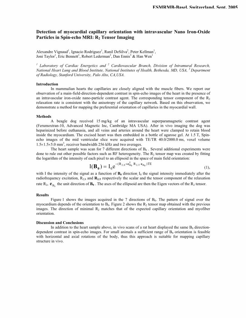
Figure 1 shows the images acquired in the 7 directions of B0. The pattern of signal over the myocardium depends of the orientation to B0. Figure 2 shows the R2 tensor map obtained with the previous images. The direction of minimal R2 matches that of the expected capillary orientation and myofiber orientation. Discussion and Conclusions
In addition to the heart sample above, in vivo scans of a rat heart displayed the same B0 direction-dependent contrast in spin-echo images. For small animals a sufficient range of B0 orientation is feasible with horizontal and axial rotations of the body, thus this approach is suitable for mapping capillary structure in vivo.
ESMRMB-Basel, Switzerland, Sept. 2005
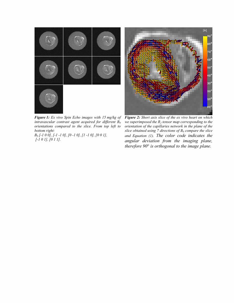
Figure 1: Ex vivo Spin Echo images with 15 mg/kg of intravascular contrast agent acquired for different B0 orientations compared to the slice. From top left to bottom right: B0 [-1 0 0], [-1 -1 0], [0 -1 0], [1 -1 0], [0 0 1], [-1 0 1], [0 1 1].
Figure 2: Short axis slice of the ex vivo heart on which we superimposed the R2 tensor map corresponding to the orientation of the capillaries network in the plane of the slice obtained using 7 directions of B0 compare the slice and Equation (1). The color code indicates the angular deviation from the imaging plane, therefore 90° is orthogonal to the image plane.