detection of sirna-mediated target mrna cleavage ... · pdf filedetection of sirna-mediated...
TRANSCRIPT
Biochemistry and Biophysics Reports 6 (2016) 16–23
Contents lists available at ScienceDirect
Biochemistry and Biophysics Reports
http://d2405-58
n CorrE-m
journal homepage: www.elsevier.com/locate/bbrep
Detection of siRNA-mediated target mRNA cleavage activities in humancells by a novel stem-loop array RT-PCR analysis
Jing Lin, Kai Xu, Jack A. Roth, Lin Ji n
Department of Thoracic and Cardiovascular Surgery, The University of Texas MD Anderson Cancer Center, 1515 Holcombe Blvd., Unit1489, Houston, TX 77030, USA
a r t i c l e i n f o
Article history:Received 30 November 2015Received in revised form16 February 2016Accepted 22 February 2016Available online 24 February 2016
Keywords:siRNA-mediated mRNA cleavageSLA-RT-PCR
x.doi.org/10.1016/j.bbrep.2016.02.01208/& 2016 The Authors. Published by Elsevier
esponding author.ail address: [email protected] (L. Ji).
a b s t r a c t
The small interfering RNA (siRNA)-mediated target mRNA cleavage activity generates cleaved mRNAfragments with varied termini, which creates major technical challenges for the accurate and efficientdetection and verification of cleavage sites on target mRNAs. Here we used a sensitive stem-loop arrayreverse transcription polymerase chain reaction (SLA-RT-PCR) approach to detect and verify the siRNA-mediated target mRNA cleavage sites by determining precise sequences at the 3′- termini of cleavedmRNA fragments in human cells under physiological conditions. Our results demonstrated the greatpotential and broad applications of using the SLA-RT-PCR as a sensitive, cost-efficient, and high-throughput tool to systematically detect siRNA-targeted mRNA cleavage sites and fragments in humancells.& 2016 The Authors. Published by Elsevier B.V. This is an open access article under the CC BY-NC-ND
license (http://creativecommons.org/licenses/by-nc-nd/4.0/).
1. Introduction
The ability of short-interfering RNA (siRNA) to silence the activityof specific genes has been proved to be very useful in dissectinggenetic function [1]. The siRNA also holds considerable promise as anovel therapeutic approach to silence disease-causing genes, parti-cularly those encoding socalled ‘non-druggable’ targets that are notamenable to conventional therapeutics such as small molecules,proteins, or monoclonal antibodies [2,3]. However, the major ob-stacles to efficiently knock-down gene expression in vivo and usesiRNAs to silence the pathogenic genes in clinical practice by RNAitechnologies are the systemic delivery of therapeutic siRNAs [3–5],their off-target effects [2,6], and the lack of accurate and efficientmethods and techniques in detection of target mRNA intermediatesand end-products resulted from siRNA activities [7,8].
It has been shown that mammalian siRNAs repress gene ex-pression by cleaving target mRNAs and leading to the sequentialdegradation through RNA-induced silencing complex (RISC) [9,10].The siRNA cleavage site on the mRNA target is usually located in themiddle of the region spanned the siRNA:target duplex [11]. Severalreports recently suggested that the efficacy of RNAiobserved in vivo could also be resulted from a general transcriptionaldown-regulation induced by the double stranded structure of siRNAwithout involving specific target mRNA cleavage [12,13]. Therefore,it is critical to confirm that an observed target gene expressionknockdown has occurred through the siRNA-induced mRNA
B.V. This is an open access article u
cleavage. However, the precise cleavage sites on the siRNA-targetedmRNA were not commonly identified and confirmed in current lit-eratures. Such efforts might partly been hampered by technicaldifficulties in detecting and verifying the cleaved mRNA inter-mediates and end products. New biological and biochemical meth-ods are much needed to efficiently and accurately identify inter-mediates and end-products resulted from the siRNA action in mul-ticellular organisms under physiological conditions. In this study, weused a novel stem-loop array RT-PCR (SLA-RT-PCR) assay to accu-rately identify siRNA-mediated target mRNA cleavage sites by effi-ciently detecting the cleaved 5′-fragments of target mRNAs underthe physiological condition in human cells. We used the syntheticsiRNAs with an ectopically expressed mRNA target templates astesting models, and the wild-type (wt) and mutant (mut)-sequencespecific siRNAs to endogenous target mRNA to evaluate the sensi-tivity, specificity, and efficacy of the SLA-RT-PCR assay for detectingsiRNA-mediated mRNA cleavage in mammalian cells under physio-logical conditions. Our results demonstrated the great potential andbroad applications of using the SLA-RT-PCR assay to systematicallydetect intermediates and end products of target mRNA cleavage inmulti-cellular organisms under physiological conditions.
2. Materials and methods
2.1. Cell culture and siRNA treatment
The human non-small cell lung cancer cell (NSCLC) lines H1299(KRAS-wild type) and H358 (KRASG12C (GGT to TGC)-mutant)were obtained from American Type Culture Collection (ATCC).
nder the CC BY-NC-ND license (http://creativecommons.org/licenses/by-nc-nd/4.0/).

Fig. 1. Schematic representation of detection siRNA-mediated target mRNA cleavage activities by SLA-RT-PCR assay. RNA-induced silencing complex (RISC) cleaves targetmRNA into two distinct fragments: a 5′ fragment with a 3′ hydroxyl group, and a 3′ fragment with a 5′ phosphate. (A) SLA-RT-PCR assay includes two steps: SLA-RT and PCR.SLA-RT efficiently detects the cleaved 5′-fragments of target mRNA. PCR is preceded with a pair of forward and reverse primer that contains an antisense sequencecomplementary to the 5′-end of target mRNA and to the 3′-end of the stem loop of RT-primer. (B) SLA-RT-Primer design and the synthetic RNA44 RNA template sequencesfor detection and verification of 3′ ends of synthetic small nucleolar RNU44 RNA by SLA-RT-PCR. The predicted full length RNU44 RNA sequences with 3′ end sequences(underlined) targeted by 3′-SLA-RT primers are shown. (C) Detection and verification of 3′ ends of RNU44 RNA by 3′-SLA-RT primers. SLA-RT-PCR products were detected byagarose gel electrophoresis (upper panel) and relative band intensity was shown in lower panel.
J. Lin et al. / Biochemistry and Biophysics Reports 6 (2016) 16–23 17
H1299 and H358 cells were grown in RPMI 1640 supplementedwith 10% fetal bovine serum in an atmosphere of humidified aircontaining 5% CO2. Cells were grown in a 6-well plate to 80%confluence and then transfected with siRNA. The transfection wasperformed using the transfection agent DharmaFECT 1 accordingto the manufacturer’s protocols (Dharmacon).
2.2. Plasmid construction
The siRNA-targeted mRNA sequences were directly derivedfrom tumor suppressor gene TUSC2 [14]. The plasmid vectorcontaining the siRNA target sequence was constructed by inserting
the siRNA targeting sequence into the reporter GFP Plasmid(Fig. 3A). All sequences in plasmid constructs were confirmed byautomated DNA sequencing.
2.3. Synthesis of materials
The small RUN44 RNAs with a predicted sequence of sixty-sixnucleotides (Fig. 2A) were synthesized using commercial source(Sigma) and purified by polyacrylamide gel electrophoresis(PAGE). The purified RUN44 RNA was used as a RNA template toevaluate SLA-RT-PCR for detection and verification of the 3′ end ofRUN44 RNA. All SLA-RT primers and PCR primers and siRNAs were
J. Lin et al. / Biochemistry and Biophysics Reports 6 (2016) 16–2318
synthesized and purchased from Sigma and their correspondingsequences were described and illustrated in individual figures.
2.4. RNA isolation
Cells were grown on 6-well plates and approximately 2�106
cells were used to isolate the total RNA using the TRIzol reagent(Life Technologies) and the additional phenol/chloroform extrac-tion was performed before ethanol precipitation according to themanufacturer's protocol. The isolated total RNA was stored in 70%ethanol at�80 °C. The purified total RNA was dissolved in RNase-free water for SLA-RT-PCR reactions.
2.5. Stem-loop array reverse transcriptase reaction (SLA-RT)
RNA samples were briefly treated with 0.04U/ml RNase-freeDNase I. RNA was reversed transcribed using the High CapacityReverse Transcription kit (Life Technologies) in combination withan array of stem-loop RT primers. The 20 ml of RT reaction con-tained 50 ng of total RNA, 5�10�12 mol of SLA-RT primer, 2 ml of
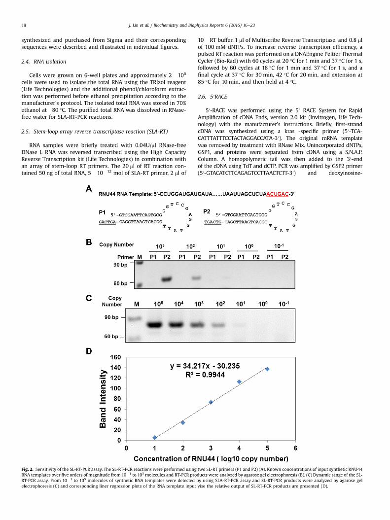
Fig. 2. Sensitivity of the SL-RT-PCR assay. The SL-RT-PCR reactions were performed usingRNA templates over five orders of magnitude from 10�1 to 103 molecules and RT-PCR proRT-PCR assay. From 10�1 to 105 molecules of synthetic RNA templates were detectedelectrophoresis (C) and corresponding liner regression plots of the RNA template input
10�RT buffer, 1 ml of Multiscribe Reverse Transcriptase, and 0.8 mlof 100 mM dNTPs. To increase reverse transcription efficiency, apulsed RT reaction was performed on a DNAEngine Peltier ThermalCycler (Bio-Rad) with 60 cycles at 20 °C for 1 min and 37 °C for 1 s,followed by 60 cycles at 18 °C for 1 min and 37 °C for 1 s, and afinal cycle at 37 °C for 30 min, 42 °C for 20 min, and extension at85 °C for 10 min, and then held at 4 °C.
2.6. 5′RACE
5′-RACE was performed using the 5′ RACE System for RapidAmplification of cDNA Ends, version 2.0 kit (Invitrogen, Life Tech-nology) with the manufacturer's instructions. Briefly, first-strandcDNA was synthesized using a kras -specific primer (5′-TCA-CATTTATTTCCTACTAGGACCATA-3′). The original mRNA templatewas removed by treatment with RNase Mix. Unincorporated dNTPs,GSP1, and proteins were separated from cDNA using a S.N.A.P.Column. A homopolymeric tail was then added to the 3′-endof the cDNA using TdT and dCTP. PCR was amplified by GSP2 primer(5′-GTACATCTTCAGAGTCCTTAACTCTT-3′) and deoxyinosine-
two SL-RT primers (P1 and P2) (A). Known concentrations of input synthetic RNU44ducts were analyzed by agarose gel electrophoresis (B). (C) Dynamic range of the SL-by using SLA-RT-PCR assay and SL-RT-PCR products were analyzed by agarose gelvise the relative output of SL-RT-PCR products are presented (D).
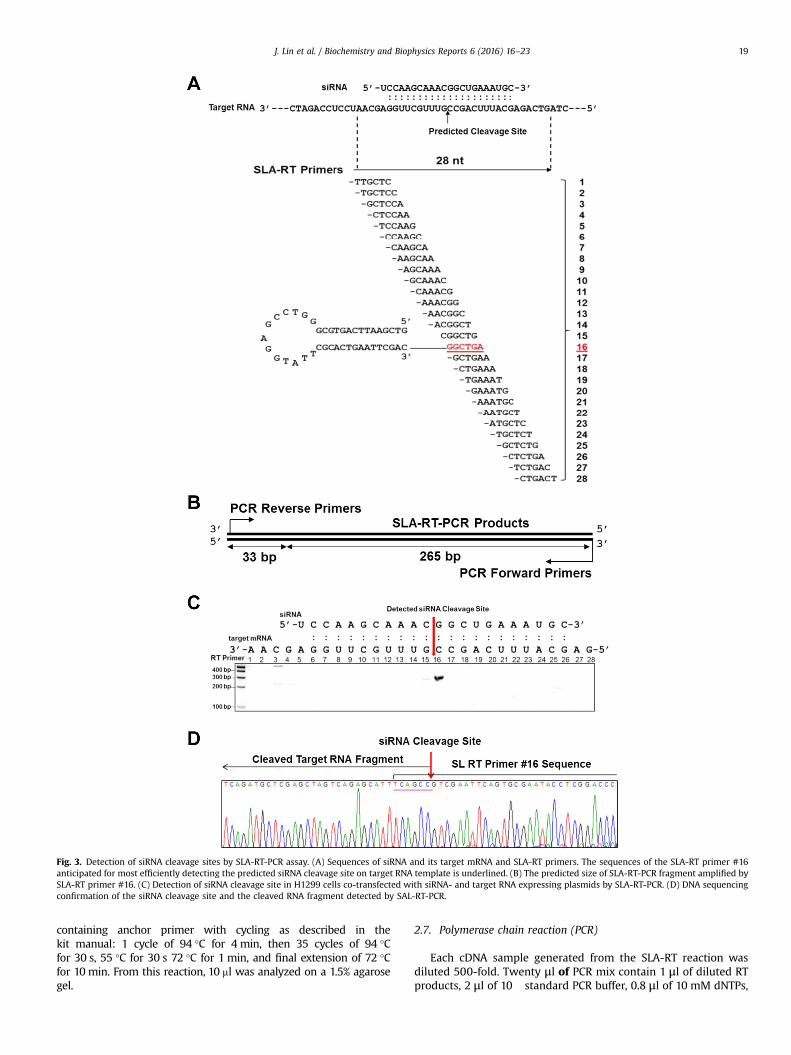
Fig. 3. Detection of siRNA cleavage sites by SLA-RT-PCR assay. (A) Sequences of siRNA and its target mRNA and SLA-RT primers. The sequences of the SLA-RT primer #16anticipated for most efficiently detecting the predicted siRNA cleavage site on target RNA template is underlined. (B) The predicted size of SLA-RT-PCR fragment amplified bySLA-RT primer #16. (C) Detection of siRNA cleavage site in H1299 cells co-transfected with siRNA- and target RNA expressing plasmids by SLA-RT-PCR. (D) DNA sequencingconfirmation of the siRNA cleavage site and the cleaved RNA fragment detected by SAL-RT-PCR.
J. Lin et al. / Biochemistry and Biophysics Reports 6 (2016) 16–23 19
containing anchor primer with cycling as described in thekit manual: 1 cycle of 94 °C for 4 min, then 35 cycles of 94 °Cfor 30 s, 55 °C for 30 s 72 °C for 1 min, and final extension of 72 °Cfor 10 min. From this reaction, 10 ml was analyzed on a 1.5% agarosegel.
2.7. Polymerase chain reaction (PCR)
Each cDNA sample generated from the SLA-RT reaction wasdiluted 500-fold. Twenty μl of PCR mix contain 1 μl of diluted RTproducts, 2 μl of 10� standard PCR buffer, 0.8 μl of 10 mM dNTPs,
J. Lin et al. / Biochemistry and Biophysics Reports 6 (2016) 16–2320
0.5 μl of 100 mM MgSO4, 0.2 μl of Taq polymerase, 1 μl of 10 μΜof forward primer and 1 μl of 10 μΜ of reverse primer. PCR re-actions were conducted at 95 °C for 5 min, followed by 35 cycles at
95 °C for 15 s, 61 °C for 30 s, and 37 °C for 30 s, and afinal extension at 72 °C for 10 min, on DNAEngine Peltier ThermalCycler.
J. Lin et al. / Biochemistry and Biophysics Reports 6 (2016) 16–23 21
The nucleotide sequences of the PCR primers are as followings:U6: sense primer, 5′-CCTGGATGATGATAAGCAAATGC-3′, anti-
sense primer, 5′-GTGCGGGTCCGAGGTATTC-3′.TUSC2: sense primer, 5′-CGGCATGGACGAGCTGTACAAGTA-3′,
antisense primer, 5′-GTGCGGGTCCGAGGTATTC-3′.KRAS: sense primer, 5′-GAAGGTGGCGGCGGCTCG-3′, antisense
primer, 5′-GTGCGGGTCCGAGGTATTC-3′.PCR products sequence analysis was performed on an ABI 3730
DNA sequencer by DNA Analysis Core Facility at MD AndersonCancer Center.
2.8. Agarose gel electrophoresis
SLA-RT-PCR products were analyzed by 1.5% agarose gel elec-trophoresis in 1�Tris-Borate-EDTA (TBE) buffer containing 89 mMof Tris Base and 89 mM of Boric Acid. Electrophoresis was per-formed at 100 V for 60 min. Gel was stained in ethidium bromidebath for 10 min prior to visualization using a UV transilluminator.
3. Results
3.1. Detection and verification of 3′ ends of synthetic RNU44 RNA bySLA-RT-PCR
We adopted a novel SLA-RT-PCR technique, originally developed byChen et al. [15], to systematically verify and detect intermediate mRNAfragments cleaved by siRNAs. This approach accurately identified un-ique nucleotide ends generated by the siRNA-mediated target cleavageactivities in human cells. The principle of works, the experimentaldesign, and assay procedures are illustrated in Fig. 1A. The SLA-RT-PCRassay includes two steps: SLA-RT and PCR. SLA-RT primers comprisedtwo unique sequence components: a short stretch of 6-base single-stranded nucleotides at the 3′ end of the primer sequences that arecomplementary to the 5′-terminal sequences of the target mRNA, anda double stranded stem at the 5′ end that forms a stem-loop tofunction as a forceps to stabilize the secondary structure of the primer.The terminal sequences of the siRNA-cleaved target mRNA fragmentsare specifically recognized by a six-nucleotide complementary exten-sion at the 3′ends of the RT-primer array, which could efficientlyprime the reverse transcription of the cleaved mRNA fragments intocDNAs. The uses of an array of stem-loop RT primers allow accuraterecognition and verification of unique ends of the mRNA fragmentsprocessed by endonucleases. If the SLA-RT primers have no perfectmatch, on both the length and the base, to the sequence of the 3′ or 5′end of the mRNA fragment the efficiency of RT reaction would havebeen dramatically reduced. The subsequent PCR is preceded with apair of forward and reverse primer that contains an antisense se-quence complementary to the 5′-end of mRNA and to the 3′-end ofthe stem loop of RT-primer, respectively. The abundance of the tran-script-specific SLA-RT-PCR amplicons represents the distribution ofcleaved RNA fragments and is determined by agarose gel electro-phoresis. The success of SLA-RT-PCR relies deeply on the differentialpriming efficiency between matched and mismatched SLA primers
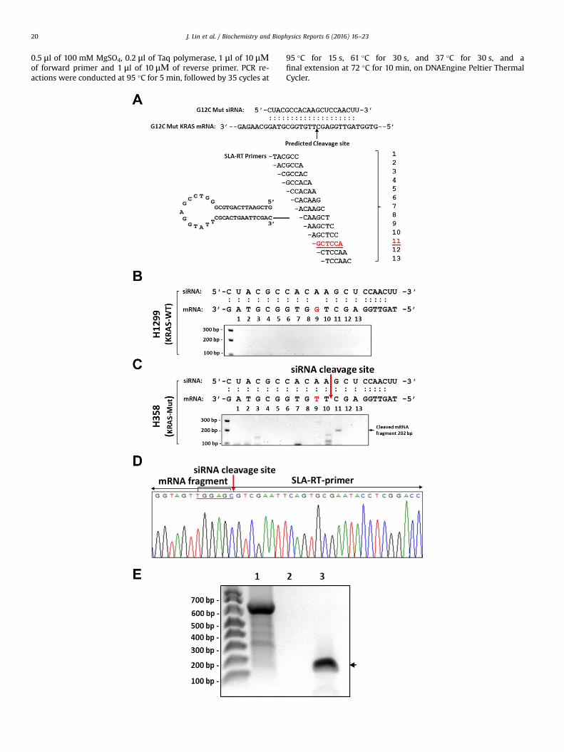
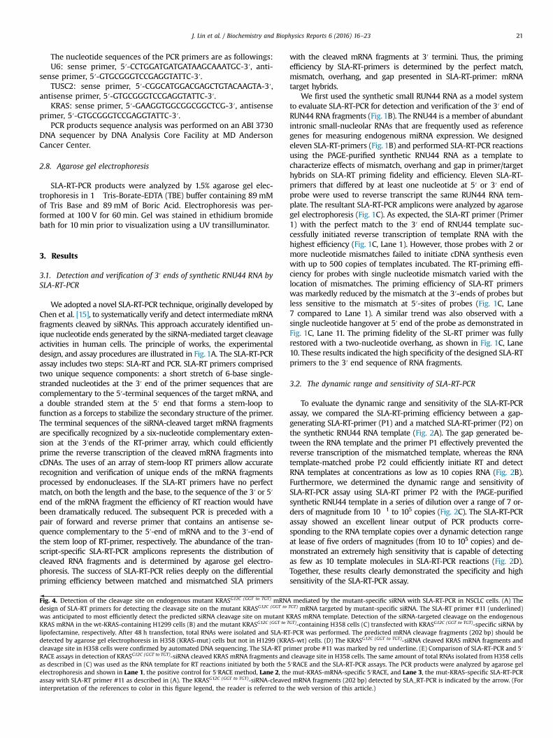
Fig. 4. Detection of the cleavage site on endogenous mutant KRASG12C (GGT to TGT) mRNAdesign of SLA-RT primers for detecting the cleavage site on the mutant KRASG12C (GGT to
was anticipated to most efficiently detect the predicted siRNA cleavage site on mutant KKRAS mRNA in the wt-KRAS-containing H1299 cells (B) and the mutant KRASG12C (GGT to T
lipofectamine, respectively. After 48 h transfection, total RNAs were isolated and SLA-RTdetected by agarose gel electrophoresis in H358 (KRAS-mut) cells but not in H1299 (KRAcleavage site in H358 cells were confirmed by automated DNA sequencing. The SLA-RT prRACE assays in detection of KRASG12C (GGT to TGT)-siRNA cleaved KRAS mRNA fragments andas described in (C) was used as the RNA template for RT reactions initiated by both theelectrophoresis and shown in Lane 1, the positive control for 5′RACE method, Lane 2, thassay with SLA-RT primer #11 as described in (A). The KRASG12C (GGT to TGT)-siRNA-cleaveinterpretation of the references to color in this figure legend, the reader is referred to t
with the cleaved mRNA fragments at 3′ termini. Thus, the primingefficiency by SLA-RT-primers is determined by the perfect match,mismatch, overhang, and gap presented in SLA-RT-primer: mRNAtarget hybrids.
We first used the synthetic small RUN44 RNA as a model systemto evaluate SLA-RT-PCR for detection and verification of the 3′ end ofRUN44 RNA fragments (Fig. 1B). The RNU44 is a member of abundantintronic small-nucleolar RNAs that are frequently used as referencegenes for measuring endogenous miRNA expression. We designedeleven SLA-RT-primers (Fig. 1B) and performed SLA-RT-PCR reactionsusing the PAGE-purified synthetic RNU44 RNA as a template tocharacterize effects of mismatch, overhang and gap in primer/targethybrids on SLA-RT priming fidelity and efficiency. Eleven SLA-RT-primers that differed by at least one nucleotide at 5′ or 3′ end ofprobe were used to reverse transcript the same RUN44 RNA tem-plate. The resultant SLA-RT-PCR amplicons were analyzed by agarosegel electrophoresis (Fig. 1C). As expected, the SLA-RT primer (Primer1) with the perfect match to the 3′ end of RNU44 template suc-cessfully initiated reverse transcription of template RNA with thehighest efficiency (Fig. 1C, Lane 1). However, those probes with 2 ormore nucleotide mismatches failed to initiate cDNA synthesis evenwith up to 500 copies of templates incubated. The RT-priming effi-ciency for probes with single nucleotide mismatch varied with thelocation of mismatches. The priming efficiency of SLA-RT primerswas markedly reduced by the mismatch at the 3′-ends of probes butless sensitive to the mismatch at 5′-sites of probes (Fig. 1C, Lane7 compared to Lane 1). A similar trend was also observed with asingle nucleotide hangover at 5′ end of the probe as demonstrated inFig. 1C, Lane 11. The priming fidelity of the SL-RT primer was fullyrestored with a two-nucleotide overhang, as shown in Fig. 1C, Lane10. These results indicated the high specificity of the designed SLA-RTprimers to the 3′ end sequence of RNA fragments.
3.2. The dynamic range and sensitivity of SLA-RT-PCR
To evaluate the dynamic range and sensitivity of the SLA-RT-PCRassay, we compared the SLA-RT-priming efficiency between a gap-generating SLA-RT-primer (P1) and a matched SLA-RT-primer (P2) onthe synthetic RNU44 RNA template (Fig. 2A). The gap generated be-tween the RNA template and the primer P1 effectively prevented thereverse transcription of the mismatched template, whereas the RNAtemplate-matched probe P2 could efficiently initiate RT and detectRNA templates at concentrations as low as 10 copies RNA (Fig. 2B).Furthermore, we determined the dynamic range and sensitivity ofSLA-RT-PCR assay using SLA-RT primer P2 with the PAGE-purifiedsynthetic RNU44 template in a series of dilution over a range of 7 or-ders of magnitude from 10�1 to 105 copies (Fig. 2C). The SLA-RT-PCRassay showed an excellent linear output of PCR products corre-sponding to the RNA template copies over a dynamic detection rangeat lease of five orders of magnitudes (from 10 to 105 copies) and de-monstrated an extremely high sensitivity that is capable of detectingas few as 10 template molecules in SLA-RT-PCR reactions (Fig. 2D).Together, these results clearly demonstrated the specificity and highsensitivity of the SLA-RT-PCR assay.
mediated by the mutant-specific siRNA with SLA-RT-PCR in NSCLC cells. (A) TheTGT) mRNA targeted by mutant-specific siRNA. The SLA-RT primer #11 (underlined)RAS mRNA template. Detection of the siRNA-targeted cleavage on the endogenousGT)-containing H358 cells (C) transfected with KRASG12C (GGT to TGT)-specific siRNA by-PCR was performed. The predicted mRNA cleavage fragments (202 bp) should beS-wt) cells. (D) The KRASG12C (GGT to TGT)-siRNA cleaved KRAS mRNA fragments andimer probe #11 was marked by red underline. (E) Comparison of SLA-RT-PCR and 5′cleavage site in H358 cells. The same amount of total RNAs isolated from H358 cells
5′RACE and the SLA-RT-PCR assays. The PCR products were analyzed by agarose gele mut-KRAS-mRNA-specific 5′RACE, and Lane 3, the mut-KRAS-specific SLA-RT-PCRd mRNA fragments (202 bp) detected by SLA_RT-PCR is indicated by the arrow. (Forhe web version of this article.)

J. Lin et al. / Biochemistry and Biophysics Reports 6 (2016) 16–2322
3.3. Detection and verification of siRNA-mediated mRNA cleavagesites by SLA-RT-PCR
In order to evaluate the potential of SLA-RT-PCR assay inidentifying siRNA targets and determining its mechanism of ac-tion, we used a synthetic siRNA and ectopically expressed artificialmRNA template as a model system to precisely detect and verifythe intermediates and end products of the siRNA activity inmammalian cells under physiological conditions. It has beenshown that mammalian siRNAs silence gene expression by cleav-ing their target mRNAs for sequential degradation [10]. The siRNAcleavage site on the mRNA target is usually located in the middleof the region spanned by the siRNA:mRNA duplex, following acanonical pattern of 10 bp from the 5′-end of siRNA in the RISC[11]. The RISC-associated argonaute 2 (AGO 2) cleaves targetmRNA into two distinct fragments: a 5′ fragment with a 3′ hy-droxyl group, and a 3′ fragment with a 5′ phosphate [16–18]. ThesiRNA and the siRNA-targeted mRNA template expression plasmidwere co-transfected into human non-small cell lung cancer(NSCLC) H1299 cells (Fig. 3). The siRNA and its target mRNA se-quences, the predicted siRNA cleavage site, and the designed set oftwenty-eight SLA-RT primers for detection of the potential siRNA-directed target RNA cleavage sites were illustrated in Fig. 3A and B.The siRNA-mediated cleavage site on the mRNA target was pre-cisely detected by the SLA-RT-PCR in the middle of siRNA targetregion at the predicted position “G”, which is 10 bp from the 5′-end of siRNA duplex, as shown in Fig. 3C and indicated by arrow.The identity of the SLA-RT-PCR products was further verified byDNA sequencing (Fig. 3D). These results clearly demonstrated thepotential applications of SLA-RT-PCR in identifying authentic siR-NA targets by precisely detecting and verifying the cleavage siteson target mRNAs mediated by siRNAs in mammalian cells.
3.4. Detection of siRNA-mediated cleavage activities on endogenousmRNA targets
To further demonstrate the utility and specificity of SLA-RT-PCRin detection the siRNA-mediated cleavage sites on endogenousmRNA targets in human cells under physiological conditions, wedetermined cleavage activities mediated by synthetic siRNAs thatspecifically target the wild-type and the mutant sites, respectively,on the endogenous KRAS mRNA sequences in human NSCLCH1299 (KRAS-wt) and H358 (KRASG12C (GGT to TGT)-mut) cells(Fig. 4). The mut-KRAS-specific siRNAs (Fig. 4A) were transfectedinto H1299 and H358 cells by DharmaFect1 transfection reagentand total RNAs were isolated 48 h after transfection. A set of SLA-RT primers covering entire siRNA complementary and the im-mediately adjacent 5′- and 3′ sequences (Fig. 4A) on wt-KRAS(Fig. 4B) or mut-KRAS (Fig. 4C) mRNA sequences were used todetect and verify the predicted cleavage site as indicated by arrow(Fig. 4A). The siRNA-cleaved KRAS mRNA fragments with a pre-dicted size of 202 bp could be detected by agarose gel electro-phoresis only in mut-KRASG12C (GGT to TGT)-containing H358 cells(Fig. 4C) but not in wt-KRAS-expressing H1299 cells (Fig. 4B)transfected by the mut-KRAS-siRNA. The KRASG12C (GGT to TGT)-siRNA cleaved mut-KRAS mRNA fragments and cleavage site inH358 cells were confirmed by automated DNA sequencing(Fig. 4D).
To further validate the utility, specificity, and sensitivity of SLA-RT-PCR assay in detection of siRNA-cleavage site and cleaved 5′-mRNA fragments, we performed a parallel comparison betweenthe SLA-RT-PCR and the 5′RACE (Invitrogen, Life Technologies)methods (Fig. 4E), using the same total RNAs isolated in H358 cellstransfected with KRASG12C (GGT to TGT)-siRNA as described in Fig. 4Cas the template for RT reactions respectively initiated by the SLA-RT and 5′RACE-RT primers specific to endogenous KRAS mRNA
transcripts. The predicted 5′RACE product was clearly detected bythe positive control provided in 5′RACE kit (Fig. 4E, Lane 1) but no5′RACE products were detected using KRAS-specific RT primer(Fig. 4E, Lane 2). The predicted KRASG12C (GGT to TGT)-siRNA-cleavedmRNA fragments (202 bp) was clearly detected by SLA_RT-PCRusing the mutant KRAS-specific SLA-RT primer #11 (A), as simi-larly shown in (C) and indicated by the arrow. These results de-monstrated the sensitivity and advantages of SLA-RT-PCR assay indetection of specific sites and 5′-fragments cleaved by siRNA ac-tivities under physiological conditions over the conventionalmethods such as the 5′RACE.
4. Discussion
siRNA has been shown to be a powerful tool for analyzing genefunction and developing novel therapeutic drugs targeting specificdisease-causing or cancer-driving genes by silencing expression ofa specific gene and encoded protein product [4]. However, for thesuccessful application of siRNA as a therapeutic for human disease,one of most critical questions need to be answered in the clinicalpractice is to detect and confirm whether the designed siRNAtherapeutic hits its predicted gene target. This question can beeffectively addressed by accurately detecting the siRNA cleavagesites and cleaved products of the target mRNA. However, suchefforts might partly be hindered by technic difficulties in detectingand verifying specific cleavage mRNA sites and the cleaved mRNAintermediates and end products.
Traditional RT-PCR cannot detect mRNA transcript termini andintermediates generated by various cellular processes and biolo-gical activities, which are important for understanding the biolo-gical significance of mRNA polymorphisms and mRNA function-alities. These mRNA variants might reflect the difference ofabundances between full length transcripts and shortened tran-scripts, produced by partial degradation, modification or cleavagesof transcripts through various RNA editing mechanisms, includingsiRNA targeted mRNA cleavages. Currently, 5′-RACE (rapid ampli-fication of cDNA ends), 3′-RACE, cRT-PCR (circulative RT-PCR) andNext Generation of Sequencing (NGS) are a few technologies tofulfill these needs for mRNA cleavage activities through RNA self-circulation or adapter addition by RNA ligase [19,20]. However,those technologies have their corresponding limits in sensitivity,accuracy, and cost- and time-efficiency. Moreover, the inevitableuse of RNA ligase in some of those methods result in enzymaticallybiased interpretation in the distribution of RNA terminus andsignificantly reduced their detection sensitivity and accuracy, aswe demonstrated in parallel comparison of the SLA-RT-PCR and 5′RACE methods.
In this study, we have evaluated the sensitivity and specificityof SLA-RT-PCR method for identifying siRNA-mediated targetmRNA cleavage sites by accurately detecting the termini of siRNA-cleaved target mRNA fragments on both the ectopically expressedin human cells under physiological conditions. Our results de-monstrated a great differential RT-priming efficiency betweenmatched and mismatched SLA-RT-primers to the 3′-termini ofsiRNA-cleaved target mRNA fragments. This method is extremelysensitive, allowing detection of RNA fragments as few as 10 tem-plate copies with a linear correlation up to 5 orders of magnitudeswith primers tested. Although the liner correlation could be al-tered by the different compositions among SLA-RT-primers, whichcould potentially alter the Tm (melting temperature) of SAL-RTprimers, the intensity of the individual SLA-RT-PCR amplicon wasprincipally determined by the RNA fragment abundance and thebase-stacking enhancement of perfectly matches between RNAterminal and SLA-RT-primer sequences. The high GC- or AT- richsequence contents at the 3′-termini of the cleaved target miRNA
J. Lin et al. / Biochemistry and Biophysics Reports 6 (2016) 16–23 23
fragments may affect the priming efficiency of SLA-RT, however,our testing results with varied base compositions at 3′-termini ofsynthetic mRNA templates and corresponding matched or un-matched SLA-RT primers demonstrated that the priming efficiencywas more depended on the degree of base matches than thecontent of bases. The SLA-RT-PCR could potentially be used tomore quantitatively determine the cleaved mRNA products byreal-time PCR with probes specific to the predicted PCR amplicons.This method could also be modified to detect the cleaved mRNAfragments with unknown ends such as 3′-urydylation, tailing, andtrimming by modifying the SLA-RT primers with “U-tract” andrandomizing 6-nt short stretch at the 3′-end of SLA-RT primers.Due to the high sensitivity and specificity of SLA-RT-PCR as de-monstrated, this method is principally sufficient and capable todetect the precise siRNA cleavage site on the targeted mRNA bytaking advantages of the high fidelity of SLA-RT primers in re-cognizing specific ends of RNA fragments generated in RISC andefficiently capturing an immediate and unmodified cleavagefragment from a population of specific-siRNA-cleaved mRNA in-termediates and end products at any given time under a physio-logical condition. By incorporating randomized stem-loop-arrayRT primer sequences and in combination with targeted deep se-quencing, the modified SLA-RT-PCR method can be used as a“high-throughput” assay to capture all potential mRNA cleavagesites and cleaved fragments by “on-target” and “off-target” activ-ities of a specific siRNA.
Our results demonstrated the great potential and broad appli-cations of using the SLA-RT-PCR as a sensitive, specific, and cost-efficient tool to identify authentic siRNA-cleavage sites in multi-cellular organisms under physiological conditions. SLA-RT-PCR canefficiently detect and confirm whether the designed siRNA ther-apeutic hits its predicted gene target and could be a useful tool forfacilitating the application of siRNA-based therapeutics in humandiseases.
Conflict of interests
The authors declare that they have no conflict of interest.
Acknowledgements
This work is supported in part by the National Institutes ofHealth/National Cancer Institute through a Specialized Program ofResearch Excellence (SPORE) Grant CA-070907 (J. Minna), R01Grants CA116322 (Ji) and CA176568-02 (Ji, Roth, Wu), a U.S. De-partment of Defense Grant W81XWH-09-02-0139 (Ji), a CancerPrevention and Research Institute of Texas (CPRIT) GrantRP130502 (Wu), a MD Anderson's Cancer Center Support GrantCA-016672-Lung Program, DNA Analysis Facility Shared Resource,and from the Tobacco Settlement Funds as appropriated by theTexas State Legislature.
Appendix A. Transparency document
Transparency document associated with this article can befound in the online version at http://dx.doi.org/10.1016/j.bbrep.2016.02.012.
References
[1] S.M. Elbashir, J. Harborth, W. Lendeckel, A. Yalcin, K. Weber, T. Tuschl, Du-plexes of 21-nucleotide RNAs mediate RNA interference in cultured mam-malian cells, Nature 411 (2001) 494–498.
[2] M. John, R. Constien, A. Akinc, M. Goldberg, Y.A. Moon, M. Spranger,P. Hadwiger, J. Soutschek, H.P. Vornlocher, M. Manoharan, M. Stoffel, R. Langer,D.G. Anderson, J.D. Horton, V. Koteliansky, D. Bumcrot, M. John, R. Constien,A. Akinc, M. Goldberg, Y.A. Moon, M. Spranger, P. Hadwiger, J. Soutschek, H.P. Vornlocher, M. Manoharan, M. Stoffel, R. Langer, D.G. Anderson, J.D. Horton,V. Koteliansky, D. Bumcrot, Effective RNAi-mediated gene silencing withoutinterruption of the endogenous microRNA pathway, Nature 449 (2007)745–747.
[3] J. Soutschek, A. Akinc, B. Bramlage, K. Charisse, R. Constien, M. Donoghue,S. Elbashir, A. Geick, P. Hadwiger, J. Harborth, M. John, V. Kesavan, G. Lavine, R.K. Pandey, T. Racie, K.G. Rajeev, I. Rohl, I. Toudjarska, G. Wang, S. Wuschko,D. Bumcrot, V. Koteliansky, S. Limmer, M. Manoharan, H.P. Vornlocher,J. Soutschek, A. Akinc, B. Bramlage, K. Charisse, R. Constien, M. Donoghue,S. Elbashir, A. Geick, P. Hadwiger, J. Harborth, M. John, V. Kesavan, G. Lavine, R.K. Pandey, T. Racie, K.G. Rajeev, I. Rohl, I. Toudjarska, G. Wang, S. Wuschko,D. Bumcrot, V. Koteliansky, S. Limmer, M. Manoharan, H.P. Vornlocher, Ther-apeutic silencing of an endogenous gene by systemic administration ofmodified siRNAs, Nature 432 (2004) 173–178.
[4] A. de Fougerolles, H.P. Vornlocher, J. Maraganore, J. Lieberman, Interferingwith disease: a Progress report on siRNA-based therapeutics, Nat. Rev. Drug.Discov. 6 (2007) 443–453.
[5] T.S. Zimmermann, A.C. Lee, A. Akinc, B. Bramlage, D. Bumcrot, M.N. Fedoruk,J. Harborth, J.A. Heyes, L.B. Jeffs, M. John, A.D. Judge, K. Lam, K. McClintock, L.V. Nechev, L.R. Palmer, T. Racie, I. Rohl, S. Seiffert, S. Shanmugam, V. Sood,J. Soutschek, I. Toudjarska, A.J. Wheat, E. Yaworski, W. Zedalis, V. Koteliansky,M. Manoharan, H.P. Vornlocher, I. MacLachlan, T.S. Zimmermann, A.C.H. Lee,A. Akinc, B. Bramlage, D. Bumcrot, M.N. Fedoruk, J. Harborth, J.A. Heyes, L.B. Jeffs, M. John, A.D. Judge, K. Lam, K. McClintock, L.V. Nechev, L.R. Palmer,T. Racie, I. Rohl, S. Seiffert, S. Shanmugam, V. Sood, J. Soutschek, I. Toudjarska,A.J. Wheat, E. Yaworski, W. Zedalis, V. Koteliansky, M. Manoharan, H.P. Vornlocher, I. MacLachlan, RNAi-mediated gene silencing in non-humanprimates, Nature 441 (2006) 111–114.
[6] D.H. Kim, J.J. Rossi, Strategies for silencing human disease using RNA inter-ference, Nat. Rev. Genet. 8 (2007) 173–184.
[7] M.A. Behlke, Progress towards in vivo use of siRNAs, Mol. Ther. 13 (2006)644–670.
[8] D.M. Dykxhoorn, D. Palliser, J. Lieberman, The silent treatment: siRNAs assmall molecule drugs, Gene Ther. 13 (2006) 541–552.
[9] S. Cheloufi, C.O. Dos Santos, M.M. Chong, G.J. Hannon, A dicer-independentmiRNA biogenesis pathway that requires Ago catalysis, Nature 465 (2010)584–589.
[10] G. Hutvagner, M.J. Simard, Argonaute proteins: key players in RNA silencing,Nat. Rev. Mol. Cell Biol. 9 (2008) 22–32.
[11] J. Martinez, A. Patkaniowska, H. Urlaub, R. Luhrmann, T. Tuschl, Single-stran-ded antisense siRNAs guide target RNA cleavage in RNAi, Cell 110 (2002)563–574.
[12] M.E. Kleinman, K. Yamada, A. Takeda, V. Chandrasekaran, M. Nozaki, J.Z. Baffi,R.J. Albuquerque, S. Yamasaki, M. Itaya, Y. Pan, B. Appukuttan, D. Gibbs, Z. Yang,K. Kariko, B.K. Ambati, T.A. Wilgus, L.A. DiPietro, E. Sakurai, K. Zhang, J.R. Smith, E.W. Taylor, J. Ambati, Sequence- and target-independent angio-genesis suppression by siRNA via TLR3, Nature 452 (2008) 591–597.
[13] M. Robbins, A. Judge, E. Ambegia, C. Choi, E. Yaworski, L. Palmer, K. McClintock,I. MacLachlan, Misinterpreting the therapeutic effects of small interfering RNAcaused by immune stimulation, Hum. Gene Ther. 19 (2008) 991–999.
[14] J. Lin, K. Xu, J. Gitanjali, J.A. Roth, L. Ji, Regulation of tumor suppressor geneFUS1 expression by the untranslated regions of mRNA in human lung cancercells, Biochem. Biophys. Res. Commun. 410 (2011) 235–241.
[15] C. Chen, D.A. Ridzon, A.J. Broomer, Z. Zhou, D.H. Lee, J.T. Nguyen, M. Barbisin,N.L. Xu, V.R. Mahuvakar, M.R. Andersen, K.Q. Lao, K.J. Livak, K.J. Guegler, Real-time quantification of microRNAs by stem-loop RT-PCR, Nucleic Acids Res. 33(2005) e179.
[16] B.A. Janowski, J. Hu, D.R. Corey, Silencing gene expression by targeting chro-mosomal DNA with antigene peptide nucleic acids and duplex RNAs, Nat.Protoc. 1 (2006) 436–443.
[17] J.A. Broderick, W.E. Salomon, S.P. Ryder, N. Aronin, P.D. Zamore, Argonauteprotein identity and pairing geometry determine cooperativity in mamma-lian RNA Silencing, RNA 17 (2011) 1858–1869.
[18] E.S. Cenik, P.D. Zamore, Argonaute proteins, Curr. Biol. 21 (2011) R446–R449.[19] J. Shendure, H. Ji, Next-generation DNA sequencing, Nat. Biotechnol. 26 (2008)
1135–1145.[20] A. Waha, M. Watzka, A. Koch, T. Pietsch, R. Przkora, N. Peters, O.D. Wiestler,
A. von Deimling, A rapid and sensitive protocol for competitive reversetranscriptase (cRT) PCR analysis of cellular genes, Brain Pathol. 8 (1998) 13–18.