development 134, 1653-1662 (2007) …dev.biologists.org/content/develop/134/9/1653.full.pdf · the...
TRANSCRIPT

DEVELO
PMENT
Development 134, 1653-1662 (2007) doi:10.1242/dev.001016
The AP2 transcription factors DORNRÖSCHEN andDORNRÖSCHEN-LIKE redundantly control Arabidopsisembryo patterning via interaction with PHAVOLUTAJohn W. Chandler*, Melanie Cole, Annegret Flier, Britta Grewe and Wolfgang Werr
DORNRÖSCHEN (DRN) (also known as ENHANCER OF SHOOT REGENERATION1; ESR1) and DRN-LIKE (DRNL; also known as ESR2) aretwo linked paralogues encoding AP2 domain-containing proteins. drn mutants show embryo cell patterning defects and, similarlyto drnl mutants, disrupt cotyledon development at incomplete penetrance. drn drnl double mutants with weak or strong drnlalleles show more highly penetrant and extreme phenotypes, including a pin-like embryo without cotyledons, confirming a highdegree of functional redundancy for the two genes in embryo patterning. Altered expression of PIN1::PIN1-GFP and DR5::GFP in drnmutant embryos places DRN upstream of auxin transport and response. A yeast two-hybrid screen with DRN followed by co-immunoprecipitation and bimolecular fluorescence complementation revealed PHAVOLUTA (PHV) to be a protein interactionpartner in planta. drn phv double mutants show an increased penetrance of embryo cell division defects. DRNL can also interactwith PHV and both DRN and DRNL can heterodimerise with additional members of the class III HD-ZIP family, PHABULOSA,REVOLUTA, CORONA and ATHB8. Interactions involve the PAS-like C-terminal regions of these proteins and the DRN/DRNL AP2domain.
KEY WORDS: Arabidopsis, Cotyledon, Embryo
INTRODUCTIONAngiosperm growth results from two developmental growth phases:an embryonic phase whereby the primary body plan is establishedtogether with the apical-basal and radial axes of symmetry, and apostembryonic phase, in which new organs are initiated via the shootand root meristems. Cotyledon development during dicotembryogenesis marks the start of organogenesis and the changefrom radial to bilateral symmetry at the transition from the globular-to heart-stage embryo. Studies of seedling-lethal Arabidopsismutants have defined three embryonic domains wherein subsequentcell fate is largely determined by position in the embryo (Mayer etal., 1991): (1) the majority of the cotyledons, together with the shootapical meristem, are derived from the apical domain, recognisableas the derivatives of cell divisions of the upper tier of cells at theoctant stage; (2) the central domain mainly gives rise to thehypocotyl and root apical initials; and (3) the basal region, derivedfrom the hypophysis, generates the root quiescent centre (Harada,1999).
Several Arabidopsis genes are important for the development ofthe apical region of the embryo and give rise to cotyledonphenotypes when mutated. Combined mutations at the CUP-SHAPED COLYLEDON (CUC1, 2 and 3) loci result in cotyledonfusion accompanied by an absence of the shoot apical meristem(SAM) (Aida et al., 1997; Vroemen et al., 2003; Hibara et al., 2006).Polar auxin transport essentially contributes to the establishment ofboth bilateral symmetry (Liu et al., 1993) and apical-basal polarity(Friml et al., 2003), and an abnormal cotyledon number at lowpenetrance is also observed in plant hormone response mutants
(Rashotte et al., 2006; Saibo et al., 2006). In Arabidopsis, auxinpolarity and embryonic patterning have been extensively studied inmutants of PIN gene family members that encode plant-specificproteins involved in auxin efflux. PIN proteins are functionallyredundant, and higher-order pin mutants reveal defects in embryoniccell division patterns that are reflected postembryonically mostly incotyledon defects such as fusion or monocotyledony (Friml et al.,2003; Furutani et al., 2004; Vieten et al., 2005). Additionally, pinoid,a mutant in a serine/threonine protein kinase that affects localisationof PIN proteins (Friml et al., 2004), or mutations in MONOPTEROS(MP) and BODENLOS (BDL), which encode the auxin responsivefactor ARF5 and its inhibitor IAA12, respectively, all causecotyledon defects and/or disrupted embryo domains (Bennet et al.,1995; Hardtke and Berleth, 1998; Hamann et al., 1999). A generalfeature of auxin signalling and cuc mutants is the incompletepenetrance of cotyledon defects, suggesting that pathways leadingto bilateral symmetry and cotyledon establishment are considerablyredundant.
Redundant control of embryo patterning is also demonstrated bythe Arabidopsis class III HD-ZIP gene family. PHAVOLUTA (PHV)and PHABULOSA (PHB) are well-known representatives of thisHD-ZIP subclass and were identified as dominant gain-of-functionalleles due to mutations in a highly conserved microRNA target site(McConnell et al., 2001; Bao et al., 2004). Amongst this family, onlyREVOLUTA (REV) has a loss-of-function phenotype (Talbert et al.,1995); however, higher-order knockouts reveal redundant functionsin embryo and cotyledon patterning (Prigge et al., 2005).
The DORNRÖSCHEN (DRN) (also known as ENHANCER OFSHOOT REGENERATION1; ESR1) gene contributes to Arabidopsismeristem organisation (Kirch et al., 2003) and cytokinin-independent shoot regeneration (Banno et al., 2001). DRNexpression is highly dynamic and is observable from the two- tofour-cell stage in the embryo proper, before focussing to theemerging cotyledons and becoming restricted to the SAM at thetorpedo stage. During postembryonic development, DRN remains
RESEARCH ARTICLE 1653
Institute of Developmental Biology, University of Cologne, Gyrhofstrasse 17, D-50923, Cologne, Germany.
*Author for correspondence (e-mail: [email protected])
Accepted 22 February 2007
DEVELO
PMENT
Development 134 (9)RESEARCH ARTICLE
was amplified by PCR, sequenced, directionally cloned into pGBKT7(Clontech) using NcoI and BamHI, and transformed into the yeast strainY187 (Clontech Palo Alto, CA). The two-hybrid screen was performed byyeast mating, according to the manufacturer’s protocol (Clontech PT3024-1), and with quadruple selection (�-glucuronidase, –Leu, –Trp, –Ade).
In planta bimolecular fluorescence complementation (BiFC)The ORFs encoding full-length DRN or DRNL and PHVs (the C-terminal451 amino acids from amino acid 391 to the end) were cloned in-frame intothe BamHI site of pUC-SPYCE or pUC-SPYNE (Walter et al., 2004). Forcontrol experiments, GFP fusions were created in the pRT-�NotI/AscIvector (Überlacker and Werr, 1996). Transient expression in leek epidermalcells was performed according to Cole et al. (Cole et al., 2006). YFP/GFPfluorescence was visualised using a MZFLIII stereomicroscope (Leica) afterUV excitation and using a GFP filter. All images were processed usingPhotoshop software (Adobe).
Co-immunoprecipitation and western blot analysisEpitope-tagged proteins or peptides were synthesised via the EasyXpress invitro transcription/translation system (Qiagen) based on the T7 promoter.Templates of the required proteins were obtained via nested PCR reactionson the respective gene ORFs in pUC-SPYCE/NE containing an HA or mycepitope-coding sequence preceding the YFP subdomains. The T7 promoterand 6xHis tag were added to DRN and DRNL coding regions by nested PCRusing primers from the EasyXpress Kit. For co-expression, the relevantamplicons were mixed. Control reactions were performed with singleamplicons. Immunoprecipitation (IP), gel electrophoretic analyses anddetection of epitope-tagged proteins essentially followed the protocols ofCole et al. (Cole et al., 2006). Peroxidase activity was detected viachemiluminescence and documented on Kodak X-Omat AR film. C-terminal fragments of class III HD-ZIP proteins, the PAS-like domain andthe AP2 domain and were amplified by PCR from cDNA derived fromvarious plant tissues, and epitope-coding sequences or the T7 promoter wereadded by nested PCR using the EasyXpress Kit. Polypeptide termini: PHV,amino acid 624 to end; PHB, amino acid 636 to end; REV, amino acid 644to end; CNA, amino acid 618 to end; AtHB8, amino acid 619 to end; andPHV PAS-like domain, amino acids 721 to 792. The AP2 domain of DRNand DRNL extended from amino acids 54 and 55 to 115 and 116,respectively.
RESULTSdrn drnl and phv mutantsThe position of the insertion in the drn-1 allele is after nucleotide+327 (relative to the ATG) of the DRN gene, within the AP2-domaincoding region. For drn-2, the insertion is after nucleotide +7 of theDRN ORF, and for drnl-1 it is after nucleotide +777. The drnl-2allele has a base substitution from C to T at position +278, resultingin an A to V substitution at amino acid 93. This conserved A residuehas recently been shown in Brassica napus ERF/AP2 proteins to beessential for DNA binding (Liu et al., 2006), suggesting that thedrnl-2 AP2 domain is unable to bind target genes. For the phv899_CO2 allele, the insertion is after nucleotide +84.
detectable in the L1 layer of the SAM, from where expressionextends into emerging lateral organs (Kirch et al., 2003). Aparalogous gene, DRNL, is linked to DRN on chromosome 1 and hasalso been named ESR2 (Ikeda et al., 2006), SOB2 (Ward et al., 2006)and BOLITA (Marsch-Martinez et al., 2006). To elucidate further therole of DRN and DRNL in Arabidopsis development, wecharacterised loss-of-function mutants and, following a yeast two-hybrid screen with DRN, established that both proteins are capableof heterodimerising with members of the class III HD-ZIP family.In view of the highly redundant control of early embryo patterninginvolving multiple independent gene pathways, we show here thatDRN and DRNL are two additional factors that control Arabidopsisembryogenesis. DRN not only acts upstream of auxin polar transportand response, but also functions redundantly with DRNL andinteracts with PHV in planta.
MATERIALS AND METHODSdrn and drnl insertion mutantsTwo independent insertion mutants in the DRN gene (At1g12980)containing a dSpm element were obtained from the SLAT lines in Columbia(Tissier et al., 1999): drn-1 (Kirch et al., 2003) and drn-2 (SM_3.35017).For the DRNL gene (At1g24590), a drnl-1 mutant in Ler was identifiedfrom an enhancer-inhibitor insertional mutagenesis screen as line ITS75(Speulman et al., 1999); the drnl-2 EMS allele in Ler was a gift from T. Jack(Dartmouth College, Hanover, NH). An insertion line (899_CO2) for thePHV gene (At1g30490) was obtained from the SAIL population (Sessionset al., 2002). All gene insertions were confirmed by PCR (primers DRNFand Spm8 for DRN; DRNLF and ITIR3 for DRNL; PHVIntR and LB3 forPHV; Table 1) followed by sequencing of the amplicons. Homozygousmutants were confirmed by the absence of the wild-type gene using primersflanking the insertion (DRNF and DRNR, DRNLF and DRNLR, or PHVand PHVIntR). For drnl-2, homozygosity was confirmed using a dCAPSmarker with primers DRNLCAPS1 and DRNLCAPS2, which give a wild-type amplicon of 184 bp that is cleaved in the mutant by AccI into twofragments of 159 and 25 bp. drn-1 and drnl-1 mutant lines were back-crossed three times.
Plants were cultivated on soil in the greenhouse or in sterile culture on0.5�MS medium supplemented with 1% sucrose under long-day conditions(16 hours light, 8 hours dark) at 22°C.
Histology and in situ hybridisationsCotyledons were cleared with cold acetone for 20 minutes and decolourisedwith 100% ethanol overnight. Ovules were dissected from siliques andcleared overnight with Hoyers Solution (2.5 g gum arabic, 100 g chloralhydrate, 5 ml glycerol and 30 ml water). Microscopy was performed usinga Zeiss Axiophot microscope equipped with an Axiocam HR CCD camerausing differential interference contrast optics.
Non-radioactive in situ hybridisations and the preparation of dioxygenin-labelled RNA probes by T7 RNA polymerase essentially followed theprotocols of Kirch et al. (Kirch et al., 2003) or Bradley et al. (Bradley et al.,1993). Probes were as follows: for DRN, from nucleotide +377 (relative tothe ATG at +1) to the stop codon and including 78 bp of the 3 UTR; forDRNL, nucleotide +348 to the stop codon; and for PHV, from nucleotide+1585 to +2475.
Confocal imagingThe DR5::GFPer and pPIN1::PIN1:GFP reporter lines (gifts from J. Friml,ZMBP, Tübingen, Germany) were crossed into the drn-1 mutantbackground. Homozygous drn-1 plants harbouring the DR5::GFPerconstruct or segregating F2 drn-1 embryos from a cross between drn-1 anda pPIN1::PIN1:GFP line were monitored for GFP expression using a Leicaconfocal microscope.
Yeast two-hybrid screenThe construction of a meristem-enriched cDNA library has been describedby Cole et al. (Cole et al., 2006). As bait, 348 bp of the DRN open readingframe (ORF) encoding the N-terminal 116 amino acids of the DRN protein
1654
Table 1. Oligonucleotide primers used for genotyping mutantsPrimer Sequence (5�-3�)
DRNLF ATGGAAGAAGCAATCATGAGADRNLIntR AACATTCCACCATTTCCGTTCITIR3 CTTGCCTTTTTTCTTGTAGTGDRNF ATGGAAAAAGCCTTGAGAAACDRNR CTATCCCCACGATCTTCGGCASpm8 GTTTTGGCCGACACTCCTTACPHVF ATGATGGCTCATCACTCCATGPHVIntR AAGTTTCAAAAGCTTAACAATLB3 TAGCATCTGAATTTCATAACCATCTCGATACACDRNLCAPS1 TACCGCAAAAGCTGCCTCDRNLCAPS2 AGCGGCGCAGTCATATGCGCAGTCT

DEVELO
PMENT
Arabidopsis embryo patterning by DORNRÖSCHEN RESEARCH ARTICLE 1655
from the fusion of two discrete primordia. Both alternatives suggestthe improper recruitment of cells into developing cotyledonprimordia, and the improper establishment or maintenance of organboundaries during organogenesis.
In view of the drn embryo mutant phenotype, we reinvestigatedthe drn-D overexpression mutant (Kirch et al., 2003) for more subtlephenotypes. The main phenotype is an enlarged SAM and vegetativemeristem arrest coupled with the initiation of radialised leaves(Kirch et al., 2003). However, ectopic DRN expression also affectsembryonic development; polycotyledony was occasionally observed(>1%; see Fig. S1A in the supplementary material). Postembryonicdevelopmental phenotypes were also observed, including the fusionof leaf margins, fusion between stems and leaves and variable floralorgan number (>2-3% of plants; see Fig. S1B-E in thesupplementary material), implying that DRN contributes toorganogenesis and patterning throughout development.
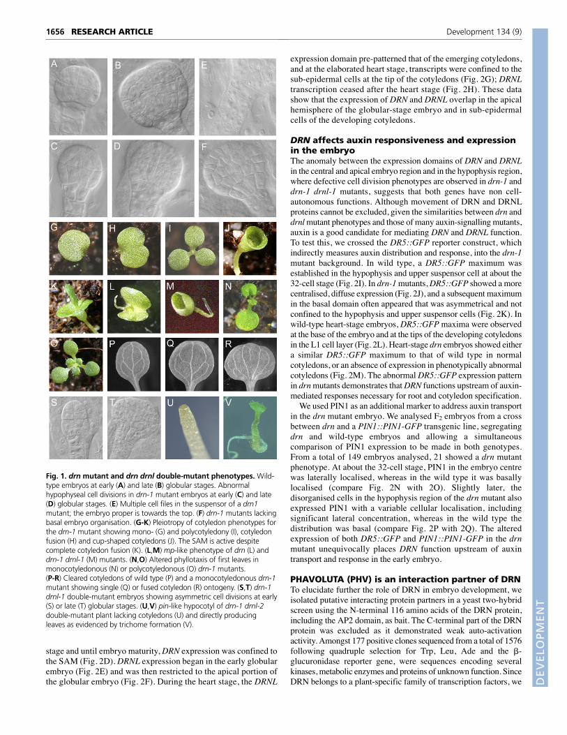
DRN and DRNL are functionally redundantThe incomplete penetrance and similarity of drn and drnl single-mutant cotyledon phenotypes suggest redundant gene functions.We therefore created double mutants between drn-1 and eitherdrnl-1 or the stronger allele drnl-2. Embryonic cell patterningdefects were observed in essentially all (90 of 96) drn-1 drnl-1double-mutant embryos (Fig. 1S,T), considerably higher than thefrequency for drn-1 single-mutant embryos (Table 2). drn-1 drnl-1 double mutants also showed pleiotropic cotyledon phenotypes,including those observed in the single mutants (Table 2).However, the most significant feature of the drn-1 drnl-1 doublemutant was a large increase in phenotypic penetrance: the mp-likephenotype was observed in 20% of mutants, and cotyledondefects in 30%, raising the total penetrance of plants with aphenotype to about 50% (Table 2). Double-homozygous drn-1drnl-2 plants are sterile, but approximately a quarter of theprogeny of drn-1 drnl-2/DRNL plants had pin-like embryos, witha complete absence of cotyledons (Fig. 1U). These plants weregenotyped as double-homozygous mutants and directly initiatedleaves from a functional SAM (Fig. 1V).
Expression patterns of DRN and DRNLIn view of the genetic redundancy observed between DRN andDRNL, we investigated whether the expression patterns of thesegenes overlapped in Arabidopsis embryos using RNA in situhybridisation. DRN is expressed from the four-cell stage (Kirchet al., 2003) and throughout the globular embryo (Fig. 2A). At thetransition stage, DRN expression was localised to the apical celltiers (Fig. 2B), and throughout the heart stage it becameincreasingly restricted to the lobes of the developing cotyledons(Fig. 2C). From the mature heart stage throughout the torpedo
drn and drnl single mutants are affected inembryonic patterning and cotyledonorganogenesisThe major phenotype of the drn mutant is abnormal cell division,observable from the globular embryo stage onwards (Table 2).The wild-type globular embryo contacts the single file of cellsknown as the suspensor through the hypophysis, which dividesasymmetrically in the globular embryo to give: (1) an upper lens-shaped cell (see Fig. 1A,B), which is the progenitor of thequiescent centre; and (2) a subtending daughter cell, which willgenerate the columella stem cells. It is at the transition fromglobular to heart stage that cell divisions parallel to the surface atlateral marginal positions give rise to the emerging cotyledonlobes (West and Harada, 1993). drnl mutants showed no embryodevelopment defects. By contrast, approximately half (48.0%) ofhomozygous drn-1 embryos exhibited a phenotype consisting ofabnormal development in the hypophysis region, or no obviousdistinction between embryo proper and suspensor. In a fewpercent of drn mutants, the hypophyseal cell divided periclinallyat an early stage (Fig. 1C), resulting in the absence of the lens-shaped cell and a subsequent haphazard cellular organisation ofthis region (Fig. 1D,F). At a similar frequency, suspensors of drnmutants had double or triple cell files (Fig. 1E), presumablyresulting from supernumerary cell divisions perpendicular to thenormal plane of division. Both drn and drnl single mutantsshowed defective cotyledon development phenotypes atincomplete penetrance, including monocotyledonous seedlings,seedlings with partially fused cotyledons, tricots or various tricotfusion combinations (Table 2 and Fig. 1G-I). Completely fused orcup-shaped cotyledons were occasionally observed (<1%) in bothdrn mutant alleles, and these plants produced a functional SAMand developed normally (Table 2 and Fig. 1J,K). Also rare (<1%)were plants with a single cotyledon-like structure, no hypocotyland a rudimentary root (Fig. 1L,M), reminiscent of mp (Berlethand Jürgens, 1993) and bdl (Hamann et al., 1999) seedlings,which are mutants in ARF5 (Hardtke and Berleth, 1998) and itsIAA12 partner protein (Hamann et al., 2002). The lowerpenetrance in drn mutants of cotyledon defects as compared withcell division defects indicates that embryo cell patterning defectsare not manifested in post-germination development.
Inappropriate cotyledon development in drn or drnl mutantsresulted in subsequent alterations of leaf phyllotaxy: a single leafinitiated opposite single fused cotyledons (Fig. 1N), or, in the caseof tricots, three leaf primordia initiated between the cotyledons (Fig.1O). Cleared monocotyledonous drn-1 and drnl-1 cotyledons eithershowed a wild-type vasculature pattern (Fig. 1P) with a single mid-vein (Fig. 1Q), or two or more mid-veins (Fig. 1R), showing that thecotyledon derived from either a single cotyledon primordium or
Table 2. Phenotypes of drn, drnl, phv and drn drnl double mutantsWild Single or two Tricots and Cup-shaped mp-like pin embryo Percentage Embryo celltype fused cotyledons various fusions cotyledons phenotype phenotype phenotype division defects*
drn-1 1804 92 13 2 15 0 6.34 48/100 (48.0%)drn-2 2229 53 6 1 4 0 2.79drnl-1 3408 44 3 0 0 0 1.36 0/69 (0.00%)drnl-2 609 46 0 0 0 0 7.6phv 317 0 0 0 0 0 0 0/35 (0.00%)drn-1 phv 2041 138 0 0 28 0 7.55 54/57 (94.7%)drn-1 drnl-1 718† 271† 0 0 398† 0 48.24 90/96 (93.8%)drn-1 drnl-2/DRNL‡ 492 42 2 0 0 192 28.07
*Embryo cell division defects refers to the proportion of embryos from cleared ovules showing cellular defects from globular stage (>32 cells) to early heart stage.†Values are pooled for progeny of eight individual double mutants.‡drn-1 drnl-2 double mutant plants are sterile.
DEVELO
PMENT
expression domain pre-patterned that of the emerging cotyledons,and at the elaborated heart stage, transcripts were confined to thesub-epidermal cells at the tip of the cotyledons (Fig. 2G); DRNLtranscription ceased after the heart stage (Fig. 2H). These datashow that the expression of DRN and DRNL overlap in the apicalhemisphere of the globular-stage embryo and in sub-epidermalcells of the developing cotyledons.
DRN affects auxin responsiveness and expressionin the embryoThe anomaly between the expression domains of DRN and DRNLin the central and apical embryo region and in the hypophysis region,where defective cell division phenotypes are observed in drn-1 anddrn-1 drnl-1 mutants, suggests that both genes have non cell-autonomous functions. Although movement of DRN and DRNLproteins cannot be excluded, given the similarities between drn anddrnl mutant phenotypes and those of many auxin-signalling mutants,auxin is a good candidate for mediating DRN and DRNL function.To test this, we crossed the DR5::GFP reporter construct, whichindirectly measures auxin distribution and response, into the drn-1mutant background. In wild type, a DR5::GFP maximum wasestablished in the hypophysis and upper suspensor cell at about the32-cell stage (Fig. 2I). In drn-1 mutants, DR5::GFP showed a morecentralised, diffuse expression (Fig. 2J), and a subsequent maximumin the basal domain often appeared that was asymmetrical and notconfined to the hypophysis and upper suspensor cells (Fig. 2K). Inwild-type heart-stage embryos, DR5::GFP maxima were observedat the base of the embryo and at the tips of the developing cotyledonsin the L1 cell layer (Fig. 2L). Heart-stage drn embryos showed eithera similar DR5::GFP maximum to that of wild type in normalcotyledons, or an absence of expression in phenotypically abnormalcotyledons (Fig. 2M). The abnormal DR5::GFP expression patternin drn mutants demonstrates that DRN functions upstream of auxin-mediated responses necessary for root and cotyledon specification.
We used PIN1 as an additional marker to address auxin transportin the drn mutant embryo. We analysed F2 embryos from a crossbetween drn and a PIN1::PIN1-GFP transgenic line, segregatingdrn and wild-type embryos and allowing a simultaneouscomparison of PIN1 expression to be made in both genotypes.From a total of 149 embryos analysed, 21 showed a drn mutantphenotype. At about the 32-cell stage, PIN1 in the embryo centrewas laterally localised, whereas in the wild type it was basallylocalised (compare Fig. 2N with 2O). Slightly later, thedisorganised cells in the hypophysis region of the drn mutant alsoexpressed PIN1 with a variable cellular localisation, includingsignificant lateral concentration, whereas in the wild type thedistribution was basal (compare Fig. 2P with 2Q). The alteredexpression of both DR5::GFP and PIN1::PIN1-GFP in the drnmutant unequivocally places DRN function upstream of auxintransport and response in the early embryo.
PHAVOLUTA (PHV) is an interaction partner of DRNTo elucidate further the role of DRN in embryo development, weisolated putative interacting protein partners in a yeast two-hybridscreen using the N-terminal 116 amino acids of the DRN protein,including the AP2 domain, as bait. The C-terminal part of the DRNprotein was excluded as it demonstrated weak auto-activationactivity. Amongst 177 positive clones sequenced from a total of 1576following quadruple selection for Trp, Leu, Ade and the �-glucuronidase reporter gene, were sequences encoding severalkinases, metabolic enzymes and proteins of unknown function. SinceDRN belongs to a plant-specific family of transcription factors, we
stage and until embryo maturity, DRN expression was confined tothe SAM (Fig. 2D). DRNL expression began in the early globularembryo (Fig. 2E) and was then restricted to the apical portion ofthe globular embryo (Fig. 2F). During the heart stage, the DRNL
Fig. 1. drn mutant and drn drnl double-mutant phenotypes. Wild-type embryos at early (A) and late (B) globular stages. Abnormalhypophyseal cell divisions in drn-1 mutant embryos at early (C) and late(D) globular stages. (E) Multiple cell files in the suspensor of a drn1mutant; the embryo proper is towards the top. (F) drn-1 mutants lackingbasal embryo organisation. (G-K) Pleiotropy of cotyledon phenotypes forthe drn-1 mutant showing mono- (G) and polycotyledony (I), cotyledonfusion (H) and cup-shaped cotyledons (J). The SAM is active despitecomplete cotyledon fusion (K). (L,M) mp-like phenotype of drn (L) anddrn-1 drnl-1 (M) mutants. (N,O) Altered phyllotaxis of first leaves inmonocotyledonous (N) or polycotyledonous (O) drn-1 mutants.(P-R) Cleared cotyledons of wild type (P) and a monocotyledonous drn-1mutant showing single (Q) or fused cotyledon (R) ontogeny. (S,T) drn-1drnl-1 double-mutant embryos showing asymmetric cell divisions at early(S) or late (T) globular stages. (U,V) pin-like hypocotyl of drn-1 drnl-2double-mutant plant lacking cotyledons (U) and directly producingleaves as evidenced by trichome formation (V).
Development 134 (9)RESEARCH ARTICLE1656

DEVELO
PMENT
ATHB-15 – The Arabidopsis Information Resource) and ATHB8(Sessa et al., 1994; Prigge et al., 2005). As these family membersact redundantly (Prigge et al., 2005), we investigated whether DRNand DRNL could interact with other class III HD-ZIP membersapart from PHV. We used equivalent C-terminal parts of all five
focussed our analyses on potential transcription factor partners. Ofnote was PHV (AtHB9; At1g30490), which was independentlyisolated eight times, based on cDNA termini sequences. All isolatedPHV clones encoded C-terminal parts of the protein, extendingmaximally from amino acid 754 to the last amino acid, 841.
The PHV expression pattern is described elsewhere (Prigge et al.,2005), but to support the functional significance of the putativeprotein-protein interactions between PHV and DRN or DRNL, wedemonstrated that PHV is temporally and spatially co-expressedwith DRN and DRNL in the proembryo, and is concentrated apicallyin the developing globular embryo before becoming localised to theadaxial side of the developing cotyledons (Fig. 2R-U).
We used two methods to substantiate the affinity of the protein-protein interaction between DRN and PHV shown by the yeast two-hybrid screen. Firstly, we performed co-immunoprecipitation (CoIP)experiments using epitope-tagged full-length DRN, DRNL andPHV. These results (Fig. 3A,B) confirmed the protein-proteininteraction between DRN and PHV and between DRNL and PHV invitro. DRNL could not be co-immunoprecipitated by DRN (Fig.4B), demonstrating that DRN and DRNL are not capable ofheterodimerisation.
Additionally, we used bimolecular fluorescencecomplementation (BiFC) (Walter et al., 2004) in a transient assayin leek epidermal cells to verify the observed biochemicalinteraction between DRN or DRNL and PHV, in vivo, using full-length DRN and PHVs as the C-terminal part of PHV was sufficientto sustain interaction with DRN in the two-hybrid screen. GFPexpression was observed in control experiments (Fig. 3C-E) andYFP expression was reproducibly obtained in multiple independentexperiments when the PHVs BiFC construct was co-bombardedwith DRN or DRNL BiFC constructs (Fig. 3F,G). A series ofnegative controls were performed, including co-bombardment ofempty YFP vectors, and no evidence for DRN heterodimerisationwith DRNL was observed (data not shown). More importantly, noYFP fluorescence was observed following co-bombardment ofDRN in pUC-SPYCE and SHOOTMERISTEMLESS (STM) inpUC-SPYNE, nor in the reciprocal cloning combination (data notshown). These results not only demonstrate that that both DRN andDRNL can form stable heterodimers with the PHV protein inplanta, but that these interactions are specific.
DRN and PHV genetically co-regulate embryopatterningConsidering the biochemical dimerisation between DRN and PHVin vitro and in planta, we asked whether genetic evidence wouldsupport the hypothesis of an active DRN-PHV protein dimer byconstructing a drn-1 phv double mutant. The phv mutant showedwild-type embryo development. However, the drn-1 phv mutantshowed embryo cell division defects similar to those observed in thedrn-1 drnl-1 double mutant and at almost complete penetrance ascompared with that of the drn single mutant (Table 2). Additionally,the penetrance of drn-1 phv cotyledon defects was higher than thatof drn single mutants, with slightly more plants with an mpphenocopy (Table1). The increased penetrance of embryo celldefects of drn-1 phv plants over drn single mutants suggests thatboth genes contribute to the same embryo developmental pathways.
DRN and DRNL can interact with all class III HD-ZIPfamily membersThe Arabidopsis class III HD-ZIP protein family also contains theclosely related homologue of PHV, PHABULOSA (PHB), inaddition to REVOLUTA (REV), CORONA (CNA; also known as
Fig. 2. RNA in situ hybridisations and alteration of DR5 and PIN1expression in drn mutants. (A-D) DRN expression on medianlongitudinal sections of wild-type embryos in early globular (A), lateglobular (B), early heart (C) and torpedo (D) stages. (E-H) DRNLexpression in wild-type embryos in early globular (E), late globular (F),early heart (G) and late heart (H) stages. (I-K) DR5::GFP expression inglobular wild-type (I), globular drn-1 mutant (J) and drn-1 transition-stage (K) embryos. The arrow in J marks the hypophysis region. Notesupernumery cell files in the suspensor (K). (L,M) DR5::GFP expressionin wild-type (L) and drn-1 (M) heart-stage embryos. Note the normaland mutant cotyledons in M. (N-Q) PIN1 expression in wild-type (N) anddrn (O) 32-cell-stage embryos, and in wild-type (P) and drn (Q) globularembryos. Arrows show basal PIN1 polarity (N,P) and abnormal lateralPIN1 expression (O,Q). (R-U) RNA in situ hybridisation for PHV onmedian longitudinal sections of wild-type embryos at early globular (R),late globular (S), heart (T) or late heart (U) stage.
Arabidopsis embryo patterning by DORNRÖSCHEN RESEARCH ARTICLE 1657

DEVELO
PMENT
The interaction between DRN and PHV isdependent on the AP2 domain and the C-terminalconserved PAS-LIKE domainThe C-terminal region of class III HD-ZIP members of Arabidopsisis highly conserved (Fig. 4A) and therefore indicative of conservedfunction. Using the SMART protein motif search of EMBL(http://smart.embl-heidelberg.de), putative homology was found toseveral characterised domains, including the PAS domain – namedafter Drosophila period (PER), vertebrate aryl hydrocarbon receptornuclear translocator (ARNT) and Drosophila single minded (SIM) –which is found in a superfamily of more than 1000 eukaryotic proteinsdirectly or indirectly involved in signal transduction (Mukherjee andBürglin, 2006). Homology to the PAS domain resides between aminoacids 722 and 792 of the PHV protein, within the sequence we haveshown to dimerise with DRN and DRNL. Although this homology isweak, probably representing the low sequence identity between PASdomains (Gilles-Gonzalez and Gonzalez, 2004), this region spans fiveconserved �-helices of the PAS domain. For this reason, weinvestigated whether this region of the PHV protein was sufficient tomaintain high affinity binding to the full-length DRN protein. In pull-down assays, DRN could dimerise with the 70-amino-acid PHV PASfragment (Fig. 4D) and, reciprocally, the PHV PAS fragment was ableto precipitate DRN or DRNL (Fig. 4D), identifying a functionalprotein-protein interaction domain within this region.
To establish which region of the DRN protein is responsible fordimerisation with PHV, we performed CoIP experiments withsubfragments of the N-terminus of DRN used for the two-hybridscreen: (1) the N-terminal polypeptide (amino acids 1-53) excludingthe AP2 domain; and (2) the conserved 61 amino acid AP2 consensusdomain (Kim et al., 2006) (DRN amino acids 54-115). The N-terminal polypeptide of DRN was not able to co-precipitate PHV(data not shown), whereas the AP2 domain could (Fig. 4F),confirming that it is sufficient to mediate heterodimer formation withPHV. The same experiment was performed using the DRNL AP2domain (amino acids 55 to 116), which was also able to precipitatethe PHV polypeptide (Fig. 4F). The AP2 domain is generallyconsidered to comprise a GCC-box DNA-binding motif (Riechmannand Meyerowitz, 1998; Sakuma et al., 2002) composed of three-stranded �-sheets and an �-helix, occurring either in tandem repeatssuch as in the founding family member APETALA2 (Okamuro et al.,1997), or as a single domain as in DRN or DRNL. The 3D structureof the Arabidopsis ERF1 AP2 domain (Allen et al., 1998; Marchler-Bauer et al., 2005) shows that the DNA-binding domain resides inthe N-terminal �-sheet region of the domain, leaving residues withinthe C-terminus to orientate on the opposite face to the DNA-bindingface so as to be sterically potentially able to interact with otherproteins. The cysteine and serine residues in DRN and DRNL AP2domains (Fig. 4E; marked in yellow Fig. 4G) are noteworthy: thecysteine conserved in both DRN and DRNL is unique amongst plantAP2 domain proteins (see Kim et al., 2006) and falls within the �-helix of the RAYD domain, considered to be a conserved structuralmotif relevant to the function of all AP2 proteins (Okamuro et al.,1997).
DISCUSSIONDRN and DRNL are functionally redundantTwo independent drn or drnl mutant alleles share similar pleiotropiccotyledon phenotypes at low penetrance; however, DRN has a morepronounced role in patterning of the hypophysis and suspensorregions because drnl mutants were unaffected in this aspect ofembryo development. The drnl-2 allele showed the highestcotyledon phenotype penetrance and is presumably a stronger allele
proteins corresponding to the PHV sequences obtained from thetwo-hybrid screen (see Materials and methods). His-tagged full-length DRN and DRNL proteins were detected following CoIP viathe HA-tagged C-terminal fragments of all five members of theclass III HD-ZIP family (Fig. 4B). DRN and DRNL can thusdimerise with all Arabidopsis class III HD-ZIP proteins, a findingsupported by their overlapping expression patterns in the embryo(Prigge et al., 2005) (Fig. 2). We also tested the specificity of PHVinteractions by investigating whether the full-length PHV proteincould co-precipitate STM, another protein whose expressiondomain overlaps that of PHV in the embryo. A full-length STM-GFP fusion protein could not be co-precipitated via the HA tag ofPHV (Fig. 4C), confirming that PHV specifically interacts withDRN and DRNL.
Fig. 3. CoIP and BiFC analysis for the interaction between DRN orDRNL and PHV. (A,B) Full-length PHV protein can precipitate full-length DRN (A) or DRNL (B). Protein mixtures were precipitated via PHV-HA. The grid summarises the IP and Co-IP lanes and the antibodiesused. Asterisks mark the co-eluted IgG light chain. (C-E) Cellularfluorescence of GFP-DRN (C), GFP-DRNL (D) or GFP-PHVs (E) in leekepidermal cells. (F,G) Bimolecular fluorescence complementationshowing YFP expression observed with a GFP filter in leek epidermalcells following co-bombardment of full-length DRN and PHVs proteins(F) or full-length DRNL and PHVs proteins (G) fused to complementaryYFP subdomains.
Development 134 (9)RESEARCH ARTICLE1658

DEVELO
PMENT
Fig. 4. The interaction between DRN and class III HD-ZIP family proteins involves the AP2 and PAS-like domains, respectively. (A) Acomparison of the C-terminal regions of the Arabidopsis class III HD-ZIP proteins. Accession numbers for the proteins are as follows: CNA,AAW88440; AtHB8, CAD29660; REV AAF42938; PHV CAD29544; PHB, NP_181018. Homology is compared over the region from PHV amino acid654 to the C-terminus. The MEKHLA domain, homology to the PAS domain from http://smart.embl-heidelberg.de and the �-helices within the PHVPAS domain are marked. (B) Full-length DRN is precipitated by C-terminal regions of all class III HD-ZIP members, but not by DRNL. HA-tagged classIII HD-ZIP proteins or DRNL were used for precipitation of DRN. The grid shows IP and Co-IP lanes and which antibodies were used. The position ofthe DRN protein is marked with an arrowhead. Asterisks represent the IgG heavy or light chains. The control lanes show IPs in the absence of eitheror both in vitro transcribed/translated products, confirming the identity of the lower IgG band. (C) Full-length PHV cannot co-precipitate full-lengthSTM-GFP. The first and third lanes contain in vitro translated proteins before precipitation. PHV-HA was used for precipitation. (D) Full-length DRNcan co-precipitate the 71 amino acid PHVPAS domain (first and second lanes) and, reciprocally, PHVPAS can co-precipitate full-length DRNL (third andfourth lanes). (E) An alignment of the AP2 domains of DRN and DRNL. Identical amino acids are shaded and the position of the unique cysteine andserine residues within the �-helix are in bold. (F) The DRN AP2 domain (IP) can precipitate PHVs (Co-IP) (first and third lanes) and the DRNL AP2domain can precipitate PHVs (second and fourth lanes). (G) 3D crystal structure of the AP2 domain of the Arabidopsis ERF1 protein (Allen et al.,1998; Marchler-Bauer et al., 2005) binding a DNA helix shown on the right, viewed from above into the helix. Cysteine/serine residues of theDRN/DRNL proteins are highlighted in yellow.
Arabidopsis embryo patterning by DORNRÖSCHEN RESEARCH ARTICLE 1659

DEVELO
PMENT
Development 134 (9)RESEARCH ARTICLE
PIN1 distribution in drn embryos unequivocally places DRN functionupstream of the auxin transport fundamental for embryo apical-basalpatterning (Friml et al., 2003; Weijers et al., 2005), a conclusion thatcould not have been derived from altered DR5 activity alone. This issupported by the pin-like phenotype of drn-1 drnl-2 embryos, whichphenocopies pin mutants deficient in polar auxin transport.
Interdomain signalling in Arabidopsis embryos is also suggestedby analyses of embryo-lethal mutants, in which normal embryodevelopment involves the inhibition of the embryonic potential of thesuspensor by the embryo proper (Marsden and Meinke, 1985).Aberrant hypophyseal cell divisions in drn single mutants rarely leadto a defective root phenotype, which suggests an ability of the lowerhypophysis cell to generate columella stem cells or organise the RAM,independent of the number of precursor cells. This supports thehypothesis that additional and partially redundant pathways areactivated as cell number increases during embryogenesis, and thatthese can compensate for early developmental defects (Laux et al.,2004). It should be remembered that stereotypic cell division patternsof Arabidopsis embryos are not representative of plants in general anda single row of suspensor cells is not the rule in dicots; for example,Phaseolus multiflorus has a massive suspensor consisting of multiplecell files merging into the embryo proper (Wardlaw, 1955).
The vasculature of monocotyledonous drn mutants shows thatcotyledons may be single or arise from cotyledon fusion, presumablyvia disruption of cell recruitment into correctly initiated cotyledonsand disruption in the maintenance of cotyledon boundaries duringorganogenesis. This, together with supernumery cotyledons in drn anddrnl mutants, shows that both genes have roles in both cotyledoninitiation and boundary maintenance. Leaf primordia initiate at auxinconcentration maxima via the dynamic expression of PIN1, which actsas an instructive signal for organ initiation (Reinhardt et al., 2003). Indrn and drnl mutants, disruption of leaf phyllotaxis suggests thatcotyledon misdevelopment at least temporarily reprogrammes thephyllotactic inductive signals for leaf initiation, perhaps via furtherdisruption of auxin responses.
DRN and DRNL form heterodimers with class III HD-ZIP proteins via a C-terminal PAS-LIKE domainWe have demonstrated biochemically and via transient in plantaassays that DRN and DRNL can heterodimerise with the C-termini ofall members of the class III HD-ZIP family. The in vivo relevance ofthese interactions is supported by the expression patterns of the genefamily members: all except ATHB8 are co-expressed with DRN andDRNL in the apical embryo domain (Emery et al., 2003). In theabsence of evidence for heterodimerisation between DRN and DRNL,both proteins may individually compete for the same class III HD-ZIPinteraction partners, which also act redundantly (Prigge et al., 2005).
The conserved C-terminus region is characteristic for class III HD-ZIP proteins, although a function has yet to be assigned to it.Mukherjee and Bürglin (Mukherjee and Bürglin, 2006) identify aMEKHLA domain within eukaryotic PAS-containing proteins,specific to class III HD-ZIP genes of higher plants andChlamydomonas reinhardtii. It contains the PAS domain and anadditional 150 amino acids and is hypothesised to represent a discretefunctional unit involved in a signalling pathway (Mukherjee andBürglin, 2006). Our data unequivocally show that a 71 amino acidregion containing five �-helices within the PAS-like domain issufficient to mediate interactions between DRN and PHV. These �-helices and the associated five- to six-stranded antiparallel �-barrelsform a pocket that may contain various prosthetic groups (Mukherjeeand Bürglin, 2006). PAS domains have been reported to mediateprotein-protein interactions (Taylor and Zhulin, 1999; Card et al.,
than drnl-1. The mutated Ala residue has been shown in Brassicanapus ERF/AP2 proteins to be essential for DNA binding (Liu et al.,2006), suggesting that the mutated drnl-2 protein is unable to bindtarget genes. Importantly, the penetrance of hypophysis or cotyledonphenotypes significantly increased in drn-1 drnl-1 double mutants,thereby demonstrating a role for DRN and DRNL in both the apicaland basal domain of the Arabidopsis embryo and that the genes actredundantly. The penetrance of drn-1 drnl-1 double-mutantphenotypes was still incomplete compared with that of drn-1 drnl-2 double mutants, probably reflecting different drnl-1 allelestrengths. The complete lack of cotyledons at almost completepenetrance in drn-1 drnl-2 mutants demonstrates that both genestogether are essential for cotyledon initiation.
Functional contributions of DRN and DRNL can be separated in theapical and basal embryo domains: loss of function of either gene in theapical domain is manifest in the cotyledon phenotype observed insingle and double mutants. This correlates with the expression patternof both genes in apical cell tiers concomitant with cotyledon initiation.No cell patterning defects were observed in the basal embryo domainup to the 16-cell stage of drn-1 drnl-1 mutants, despite transcriptionalactivity of both genes throughout the proembryo. However, about50% of drn single-mutant embryos and essentially all drn-1 drnl-1double-mutant embryos exhibited subsequent cell division defects inthe hypophyseal region and subtending suspensor cells in the absenceof either DRN or DRNL transcription in these cells. The spatialseparation of gene expression and hypophyseal phenotype, therefore,must involve either mobile DRN or DRNL proteins or otherinterdomain signaling components. This is similar to the situationobserved for BDL and MP (Berleth and Jürgens, 1993; Hamann et al.,1999), which are expressed in proembryo cells but not in thehypophysis, which in mp and bdl mutants fails to undergo theasymmetric division that gives rise to the quiescent centre precursorsand columella initials. MP and BDL therefore affect the hypophysisin a non-cell-autonomous manner and signalling between embryoproper and underlying hypophysis is essential for normal rootdevelopment. A transient indirect response to auxin together with apostulated additional factor operating downstream of MP and BDunderlies this cell-to-cell signalling (Weijers et al., 2006). Expressionof DR5::GFP in drn mutants is more informative in explaining thespatial anomaly between DRN/DRNL gene expression domains andphenotype. In wild type, an auxin maximum is established in thehypophysis and upper suspensor cell at the late globular stage and isnecessary for hypophysis cell fate specification (Friml et al., 2003).This maximum as reported by DR5::GFP is absent in drn mutantglobular embryos, similar to the situation in bdl mutants (Weijers etal., 2006), and might explain the cell division defects in thehypophysis region. The absence of local DR5::GFP response at thecotyledon tips and in provascular strands in drn mutant cotyledonscorrelates with phenotypic defects in cotyledon initiation.
PIN1, an additional auxin marker, marks all cell boundaries up tothe 16-cell stage in wild type, before the polarity in expression isestablished that concentrates PIN1 basally in the provascular cellsfacing the hypophysis (Steinman et al., 1999; Friml et al., 2003). Thevariable polarity of PIN1 in cells of the hypophysis region of drnmutants suggests an alteration in directed auxin flow, which mightexplain the absence of DR5::GFP accumulation here at an appropriatetemporal phase. Although variable PIN1 polarity correlates with theabnormal cell divisions in the hypophysis region, it is distinct from thedomain of DRN transcription. This strengthens our conclusion that drncell division phenotypes arise either from movement of the DRNprotein or that DRN functions upstream of auxin transport andinvolves additional interdomain signaling components. The altered
1660

DEVELO
PMENT
Arabidopsis embryo patterning by DORNRÖSCHEN RESEARCH ARTICLE 1661
required for normal cotyledon development. PHV and DRN do notpromiscuously interact with other proteins, such as STM, that are co-expressed in the early embryo, supporting the specificity of complexesinvolving DRN and PHV.
We show in this paper that two Arabidopsis AP2-domaincontaining paralogues control embryo development in specific andearly embryo expression domains. Both proteins can form protein-protein interactions with class III HD-ZIP proteins and with PHV viaa PAS-like domain in the C-terminal region of PHV and PHB, and theAP2 domain of DRN and DRNL. Our data suggest that transcriptionalcomplexes involving DRN and DRNL act redundantly with their classIII HD-ZIP partners to control embryo organogenesis and patterning.The robustness of embryonic patterning as suggested by the lowpenetrance of mutant phenotypes therefore finds a biochemical basisin the promiscuity of transcription factor interactions. Theidentification of DRN and DRNL as partners of class III HD-ZIPproteins therefore enables biochemical access to signal transductioncascades in the embryo on the basis of genetic pathways.
We thank Thomas Jack, Anwesha Nag and Yingzhen Yang for the drnl-2 alleleprior to publication; Hans Sommer for the library for two-hybrid screening;Klaus Harter for providing vectors for BiFC; and J. Friml for the DR5::GFPer andPIN1::PIN-GFP constructs. This project was funded by the DeutscheForschungsgemeinschaft through SFB 572.
Supplementary materialSupplementary material for this article is available athttp://dev.biologists.org/cgi/content/full/134/9/1653/DC1
ReferencesAida, M., Ishida, T., Fukaki, H., Fujisawa, H. and Tasaka, M. (1997). Genes
involved in organ separation in Arabidopsis: an analysis of the cup-shapedcotyledon mutant. Plant Cell 9, 841-857.
Allen, M. D., Yamasaki, K., Ohme-Takagi, M., Tateno, M. and Suzuki, M.(1998). A novel mode of DNA recognition by a �-sheet revealed by the solutionstructure of the GCC-box binding domain in complex with DNA. EMBO J. 17,5484-5496.
Bao, N., Lye, K.-W. and Barton, M. K. (2004). MicroRNA binding sites inArabidopsis class III HD-ZIP mRNAs are required for methylation of the templatechromosome. Dev. Cell 7, 653-662.
Banno, H., Ikeda, Y., Niu, Q. W. and Chua, N.-H. (2001). Overexpression ofArabidopsis ESR1 induces initiation of shoot regeneration. Plant Cell 13, 2609-2618.
Bennet, S. R. M., Alvarez, J., Bossinger, G. and Smyth, D. R. (1995).Morphogenesis in pinoid mutants of Arabidopsis thaliana. Plant J. 8, 505-520.
Berleth, T. and Jürgens, G. (1993). The role of the monopteros gene inorganising the basal body region of the Arabidopsis embryo. Development 118,575-587.
Bradley, D., Carpenter, R., Sommer, H., Hartley, N. and Coen, E. (1993).Complementary floral homeotic phenotypes result from opposite orientations ofa transposon at the plena locus of Antirrhinum. Cell 72, 85-95.
Card, P. B., Erbel, P. J. and Gardner, K. H. (2005). Structural basis of ARNT PAS-Bdimerization: use of a common beta-sheet interface for hetero- andhomodimerization. J. Mol. Biol. 353, 664-677.
Cole, M., Nolte, C. and Werr, W. (2006). Nuclear import of the transcriptionfactor SHOOTMERISTEMLESS depends on heterodimerisation with BLH proteinsexpressed in discrete sub-domains of the shoot apical meristem of Arabidopsisthaliana. Nucleic Acids Res. 34, 1281-1292.
Elliott, R. C., Betzner, A. S., Huttner, E., Oakes, M. P., Tucker, W. Q., Gerentes,D., Perez, P. and Smyth, D. R. (1996). AINTEGUMENTA, an APETALA2-likegene of Arabidopsis with pleiotropic roles in ovule development and floral organgrowth. Plant Cell 8, 155-168.
Emery, J. F., Floyd, S. K., Alvarez, J., Eshed, Y., Hawker, N. P., Izhaki, A.,Baum, S. F. and Bowman, J. L. (2003). Radial patterning of Arabidopsis shootsby class III HD-ZIP and KANADI genes. Curr. Biol. 13, 1768-1774.
Friml, J., Vieten, A., Sauer, M., Weijers, D., Schwarz, H., Hamann, T.,Offringa, R. and Jürgens, G. (2003). Efflux-dependent auxin gradientsestablish the apical-basal axis of Arabidopsis. Nature 426, 147-152.
Friml, J., Yang, X., Michniewicz, M., Weijers, D., Quint, A., Tietz, O.,Benjamins, R., Ouwerkerk, P. B., Ljung, K., Sandberg, G. et al. (2004). aPINOID-dependent binary switch in apical-basal PIN polar targeting directs auxinefflux. Science 306, 862-865.
Furutani, M., Vernoux, T., Traas, J., Kato, T., Tasaka, M. and Aida, M. (2004).
2005) and contain two highly conserved S1 and S2 regions (Zhulin etal., 1997). Bacterial PAS domains sense oxygen via a bound hememolecule and propagate a signal via the His kinase pathway (Hao etal., 2002). Plant PAS proteins possibly function similarly, or theMEKHLA domain might provide a docking structure to recruit otherproteins into a transcriptionally functional complex. Although plantHD-ZIP proteins efficiently bind DNA as homo- or heterodimers(Sessa et al., 1993; Johannesson et al., 2001), it can be envisaged thathigher order protein complexes containing DRN and/or DRNL andpossibly other proteins are formed and co-ordinately act as atranscriptional unit in the control of embryo patterning. In vitro CoIPexperiments demonstrate heterodimerisation between full-lengthDRN and PHV proteins, and BiFC experiments performed with thePHV C-terminus and full-length DRN further confirm the interactionin planta. PHV and PHB are involved in adaxial/abaxial leafpatterning (McConnell et al., 2001; Emery et al., 2003) and are alsoexpressed in the SAM and during early stages of leaf developmentwhere DRN remains active postembryonically (McConnell et al.,2001; Kirch et al., 2003), suggesting that class III HD-ZIP proteinsand AP2-class transcription factors such as DRN or DRNL might bepartners throughout the plant life cycle.
DRN and PHV act in a common embryonicpatterning pathwayWe have supported DRN-PHV protein interaction data with geneticdata showing a combined genetic effect of PHV and DRN inembryonic patterning: enhanced embryo cell division defects of thedrn-1 phv double mutant show that both genes contribute to the sameembryonic patterning pathways. This might reflect partial redundancybetween both gene functions; but if DRN and PHV act in a commonprotein complex, and as both DRN and DRNL are functionallyredundant and can interact with other partially redundant class III HD-ZIP family members, it is more likely to reflect redundancy involvingother heterodimer combinations between different members of bothprotein families.
The AP2 domain is responsible for protein-proteininteractionsOur finding that the DRN and DRNL AP2 domain alone is sufficientto mediate heterodimerisation with PHV is to the best of ourknowledge the first experimental evidence that plant AP2 domainshave a role in protein dimerisation as well as in DNA binding. AP2proteins are plant-specific and with 144 AP2/ERF members, compriseone of the largest transcription factor families in Arabidopsis (Sakumaet al., 2002). They are key regulators in diverse developmentalprocesses such as flower formation [AP2 (Jofuku et al., 1994)], ovuledevelopment [AINTEGUMENTA (Elliott et al., 1996; Klucher et al.,1996)] and abiotic stress [TaDREB1 (Shen et al., 2003)]. Both thetandem repeat unit and the single AP2 domain comprise functionalDNA-binding motifs. The cysteine and serine residues in the DRNLand DRN AP2 domains, unique among Arabidopsis AP2 domains,reside within the core �-helix of the RAYD element, which has beenproposed to mediate protein-protein interactions (Okamuro et al.,1997). Two alternative effects of dimerisation which would affect theregulation of target genes are that either the interaction between DRNor DRNL and class III HD ZIP proteins sterically interferes with theDNA-binding activity of the AP2 domain, or it contributes to theDNA-binding specificity/affinity of the AP2 domain, as is known forprotein-DNA interactions mediated by the homeodomain (Moens andSelleri, 2006). Based on overlapping transcription patterns, aheterodimeric complex between DRN-PHV or DRNL-PHV or relatedHD-ZIP III proteins could control the transcription of target genes

DEVELO
PMENT
Development 134 (9)RESEARCH ARTICLE
Kieber, J. (2006). A subset of Arabidopsis AP2 transcription factors mediatescytokinin responses in concert with a two-component pathway. Proc. Natl. Acad.Sci. USA 103, 11081-11085.
Reinhardt, D., Pesce, E. R., Steiger, P., Mandel, T., Baltensperger, K., Bennett,M., Traas, J., Friml, J. and Kuhlemeier, C. (2003). Regulation of phyllotaxis bypolar auxin transport. Nature 426, 255-260.
Riechmann, J. L. and Meyerowitz, E. M. (1998). The AP2/EREBP family of planttranscription factors. Biol. Chem. 379, 633-646.
Saibo, N. J. M., Vriezen, W. H., De Grauwe, L., Azmi, A., Prinsen, E. and Vander Straeten, D. (2006). A comparative analysis of the Arabidopsis mutantamp1-1 and a novel weak amp1 allele reveals new functions of the AMP1protein. Planta 225, 831-842.
Sakuma, Y., Liu, Q., Dubouzet, J. G., Abe, H., Shinozaki, K. and Yamaguchi-Shinozaki, K. (2002). DNA-binding specificity of the ERF/AP2 domain ofArabidopsis DREBs, transcription factors involved in dehydration- and cold-inducible gene expression. Biochem. Biophys. Res. Commun. 290, 998-1009.
Sessa, G., Morelli, G. and Ruberti, I. (1993). The Athb-1 and -2 HD-Zip domainshomodimerize forming complexes of different DNA binding specificities. EMBOJ. 12, 3507-3517.
Sessa, G., Carabelli, M., Ruberti, I., Lucchetti, S., Baima, S. and Morelli, G.(1994). Identification of distinct families of HD-Zip proteins in Arabidopsisthaliana. In Molecular-Genetic Analysis of Plant Development and Metabolism(ed. C. Puigdomenèch and G. Coruzzi), pp. 411-426. Berlin: Springer Verlag.
Sessions, A., Burke, E., Presting, G., Aux, G., McElver, J., Patton, D., Dietrich,B., Ho, P., Bacwaden, J., Ko, C. et al. (2002). A high-throughput Arabidopsisreverse genetics system. Plant Cell 14, 2985-2994.
Shen, Y. G., Zhang, W. K., He, S. J., Zhang, J. S., Liu, Q. and Chen, S. Y.(2003). An EREBP/AP2-type protein in Triticum aestivum was a DRE-bindingtranscription factor induced by cold, dehydration and ABA stress. Theor. Appl.Genet. 106, 923-930.
Speulman, E., Metz, P. L. J., van Arkel, G., te Lintel Hekkert, B., Stiekema, W.J. and Pereira, A. (1999). A two-component Enhancer-Inhibitor transposonmutagenesis system for functional analysis of the Arabidopsis genome. Plant Cell11, 1853-1866.
Steinmann, T., Geldner, N., Grebe, M., Mangold, S., Jackson, C. L., Paris, S.,Gälweiler, L., Palme, K. and Jürgens, G. (1999). Coordinated polar localizationof auxin efflux carrier PIN1 by GNOM ARF GEF. Science 286, 316-318.
Talbert, P. B., Adler, H. T., Parks, D. W. and Comai, L. (1995). The REVOLUTAgene is necessary for apical meristem development and for limiting celldivisions in the leaves and stems of Arabidopsis thaliana. Development 121,2723-2735.
Taylor, B. L. and Zhulin, I. B. (1999). PAS domains: internal sensors of oxygen,redox potential, and light. Microbiol. Mol. Biol. Rev. 63, 479-506.
Tissier, A. F., Marillonnet, S., Klimyuk, V., Patel, K., Torres, M. A., Murphy, G.and Jones, J. D. (1999). Multiple independent defective suppressor-mutatortransposon insertions in Arabidopsis: a tool for functional genomics. Plant Cell11, 1841-1852.
Überlacker, B. and Werr, W. (1996). Optimized vectors for expression andtransfer of large open reading frames in transgenic plants. Mol. Breed. 2, 293-295.
Vieten, A., Vanneste, S., Wisniewska, J., Benkova, E., Benjamins, R.,Beeckman, T., Luschnig, C. and Friml, J. (2005). Functional redundancy of PINproteins is accompanied by auxin-dependent cross-regulation of PIN expression.Development 132, 4521-4531.
Vroeman, C. W., Mordhorst, A. P., Albrecht, C., Kwaaitaal, M. A. and deVries, S. C. (2003). The CUP-SHAPED COTYLEDON3 gene is required forboundary and shoot apical meristem formation in Arabidopsis. Plant Cell 15,1563-1577.
Walter, M., Chaban, C., Schütze, K., Batistic, O., Weckermann, K., Näke, C.,Blazevic, D., Grefen, C., Schumacher, K., Oecking, C. et al. (2004).Visualization of protein interactions in living plant cells using bimolecularfluorescence complementation. Plant J. 40, 428-438.
Ward, J. M., Smith, A. M., Shah, P. K., Galanti, S. E., Yi, H., Demianski, A. J.,van der Graaff, E., Keller, B. and Neff, M. M. (2006). A new role for theArabidopsis AP2 transcription factor, LEAFY PETIOLE, in gibberellin-inducedgermination is revealed by the misexpression of a homologous gene, SOB2/DRN-LIKE. Plant Cell 18, 29-39.
Wardlaw, C. W. (1955). Embryogenesis in Plants. London: Methuen.Weijers, D., Sauer, M., Meurette, O., Frimil, J., Ljung, K., Sandberg, G.,
Hooykaas, P. and Offringa, R. (2005). Maintenance of embryonic auxindistribution for apical-basal patterning by PIN-FORMED-dependent auxintransport in Arabidopsis. Plant Cell 17, 2517-2526.
Weijers, D., Schlereth, A., Ehrismann, J. S., Schwank, G., Kientz, M. andJürgens, G. (2006). Auxin triggers transient local signaling for cell specificationin Arabidopsis embryogenesis. Dev. Cell 10, 265-270.
West, M. A. L. and Harada, J. (1993). Embryogenesis in higher plants: anoverview. Plant Cell 5, 1361-1369.
Zhulin, I. B., Taylor, B. L. and Dixon, R. (1997). PAS domain S-boxes in Archaea,Bacteria and sensors for oxygen and redox. Trends Biochem. Sci. 22, 331-333.
PIN-FORMED1 and PINOID regulate boundary formation and cotyledondevelopment in Arabidopsis embryogenesis. Development 131, 5021-5030.
Gilles-Gonzalez, M. A. and Gonzalez, G. (2004). Signal transduction by heme-containing PAS-domain proteins. J. Appl. Physiol. 96, 774-783.
Hamann, T., Mayer, U. and Jürgens, G. (1999). The auxin-insensitive bodenlosmutation affects primary root formation and apical-basal patterning in theArabidopsis embryo. Development 126, 1387-1395.
Hamann, T., Benkova, E., Baurle, I., Kientz, M. and Jürgens, G. (2002). TheArabidopsis BODENLOS gene encodes an auxin response protein inhibitingMONOPTEROS-mediated embryo patterning. Genes Dev. 16, 1610-1615.
Hao, B., Isazza, C., Arndt, J., Soltis, M. and Chan, M. K. (2002). Structure-based mechanism of O2 sensing and ligand discrimination by the FixL hemedomain of Bradyrhizobium japonicum. Biochemistry 41, 12952-12958.
Harada, J. J. (1999). Signaling in plant embryogenesis. Curr. Opin. Plant Biol. 2,23-27.
Hardtke, C. S. and Berleth, T. (1998). The Arabidopsis gene MONOPTEROSencodes a transcription factor mediating embryo axis formation and vasculardevelopment. EMBO J. 17, 1405-1411.
Hibara, K., Karim, M. R., Takada, S., Taoka, K., Rurutani, M., Aida, M. andTasaka, M. (2006). Arabidopsis CUP-SHAPED COTYLEDON3 regulatespostembryonic shoot meristem and organ boundary formation.
Ikeda, Y., Banno, H., Niu, Q.-W., Howell, S. and Chua, N.-H. (2006). TheENHANCER OF SHOOT REGENERATION 2 gene of Arabidopsis regulates CUPSHAPED COTYLEDON 1 at the transcriptional level and controls cotyledondevelopment. Plant Cell Physiol. 47, 1443-1456.
Jofuku, K. D., den Boer, B. G., Van Montagu, M. and Okamuro, J. K. (1994).Control of Arabidopsis flower and seed development by the homeotic geneAPETALA2. Plant Cell 6, 1211-1225.
Johannesson, H., Wang, Y. and Engstrom, P. (2001). DNA-binding anddimerization preferences of Arabidopsis homeodomain-leucine zippertranscription factors in vitro. Plant Mol. Biol. 45, 63-73.
Kim, S., Soltis, P. S., Wall, K. and Soltis, D. E. (2006). Phylogeny and domainevolution in the APETALA2-like gene family. Mol. Biol. Evol. 23, 107-120.
Kirch, T., Simon, R., Grunewald, M. and Werr, W. (2003). TheDORNROESCHEN/ENHANCER OF SHOOT REGENERATION1 gene of Arabidopsisacts in the control of meristem cell fate and lateral organ development. PlantCell 15, 694-705.
Klucher, K. M., Chow, H., Reiser, L. and Fischer, R. L. (1996). TheAINTEGUMENTA gene of Arabidopsis required for ovule and femalegametophyte development is related to the floral homeotic gene APETALA2.Plant Cell 8, 137-153.
Laux, T., Würschum, T. and Breuninger, H. (2004). genetic regulation ofembryonic pattern formation. Plant Cell 16, S190-S202.
Liu, C.-M., Xu, Z.-H. and Chua, N.-H. (1993). Auxin polar transport is essentialfor the establishment of bilateral symmetry during early plant embryogenesis.Plant Cell 5, 621-630.
Liu, Y., Zhao, T. J., Liu, J. M., Liu, Q., Yan, Y. B. and Zhou, H. M. (2006). Theconserved Ala37 in the ERF/AP2 domain is essential for binding with the DREelement and the GCC box. FEBS Lett. 580, 1303-1308.
Marchler-Bauer, A., Anderson, J. B., Cherukuri, P. F., DeWeese-Scott, C.,Geer, L. Y., Gwadz, M., He, S., Hurwitz, D. I., Jackson, J. D., Ke, Z. et al.(2005). CDD: a Conserved Domain Database for protein classification. NucleicAcids Res. 33, D192-D196.
Marsch-Martinez, N., Greco, R., Becker, J. D., Dixit, S., Bergervoet, J. H. W.,Karaba, A., de Folter, S. and Pereira, A. (2006). BOLITA, an ArabidopsisAP2/ERF-like transcription factor that affects cell expansion andproliferation/differentiation pathways. Plant Mol. Biol. 62, 825-843.
Marsden, M. P. F. and Meinke, D. (1985). Abnormal development of thesuspensor in an embryo-lethal mutant of Arabidospis thaliana. Am. J. Bot. 72,1801-1812.
Mayer, U., Torres Ruiz, R. A., Berleth, T., Misear, S. and Jürgens, G. (1991).Mutations affecting body organization in the Arabidopsis embryo. Nature 353,402-407.
McConnell, J. R., Emery, J., Eshed, Y., Bao, N., Bowman, J. and Barton, M. K.(2001). Role of PHABULOSA and PHAVOLUTA in determining radial patterningin shoots. Nature 411, 709-713.
Moens, C. B. and Selleri, L. (2006). Hox cofactors in vertebrate development.Dev. Biol. 291, 193-206.
Mukherjee, K. and Burglin, T. R. (2006). MEKHLA, a novel domain with similarityto PAS domains, is fused to plant homeodomain lecine zipper III proteins. PlantPhysiol. 140, 1142-1150.
Okamuro, J. K., Caster, B., Villarroel, R., Van Montagu, M. and Joufuku, K.D. (1997). The AP2 domain of APETALA2 defines a large new family of DNAbinding proteins in Arabidopsis. Proc. Natl. Acad. Sci. USA 94, 7076-7081.
Prigge, M. J., Otsuga, D., Alonso, J. M., Ecker, J. R., Drews, G. N. and Clarke,S. E. (2005). Class III homeodomain-leucine zipper gene family members haveoverlapping, antagonistic and distinct roles in Arabidopsis development. PlantCell 17, 61-76.
Rashotte, A., Mason, M. G., Hutchison, C., Ferreira, F. J., Schaller, G. E. and
1662