development 135, 4015-4024 (2008) …dev.biologists.org/content/develop/135/24/4015.full.pdf ·...
TRANSCRIPT
4015RESEARCH ARTICLE
INTRODUCTIONWnt signaling controls diverse developmental processes such as axisformation, anteroposterior patterning and the development of theneural crest (Moon et al., 1997; Raible and Ragland, 2005; Wodarzand Nusse, 1998). Precise regulation is necessary for manydevelopmental processes and misregulation of several componentsof the canonical Wnt signaling pathway has been implicated incancer formation (Polakis, 2007). Conversely, reduction or loss offunction of Wnt signaling leads to general developmental defects orloss of organs, demonstrating the need for a tight regulation of thelevels of Wnt signaling (Logan and Nusse, 2004).
The Wnt signaling pathway is highly conserved among allmetazoans. Wnt ligands bind to frizzled (fz) transmembranereceptors leading to accumulation and nuclear localization of β-catenin, which serves as a transcriptional co-activator for TCF/Leftranscription factors (Logan and Nusse, 2004; Wodarz and Nusse,1998). In addition to this so called ‘canonical’ Wnt signalingpathway, fz receptors also activate alternative signaling pathwayslike the planar cell polarity (PCP) pathway, which defines theorientation of cells in the plane of an epithelium (Klein andMlodzik, 2005; Seifert and Mlodzik, 2007). PCP signaling hasbeen best characterized in Drosophila, where it determines forexample the ommatidia organization in the eye and the bristle hairorientation in the wing (Axelrod and McNeill, 2002; Klein andMlodzik, 2005). In vertebrates, PCP signaling is necessary for theorientation of the stereocilia bundles in the neurosensoryepithelium of the inner ear, and dynamic convergent extensionmovements during gastrulation and neurulation (Wallingford etal., 2002; Wang and Nathans, 2007).
Dishevelled (dsh) is a key regulator of both the canonical as wellas the PCP signaling pathway and regulates cell fate specification aswell as cell movements such as convergent extension. Dsh consistsof three major conserved domains, the DIX, PDZ and DEP domain,that have been implicated in different downstream signaling events(Boutros and Mlodzik, 1999; Wallingford and Habas, 2005). TheDIX domain is used for canonical Wnt signaling, whereas the DEPdomain is involved in PCP signaling (Boutros et al., 1998; Habas etal., 2003; Itoh et al., 2000; Rothbacher et al., 2000). By contrast, thePDZ domain is shared by both pathways. As downstream effectorsof dsh are distinct for canonical and PCP signaling, it remainsunclear how dsh selectively activates one or the other pathway. Inthe vertebrate PCP pathway, the signaling mechanisms affecting thesubcellular localization and choice of effectors proteins of dsh arenot well defined.
PTK7 (protein tyrosine kinase 7) is a regulator of PCP signalingthat could modulate the dsh localization as well as the interactionwith pathway-specific effector proteins. PTK7 regulates PCP in theinner ear hair cells and during neural tube closure in mice (Lu et al.,2004). In Xenopus, PTK7 is required for neural convergentextension (Lu et al., 2004). PTK7 is a transmembrane proteincontaining seven extracellular immunoglobulin domains and akinase homology domain. Although the kinase domain lacks theDFG triplet necessary for catalytic activity, its overall structure isevolutionary conserved from Hydra to humans (Kroiher et al., 2001;Miller and Steele, 2000). The signaling mechanism of PTK7 has notbeen characterized, but considering the structure and function ofPTK7, it could affect the localization of dsh as well as itsdownstream signaling.
Neural crest migration is a new system to analyze the mechanisticrole of PTK7 in vertebrate PCP signaling. Neural crest cells areinduced at the border region of the neural plate and migrate ondefined routes throughout the embryo, where they give rise to avariety of derivatives ranging from neurons and glia cells of theperipheral nervous system, to cartilage and pigment cells. Althoughcanonical Wnt signaling plays a role in the induction, delaminationand differentiation of neural crest cells (Schmidt and Patel, 2005;
PTK7 recruits dsh to regulate neural crest migrationIryna Shnitsar and Annette Borchers*
PTK7 regulates planar cell polarity (PCP) signaling during vertebrate neural tube closure and establishment of inner ear hair cellpolarity; however, its signaling mechanism is unknown. Here, we demonstrate a new function for PTK7 in Xenopus neural crestmigration and use this system in combination with in vitro assays to define the intersection of PTK7 with the non-canonical Wntsignaling pathway that regulates PCP. In vitro, using Xenopus ectodermal explants, we show that PTK7 recruits dishevelled (dsh) tothe plasma membrane, a function that is dependent on the PDZ domain of dsh, as well as on the conserved kinase domain of PTK7.Furthermore, endogenous PTK7 is required for frizzled7-mediated dsh localization. Immunoprecipitation experiments confirm thatPTK7 can be found in a complex with dsh and frizzled7, suggesting that it cooperates with frizzled to localize dsh. To evaluate thein vivo relevance of the PTK7-mediated dsh localization, we analyzed Xenopus neural crest migration, as loss-of-function of PTK7inhibits neural crest migration in whole embryos as well as in transplanted neural crest cells. Supporting the in vivo role of PTK7 inthe localization of dsh, a PTK7 deletion construct deficient in dsh binding inhibits neural crest migration. Furthermore, the PTK7-mediated membrane localization of a dsh deletion mutant lacking PCP activity inhibits neural crest migration. Thus, PTK7 regulatesneural crest migration by recruiting dsh, providing molecular evidence of how PTK7 intersects with the PCP signaling pathway toregulate vertebrate cell movements.
KEY WORDS: Planar cell polarity, Neural crest migration, Xenopus, dsh, fz7
Development 135, 4015-4024 (2008) doi:10.1242/dev.023556
Department of Developmental Biochemistry, Center for Molecular Physiology of the Brain (CMPB), GZMB, University of Goettingen, Justus-von-Liebig-Weg 11,37077 Goettingen, Germany.
*Author for correspondence (e-mail: [email protected])
Accepted 7 October 2008 DEVELO
PMENT
4016
Yanfeng et al., 2003), PCP signaling seems to be required for neuralcrest migration. De Calisto et al. have shown that a dsh mutantlacking PCP activity inhibits neural crest migration. Conversely,inhibition of neural crest migration by loss of Wnt11 function canbe rescued by expressing a dsh mutant, which activates PCPsignaling in neural crest cells (De Calisto et al., 2005). Furthermore,‘core-PCP signals’ such as Van Gogh/strabismus, prickle and daamare expressed in cranial neural crest cells (Bekman and Henrique,2002; Darken et al., 2002; Goto and Keller, 2002; Nakaya et al.,2004), indicating that the PCP signaling cascade is active duringneural crest migration.
Here, we identify a new function for PTK7 in neural crestmigration. By analyzing the signaling mechanism of Xenopus PTK7in vitro in animal cap explants and in vivo in migrating neural crestcells, we provide evidence that PTK7 localizes dsh to the plasmamembrane and that this function is required for neural crestmigration.
MATERIALS AND METHODSConstruct designMyc-tagged PTK7 (PTK7-myc) was amplified by PCR (forward primers5�CGGGATCCATGGGGCCGATTGTGCTC3�, reverse primers5�CCATCGATACCCTTGTGTCTTGCTGCC3�), cut with BamHI andClaI, and ligated into the respective restriction sites of pCS2-MT. HA-taggedPTK7 (PTK7-HA) was obtained from PTK7-myc by removing the myc-tagwith ClaI and XbaI, and inserting an HA-tag generated using the followingprimers: forward primer, 5�CGATATCCCTACGATGT TCCAGAT -TATGCATGATAA3�; reverse primer 5�CTAGTTGCGTAATCCG -GTACATCGTAAGGGTAGT3�. The deletion of the predicted kinasedomain of PTK7 (�kPTK7-myc) was introduced by PCR amplification of PTK7-myc using the following primers: forward,5�CTTGTCGCCAGAGCTGTGTC3�; reverse, 5�TCTTCTGGCAGCA -AGACACAAG3�. RNA transcribed from PTK7 constructs cloned in pCS2is significantly more potent than RNA generated from the constructspublished by Lu et al. (Lu et al., 2004); therefore lower concentrations wereinjected.
For neural crest-specific expression, a minimal slug promoter, a700BA(Vallin et al., 2001), was excised with SpeI/BamHI from a700BA-GFP(Vallin et al., 2001) and ligated into the SpeI/BamHI sites of BS-I-Sce-II KS(Pan et al., 2006) generating slug-BS-I-Sce-II KS. To express PTK7 underthe control of the slug promoter, full-length PTK7 was excised usingSal/BspE1, blunted and ligated into the BamHI site of slug-BS-I-Sce-II KSgenerating slug-PTK7. For cloning of the kinase deletion mutant (slug-�kPTK7), full-length PTK7 was cut with SalI/Bsu361, blunted and ligatedinto the BamHI site of slug-BS-I-Sce-II KS. To express GFP in neural crestcells, the minimal slug promoter and the GFP-coding sequence were excisedwith SpeI/NotI from a700BA-GFP (Vallin et al., 2001) and inserted into theSpeI/NotI sites of BS-I-Sce-II KS. All constructs cloned in slug-BS-I-Sce-II KS were purified using QIAprep Spin Miniprep Kit (Qiagen) previous toinjection in Xenopus embryos.
Xenopus injection and neural crest transplantationXenopus injection experiments were performed as previously described(Borchers et al., 2001). The PTK7 MO sequences were published elsewhere(Lu et al., 2004). Here, a 1:1 mixture of MO2 and MO3 was used at totalconcentrations indicated in the text. As a toxicity control, identicalconcentrations of control MO (Gene Tools) were used. To evaluate neural crestmigration independently of earlier neural tube closure defects, PTK7 MOinjected embryos with severe neural tube closure phenotypes were excluded.
For RNA injection experiments, capped mRNA was synthesized usingthe mMESSAGE mMACHINE Kit (Ambion) according to themanufacturer’s instructions. The following published plasmids were used tosynthesize RNA for dsh-GFP (Yang-Snyder et al., 1996), dsh�DIX-GFP,dsh�PDZ-GFP, dsh�DEP-GFP (Miller et al., 1999), dsh-myc (Sokol, 1996),fz7-myc (Winklbauer et al., 2001) and lacZ (Smith and Harland, 1991). Forin situ hybridization, embryos were injected with RNA, plasmids or MO in
one blastomere at the two-cell stage. GFP or lacZ RNA were used as lineagetracers as indicated in the text. To analyze protein localization in animal capassays, embryos were injected in the animal pole at the one-cell stage.
Transplantation of neural crest cells was carried out as previouslydescribed (Borchers et al., 2000). Images were taken using a Leica MZFLIIImicroscope and a Leica DC500 camera (Leica Fire Cam 1.2.0 software forMacintosh).
Whole-mount in situ hybridization and immunostainingβ-Galactosidase staining and whole-mount in situ hybridization wereperformed as previously described (Borchers et al., 2002; Harland, 1991).Antisense probes were synthesized from the following published plasmidsusing a DIG-RNA labeling kit (Roche): AP-2 (Winning et al., 1991),engrailed (Brivanlou and Harland, 1989), twist (Hopwood et al., 1989) andSox10 (Aoki et al., 2003). The PTK7 antisense probe was generated fromfull-length PTK7 (RZPD, IMAGp998I099552) linearized with SexA1.
For immunostaining on vibratome sections, embryos were fixed for 1 hourin MEMFA (3.7% formaldehyde, 0.1 M MOPS, 2 mM EGTA, 1 mM MgSO4).Sectioning and immunostaining was performed as previously described(Borchers et al., 2000). Myc-tagged PTK7 was detected with anti-c-myc-Cy3antibody (C6594, Sigma) and sections were imaged using an Axioplan 2microscope (Zeiss) equipped with an AxioCam HRc digital camera.
Animal cap localization experimentEctodermal explants (animal caps) were prepared as described byWallingford and Harland (Wallingford and Harland, 2001). At stage 10.5-11, animal caps were fixed in MEMFA and processed for immunostaining.Antibodies were used in the following dilutions: anti-c-myc-Cy3 (Sigma,C6594) 1:100, anti-HA (MMS-101P, Covance) 1:150, anti-GFP (AB290,Abcam) 1:1000, goat anti-mouse Alexa-fluor 488 nm (A11029, Invitrogen)1:200, goat anti-mouse Cy5 (AB6563, Abcam) 1:200, and goat anti-rabbitIgG fluorescein isothiocyanate (FITC) (F7367, Sigma) 1:200. Afterimmunostaining, protein localization was imaged by confocal microscopyusing a LSM510 META confocal microscope with LSM510 META software(Carl Zeiss, MicroImaging).
ImmunoprecipitationFor immunoprecipitation, 50 stage 11 embryos were homogenized in 500 μlNOP buffer (10 mM TrisHCl pH 7.8, 150 mM NaCl, 5% NP40)supplemented with protease and phosphatase inhibitors to a finalconcentration of 1 mM PMSF, 0.1 mM NaVO4, 1 mM NaF and 1 mM β-glycerolphosphate. Inhibitor tablets (Roche) were added according to themanufacturer’s instructions. Lysates were centrifuged at 16,000 g for 20minutes at 4°C and the supernatant was pre-cleaned by a 30 minuteincubation with Protein A sepharose (Amersham). After incubation withanti-myc antibodies (9E10, Sigma) for 1.5 hours at 4°C, protein A sepharose(15 μl of the bead volume) was added for 1 hour and protein complexes wereprecipitated by low-speed centrifugation (400 g). Sepharose beads werewashed three times with NOP buffer, boiled in Laemmli sample buffer andanalyzed by western blotting using anti-HA antibodies (Covance).
RESULTSPTK7 leads to membrane localization of dshPTK7 is necessary for disparate processes regulated by PCPsignaling such as neural tube closure and hair cell polarity in theinner ear (Lu et al., 2004); however, its precise signaling mechanismis unknown. Although the genetic interaction of PTK7 with Vangl2(Lu et al., 2004) suggests that PTK7 is an integral part of the PCPsignaling cascade, so far its intersection with it has not been defined.One possibility is that PTK7 signals via dsh, which like PTK7regulates neural convergent extension (Wallingford and Harland,2001; Wang et al., 2006). A prerequisite for this would be that dshco-localizes with PTK7 at the plasma membrane. To analyzewhether this is the case, we employed the Xenopus animal cap assay,which has previously been used to demonstrate the interaction andco-localization of dsh with fz at the plasma membrane (Axelrod etal., 1998; Medina et al., 2000; Medina and Steinbeisser, 2000;
RESEARCH ARTICLE Development 135 (24)
DEVELO
PMENT
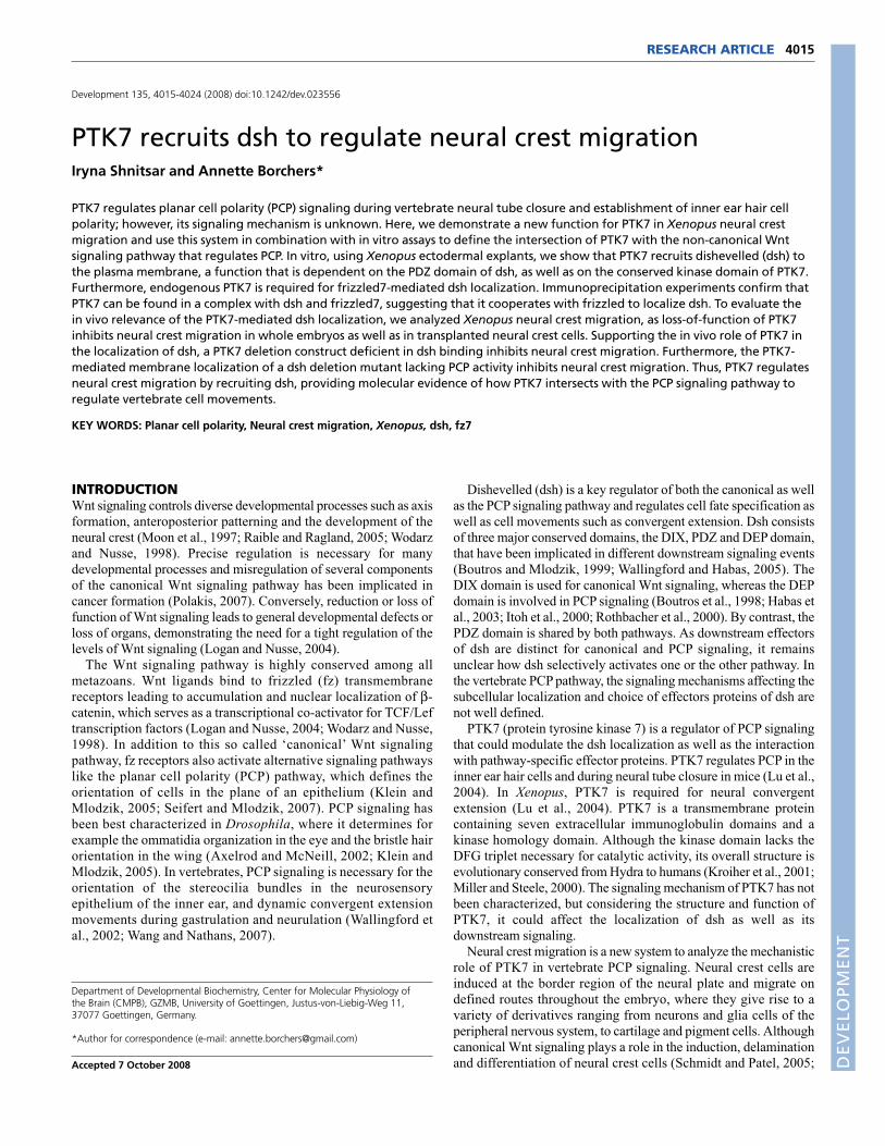
Sheldahl et al., 1999; Yanagawa et al., 1995). Animal caps areectodermal explants of blastula stage Xenopus embryos that allowthe analysis of the cellular localization of overexpressed taggedproteins. For colocalization assays, Xenopus embryos were injectedwith RNA coding for the respective tagged constructs in the animalpole of a one-cell stage embryo. At blastula stage, the animal capwas dissected and cultured until gastrula stages, when cellularprotein localization was determined using a laser scanningmicroscope. GFP-tagged dsh is predominantly localized in thecytoplasm of animal caps (Fig. 1A,G), but is translocated to theplasma membrane if myc-tagged fz7 is co-expressed (Fig. 1C,G).To examine whether PTK7 translocates dsh to the plasmamembrane, myc-tagged PTK7 was co-expressed with GFP-taggeddsh. Like fz7, PTK7 can recruit dsh-GFP from the cytoplasm to themembrane (Fig. 1E,G). To further confirm the co-localization ofPTK7 with dsh in a biochemical assay, we used glycerol gradientdensity centrifugation (see Fig. S1 in the supplementary material).This method allows the separation of proteins according to theirmolecular weight, and protein complex formation can be detectedas a shift to higher molecular weight fractions. Indeed, the dshprotein tailed into higher density fractions in the presence of PTK7(see Fig. S1 in the supplementary material), indicating complexformation of dsh with PTK7. Thus, the animal cap localization assayas well as the glycerol gradient density centrifugation indicate thatPTK7 connects with the Wnt signaling pathway at the level of dsh.
The kinase domain of PTK7 and the PDZ domain ofdsh are required for co-localizationTo identify the molecular domains necessary for the interaction ofPTK7 with dsh, deletion mutants were tested for co-localization inanimal caps. As the intracellular domain of PTK7 contains aconserved tyrosine kinase motif, we analyzed whether its deletionabolishes translocation of dsh. Indeed, in contrast to overexpressionof wild-type PTK7, this deletion mutant (�kPTK7) was not able totranslocate dsh to the plasma membrane (Fig. 1F,G). Furthermore,this mutant also failed to shift dsh to higher molecular weightfractions in Xenopus lysates separated by glycerol gradientcentrifugation (see Fig. S1C,D in the supplementary material).Taken together, this indicates that the kinase motif of PTK7 isrequired for dsh recruitment to the plasma membrane.
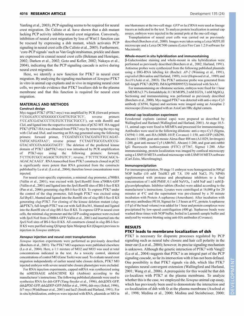
Dsh contains different functional domains that are involved incanonical and non-canonical Wnt signaling, respectively (Wallingfordand Habas, 2005). To identify which of these domains are necessaryfor the PTK7-dependent dsh-translocation, we expressed GFP-taggeddeletion mutants of the DIX, the PDZ and the DEP domain of dsh(Fig. 2A) in animal caps. In the absence of PTK7 expression, allmutant dsh proteins were mainly localized in the cytoplasm (Fig.2B,D,F,H). However, co-expression of PTK7 transferred �DIX- aswell as �DEP-dsh to the membrane (Fig. 2C,G,H). In the case of the�DIX-dsh-injected caps, a residual cytoplasmic staining remainedthat was not apparent in �DEP-dsh injected caps. By contrast, �PDZ-dsh was not translocated to the plasma membrane in the presence ofPTK7 (Fig. 2E,H), indicating that the PDZ domain is required forfunction. In summary, these data show that the tyrosine kinase domainof PTK7 as well as the PDZ domain of dsh are necessary for thetranslocation of dsh to the plasma membrane.
PTK7 is part of a fz7/dsh complex and is requiredfor fz7-mediated dsh localizationThe ability of PTK7 to control dsh localization suggests that the twoproteins might interact. We tested for binding by co-expressing HA-tagged PTK7 with myc-tagged dsh and immunoprecipitating with
4017RESEARCH ARTICLEPTK7 recruits dsh
Fig. 1. PTK7 recruits dsh to the plasma membrane. Animal capswere injected with different tagged RNAs, and protein localization wasanalyzed by confocal microscopy. The GFP-tagged dsh (green, left), theco-expressed myc-tagged protein (red, middle) and the merged pictures(right) are shown. (A) GFP-tagged dsh is localized to the cytoplasm ofanimal caps injected with 100 pg dsh-GFP RNA. (B) Myc-tagged fz7 ispredominantly membrane localized in animal caps injected with 100 pgfz7-myc RNA. (C) Co-injection of 100 pg dsh-GFP and 100 pg fz7-mycRNA leads to membrane recruitment of dsh. (D) PTK7 is membranelocalized in animal caps injected with 500 pg myc-tagged PTK7 RNA.(E) Animal caps co-injected with 100 pg dsh-GFP RNA and 500 pgPTK7-myc RNA show membrane-recruitment of dsh. Cells that do notexpress PTK7 in the membrane do not show membrane-localization ofdsh (white arrowhead). (F) Animal caps injected with 250 pg RNAcoding for a PTK7 mutant lacking the conserved kinase domain(�kPTK7) and 100 pg dsh-GFP RNA do not show membranelocalization of dsh. (G) Graph summarizing the percentage of cells withmembrane-localized dsh. For colocalization assays using PTK7 or fz7,only cells in which these proteins were membrane localized wereanalyzed. To determine the number of cells with cytoplasmic dshlocalization DAPI co-staining was used. The total number of cells isindicated above each column. D
EVELO
PMENT
4018
either myc- or HA-antibodies. Independent of the antibodies used,we could not detect co-immunoprecipitation of PTK7 and dsh (datanot shown), indicating that additional molecules are required forPTK7-mediated dsh localization. A likely candidate is fz7, which,like PTK7, is also able to recruit dsh to the plasma membrane. Totest whether PTK7 forms a complex with dsh and fz7, we expressedHA-tagged PTK7 with either myc-tagged dsh or myc-tagged fz7, ora combination of the two (see Fig. S2 in the supplementarymaterial). Although HA-tagged PTK7 was co-precipitated withmyc-tagged fz7 in one out of three experiments, we detected onlyrobust co-precipitation in combination with myc-tagged dsh and
myc-tagged fz7 (see Fig. S2 in the supplementary material),indicating that PTK7 is part of a protein complex that includes fz7and dsh.
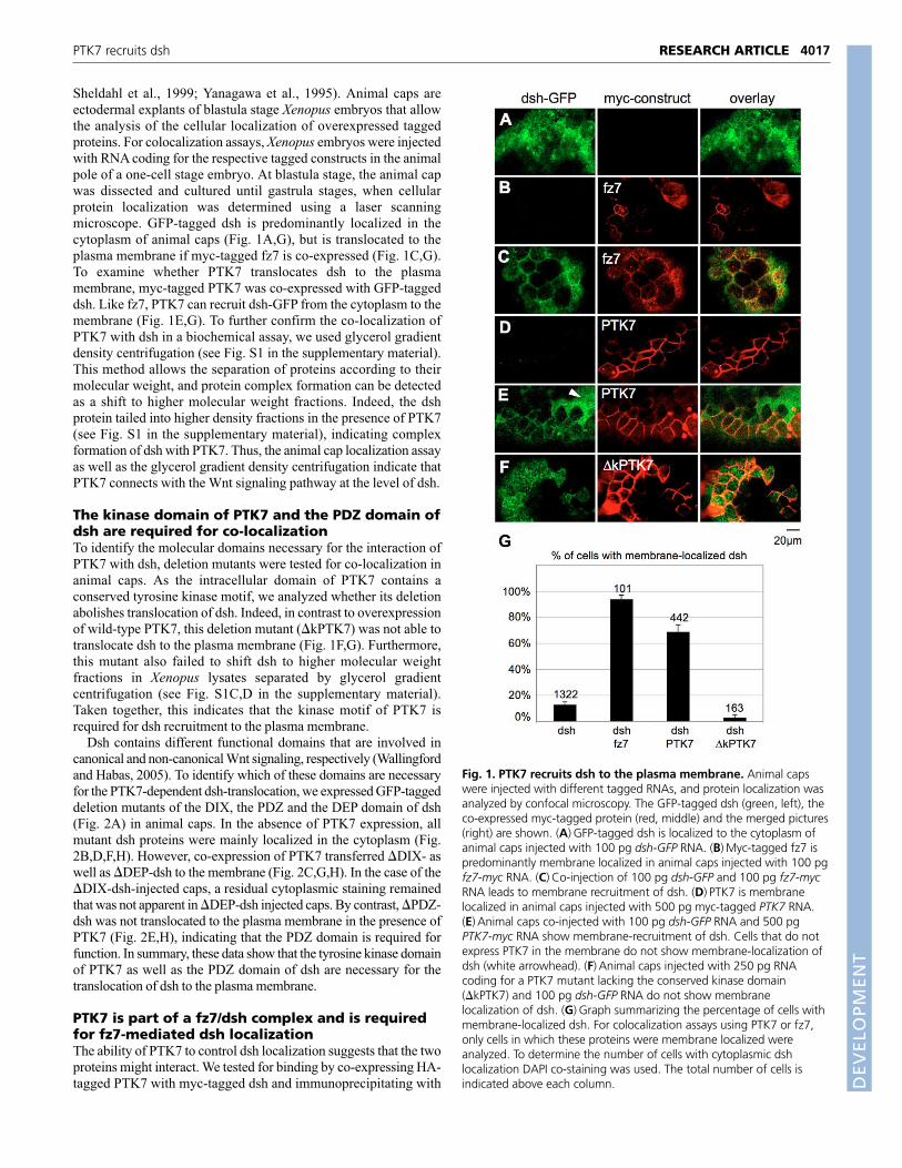
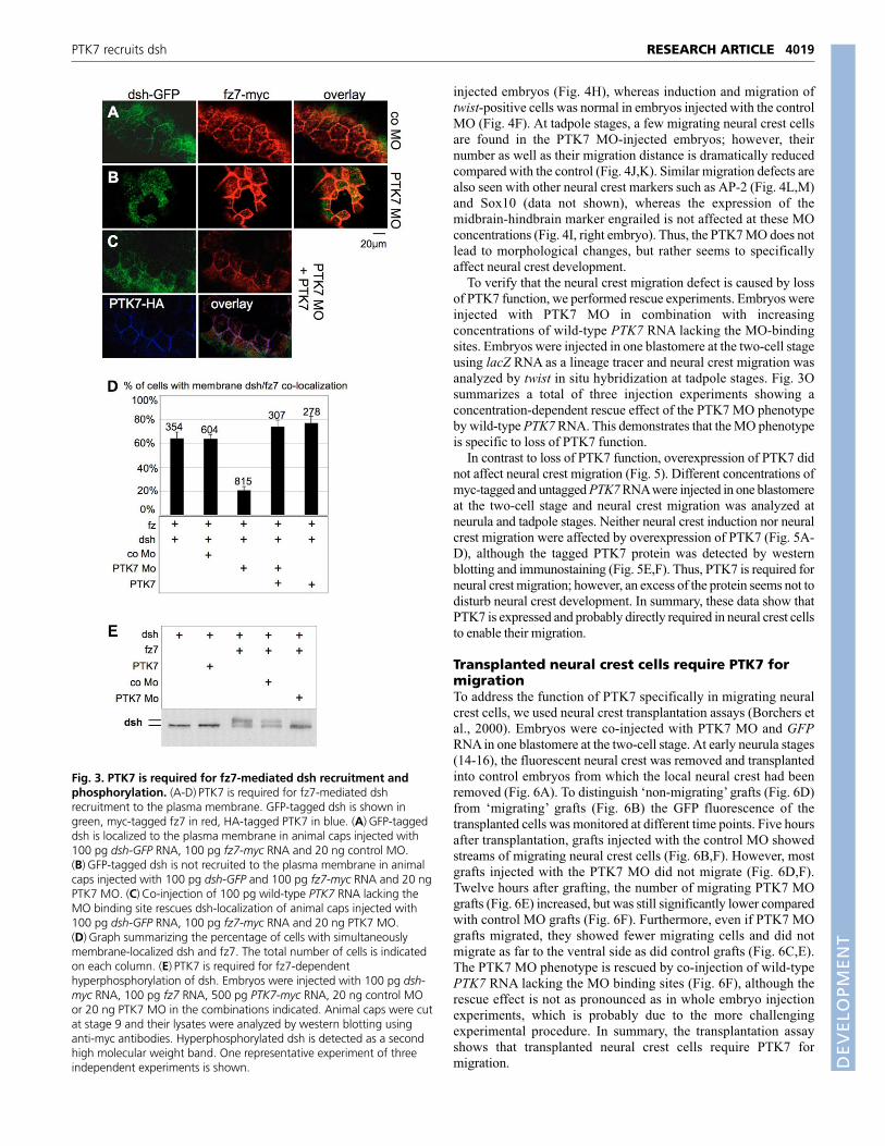
If PTK7 forms a complex with fz7 and dsh, PTK7 could affect thefz7-mediated dsh recruitment. To test this, we analyzed animal caps,which endogenously express PTK7 (see Fig. S3 in thesupplementary material), how loss of PTK7 function interferes withthe fz7-mediated dsh recruitment. In the presence of controlmorpholino oligonucleotides (MOs), fz7 recruits GFP-tagged dsh tothe plasma membrane (Fig. 3A,D). However, the PTK7 MOabolished the fz7-mediated dsh translocation (Fig. 3B,D). This couldbe rescued by co-injection of PTK7 RNA lacking the MO bindingsite (Fig. 3C,D).
As fz-mediated dsh membrane localization correlates withhyperphosphorylation of dsh (Rothbacher et al., 2000), we furtherexamined whether PTK7 loss of function also affects thephosphorylation status of dsh. Animal caps expressing myc-taggeddsh alone or in combination with PTK7 show only a single band inwestern blots using anti-myc antibodies. However, an additionalhigh molecular weight band representing hyperphosphorylated dshis detected in lysates expressing dsh with fz7 (Fig. 3E). Interestingly,this fz7-mediated hyperphosphorylation of dsh is inhibited by thePTK7 MO, indicating that PTK7 is required for the fz7-mediateddsh hyperphosphorylation. Thus, these data support that PTK7 ispart of a dsh-fz7 complex required for dsh localization andphosphorylation.
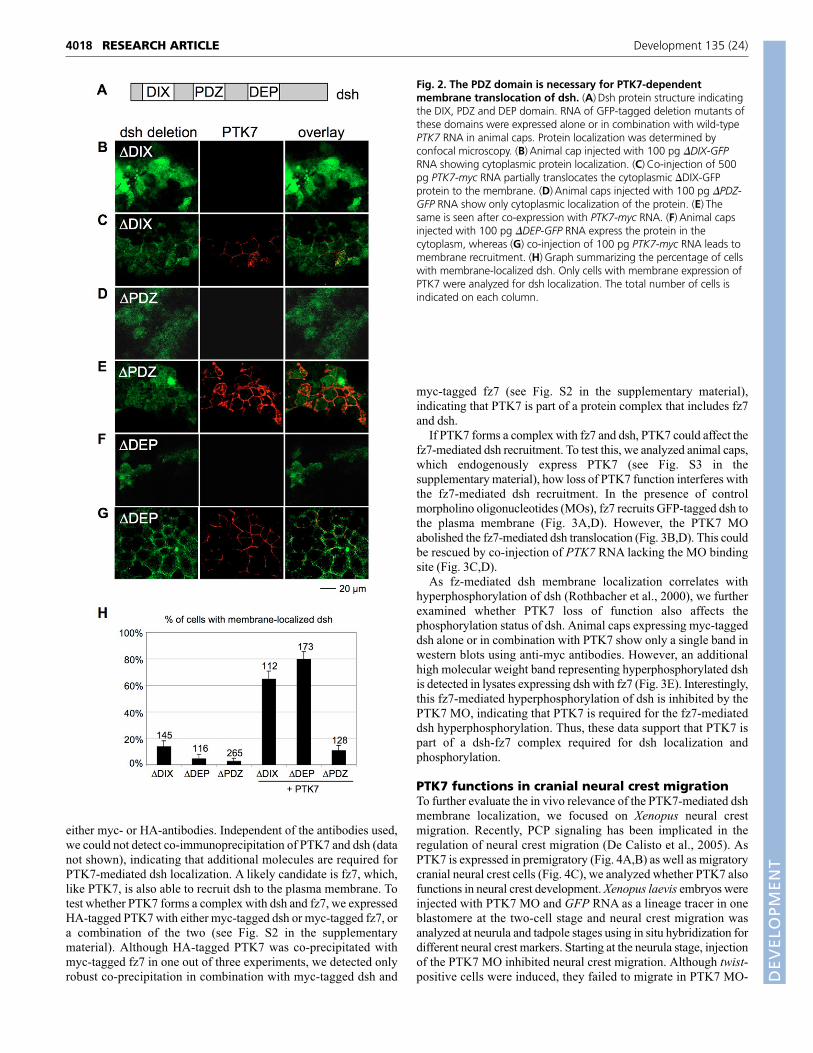
PTK7 functions in cranial neural crest migrationTo further evaluate the in vivo relevance of the PTK7-mediated dshmembrane localization, we focused on Xenopus neural crestmigration. Recently, PCP signaling has been implicated in theregulation of neural crest migration (De Calisto et al., 2005). AsPTK7 is expressed in premigratory (Fig. 4A,B) as well as migratorycranial neural crest cells (Fig. 4C), we analyzed whether PTK7 alsofunctions in neural crest development. Xenopus laevis embryos wereinjected with PTK7 MO and GFP RNA as a lineage tracer in oneblastomere at the two-cell stage and neural crest migration wasanalyzed at neurula and tadpole stages using in situ hybridization fordifferent neural crest markers. Starting at the neurula stage, injectionof the PTK7 MO inhibited neural crest migration. Although twist-positive cells were induced, they failed to migrate in PTK7 MO-
RESEARCH ARTICLE Development 135 (24)
Fig. 2. The PDZ domain is necessary for PTK7-dependentmembrane translocation of dsh. (A) Dsh protein structure indicatingthe DIX, PDZ and DEP domain. RNA of GFP-tagged deletion mutants ofthese domains were expressed alone or in combination with wild-typePTK7 RNA in animal caps. Protein localization was determined byconfocal microscopy. (B) Animal cap injected with 100 pg �DIX-GFPRNA showing cytoplasmic protein localization. (C) Co-injection of 500pg PTK7-myc RNA partially translocates the cytoplasmic �DIX-GFPprotein to the membrane. (D) Animal caps injected with 100 pg �PDZ-GFP RNA show only cytoplasmic localization of the protein. (E) Thesame is seen after co-expression with PTK7-myc RNA. (F) Animal capsinjected with 100 pg �DEP-GFP RNA express the protein in thecytoplasm, whereas (G) co-injection of 100 pg PTK7-myc RNA leads tomembrane recruitment. (H) Graph summarizing the percentage of cellswith membrane-localized dsh. Only cells with membrane expression ofPTK7 were analyzed for dsh localization. The total number of cells isindicated on each column.
DEVELO
PMENT
injected embryos (Fig. 4H), whereas induction and migration oftwist-positive cells was normal in embryos injected with the controlMO (Fig. 4F). At tadpole stages, a few migrating neural crest cellsare found in the PTK7 MO-injected embryos; however, theirnumber as well as their migration distance is dramatically reducedcompared with the control (Fig. 4J,K). Similar migration defects arealso seen with other neural crest markers such as AP-2 (Fig. 4L,M)and Sox10 (data not shown), whereas the expression of themidbrain-hindbrain marker engrailed is not affected at these MOconcentrations (Fig. 4I, right embryo). Thus, the PTK7 MO does notlead to morphological changes, but rather seems to specificallyaffect neural crest development.
To verify that the neural crest migration defect is caused by lossof PTK7 function, we performed rescue experiments. Embryos wereinjected with PTK7 MO in combination with increasingconcentrations of wild-type PTK7 RNA lacking the MO-bindingsites. Embryos were injected in one blastomere at the two-cell stageusing lacZ RNA as a lineage tracer and neural crest migration wasanalyzed by twist in situ hybridization at tadpole stages. Fig. 3Osummarizes a total of three injection experiments showing aconcentration-dependent rescue effect of the PTK7 MO phenotypeby wild-type PTK7 RNA. This demonstrates that the MO phenotypeis specific to loss of PTK7 function.
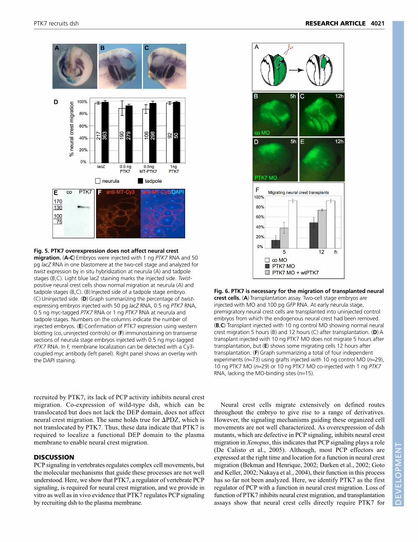
In contrast to loss of PTK7 function, overexpression of PTK7 didnot affect neural crest migration (Fig. 5). Different concentrations ofmyc-tagged and untagged PTK7RNA were injected in one blastomereat the two-cell stage and neural crest migration was analyzed atneurula and tadpole stages. Neither neural crest induction nor neuralcrest migration were affected by overexpression of PTK7 (Fig. 5A-D), although the tagged PTK7 protein was detected by westernblotting and immunostaining (Fig. 5E,F). Thus, PTK7 is required forneural crest migration; however, an excess of the protein seems not todisturb neural crest development. In summary, these data show thatPTK7 is expressed and probably directly required in neural crest cellsto enable their migration.
Transplanted neural crest cells require PTK7 formigrationTo address the function of PTK7 specifically in migrating neuralcrest cells, we used neural crest transplantation assays (Borchers etal., 2000). Embryos were co-injected with PTK7 MO and GFPRNA in one blastomere at the two-cell stage. At early neurula stages(14-16), the fluorescent neural crest was removed and transplantedinto control embryos from which the local neural crest had beenremoved (Fig. 6A). To distinguish ‘non-migrating’ grafts (Fig. 6D)from ‘migrating’ grafts (Fig. 6B) the GFP fluorescence of thetransplanted cells was monitored at different time points. Five hoursafter transplantation, grafts injected with the control MO showedstreams of migrating neural crest cells (Fig. 6B,F). However, mostgrafts injected with the PTK7 MO did not migrate (Fig. 6D,F).Twelve hours after grafting, the number of migrating PTK7 MOgrafts (Fig. 6E) increased, but was still significantly lower comparedwith control MO grafts (Fig. 6F). Furthermore, even if PTK7 MOgrafts migrated, they showed fewer migrating cells and did notmigrate as far to the ventral side as did control grafts (Fig. 6C,E).The PTK7 MO phenotype is rescued by co-injection of wild-typePTK7 RNA lacking the MO binding sites (Fig. 6F), although therescue effect is not as pronounced as in whole embryo injectionexperiments, which is probably due to the more challengingexperimental procedure. In summary, the transplantation assayshows that transplanted neural crest cells require PTK7 formigration.
4019RESEARCH ARTICLEPTK7 recruits dsh
Fig. 3. PTK7 is required for fz7-mediated dsh recruitment andphosphorylation. (A-D) PTK7 is required for fz7-mediated dshrecruitment to the plasma membrane. GFP-tagged dsh is shown ingreen, myc-tagged fz7 in red, HA-tagged PTK7 in blue. (A) GFP-taggeddsh is localized to the plasma membrane in animal caps injected with100 pg dsh-GFP RNA, 100 pg fz7-myc RNA and 20 ng control MO.(B) GFP-tagged dsh is not recruited to the plasma membrane in animalcaps injected with 100 pg dsh-GFP and 100 pg fz7-myc RNA and 20 ngPTK7 MO. (C) Co-injection of 100 pg wild-type PTK7 RNA lacking theMO binding site rescues dsh-localization of animal caps injected with100 pg dsh-GFP RNA, 100 pg fz7-myc RNA and 20 ng PTK7 MO.(D) Graph summarizing the percentage of cells with simultaneouslymembrane-localized dsh and fz7. The total number of cells is indicatedon each column. (E) PTK7 is required for fz7-dependenthyperphosphorylation of dsh. Embryos were injected with 100 pg dsh-myc RNA, 100 pg fz7 RNA, 500 pg PTK7-myc RNA, 20 ng control MOor 20 ng PTK7 MO in the combinations indicated. Animal caps were cutat stage 9 and their lysates were analyzed by western blotting usinganti-myc antibodies. Hyperphosphorylated dsh is detected as a secondhigh molecular weight band. One representative experiment of threeindependent experiments is shown. D
EVELO
PMENT

4020
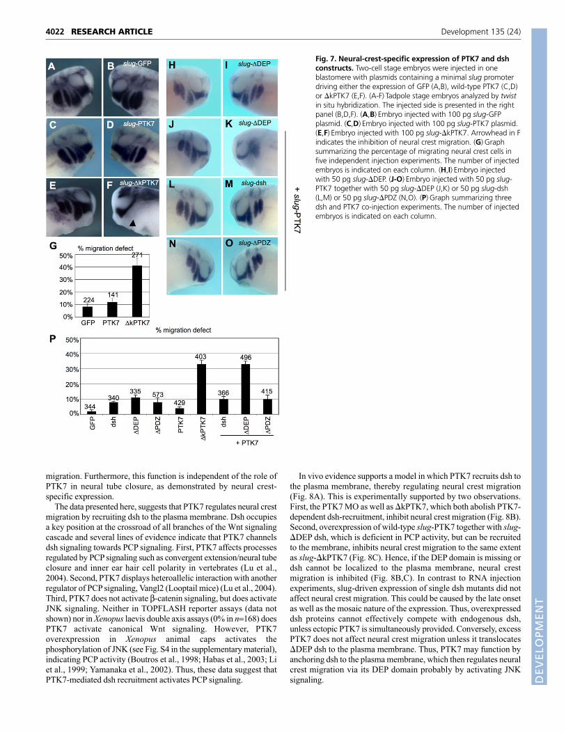
Neural-crest-specific expression of �kPTK7 inhibitsneural crest migrationTo study the function of PTK7 in neural crest migration isolatedfrom earlier developmental defects, we targeted the expression ofPTK7 to neural crest cells. PTK7 constructs were expressed underthe control of the neural crest-specific slug promoter, so that only thecells that are already specified to become neural crest cells areaffected. Regions of the slug promoter that are sufficient to driveneural-crest-specific expression (Vallin et al., 2001) were used toexpress GFP (Fig. 7A,B), full-length PTK7 (Fig. 7C,D) and adeletion mutant of the kinase motif, �kPTK7 (Fig. 7E,F). �kPTK7was used as a PTK7 antimorph, as it failed to recruit dsh to theplasma membrane in animal cap assays (Fig. 1F) and caused similarneural crest migration defects as the PTK7 MO in whole embryos(injection of 50 pg �kPTK7 RNA caused 44% neural crest migrationdefect, n=122). Plasmids were injected together with lacZ RNA asa lineage tracer in one blastomere at the two-cell stage and embryos
were analyzed for twist expression at neurula and tadpole stages. Asplasmid injections result in mosaic expression, the effects aregenerally not as strong as MO or RNA injections. Nevertheless,neural crest-specific expression of �kPTK7 significantly inhibitedthe migration of twist-expressing neural crest cells (Fig. 7E,F),whereas the expression of GFP or PTK7 rarely affected neural crestmigration (Fig. 7A-D,G). Although we cannot completely rule outthe possibility that PTK7 does also affect neural crest induction,these data demonstrate that PTK7 has an independent function inneural crest migration.
Recruitment of the DEP domain by PTK7 isrequired for neural crest migrationHaving established that PTK7 is required for neural crestmigration, we can use this system to analyze the in vivo relevanceof the PTK7-mediated dsh membrane localization. By targetingthe kinase deletion mutant of PTK7, �kPTK7, to neural crestcells, we have already demonstrated that failure of dshlocalization (Fig. 1F,G) inhibits neural crest migration (Fig. 7F,P).Next, to characterize the genetic interaction of PTK7 with dsh inmigrating neural crest cells, we targeted the expression of PTK7,as well as of different dsh mutants, to neural crest cells using theslug promoter system. First, we analyzed how dsh, �PDZ dsh and�DEP dsh affect neural crest migration. We used �PDZ dsh,because this mutant abolishes the PTK7-mediated translocation,and �DEP dsh, because the DEP domain is required for PCPactivity and affects neural crest migration (De Calisto et al., 2005;Wallingford and Habas, 2005). Single injections of all dshconstructs and a single injection of slug-PTK7 produced only verymild defects (Fig. 7H,I,P). The same holds true for co-injection ofslug-PTK7 with slug-dsh or slug-�PDZ (Fig. 7L-P). However, thecombination of slug-PTK7 with slug-�DEP inhibited neural crestmigration to an extent that is comparable with expression of slug-�kPTK7 (Fig. 7J,K,P), which cannot recruit dsh to the plasmamembrane. This suggests that, although �DEP dsh can be
RESEARCH ARTICLE Development 135 (24)
Fig. 4. PTK7 is expressed in cranial neural crest cells and requiredfor their migration. (A-C) PTK7 expression pattern in Xenopus laevisdetected by whole-mount in situ hybridization. (A,B) PTK7 is broadlyexpressed in premigratory neural crest cells at early neurula stages(black arrow). (C) At stage 26, PTK7 expression is detected in migratingcranial neural crest cells (black arrow). (D-M) Embryos injected in oneblastomere at the two-cell stage with different constructs incombination with 100 pg GFP RNA as a lineage tracer. Neural crestmigration was analyzed at neurula stages using the neural crest markertwist (D,F,H) or the midbrain-hindbrain marker engrailed (E,G,I). Theinjected side is shown on the right. (D,E) GFP-injected embryos.(F,G) Embryos co-injected with 10 ng control MO and GFP RNA.(H,I) Embryos co-injected with 10 ng PTK7 MO and GFP RNA. (J-M) Tadpole embryos analyzed by in situ hybridization with the neuralcrest markers twist (J,K) or AP-2 (L,M). The injected side is shown onthe right. (J,L) Tadpoles injected with 10 ng control MO und 100 pg GFPRNA. (K,M) Tadpoles injected with 10 ng PTK7 MO and 100 pg GFPRNA. Inhibition of cranial neural crest migration is marked by arrows inH,K,M. (N) Graph summarizing the MO injection experiments. Leftgraph summarizes five independent experiments (analyzing neurulastages) and six independent experiments (analyzing tadpole stages),respectively. The right graph summarizes three independent rescueexperiment. The percentage of migrating neural crest cells wasdetermined by twist in situ hybridization. Columns are labeled with thenumber of injected embryos.
DEVELO
PMENT
recruited by PTK7, its lack of PCP activity inhibits neural crestmigration. Co-expression of wild-type dsh, which can betranslocated but does not lack the DEP domain, does not affectneural crest migration. The same holds true for �PDZ, which isnot translocated by PTK7. Thus, these data indicate that PTK7 isrequired to localize a functional DEP domain to the plasmamembrane to enable neural crest migration.
DISCUSSIONPCP signaling in vertebrates regulates complex cell movements, butthe molecular mechanisms that guide these processes are not wellunderstood. Here, we show that PTK7, a regulator of vertebrate PCPsignaling, is required for neural crest migration, and we provide invitro as well as in vivo evidence that PTK7 regulates PCP signalingby recruiting dsh to the plasma membrane.
Neural crest cells migrate extensively on defined routesthroughout the embryo to give rise to a range of derivatives.However, the signaling mechanisms guiding these organized cellmovements are not well characterized. As overexpression of dshmutants, which are defective in PCP signaling, inhibits neural crestmigration in Xenopus, this indicates that PCP signaling plays a role(De Calisto et al., 2005). Although, most PCP effectors areexpressed at the right time and location for a function in neural crestmigration (Bekman and Henrique, 2002; Darken et al., 2002; Gotoand Keller, 2002; Nakaya et al., 2004), their function in this processhas so far not been analyzed. Here, we identify PTK7 as the firstregulator of PCP with a function in neural crest migration. Loss offunction of PTK7 inhibits neural crest migration, and transplantationassays show that neural crest cells directly require PTK7 for
4021RESEARCH ARTICLEPTK7 recruits dsh
Fig. 5. PTK7 overexpression does not affect neural crestmigration. (A-C) Embryos were injected with 1 ng PTK7 RNA and 50pg lacZ RNA in one blastomere at the two-cell stage and analyzed fortwist expression by in situ hybridization at neurula (A) and tadpolestages (B,C). Light blue lacZ staining marks the injected side. Twist-positive neural crest cells show normal migration at neurula (A) andtadpole stages (B,C). (B) Injected side of a tadpole stage embryo.(C) Uninjected side. (D) Graph summarizing the percentage of twist-expressing embryos injected with 50 pg lacZ RNA, 0.5 ng PTK7 RNA,0.5 ng myc-tagged PTK7 RNA or 1 ng PTK7 RNA at neurula andtadpole stages. Numbers on the columns indicate the number ofinjected embryos. (E) Confirmation of PTK7 expression using westernblotting (co, uninjected controls) or (F) immunostaining on transversesections of neurula stage embryos injected with 0.5 ng myc-taggedPTK7 RNA. In F, membrane localization can be detected with a Cy3-coupled myc antibody (left panel). Right panel shows an overlay withthe DAPI staining.
Fig. 6. PTK7 is necessary for the migration of transplanted neuralcrest cells. (A) Transplantation assay. Two-cell stage embryos areinjected with MO and 100 pg GFP RNA. At early neurula stage,premigratory neural crest cells are transplanted into uninjected controlembryos from which the endogenous neural crest had been removed.(B,C) Transplant injected with 10 ng control MO showing normal neuralcrest migration 5 hours (B) and 12 hours (C) after transplantation. (D) Atransplant injected with 10 ng PTK7 MO does not migrate 5 hours aftertransplantation, but (E) shows some migrating cells 12 hours aftertransplantation. (F) Graph summarizing a total of four independentexperiments (n=73) using grafts injected with 10 ng control MO (n=29),10 ng PTK7 MO (n=29) or 10 ng PTK7 MO co-injected with 1 ng PTK7RNA, lacking the MO-binding sites (n=15).
DEVELO
PMENT
4022
migration. Furthermore, this function is independent of the role ofPTK7 in neural tube closure, as demonstrated by neural crest-specific expression.
The data presented here, suggests that PTK7 regulates neural crestmigration by recruiting dsh to the plasma membrane. Dsh occupiesa key position at the crossroad of all branches of the Wnt signalingcascade and several lines of evidence indicate that PTK7 channelsdsh signaling towards PCP signaling. First, PTK7 affects processesregulated by PCP signaling such as convergent extension/neural tubeclosure and inner ear hair cell polarity in vertebrates (Lu et al.,2004). Second, PTK7 displays heteroallelic interaction with anotherregulator of PCP signaling, Vangl2 (Looptail mice) (Lu et al., 2004).Third, PTK7 does not activate β-catenin signaling, but does activateJNK signaling. Neither in TOPFLASH reporter assays (data notshown) nor in Xenopus laevis double axis assays (0% in n=168) doesPTK7 activate canonical Wnt signaling. However, PTK7overexpression in Xenopus animal caps activates thephosphorylation of JNK (see Fig. S4 in the supplementary material),indicating PCP activity (Boutros et al., 1998; Habas et al., 2003; Liet al., 1999; Yamanaka et al., 2002). Thus, these data suggest thatPTK7-mediated dsh recruitment activates PCP signaling.
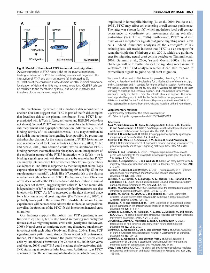
In vivo evidence supports a model in which PTK7 recruits dsh tothe plasma membrane, thereby regulating neural crest migration(Fig. 8A). This is experimentally supported by two observations.First, the PTK7 MO as well as �kPTK7, which both abolish PTK7-dependent dsh-recruitment, inhibit neural crest migration (Fig. 8B).Second, overexpression of wild-type slug-PTK7 together with slug-�DEP dsh, which is deficient in PCP activity, but can be recruitedto the membrane, inhibits neural crest migration to the same extentas slug-�kPTK7 (Fig. 8C). Hence, if the DEP domain is missing ordsh cannot be localized to the plasma membrane, neural crestmigration is inhibited (Fig. 8B,C). In contrast to RNA injectionexperiments, slug-driven expression of single dsh mutants did notaffect neural crest migration. This could be caused by the late onsetas well as the mosaic nature of the expression. Thus, overexpresseddsh proteins cannot effectively compete with endogenous dsh,unless ectopic PTK7 is simultaneously provided. Conversely, excessPTK7 does not affect neural crest migration unless it translocates�DEP dsh to the plasma membrane. Thus, PTK7 may function byanchoring dsh to the plasma membrane, which then regulates neuralcrest migration via its DEP domain probably by activating JNKsignaling.
RESEARCH ARTICLE Development 135 (24)
Fig. 7. Neural-crest-specific expression of PTK7 and dshconstructs. Two-cell stage embryos were injected in oneblastomere with plasmids containing a minimal slug promoterdriving either the expression of GFP (A,B), wild-type PTK7 (C,D)or �kPTK7 (E,F). (A-F) Tadpole stage embryos analyzed by twistin situ hybridization. The injected side is presented in the rightpanel (B,D,F). (A,B) Embryo injected with 100 pg slug-GFPplasmid. (C,D) Embryo injected with 100 pg slug-PTK7 plasmid.(E,F) Embryo injected with 100 pg slug-�kPTK7. Arrowhead in Findicates the inhibition of neural crest migration. (G) Graphsummarizing the percentage of migrating neural crest cells infive independent injection experiments. The number of injectedembryos is indicated on each column. (H,I) Embryo injectedwith 50 pg slug-�DEP. (J-O) Embryo injected with 50 pg slug-PTK7 together with 50 pg slug-�DEP (J,K) or 50 pg slug-dsh(L,M) or 50 pg slug-�PDZ (N,O). (P) Graph summarizing threedsh and PTK7 co-injection experiments. The number of injectedembryos is indicated on each column.
DEVELO
PMENT
The mechanism by which PTK7 mediates dsh recruitment isunclear. Our data suggest that PTK7 is part of the fz-dsh complexthat localizes dsh to the plasma membrane. First, PTK7 is co-precipitated with fz7/dsh in Xenopus lysates and HEK293 cells (datanot shown). Second, PTK7 loss of function inhibits the fz7-mediateddsh recruitment and hyperphosphorylation. Alternatively, as thebinding activity of PTK7/fz7/dsh is weak, PTK7 may contribute tothe fz/dsh interaction at the signaling level possibly by promotingdsh phosphorylation. As the kinase domain of PTK7 lacks aminoacid residues crucial for kinase activity (Kroiher et al., 2001; Millerand Steele, 2000), this scenario could involve additional PTK7-binding partners that mediate dsh phosphorylation. Independent ofthe molecular nature of the PTK7 interaction with fz/dsh – be itbinding, signaling or both – it also remains to be seen whether PTK7exclusively interacts with fz7 or whether other fz family memberscan replace it. The latter is supported by the finding that animal capsexpress other fz family members, including fz8 (see Fig. S3 in thesupplementary material), which, like fz7, recruits dsh to the plasmamembrane (Rothbacher et al., 2000). Furthermore, loss of functionof fz7 does not affect the PTK7-mediated dsh localization in animalcaps (data not shown), suggesting that either PTK7 can recruit dshindependently of fz7 or indeed that other fz family members can alsointeract with PTK7. As fz7 is expressed at the right time and placefor a function in neural crest migration (De Calisto et al., 2005), itprobably takes part in the in vivo PTK7-fz-dsh interaction. Futureexperiments will be needed to address the molecular composition,as well as the function, of the PTK7-fz-dsh interaction in neural crestmigration.
Our findings supports the notion that PCP signaling is notlimited to epithelia, but is also found in moving mesenchymaltissues such as migrating neural crest cells (Kuriyama and Mayor,2008). Neural crest cells migrate over long distances, but also stayin contact with each other (Teddy and Kulesa, 2004). Thus, PCPsignaling may pattern migrating cells or orient cell protrusions.Indeed, PCP factors determine the polarization of neural crestcells by lamellipodia formation (De Calisto et al., 2005; Kuriyamaand Mayor, 2008) and PTK7 could mediate this by activating dsh-JNK signaling at precise cellular locations. Additionally, as PTK7contains extracellular immunoglobulin domains, which have been
implicated in homophilic binding (Lu et al., 2004; Pulido et al.,1992), PTK7 may affect cell clustering or cell contact persistence.The latter was shown for fz7, which modulates local cell contactpersistence to coordinate cell movements during zebrafishgastrulation (Witzel et al., 2006). Furthermore, PTK7 could alsofunction as a receptor for signals that guide migrating neural crestcells. Indeed, functional analyses of the Drosophila PTK7ortholog (otk, off-track) indicate that PTK7 is a co-receptor forsemaphorin/plexins (Winberg et al., 2001), which are guidancecues for migrating neural crest cells in vertebrates (Gammill et al.,2007; Gammill et al., 2006; Yu and Moens, 2005). The nextchallenge will be to further dissect the signaling mechanism ofvertebrate PTK7 and analyze whether it can also respond toextracellular signals to guide neural crest migration.
We thank R. Moon and H. Steinbeisser for providing plasmids, D. Frank, A.Hufton, H. Peradziryi and M. Podleschny for critical reading of the manuscript,and H. Steinbeisser and A. Wodarz for helpful critical discussion. Furthermore,we thank H. Steinbeisser for the fz7 MO and A. Wodarz for providing the laserscanning microscope and technical support, and I. Wunderlich for technicalassistance. Finally, we thank T. Pieler for infrastructure and support. This workwas supported by grants to A.B. by the Deutsche Forschungsgemeinschaft(DFG) and the DFG Center for Molecular Physiology of the Brain (CMPB). I.S.was supported by a stipend from the Christiane-Nüsslein-Volhard-Foundation.
Supplementary materialSupplementary material for this article is available athttp://dev.biologists.org/cgi/content/full/135/24/4015/DC1
ReferencesAoki, Y., Saint-Germain, N., Gyda, M., Magner-Fink, E., Lee, Y. H., Credidio,
C. and Saint-Jeannet, J. P. (2003). Sox10 regulates the development of neuralcrest-derived melanocytes in Xenopus. Dev. Biol. 259, 19-33.
Axelrod, J. D. and McNeill, H. (2002). Coupling planar cell polarity signaling tomorphogenesis. Scientific World J. 2, 434-454.
Axelrod, J. D., Miller, J. R., Shulman, J. M., Moon, R. T. and Perrimon, N.(1998). Differential recruitment of Dishevelled provides signaling specificity in theplanar cell polarity and Wingless signaling pathways. Genes Dev. 12, 2610-2622.
Bekman, E. and Henrique, D. (2002). Embryonic expression of three mousegenes with homology to the Drosophila melanogaster prickle gene. Mech. Dev.119 Suppl. 1, S77-S81.
Borchers, A., Epperlein, H. H. and Wedlich, D. (2000). An assay system to studymigratory behavior of cranial neural crest cells in Xenopus. Dev. Genes Evol. 210,217-222.
Borchers, A., David, R. and Wedlich, D. (2001). Xenopus cadherin-11 restrainscranial neural crest migration and influences neural crest specification.Development 128, 3049-3060.
Borchers, A. G., Hufton, A. L., Eldridge, A. G., Jackson, P. K., Harland, R. M.and Baker, J. C. (2002). The E3 ubiquitin ligase GREUL1 anteriorizes ectodermduring Xenopus development. Dev. Biol. 251, 395-408.
Boutros, M. and Mlodzik, M. (1999). Dishevelled: at the crossroads of divergentintracellular signaling pathways. Mech. Dev. 83, 27-37.
Boutros, M., Paricio, N., Strutt, D. I. and Mlodzik, M. (1998). Dishevelledactivates JNK and discriminates between JNK pathways in planar polarity andwingless signaling. Cell 94, 109-118.
Brivanlou, A. H. and Harland, R. M. (1989). Expression of an engrailed-relatedprotein is induced in the anterior neural ectoderm of early Xenopus embryos.Development 106, 611-617.
Darken, R. S., Scola, A. M., Rakeman, A. S., Das, G., Mlodzik, M. and Wilson,P. A. (2002). The planar polarity gene strabismus regulates convergent extensionmovements in Xenopus. EMBO J. 21, 976-985.
De Calisto, J., Araya, C., Marchant, L., Riaz, C. F. and Mayor, R. (2005).Essential role of non-canonical Wnt signalling in neural crest migration.Development 132, 2587-2597.
Gammill, L. S., Gonzalez, C., Gu, C. and Bronner-Fraser, M. (2006). Guidanceof trunk neural crest migration requires neuropilin 2/semaphorin 3F signaling.Development 133, 99-106.
Gammill, L. S., Gonzalez, C. and Bronner-Fraser, M. (2007). Neuropilin2/semaphorin 3F signaling is essential for cranial neural crest migration andtrigeminal ganglion condensation. Dev. Neurobiol. 67, 47-56.
Goto, T. and Keller, R. (2002). The planar cell polarity gene strabismus regulatesconvergence and extension and neural fold closure in Xenopus. Dev. Biol. 247,165-181.
4023RESEARCH ARTICLEPTK7 recruits dsh
Fig. 8. Model of the role of PTK7 in neural crest migration.(A) Overexpression of PTK7 recruits dsh to the plasma membrane,leading to activation of PCP and enabling neural crest migration. Theinteraction of PTK7 and dsh may involve fz7 (indicated as ?).(B) Deletion of the conserved kinase domain of PTK7 inhibits membranelocalization of dsh and inhibits neural crest migration. (C) �DEP dsh canbe recruited to the membrane by PTK7, but lacks PCP activity andtherefore blocks neural crest migration.
DEVELO
PMENT
4024
Habas, R., Dawid, I. B. and He, X. (2003). Coactivation of Rac and Rho byWnt/Frizzled signaling is required for vertebrate gastrulation. Genes Dev. 17,295-309.
Harland, R. M. (1991). In situ hybridization: an improved whole-mount methodfor Xenopus embryos. Methods Cell Biol. 36, 685-695.
Hopwood, N. D., Pluck, A. and Gurdon, J. B. (1989). A Xenopus mRNA relatedto Drosophila twist is expressed in response to induction in the mesoderm andthe neural crest. Cell 59, 893-903.
Itoh, K., Antipova, A., Ratcliffe, M. J. and Sokol, S. (2000). Interaction ofdishevelled and Xenopus axin-related protein is required for wnt signaltransduction. Mol. Cell. Biol. 20, 2228-2238.
Klein, T. J. and Mlodzik, M. (2005). Planar cell polarization: an emerging modelpoints in the right direction. Annu. Rev. Cell Dev. Biol. 21, 155-176.
Kroiher, M., Miller, M. A. and Steele, R. E. (2001). Deceiving appearances:signaling by ‘dead’ and ‘fractured’ receptor protein-tyrosine kinases. BioEssays23, 69-76.
Kuriyama, S. and Mayor, R. (2008). Molecular analysis of neural crest migration.Philos. Trans. R. Soc. Lond. B Biol. Sci. 363, 1349-1362.
Li, L., Yuan, H., Xie, W., Mao, J., Caruso, A. M., McMahon, A., Sussman, D. J.and Wu, D. (1999). Dishevelled proteins lead to two signaling pathways:regulation of LEF-1 and c-Jun N-terminal kinase in mammalian cells. J. Biol.Chem. 274, 129-134.
Logan, C. Y. and Nusse, R. (2004). The Wnt signaling pathway in developmentand disease. Annu. Rev. Cell Dev. Biol. 20, 781-810.
Lu, X., Borchers, A. G., Jolicoeur, C., Rayburn, H., Baker, J. C. and Tessier-Lavigne, M. (2004). PTK7/CCK-4 is a novel regulator of planar cell polarity invertebrates. Nature 430, 93-98.
Medina, A. and Steinbeisser, H. (2000). Interaction of Frizzled 7 and Dishevelledin Xenopus. Dev. Dyn. 218, 671-680.
Medina, A., Reintsch, W. and Steinbeisser, H. (2000). Xenopus frizzled 7 canact in canonical and non-canonical Wnt signaling pathways: implications onearly patterning and morphogenesis. Mech. Dev. 92, 227-237.
Miller, J. R., Rowning, B. A., Larabell, C. A., Yang-Snyder, J. A., Bates, R. L.and Moon, R. T. (1999). Establishment of the dorsal-ventral axis in Xenopusembryos coincides with the dorsal enrichment of dishevelled that is dependenton cortical rotation. J. Cell Biol. 146, 427-437.
Miller, M. A. and Steele, R. E. (2000). Lemon encodes an unusual receptorprotein-tyrosine kinase expressed during gametogenesis in Hydra. Dev. Biol. 224,286-298.
Moon, R. T., Brown, J. D. and Torres, M. (1997). WNTs modulate cell fate andbehavior during vertebrate development. Trends Genet. 13, 157-162.
Nakaya, M. A., Habas, R., Biris, K., Dunty, W. C., Jr, Kato, Y., He, X. andYamaguchi, T. P. (2004). Identification and comparative expression analyses ofDaam genes in mouse and Xenopus. Gene Expr. Patterns 5, 97-105.
Pan, F. C., Chen, Y., Loeber, J., Henningfeld, K. and Pieler, T. (2006). I-SceImeganuclease-mediated transgenesis in Xenopus. Dev. Dyn. 235, 247-252.
Polakis, P. (2007). The many ways of Wnt in cancer. Curr. Opin. Genet. Dev. 17,45-51.
Pulido, D., Campuzano, S., Koda, T., Modolell, J. and Barbacid, M. (1992).Dtrk, a Drosophila gene related to the trk family of neurotrophin receptors,encodes a novel class of neural cell adhesion molecule. EMBO J. 11, 391-404.
Raible, D. W. and Ragland, J. W. (2005). Reiterated Wnt and BMP signals inneural crest development. Semin. Cell Dev. Biol. 16, 673-682.
Rothbacher, U., Laurent, M. N., Deardorff, M. A., Klein, P. S., Cho, K. W. andFraser, S. E. (2000). Dishevelled phosphorylation, subcellular localization andmultimerization regulate its role in early embryogenesis. EMBO J. 19, 1010-1022.
Schmidt, C. and Patel, K. (2005). Wnts and the neural crest. Anat. Embryol. 209,349-355.
Seifert, J. R. and Mlodzik, M. (2007). Frizzled/PCP signalling: a conservedmechanism regulating cell polarity and directed motility. Nat. Rev. Genet. 8, 126-138.
Sheldahl, L. C., Park, M., Malbon, C. C. and Moon, R. T. (1999). Protein kinaseC is differentially stimulated by Wnt and Frizzled homologs in a G-protein-dependent manner. Curr. Biol. 9, 695-698.
Smith, W. C. and Harland, R. M. (1991). Injected Xwnt-8 RNA acts early inXenopus embryos to promote formation of a vegetal dorsalizing center. Cell 67,753-765.
Sokol, S. Y. (1996). Analysis of Dishevelled signalling pathways during Xenopusdevelopment. Curr. Biol. 6, 1456-1467.
Tahinci, E., Thorne, C. A., Franklin, J. L., Salic, A., Christian, K. M., Lee, L. A.,Coffey, R. J. and Lee, E. (2007). Lrp6 is required for convergent extensionduring Xenopus gastrulation. Development 134, 4095-4106.
Teddy, J. M. and Kulesa, P. M. (2004). In vivo evidence for short- and long-rangecell communication in cranial neural crest cells. Development 131, 6141-6151.
Vallin, J., Thuret, R., Giacomello, E., Faraldo, M. M., Thiery, J. P. and Broders,F. (2001). Cloning and characterization of three Xenopus slug promoters revealdirect regulation by Lef/beta-catenin signaling. J. Biol. Chem. 276, 30350-30358.
Wallingford, J. B. and Harland, R. M. (2001). Xenopus Dishevelled signalingregulates both neural and mesodermal convergent extension: parallel forceselongating the body axis. Development 128, 2581-2592.
Wallingford, J. B. and Habas, R. (2005). The developmental biology ofDishevelled: an enigmatic protein governing cell fate and cell polarity.Development 132, 4421-4436.
Wallingford, J. B., Fraser, S. E. and Harland, R. M. (2002). Convergentextension: the molecular control of polarized cell movement during embryonicdevelopment. Dev. Cell 2, 695-706.
Wang, J., Hamblet, N. S., Mark, S., Dickinson, M. E., Brinkman, B. C., Segil,N., Fraser, S. E., Chen, P., Wallingford, J. B. and Wynshaw-Boris, A. (2006).Dishevelled genes mediate a conserved mammalian PCP pathway to regulateconvergent extension during neurulation. Development 133, 1767-1778.
Wang, Y. and Nathans, J. (2007). Tissue/planar cell polarity in vertebrates: newinsights and new questions. Development 134, 647-658.
Winberg, M. L., Tamagnone, L., Bai, J., Comoglio, P. M., Montell, D. andGoodman, C. S. (2001). The transmembrane protein Off-track associates withPlexins and functions downstream of Semaphorin signaling during axonguidance. Neuron 32, 53-62.
Winklbauer, R., Medina, A., Swain, R. K. and Steinbeisser, H. (2001). Frizzled-7 signalling controls tissue separation during Xenopus gastrulation. Nature 413,856-860.
Winning, R. S., Shea, L. J., Marcus, S. J. and Sargent, T. D. (1991).Developmental regulation of transcription factor AP-2 during Xenopus laevisembryogenesis. Nucleic Acids Res. 19, 3709-3714.
Witzel, S., Zimyanin, V., Carreira-Barbosa, F., Tada, M. and Heisenberg, C. P.(2006). Wnt11 controls cell contact persistence by local accumulation of Frizzled7 at the plasma membrane. J. Cell Biol. 175, 791-802.
Wodarz, A. and Nusse, R. (1998). Mechanisms of Wnt signaling in development.Annu. Rev. Cell Dev. Biol. 14, 59-88.
Yamanaka, H., Moriguchi, T., Masuyama, N., Kusakabe, M., Hanafusa, H.,Takada, R., Takada, S. and Nishida, E. (2002). JNK functions in the non-canonical Wnt pathway to regulate convergent extension movements invertebrates. EMBO Rep. 3, 69-75.
Yanagawa, S., van Leeuwen, F., Wodarz, A., Klingensmith, J. and Nusse, R.(1995). The dishevelled protein is modified by wingless signaling in Drosophila.Genes Dev. 9, 1087-1097.
Yanfeng, W., Saint-Jeannet, J. P. and Klein, P. S. (2003). Wnt-frizzled signalingin the induction and differentiation of the neural crest. BioEssays 25, 317-325.
Yang-Snyder, J., Miller, J. R., Brown, J. D., Lai, C. J. and Moon, R. T. (1996). Afrizzled homolog functions in a vertebrate Wnt signaling pathway. Curr. Biol. 6,1302-1306.
Yu, H. H. and Moens, C. B. (2005). Semaphorin signaling guides cranial neuralcrest cell migration in zebrafish. Dev. Biol. 280, 373-385.
RESEARCH ARTICLE Development 135 (24)
DEVELO
PMENT