development and comparisons of efficient gas-cultivation systems for anaerobic carbon...
TRANSCRIPT

Available online at www.sciencedirect.com
Bioresource Technology 99 (2008) 638–643
Development and comparisons of efficient gas-cultivation systemsfor anaerobic carbon monoxide-utilizing microorganisms
Jack Ford a, W. Todd French a,*, Rafael Hernandez a, Emily Easterling a, Mark Zappi b,Christine Morrison a, Margarita Licha a, Lewis R. Brown c
a Dave C. Swalm School of Chemical Engineering, P.O. Box 9595, Mississippi State University, Mississippi State, MS 39762, USAb Chemical Engineering Department, P.O. Box 44130, University of Louisiana Lafayette, Lafayette, LA 70504, USAc Department of Biological Sciences, P.O. Box GY, Mississippi State University, Mississippi State, MS 39762, USA
Received 7 September 2006; received in revised form 6 December 2006; accepted 9 December 2006Available online 6 March 2007
Abstract
We describe a system for the cultivation of gaseous substrate utilizing microorganisms that overcomes some of the limitations of fixedvolume culture vessels and the costs associated with sparging. Cali-5-BondTM gas-sampling bag was used as the culture vessel. The bagscontain approximately six times more mass of CO than the 40 mL vials at 1 atm of pressure and performed equally to the 40 mL vials interms of their ability to maintain the composition of the gas over extended incubation times. Experiments using Clostridium ljungdahlii
and CO as the sole carbon and energy source in both the gas sampling bag cultivation system and the traditional vial system demon-strated that this culture had a 15· increase in optical density in 24 h of incubation. The gas-sampling bags offer a viable alternativeto gas sparging while overcoming the limitations of fixed volume culture vessels.� 2007 Elsevier Ltd. All rights reserved.
Keywords: Anaerobic microorganisms; Cultivation; CO; CO2; H2; Sparging; C. lungdahlii; Culture vessels
1. Introduction
The earth contains a wide variety of carbon reservoirsthat can be harnessed to meet the societal power require-ments in the form of gaseous, liquid, and solid fuels withliquid fuels being of most importance. A renewable sourceof fuels is required for meeting the future energy needs ofthe United States and the world.
A renewable source feedstock from which liquid fuelscan be produced is biomass. Traditional biomass derivedfuels such as ethanol utilize only a small percentage ofavailable biomass because these processes are limited tothe starches and sugar monomers contained within the fruitof the plant. Sugars contained in the rest of the biomass are
0960-8524/$ - see front matter � 2007 Elsevier Ltd. All rights reserved.
doi:10.1016/j.biortech.2006.12.041
* Corresponding author. Tel.: +1 662 325 4308; fax: +1 662 325 2482.E-mail address: [email protected] (W. T. French).
in the polymerized form of cellulose, lignocellulose, andhemicellulose. These sugar polymers can be fermented onlyafter an expensive depolymerization process such as acidhydrolysis or through enzymatic action with cellulases.An alternative to the depolymerization of the celluloseand hemicellulose is gasification of the biomass. Gasifica-tion of the biomass produces a synthesis gas (also knownas syngas). Gasification is a thermal process that convertsbiomass, including lignocellulosic material, into synthesisgas (Maschio et al., 1994). Syngas is composed of varyingamounts of CO, CO2, H2, CH4, and trace amounts of S(Phillips et al., 1993).
Anaerobic microorganisms that utilize gaseous com-pounds such as CO, CO2, and H2 as their source of carbonand energy are known as acetogens and methanogens. Thecultivation of these gaseous-utilizing-microorganisms istraditionally carried out by either displacing the headspaceabove or bubbling gases through the medium in a fixed

J. Ford et al. / Bioresource Technology 99 (2008) 638–643 639
volume culture vessel such as test tubes or flask and stop-pered with a butyl-rubber stopper, as was the case withOhwarki and Hungate (1977), Svetlitchnyi et al. (2001),and Sokolova et al. (2002). A similar technique to theone used here was described by Kafkewitz et al. (1973)for the cultivation of H2-producing Ruminococcus albus.In their study, a chemostat was modified to include a foot-ball bladder for the collection of the H2 produced. Culti-vating microorganisms in this fashion requires a greatdeal of safety precautions especially when CO, a toxicsubstance, or H2, a flammable substance, is the gas beingsparged through the reactors. Henstra and Stamscultivated their CO-utilizing microorganisms, Carboxydo-
thermus hydrogenformans and Thermoterrabacterium ferri-reducens, using 120 mL bottles stoppered with butylrubber and pressurized to �1.7 atm with a mixture ofCO2 and either CO, N2, or H2 (Henstra and Stams,2004).
Cultivation of anaerobic microorganisms requires spe-cialized techniques from those used to culture aerobicmicroorganisms. Obviously, oxygen in the system musteither be reduced to a level acceptable by target microor-ganisms or removed from the system. Doan et al. (1999)evaluated the Coy chamber, GasPak system, and Anaero-Pack system for maintaining anaerobic conditions. Resultsof their study did not show any appreciable difference ingrowth of the seven cultures tested. These types of systemswork well for cultivation of anaerobic microorganisms thatutilize carbon sources that are liquid or solid at room tem-perature and pressure. However, when a carbon source isin the gaseous form, cultivation of these microorganismsbecomes more difficult.
Although these techniques have been successfully usedto cultivate many types of microorganisms, there are limi-tations and concerns associated with these methods. Only adefined amount of substrate can be delivered at a timeusing fixed volume culture vessels with limited pressurizingcapacities. Then, as the substrate is transferred into theliquid phase and consumed by microorganisms, its partialpressure decreases, and consequently, the mass transferrate. Fast reactions could become mass transfer limitedas pressure in the culture vessel decreases. Use of masstransfer limited data to calculate reaction kinetics (e.g.,reaction order, rate constant, and activation energy) resultsin incorrect values for industrial scale-up of chemical andbiological processes.
A new method of cultivation that operates at atmo-spheric pressure (safer to handle) and capable of minimiz-ing or eliminating mass transfer limitation would add animportant tool to the arsenal of techniques utilized bymicrobiologists with gas substrates. The system describedbelow utilizes Cali-5-BondTM gas-sampling bags to cultureCO-, CO2-, and H2-utilizing microorganisms without sub-strate limitations and/or reduced substrate mass transferrates experienced with fixed volume vessels. This systemalso decreases the high cost typically associated with vialssparged with the substrate.
2. Methods
2.1. Medium
All chemicals used in the medium were obtained intechnical or laboratory grades from Fisher Scientific(Hampton, New Hampshire) or Sigma-Aldrich (St. Louis,Missouri).
The Acetate Production Medium (APM) utilized in thisstudy was prepared as described in a patent by Gaddy andClausen (1992). After autoclaving, the bottles were placedin an anaerobic glove bag (Coy Laboratory Products)where the medium cooled to room temperature and anydissolved oxygen was removed. Medium was dispensedinto culture 40 mL Fisherbrand EPA vials (FisherScien-tific) using a Wheaton Adjustable Volume Self-RefillingRepetitive Syringe.
2.2. Culture vials
Fisherbrand 40 mL EPA vials were used with each ofthree types of caps: crimp, Si/PTFE, and Mininert�. Crimptops (FisherScientific) included Teflon/Butyl stoppers. TheSi/PTFE caps (FisherScientific) consisted of open-top poly-propylene closures with 0.005’’ PTFE/0.120’’ silicone rub-ber septa with the PTFE side of the septum facing intothe vial. The Mininert� valve caps from VICI PrecisionSampling (Baton Rouge, Louisiana) included silicone nee-dle septa with PTFE faced caps.
Tedlar HandyGrabTM gas-sampling bags were purchasedfrom Zefon International, Inc. (Ocala, FL). The 0.6 L,2 mil thick pillows (bags) featured one polypropylene fit-ting that combines valve and septum. Five mil thick,0.5 L capacity teflon gas-sampling bags were purchasedfrom Fisher Scientific. Each teflon bag had a septum fittingin a polypropylene housing. Cali-5-BondTM gas-samplingbags were purchased from Calibrated Instruments, Inc.(Hawthorne, New York) and are shown in Fig. 1. The5 mil thick, 0.5 L pillows were equipped with one replace-able septum holder and one twist on/off valve per bag.Si/PTFE septa were used in an all bags with the PTFE sidefacing the inside of the bag.
2.3. Headspace gas analysis
Headspace gases were analyzed using an Agilent 6890NNetwork Gas Chromatograph System with a ThermalConductivity Detector and a manual injection port. Thesystem used a column selection method fitted with twoSupelco columns (Bellefonte, Pennsylvania): a 45/60Molecular Sieve 5A (10 ft · 118 in Stainless Steel) and an80/100 Porapak Q (6 ft · 1/8 in Stainless Steel). Calibrationcurves for N2, O2, and CO were prepared by analyzingknown volumes of pure N2, O2, and CO samples purchasedfrom Nexair in Columbus, MS using a Hamilton� 100 lLgas-tight sampling syringe.

Fig. 1. Five mil thick, 0.5 L Cali-5-BondTM pillows equipped with one replaceable septum holder and one twist on/off valve per bag.
Fig. 2. Comparison of four capping materials used to prevent O2 fromentering the vessel over 168 h of incubation. Symbols: black bars, staticincubation conditions; white bars, shaken incubation conditions.
640 J. Ford et al. / Bioresource Technology 99 (2008) 638–643
2.4. Vial cap testing
This experiment was conducted to determine which of 3septum types would perform best with respect to maintain-ing the headspace gas composition. A 1% resazurin inwater solution was prepared for use as an oxygen indicatorin this experiment. Resazurin solution reacts with oxygen,causing the solution color to change from colorless to pinkwhen low levels of oxygen are present. Simultaneously boil-ing and sparging nitrogen into the solution prior to theexperiment removed dissolved oxygen. Six vials were pre-pared using each of three types of vial cap: crimp tops,Si/PTFE screw caps, and Mininert� valve screw caps.Crimp tops with Teflon/butyl rubber stoppers were usedwith matching 20 mL vials, while the other caps wereused with 40 mL vials. Vials were filled halfway with 1%resazurin solution (10 mL for crimp vials, 20 mL forothers) in the anaerobic glove bag. All vials were cappedwith the appropriate tops and removed from the glovebag for gassing.
The vials with the septum materials to be evaluated weregassed with 100% CO using the technique described below.Gases were introduced into the vials using a sterile gassingsyringe. The gassing syringe consisted of a pre-sterilized10 mL glass syringe stuffed with glass wool with a rubberstopper on the plunger end. A glass tube inserted througha hole in the stopper connected the syringe to the tube leav-ing the gas mixing apparatus.
All gassing procedures were performed under a fumehood. Gas was allowed to flow into the sealed vial for10–15 s to create a slightly positive pressure inside the vialand prevent oxygen from entering the headspace. At thattime, the vial cap was loosened to purge the headspace ofnitrogen. Gas flowed continuously into the vial for 2 min,and then the vial cap was tightened for the final 10–15 sto create a slight positive pressure within the vialheadspace.
Three vials for each cap type were placed in the shakerincubator at 37 �C and shaken at 100 rpm. Three addi-tional vials for each cap type were incubated statically at37 �C incubator. Gas samples of all vials were taken every24 h for 3 days with one final sampling after 7 days ofincubation.
3. Results
3.1. Vial cap and culture vial evaluations
Determining the rate of gaseous substrate consumptionby microorganisms on a microcosm scale, i.e. less than100 mL, without the introduction of oxygen through aworn septum or substrate becoming rate limiting can bechallenging. To this end a series of experiments was con-ducted to determine if septa enclosed 40-mL vials couldover come these issues.
The first objective was to compare the ability of differentsepta to prevent loss of substrate and intrusion of oxygenfrom the atmosphere over time. In this experiment, threesepta were evaluated. Teflon/Butyl septa (Fig. 2) per-formed well in the first 24 h of the experiment. It shouldbe pointed out that error bars are magnified by the shorty-axis (2%). The reason for the short y-axis was that usinga full scale would not have allowed for viewing differences.However, subsequent headspace samplings at 48, 72 and168 h post setup showed a steady increase in the oxygenconcentration in the septa enclosed vials. Another septumevaluated was a Si/PTFE rubber septum. From the data,

Fig. 3. Comparison of four capping materials used to maintain anatmosphere of N2 over 168 h of incubation. Symbols: black bars, staticincubation conditions; white bars, shaken incubation conditions.
Fig. 4. Comparison of four capping materials used to maintain anatmosphere of CO over 168 h of incubation. Symbols: black bars, staticincubation conditions; white bars, shaken incubation conditions.
J. Ford et al. / Bioresource Technology 99 (2008) 638–643 641
the ability of these septa to maintain anaerobic conditionsover 72 h is clearly demonstrated for both incubation con-ditions. This type of septum was able to maintain strictanaerobic condition for 168 h when vials were allowed toremain static. Some oxygen (>1%) was present in samplestaken after 7 days of incubation for those septa enclosedvials shaken at 150 rpm. Mininert� tops were the third typeof closures evaluated. They outperformed the Teflon/Butylrubber tops and performed similar to Si/PTFE rubber sep-tum over a 3 day incubation period independent of theincubation conditions, i.e. shaken at 150 rpm or static.Headspace analysis on the vials closed with Mininert� topsafter 168 h of incubation revealed oxygen concentrations inthese culture vessels had only changed approximately 1%for those vessels incubated statically or shaken.
In addition to 40-mL vials and cap types, 2 mil and 5 milgas-sampling bags manufactured from Teflon, and 0.5 L,5 mil Cali-5-Bond gas-sampling bags were evaluated. Nei-ther of the gas-sampling bags manufactured from Teflonwere able to maintain the CO concentration for 24 h andwere therefore removed from any further evaluations.The 5 mil gas-sampling bags outperformed all of the vialstested using static incubating conditions. Gas-samplingbags when shaken, demonstrated an increase in oxygencontent in as little as 24 h. However, oxygen levelsremained at the 24 h level for the next 48 h as shown inthe 72 h analysis. The 168 h headspace analysis revealed aslight increase in oxygen but the overall change in oxygenin the headspace was less than 1%.
Effects of incubating conditions can be seen from thedata presented in Fig. 2. The 40 mL vials sealed with theSi/PTFE rubber septum or Mininert� tops and the gas-sampling bags statically incubated showed no detectableoxygen after 48 h of incubation and only a trace amountafter 72 h of incubation for those vessels sealed with Min-inert� tops. Incubation conditions appear to have littleimpact on vessels sealed with Teflon/Butyl rubber stoppersafter 72 h of incubation. Although oxygen entered the gas-sampling bags that were shaken, these values remainedconstant after 72 h of incubation.
Changes in N2 and CO over time for these culture ves-sels were also measured and are presented in Figs. 3 and4. The differences in performance of the septa evaluatedbecome apparent. As before in Fig. 2, short y-axis wereused to demonstrate differences but give the appearanceof large errors. The vials sealed with the Teflon/Butyl rub-ber and Si/PTFE rubber demonstrated changes in the con-centration of N2 and CO within 48 h of incubation for boththe vials shaken and incubated static. Change in N2 andCO for 40 mL vials sealed with Mininert� tops was lessthan 1% through 72 h of incubation. However, headspaceanalysis after 168 h of incubation showed that averagechanges in N2 and CO ranged between 4–6% from 95%for N2 and 5–7% from 100% for CO. Variations withinthese sample sets also increases sharply. Gas-sampling bagson the other hand showed less than a 1% change in N2 forall sampling times and incubation conditions. Change in
CO was approximately a 1% change for all sampling times.Those gas-sampling bags that were shaken during incuba-tion had the greatest change in CO after 24 h of incubationbut maintained the CO for the duration of the experiment.
3.2. Mass determination
Unlike highly water-soluble substrates like sugars andpeptides, CO is not very water-soluble and the mass thatcan be applied is limited by the headspace of the culturevessel. For example, a 40 mL vial with 20 mL of cell sus-pension is limited to 20 mL of CO at 1 atm of pressure.Obviously, pressurizing the 40 mL vials can increase themass of CO and its water solubility in these fixed volumevials. Unfortunately, there is little information availableas to the amount of pressure that can be safely applied tothese types of vials.
0
2
4
6
8
10
12
14
16
18
20
22
24
1 2 3 4 5 6 7Pressure (atm)
Mas
s of
Car
bon
Mon
oxid
e (m
ole)
Fixed Volume CultureVesselsGas Bag Culture Vessels
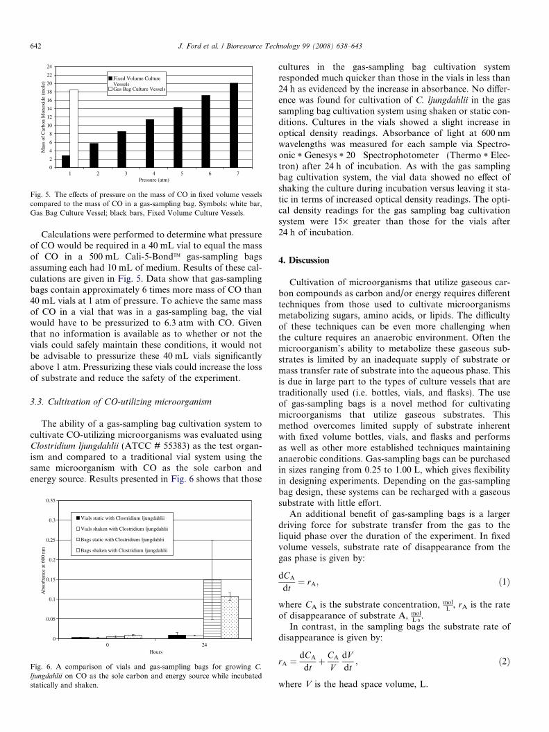
Fig. 5. The effects of pressure on the mass of CO in fixed volume vesselscompared to the mass of CO in a gas-sampling bag. Symbols: white bar,Gas Bag Culture Vessel; black bars, Fixed Volume Culture Vessels.
642 J. Ford et al. / Bioresource Technology 99 (2008) 638–643
Calculations were performed to determine what pressureof CO would be required in a 40 mL vial to equal the massof CO in a 500 mL Cali-5-BondTM gas-sampling bagsassuming each had 10 mL of medium. Results of these cal-culations are given in Fig. 5. Data show that gas-samplingbags contain approximately 6 times more mass of CO than40 mL vials at 1 atm of pressure. To achieve the same massof CO in a vial that was in a gas-sampling bag, the vialwould have to be pressurized to 6.3 atm with CO. Giventhat no information is available as to whether or not thevials could safely maintain these conditions, it would notbe advisable to pressurize these 40 mL vials significantlyabove 1 atm. Pressurizing these vials could increase the lossof substrate and reduce the safety of the experiment.
3.3. Cultivation of CO-utilizing microorganism
The ability of a gas-sampling bag cultivation system tocultivate CO-utilizing microorganisms was evaluated usingClostridium ljungdahlii (ATCC # 55383) as the test organ-ism and compared to a traditional vial system using thesame microorganism with CO as the sole carbon andenergy source. Results presented in Fig. 6 shows that those
0
0.05
0.1
0.15
0.2
0.25
0.3
0.35
0 24Hours
Abs
orba
nce
at 6
00 n
m
Vials static with Clostridium ljungdahlii
Vials shaken with Clostridium ljungdahlii
Bags static with Clostridium ljungdahlii
Bags shaken with Clostridium ljungdahlii
Fig. 6. A comparison of vials and gas-sampling bags for growing C.
ljungdahlii on CO as the sole carbon and energy source while incubatedstatically and shaken.
cultures in the gas-sampling bag cultivation systemresponded much quicker than those in the vials in less than24 h as evidenced by the increase in absorbance. No differ-ence was found for cultivation of C. ljungdahlii in the gassampling bag cultivation system using shaken or static con-ditions. Cultures in the vials showed a slight increase inoptical density readings. Absorbance of light at 600 nmwavelengths was measured for each sample via Spectro-onic * Genesys * 20 Spectrophotometer (Thermo * Elec-tron) after 24 h of incubation. As with the gas samplingbag cultivation system, the vial data showed no effect ofshaking the culture during incubation versus leaving it sta-tic in terms of increased optical density readings. The opti-cal density readings for the gas sampling bag cultivationsystem were 15· greater than those for the vials after24 h of incubation.
4. Discussion
Cultivation of microorganisms that utilize gaseous car-bon compounds as carbon and/or energy requires differenttechniques from those used to cultivate microorganismsmetabolizing sugars, amino acids, or lipids. The difficultyof these techniques can be even more challenging whenthe culture requires an anaerobic environment. Often themicroorganism’s ability to metabolize these gaseous sub-strates is limited by an inadequate supply of substrate ormass transfer rate of substrate into the aqueous phase. Thisis due in large part to the types of culture vessels that aretraditionally used (i.e. bottles, vials, and flasks). The useof gas-sampling bags is a novel method for cultivatingmicroorganisms that utilize gaseous substrates. Thismethod overcomes limited supply of substrate inherentwith fixed volume bottles, vials, and flasks and performsas well as other more established techniques maintaininganaerobic conditions. Gas-sampling bags can be purchasedin sizes ranging from 0.25 to 1.00 L, which gives flexibilityin designing experiments. Depending on the gas-samplingbag design, these systems can be recharged with a gaseoussubstrate with little effort.
An additional benefit of gas-sampling bags is a largerdriving force for substrate transfer from the gas to theliquid phase over the duration of the experiment. In fixedvolume vessels, substrate rate of disappearance from thegas phase is given by:
dCA
dt¼ rA; ð1Þ
where CA is the substrate concentration, molL
, rA is the rateof disappearance of substrate A, mol
L�s .In contrast, in the sampling bags the substrate rate of
disappearance is given by:
rA ¼dCA
dtþ CA
VdVdt; ð2Þ
where V is the head space volume, L.

J. Ford et al. / Bioresource Technology 99 (2008) 638–643 643
As can be observed in Eqs. (1) and (2), the reduction involume of the gas-sampling bags causes a faster substraterate of disappearance. Gas-sampling bag systems favorkinetically limited condition compared to fixed volume ves-sels. This condition is critical to measure reliable kineticparameters. In other words, as microorganisms consumesubstrate from the gas-sampling bags the volume reductionenhances substrate transfer into solution compared to fixedvolume systems.
Sterilization and reuse of gas-sampling bags is an issuethat could not be addressed by autoclaving in this study.It was observed that after autoclaving the 5 mil gas-sam-pling bags they were not capable of maintaining a head-space of CO due to micro-tears in the bags or damagesto the seams. Bags could be washed to remove residuesfrom a previous experiment but would need to be sterilizedby means other than autoclaving such as ethylene oxide orc radiation.
It should be pointed out that gas-sampling bags manu-factured from Teflon used in this study failed to maintainCO in the bag for 24 h. The other systems evaluated werethe 2 mil Tedlar gas-sampling bags and their performancewas determined to be unacceptable. Based on the findingsof this study it is suggested that gas-sampling bags manu-factured with a thickness of 5 mil or greater be utilized toculture microorganisms on this type of gaseous substrate.
Cultivation experiments using C. ljungdahlii demon-strated the effectiveness of the gas sampling bag cultivationsystem to cultivate CO-utilizing microorganisms. This sys-tem produces a greater concentration of cells in 24 h ofincubation than traditional fixed volume system produced.Wide deviation with the gas sampling bag cultivation sys-tem was from one sample at the 24 h readings with an opti-cal density reading of 0.26 while the other two samples hadoptical density measurements of 0.104 and 0.078. A possi-ble explanation to this observation is that a clump of cellswas retrieved from the gas sampling bag cultivation systemand summarily dispersed prior to taking the optical densityreading. In all, the optical density readings were 15·greater than those observed for the C. ljungdahlii cultivatedin the vials. This strongly suggests that the gas-samplingbag cultivation system is superior to traditional vial culti-vation system for C. ljungdahlii using CO as the sole carbonand energy source.
This work has demonstrated the feasibility of using gas-sampling bags as vessels to cultivate gaseous substrate-uti-lizing-microorganisms. This system can overcome the sup-ply limitations that are inherent with fixed volume vessels.Operation at atmospheric pressure and the capability ofutilizing larger amounts of gas at the initiation of an exper-iment increases safety, minimizes handling to toxic gases,and enhances the reliability of the measured kineticparameters.
Acknowledgements
This work was supported through funding provided bythe Department of Energy’s Office of Science EPSCoRProgram contract #DEFG0200ER45830 and United StatesDepartment of Agriculture’s Special Research PrimeAgreement contract #2005-34447-15711.
References
Doan, N., Contreras, A., Flynn, J., Morrison, J., Slots, J., 1999.Proficiencies of three anaerobic culture systems for recoveringperiodontal pathogenic bacteria. J. Clin. Microbiol. 37, 171–174.
Gaddy, J.L., Clausen, E.C., 1992. A Clostridium ljungdahlii, An AnaerobicEthanol and Acetate Producing Microorganism. US Patent #5,173,429, issued on December 22, 1992.
Henstra, A.M., Stams, A.J.M., 2004. Novel physiological features ofCarboxydothermus hydrogenoformans and Thermoterrabacterium ferri-
reducens. Appl. Environ. Microbiol. 70, 7236–7240.Kafkewitz, D., Iannotti, E.L., Wolin, M.J., Bryant, M.P., 1973. An
anaerobic chemostat that permits the collection and measurement offermentation gases. Appl. Environ. Microbiol. 25, 612–614.
Maschio, G., Lucchesi, A., Stoppato, G., 1994. Production of syngas frombiomass. Bioresource Technol. 48, 119–126.
Ohwarki, K., Hungate, R.E., 1977. Hydrogen utilization by Clostridia insewage sludge. Appl. Environ. Microbiol. 33, 1270–1274.
Phillips, J.R., Klasson, K.T., Clausen, E.C., Gaddy, J.L., 1993. Biologicalproduction of ethanol from coal synthesis gas: medium developmentstudies. Appl. Biochem. Biotech. 39–40, 559–571.
Sokolova, T.G., Kostrikina, N.A., Chernyh, N.A., Tourova, T.P.,Kolganova, T.V., Bonch-Osmolovshaya, E.A., 2002. Carboxydocella
thermautotrophica gen. nov., sp. nov., a novel anaerobic, CO-utilizingthermophile from a Kamchatkan hot spring. Int. J. Syst. Evol.Microbiol. 52, 1961–1967.
Svetlitchnyi, V., Peschel, C., Acker, G., Meyer, O., 2001. Two membrane-associated NiFeS–carbon monoxide dehydrogenases from the anaer-obic carbon-monoxide-utilizing eubacterium Carboxydothermus hydro-
genoformans. J. Bacteriol. 183, 5134–5144.