development and cytochemical studies of …4)/pjb42(4)2711.pdf · development and cytochemical...
TRANSCRIPT
Pak. J. Bot., 42(4): 2711-2718, 2010.
DEVELOPMENT AND CYTOCHEMICAL STUDIES OF EMBRYO SAC, EMBRYO AND ENDOSPERM IN ALLIUM PERONINIANUM
IŞIL İSMAİLOĞLU*, ZEYNEP MİNE COŞKUN,
MUSTAFA KESKİN AND MERAL ÜNAL
Department of Biology, Science and Art Faculty, Marmara University, Goztepe 34730 Istanbul, Turkey.
Abstract
The development of the embryo in Allium peroninianum Aznav., is of the Onagrad type. The
developmental stages include the followings: Proembryo, globular embryo, spheroidal, stick-shaped and cylindrical mature embryo. The endosperm of A. peronnianum follows nuclear type of development. The endosperm becomes completely cellular at the stage of globular embryo. The chalazal end of the embryo sac forms a coenocytic haustorium. The nuclei in haustorium are hypertrophic. However, some features of endosperm development show striking differences from the nuclear type. The embryo sac is divided into two chambers at free nuclear stage: small micropylar and large chalazal chambers. Both the chambers are firstly free nuclear, later they become cellular. Cytochemical tests indicate the presence of higher amounts of insoluble and acidic polysaccharides in haustorium than that of endosperm but there were no lipids in both. Introduction
The genus Allium consists of remarkable embryological features e.g. sexual or asexual reproduction, diplospory, parthenogenesis, autonomous endosperm, endosperm haustoria, hypertrophied synergids, the occurrences of antipodal, integumentary and synergid embryos (Syamasundar & Panchaksharappa, 1975; Kojima & Nagato, 1992a, b). In A. neopolitanum and A. roseum the volume of persistent synergids increases due to the endoreduplication cycles (Fazla-Yıldırım, 2004). Antipodal embryony frequently occurs in apomictic Allium species but Allium amphimictics are characterized by ephemeral antipodals. The development of endosperm is of the nuclear type and embryo development conforms to the Asterad type in A. fistulosum L. (Xiang-Yuan, 1987). Musial et al., (2005) studied the development of A. cepa embryo sacs In vitro and observed an autonomous, free nuclear endosperm. Polytene chromosomes were recorded in the endosperm nuclei of A. ursinum (Turala, 1966). A. tuberosum is a pseudogamous apomictic and egg and antipodal cells have the ability to start embryogenesis independently of pollination (Kojima & Nagato, 1992a). Therefore, it seemed interesting to examine the development of endosperm and embryo of A. peroninianum which is an endemic for Turkey. Materials and Methods
The flower buds were collected at Basibuyuk, Istanbul, Turkey and they were fixed in acetic-alcohol (1:3, v/v). After dehydration in a graduated series of ethyl alcohol, the material was embedded in paraffin. The 8-12 µm thick microtome sections were stained with Regaud’s haematoxylin. For cytochemical observations, sections were stained with periodic acid-Schiff (PAS) for the localization of insoluble polysaccharides (O’Brien & McCully, 1981), with Alcian blue (1% Alcian blue in 3% acetic acid) for acidic polysaccharides and pectins (Heslop-Harrison, 1979), with Coomassie brilliant blue (in mixture of water, methanol and acetic acid (v:v:v, 87:10:3)) for proteins (Fisher, 1968) and with Sudan black B (in 70% ethanol) for lipids (Pearse, 1968). *Corresponding author E-mail: [email protected]
IŞIL İSMAİLOĞLU ET AL.,
2712
The preparations were analyzed with the Image pro-express software, assisted by an Evolution LC color camera and an Olympus BH-2 microscope. Results
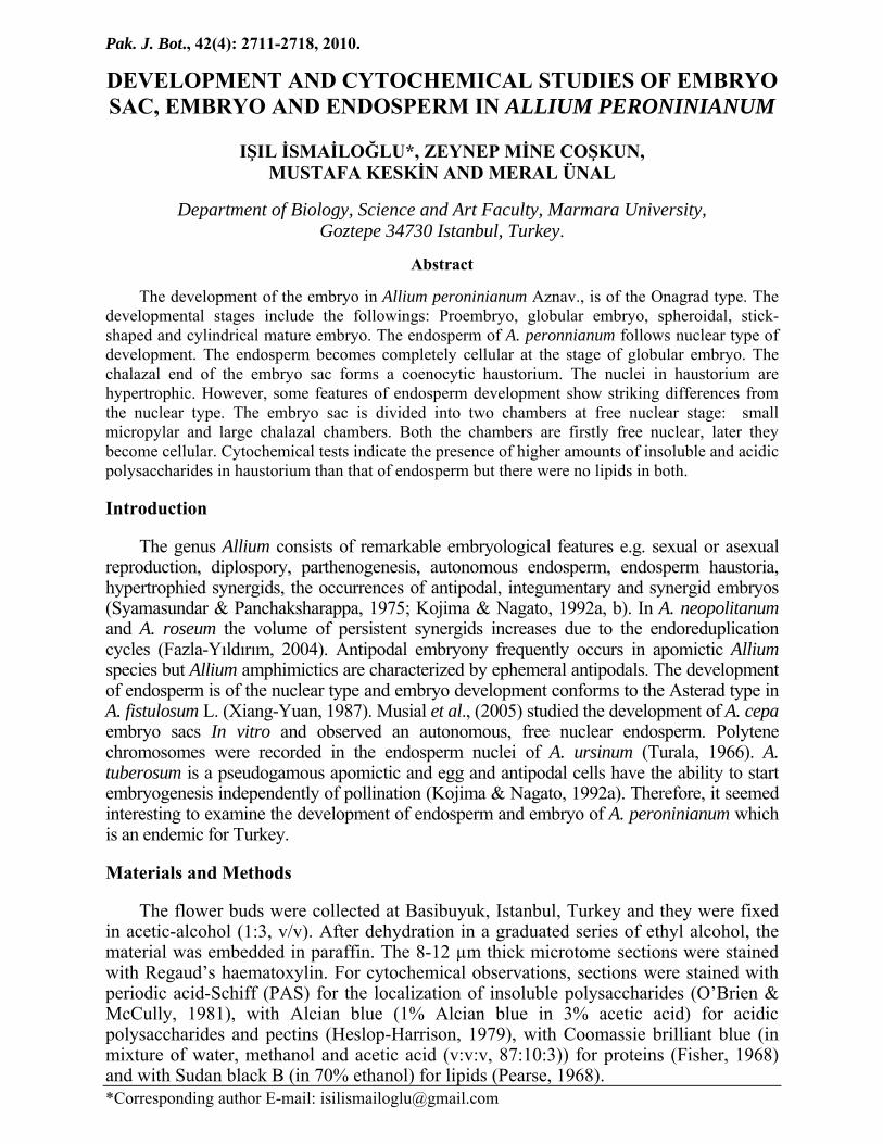
Two ovules are attached to the placenta in each of three ovarian locules. Each ovule contains one embryo sac (ES), no ovule are ever found in which more than one ES is formed (Fig. 1a). ES development conforms to bisporic Allium type. Mature ES contains three antipodal cells degenerating in early stage, two polars fusing to from secondary nucleus, two synergids and one egg cell. One of synergids degenerates in early stage, the other persists (Fig. 1b). The synergids are hooked and exhibit filiform apparatus which gives strong PAS positive reaction (Fig. 1c). It is the most conspicuous structure in the sinergid and extends from the wall in proximal portion of the cell up to almost its center. The filiform apparatus stains intensely for insoluble polysaccharides and composed of fibrillar projections. The wall of ES strongly stained with PAS and Alcian blue (Fig. 5b, c).
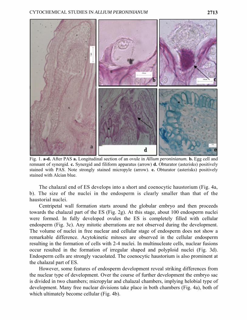
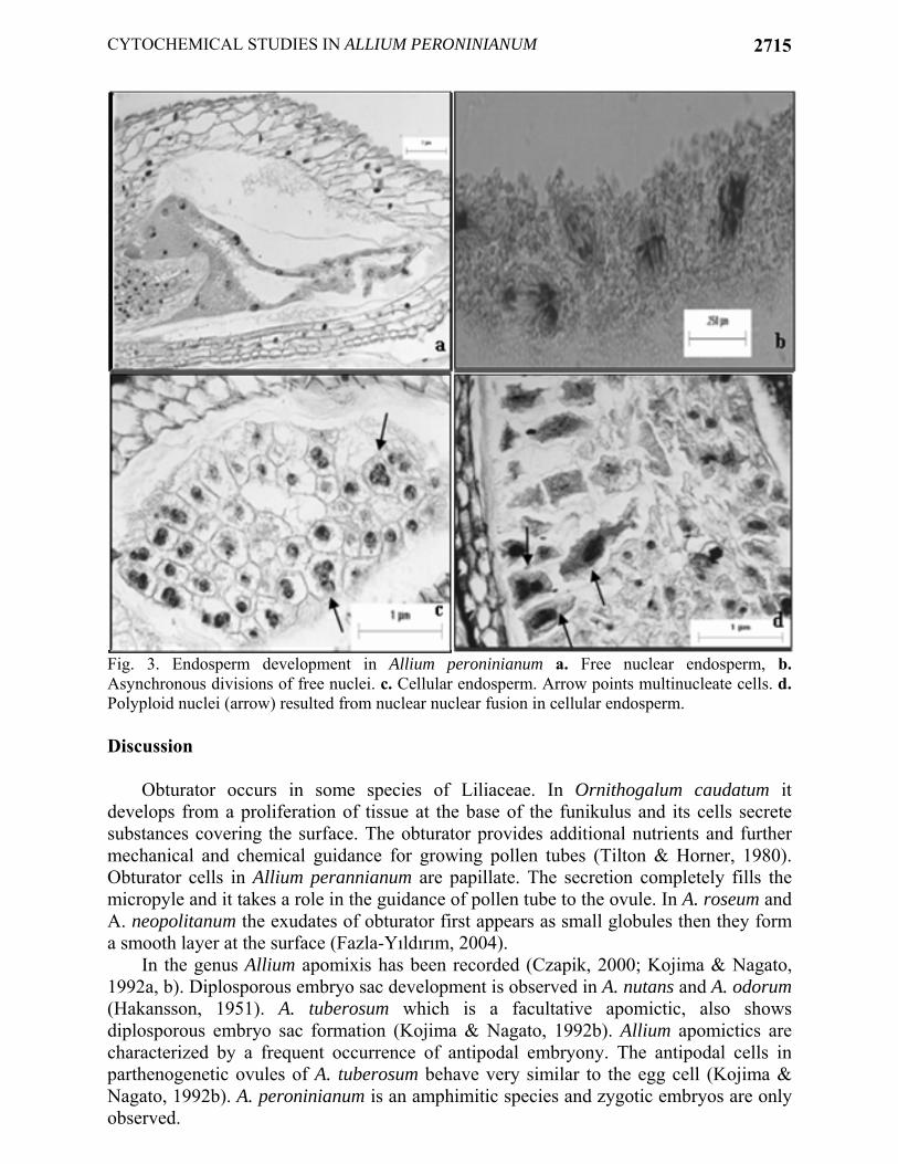
In A. peroninianum obturator develops at the base of the funiculus and it consists of thick-walled, greatly elongated papillate cells. A large vacuole commonly is found in the distal end of the cell. The cells of obturator are secretory and produce substances which coat their surface. By cell maturity, the secretory substances form a smooth thick layer at the surface. Cytochemical tests indicate the presence of insoluble and acidic polysaccharides in the secretory cells and on the surface of obturator (Fig. 1d, e). It seems, therefore, to take a role in guidance of pollen tubes toward the micropyle. Micropylar part of the ovule is also strongly positive with PAS (Fig. 1d). The development of embryo: Embryo development is of the Onagrad type. Zygote is present at the micropylar pole of the ES and smaller than the egg cell (Fig. 2a). The volume decrement is due to the disappearance of vacuole. When zygote is at metaphase or anaphase stage of the first division, the endosperm has about 6-10 free nuclei (Fig. 2b, c). The remnants of pollen tube can be still seen in that stage. The first division of zygote is transversal giving rise to a terminal cell and a basal cell almost equal in size (Fig. 2d). Apical cell divides vertically to form two juxtaposed cells. In this way a proembryo of four cells is formed (Fig. 2e). Suspensor is short and it usually consists of 2 or 4 cells. When the globular embryo is 16-celled, endosperm is still free nuclear the number of nuclei ranged from 18 to 22 (Fig. 2f). When the embryo is about 50-celled stage, the cell wall formation starts around globular embryo (Fig. 2g). At the cellular endosperm stage, the embryo reaches stick-shaped stage and grows in length. At that stage, a clear zone around embryo is very prominent on account of digestion of endosperm cells. In the oldest ovules, the cotyledon elongates. At the mature cylindrical embryo stage multicellular endosperm fills the ES (Fig. 2h). The development of endosperm: Endosperm is predominantly nuclear. The primary endosperm nucleus is formed by the fusion of a sperm with the secondary nucleus and divides soon thereafter. Its position is either in the center or toward the chalazal end of the ES. Free nuclear divisions are usually synchronous, the nuclei are spherical or elliptical and generally of the same size and embedded in a cytoplasmic sheath around the central vacuole. At first, the nuclei are distributed uniformly but later they aggregate at the chalazal part (Fig. 3a). In the advanced stages of development mitotic divisions are not synchronous (Fig. 3b).
CYTOCHEMICAL STUDIES IN ALLIUM PERONINIANUM
2713
Fig. 1. a-d. After PAS a. Longitudinal section of an ovule in Allium peroninianum. b. Egg cell and remnant of synergid. c. Synergid and filiform apparatus (arrow) d. Obturator (asterisks) positively stained with PAS. Note strongly stained micropyle (arrow). e. Obturator (asterisks) positively stained with Alcian blue.
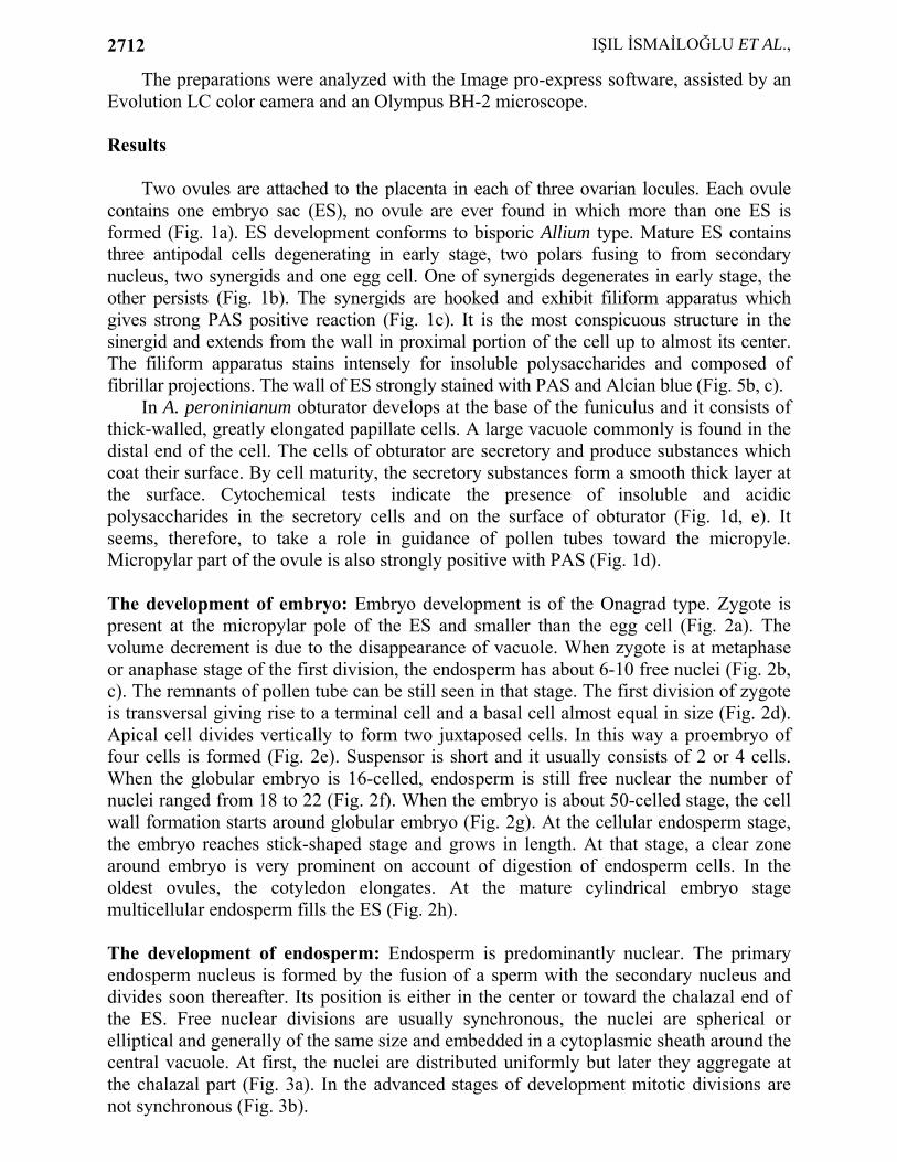
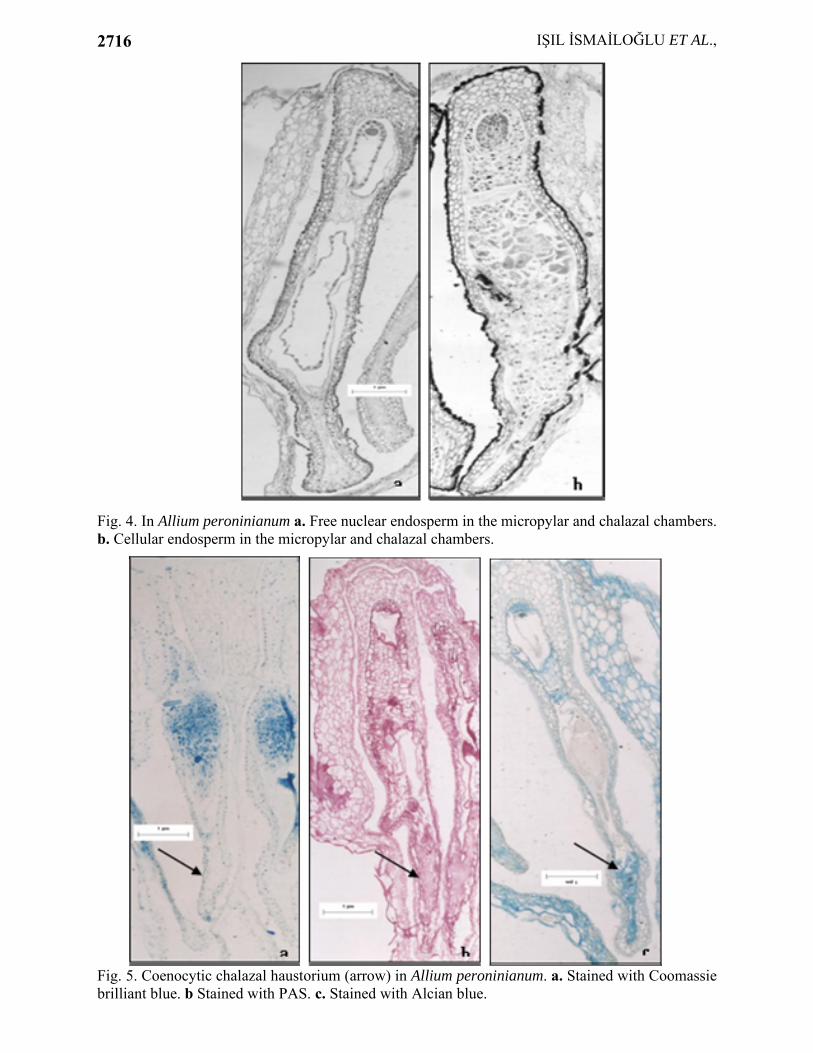
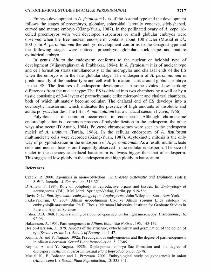
The chalazal end of ES develops into a short and coenocytic haustorium (Fig. 4a, b). The size of the nuclei in the endosperm is clearly smaller than that of the haustorial nuclei.
Centripetal wall formation starts around the globular embryo and then proceeds towards the chalazal part of the ES (Fig. 2g). At this stage, about 100 endosperm nuclei were formed. In fully developed ovules the ES is completely filled with cellular endosperm (Fig. 3c). Any mitotic aberrations are not observed during the development. The volume of nuclei in free nuclear and cellular stage of endosperm does not show a remarkable difference. Acytokinetic mitoses are observed in the cellular endosperm resulting in the formation of cells with 2-4 nuclei. In multinucleate cells, nuclear fusions occur resulted in the formation of irregular shaped and polyploid nuclei (Fig. 3d). Endosperm cells are strongly vacuolated. The coenocytic haustorium is also prominent at the chalazal part of ES.
However, some features of endosperm development reveal striking differences from the nuclear type of development. Over the course of further development the embryo sac is divided in two chambers; micropylar and chalazal chambers, implying helobial type of development. Many free nuclear divisions take place in both chambers (Fig. 4a), both of which ultimately become cellular (Fig. 4b).
IŞIL İSMAİLOĞLU ET AL.,
2714
Fig. 2. Embryo development in Allium peroninianum. a. Zygote. b. Zygote at metaphase. c. Zygote at anaphase. d. Two-celled proembryo. e. Six-celled proembryo and suspensor at free nuclear endosperm stage. f. Early globular embryo with a short suspensor. g. Late globular embryo surrounded by a clear space at cellular endosperm stage. h. Cylindrical mature embryo.
Cytochemical analysis indicated that free nuclear and cellular endosperm show weak stainability for protein, insoluble and acidic polysaccharides as revealed by staining Coomassie brilliant blue, PAS reagent, Alcian blue, respectively (Fig. 5a,b,c). There is no reaction with Sudan black B. It means no lipid is present in endosperm. Haustorium shows an increase in the PAS positiviness and very rich stainability for acidic polysaccharides but not at all for lipids and proteins.
CYTOCHEMICAL STUDIES IN ALLIUM PERONINIANUM
2715
Fig. 3. Endosperm development in Allium peroninianum a. Free nuclear endosperm, b. Asynchronous divisions of free nuclei. c. Cellular endosperm. Arrow points multinucleate cells. d. Polyploid nuclei (arrow) resulted from nuclear nuclear fusion in cellular endosperm. Discussion
Obturator occurs in some species of Liliaceae. In Ornithogalum caudatum it develops from a proliferation of tissue at the base of the funikulus and its cells secrete substances covering the surface. The obturator provides additional nutrients and further mechanical and chemical guidance for growing pollen tubes (Tilton & Horner, 1980). Obturator cells in Allium perannianum are papillate. The secretion completely fills the micropyle and it takes a role in the guidance of pollen tube to the ovule. In A. roseum and A. neopolitanum the exudates of obturator first appears as small globules then they form a smooth layer at the surface (Fazla-Yıldırım, 2004).
In the genus Allium apomixis has been recorded (Czapik, 2000; Kojima & Nagato, 1992a, b). Diplosporous embryo sac development is observed in A. nutans and A. odorum (Hakansson, 1951). A. tuberosum which is a facultative apomictic, also shows diplosporous embryo sac formation (Kojima & Nagato, 1992b). Allium apomictics are characterized by a frequent occurrence of antipodal embryony. The antipodal cells in parthenogenetic ovules of A. tuberosum behave very similar to the egg cell (Kojima & Nagato, 1992b). A. peroninianum is an amphimitic species and zygotic embryos are only observed.
IŞIL İSMAİLOĞLU ET AL.,
2716
Fig. 4. In Allium peroninianum a. Free nuclear endosperm in the micropylar and chalazal chambers. b. Cellular endosperm in the micropylar and chalazal chambers.
Fig. 5. Coenocytic chalazal haustorium (arrow) in Allium peroninianum. a. Stained with Coomassie brilliant blue. b Stained with PAS. c. Stained with Alcian blue.
CYTOCHEMICAL STUDIES IN ALLIUM PERONINIANUM
2717
Embryo development in A. fistulosum L. is of the Asterad type and the development follows the stages of proembryo, globular, spheroidal, laterally concave, stick-shaped, curved and mature embryo (Xiang-Yuan, 1987). In the pollinated ovary of A. cepa 16-celled proembryos with well developed suspensors or small globular embryos were observed when the free nuclear endosperm contains about 100 nuclei (Musial et al., 2001). In A. peroninianum the embryo development conforms to the Onagrad type and the following stages were noticed: proembryo, globular, stick-shape and mature cylindrical embryo.
In genus Allium the endosperm conforms to the nuclear or helobial type of development (Vijayaraghavan & Prabhakar, 1984). In A. fistulosum it is of nuclear type and cell formation starts simultaneously at the micropylar and chalazal ends of the ES when the embryo is in the late globular stage. The endosperm of A. peroninianum is predominantly of the nuclear type and cell wall formation starts around globular embryo in the ES. The features of endosperm development in some ovules show striking differences from the nuclear type: The ES is divided into two chambers by a wall or by a tissue consisting of 2-4 layers of parenchymatic cells: micropylar and chalazal chambers, both of which ultimately become cellular. The chalazal end of ES develops into a coenocytic haustorium which indicates the presence of high amounts of insoluble and acidic polysaccharides. The ES in A. paniculatum has a chalazal caecum (Davis, 1966).
Polyploid is of common occurrence in endosperm. Although chromosome endoreduplication is a common process of polyploidization in the endosperm, the other ways also occur (D’Amato, 1984). Polytene chromosomes were seen in the endosperm nuclei of A. ursinum (Turala, 1966). In the cellular endosperm of A. fistulosum multinucleate cells were recorded (Xiang-Yuan, 1987). Acytokinetic mitosis is the only way of polyploidization in the endosperm of A. peroninianum. As a result, multinucleate cells and nuclear fusions are frequently observed in the cellular endosperm. The size of nuclei in the coenocytic chalazal haustorium is always bigger than that of endosperm. This suggested low ploidy in the endosperm and high ploidy in haustorium. References Czapik, R. 2000. Apomixis in monocotyledons. In: Grasses Systematic and Evolution. (Eds.):
S.W.L. Jacoolas. J. Euerest., pp. 316-321. D’Amato, F. 1984. Role of poliploidy in reproductive organs and tissues. In: Embryology of
Angiosperms. (Ed.): B.M. Johri. Springer-Verlag, Berlin, pp. 519-566. Davis, G.L. 1966. Systematic embryology of the Angiosperms. John Wiley and Sons, New York. Fazla-Yıldırım, C. 2004. Allium neopolitanum Cry. ve Allium roseum L.’da sitolojik ve
embriyolojik araştırmalar. Ph.D. Thesis. Marmara University, Institute for Graduate Studies in Pure and Applied Sciences.
Fisher, D.B. 1968. Protein staining of ribboned epon section for light microscopy. Histochemie, 16: 92-96.
Hakansson, A. 1951. Parthenogenesis in Allium. Botaniska Notiser, 195: 143-179. Heslop-Harrison, J. 1979. Aspects of the structure, cytochemistry and germination of the pollen of
rye (Secale cereale L.). Annals of Botany, 44: 1-47. Kojima, A. and Y. Nagato. 1992a. Pseudogamous embyogenesis and the degree of parthenogenesis
in Allium tuberosum. Sexual Plant Reproduction, 5: 79-85. Kojima, A. and Y. Nagato. 1992b. Diplosporous embryo–Sac formation and the degree of
diplospory in Allium tuberosum. Sexual Plant Reproduction, 5: 72-78. Musial, K., B. Bohanec and L. Przywara. 2001. Embryological study on gynogenesis in onion
(Allium cepa L.). Sexual Plant Reproduction, 13: 335-341.
IŞIL İSMAİLOĞLU ET AL.,
2718
Musial, K., B. Bohanec, M. Jakse and L. Przywara. 2005. The development of onion (Allium cepa L.) embryo sacs in vitro and gynogenesis induction in relation to flower size. In vitro Cellular & Developmental Biology, 41:446-452.
O’Brien, T.P. and M.E. McCully. 1981. The study of plant structure: Principles and selected methods. Melbourne, Termacarphi.
Pearse, A.G.E. 1968. Histochemisrtry-Theoretical and applied Vol.2. London, Churchill. Syamasundar, J. and M.G. Panchaksharappa. 1975. A study of the hypertrophied synergid in Allium
cepa L. Cytologia, 40: 371-376. Tilton, V.R. and H.T. Horner. 1980. Stigma, style and obturator of Ornithogalum caudatum
(Liliaceae) and their function in the reproductive process. American Journal of Botany, 67(7): 1113-1131.
Turala, K. 1966. Strukter endopolyploider kerne im bereich der samenanlage einiger monokotylen. Oesterreichische Botanische Zeitschrift, 113: 529-541.
Vijayaraghavan, M.R. and K. Prabhaker. 1984. The Endosperm. In: Embryology of Angiosperms. (Ed.): B.M. Johri. Springer-Verlag, Berlin, pp. 319-376.
Xiang-Yuan, X. 1987. Embryo and endosperm development in green onion, Allium fistulosum L. Phytomorphology, 37(2-3): 225-233.
(Received for publication 11 October 2008)