development and evaluation of colloidal gold
TRANSCRIPT

African Journal of Microbiology Research Vol. 4(9), pp. 663-670, 4 May, 2010 Available online http://www.academicjournals.org/ajmr ISSN 1996-0808 © 2010 Academic Journals Full Length Research Paper
Development and evaluation of colloidal gold immunochromatographic strip for detection of
Escherichia coli O157
Xihong Zhao1, Xiaowei He1*, Wenmei Li 1,3, Yao Liu2, Liansheng Yang1 and Jihua Wang3
1College of Light Industry and Food Sciences, South China University of Technology, Guangzhou 510640, China.
2Zhongshan Supervision Testing Institute of Quality and Metrology, Zhongshan 528403, China. 3Guangzhou Wondfo Biotech CO., LTD, 510530, Guangzhou, China.
Accepted 24 February, 2010
Escherichia coli O157:H7 is a serious and common human pathogen that can cause diarrhoea, haemorrhagic colitis, and haemolytic uraemic syndrome (HUS). In this study, the synthesis and identification of colloidal gold particles and antibody-colloidal gold conjugates probe specific to E. coli O157:H7 were performed, and the preparation of colloidal gold immunochromatographic strip based the biotin-streptavidin system was developed for detection of E. coli O157:H7.Monodispersional nanogold colloid was synthesized and preparation of nanogold-labeled polyclonal antibody probe to E. coli O157:H7 by citrate method. Combination of antibody with nanogold particles was also characterized by UV-visible (UV-vis) light absorption spectra and transmission electron microscopy (TEM). Furthermore, nanogold-labeled probe was used to develop an immunochromatographic (IC) strip for E. coli O157:H7 analysis. With this method, analysis could be completed in less than 10 min. Examination of the 65 known strains (36 E. coli O157 strains and 29 serotypes other than E. coli) showed 98.5% specifity and 100% sensitivity, only yield a false-positive reaction with Salmonella choleraesuis. The sensitivity of the IC strip was tested using 10-fold dilution E. coli O157 in foods, could be detected at a minimum of 2.3 × 10³ CFU/ml without enrichment and 2.3 CFU/ml after enrichment. Application of IC strip test were performed on 265 water samples, 340 beef samples, 208 milk samples and 120 cake samples after enrichment, the specificity of the strip was 99.2, 97.9, 94.6 and 94.9%, respectively. The sensitivity of the strip was 100% agreement with tradition culture method. The established method is very useful for monitoring E. coli O157 containment in food samples. Key words: Escherichia coli O157, colloidal gold immunochromatographic assay, rapid test.
INTRODUCTION Escherichia coli O157:H7, member of Enterohemorrhagic E. coli (EHEC), is a bacterium that causes diarrhea that is often bloody. The diarrhea can be accompanied by abdominal cramps. Fever may be absent or mild. Symptoms usually occur within 2 - 3 days following exposure, but may occur as soon as 1 day following exposure or up to one week following exposure (Reinstein et al., 2007; Erdogan et al., 2008). Healthy adults can typically recover completely from E. coli *Corresponding author. E-mail address: [email protected]. Tel: +86-20-87058470. Fax: +86-20-87059715.
O157:H7 exposure within a week. However, some people, especially young children and the elderly, can develop Hemolytic Uremic Syndrome (HUS) as a result of exposure to E. coli O157:H7, a condition that can lead to serious kidney damage and even death (Sanchez et al., 2008, Baudouin et al., 2008).
The contamination of food with E. coli O157:H7 is of increasing concern internationally. However, in a traditional method, the contaminated food sample or feces is allowed to grow in an enriched media for 6 - 18 h a portion of the broth then plated on agar media and analysed by biochemical tests and serological reactions which are time-consuming, laborious, and inefficient. Other methods such as the immunomagnetic beads

664 Afr. J. Microbiol. Res. coated with antibodies of E. coli O157:H7(Crawford et al., 2000; Shelton and Karns, 2001; Tu et al., 2003; Gehring and Tu, 2005a) or the more established polymerase chain reaction (PCR) (Elizaquivel and Aznar, 2008; Gooding and Choudary, 1998; Takeshi et al., 1997), loop-mediated amplification (LAMP) (Hara-Kudo et al., 2008; Xihong Zhao, 2010), where the relevant genes are amplified, require trained personnel and clean samples that may need several hours of incubation to obtain detectable amounts of the target. Although enzyme immunoassay has been used widely in many laboratories (Gehring et al., 2006; Chapman et al., 1997; Fratamico and Strobaugh, 1998; Feldsine et al., 2002; Bai et al., 2007), this method still requires various equipments and trained personnel. Thus, a simple and inexpensive immunochromatographic strip could have diagnostic applications without the requirement of complicated equipment. Jung, et al (Jung et al., 2005) developed the colloidal immunochromatographic strip for detection of E. coli O157 in enriched samples, reported that the minimum limit was 1.8×10⁵ CFU/ml without enrichment and 1.8 CFU/ml after enrichment. However, there were some problems with the development of immunoassay methods, such as the limit of detection and false positive (Gehring and Tu, 2005b).
Avidin and streptavidin are widely used in biotin - (strept) avidin system technology, which is based on their tight biotin-binding capability (Laitinen et al., 2007). These biotin-(strept) avidin based methods enable both signal amplification and a reduction in background activity (Hartley, 1996) .The immunoassay method based biotin-streptavidin signal amplification system was developed suitable analytical techniques to be used in many fields detection. (Paffard et al., 1997; Roth and Jeltsch, 2000; Matalka et al., 2001; Rambozzi et al., 2004; Yu et al., 2007). But there were no articles about the colloidal gold immunochromatographic strip based biotin-streptavidin system for detection E. coli O157.
We have developed a rapid two-step procedure, coated streptavidin (SA) in Test line of nitrocellulose menbrane targeting moiety, colloidal gold labeled the anti- E. coli O157 monoclonal antibodies (MAbs) and biotinylated anti- E. coli O157 polyclonal antibody were impregnated in the conjugated pad and sample pad, respectively. The advantage of SA coating in the Test line, compared to the use of directly MAbs, was a reduction in background levels and amplification sign due to rapid clearance of biotinylated antibody and capture four times molecular biotin. Also, we presented details of our investigation into the formation and characterization of an antibody–colloidal gold probe (conjugate) specific to E. coli O157:H7 and its use in developing a rapid E. coli O157:H7 diagnostic method. The formation of antibody and colloidal gold conjugate was accomplished at optimal condition. The processes of formation of colloidal gold and conjugate were monitored by UV-visible (UV-vis) light measurements, whereas transmission electron microscopy (TEM) images were used to characterize the particle
size of the conjugates. Furthermore, nanogold probe was applied in developing an immunochroma-tographic assay based the biotin-streptavidin system for E. coli O157:H7. We also estimated the specificity and sensitivity of the immunochromatographic strip with pure cultured bacteria and various enriched food samples. MATERIALS AND METHODS Bacteria strains and reagents E. coli O157 strains, E. coli Non-O157 strains, Non-E. coli strains were obtained from the Zhongshan Supervision Testing Institute of Quality and Metrology at Zhongshan city, Guangdong province, China. All cultures were maintained on Tryptic Soy Agar (Remel, Lenexa, KS) slants or plates. Food samples (water, beef, cake and milk) were bought from an open market near the campus of South China University of Technology in the city of Guangzhou, China. Cultures for the assays were grown on tryptic soy broth for 18 h at 37°C with aeration and serially diluted 10 fold in sterile 10 mmol/l phosphate buffered saline (PBS) at pH 7.4. The number of colony forming units (CFU), for each dilution was confirmed by plating 100 �l of diluted sample onto tryptic soy agar plates and incubated at 37°C for 24 h. The colonies formed on the plates were then counted.
Nitrocellulose membrane, glass fiber and absorption pad were obtained from sartorius stedim (Germany). Chloroauric acid (HAuCl4·3H2O) and sodium citrate were obtained from the chemicals and reagents of Shanghai and used without further purification. EZ-link sulfo-NHS-LC-Biotin and streptavidin (SA) were obtained from Pierce Chemical Co. (Rockford, IL, USA). Deionized and distilled water was used in this experiment. All other chemicals used in the present study were either analytical pure or with highest quality. Preparation of biotinylated antibody Preparation of biotinylated rabbit anti- E. coli O157 polyclonal antibody was carried out using EZ-link sulfo-NHSLC-Biotin according to the manufacturer’s instructions for biotinylation of proteins. Synthesis and characterization of colloidal gold Colloidal gold particles with a mean diameter of 40 nm were prepared according to the method by Xu et al (Huo et al., 2006). Briefly, an aqueous solution of chloroauric acid (30 ml of 0.01% (W/V) HAuCl4·3H2O) was heated to boiling point, followed by 300 �l of 1.0% (W/V) sodium citrate solution added into it (Ji et al., 2007). The reaction solution was stirred simultaneously and gently boiling it for 15 min until the color of the solution turned from straw yellow into black and eventually red. The obtained colloidal gold solution was used for conjugation with monoclonal antibody (MAb) to E. coli O157:H7. Minimum amount of MAb for stabilizing colloidal gold particles A murine monoclonal antibody against E. coli O157:H7 lipopolysaccharide was used in this research .For the conjugation of colloidal gold, a minimum amount of MAb concentration was needed for stabilizing colloidal gold particles. Colloid gold suspension was adjusted to the same pH (ranges about 7 - 8) with 0.01 mol/L Na2CO3 solution firstly and pipetted into a series of tubes

Zhao et al. 665
Figure 1. Schematic representation of the lateral flow immunochromatographic test strip. (A) Top view; (B) cross-section.
(tube/0.95 ml). Then, monoclonal antibody solution (0.01 - 0.07 mg/ml, 0.95 ml) was added to the above each colloidal gold solution. After that, each tube received 0.1ml of 10% NaCl and was shaken for observation of color change after 2 h. In the experiment, 0.03 mg/ml of MAb was confirmed to be the minimum amount for stabilizing colloidal gold solution. Therefore, 2.5 ml MAb (0.03 mg/ml prepared in pH 7.4, PBS 0.01 mol/L) was added drop-wise to 2.5 ml of colloidal gold solution. The solution was stored for 2 h at 4°C and then, was centrifuged to remove unconjugated MAb. The centrifugation was undertaken at 14,000 r/min at 4°C followed by the pellet redispersed in total 5 ml of pH 7.4 PBS and stored at 4°C for further experiments. Characterization of conjugates UV-vis spectroscopy studies The formation of antibody - colloidal gold conjugates was monitored by UV-vis spectroscopy (200 - 700 nm) using a double-beam spectrophotometer (model 1.70, GBC) operated at 1 nm. The gold solutions were monitored immediately after addition of the antibody and after centrifugation and resuspension of the conjugates in appropriate buffers. Transmission electron microscopy measurements TEM measurements were performed on a Hitachi H-7000 microscope operating at an acceleration voltage of 100 kv at a magnification of 100,000. The sample was prepared by placing a drop of colloidal gold and centrifuged and resuspended antibody - colloidal gold solution on a carbon-coated TEM copper grid. The film was allowed to dry for 5 min, and the excess solution was removed using a blotting paper. Samples preparation Samples of ground beef were prepared by mixing 5 g of ground beef with 45 ml of PBS spiked with E. coli O157:H7 at concentrations ranging from 101 - 107 CFU/ml. The samples were homogenized by stomaching for 1 min so that the heavier
particulates could settle before pipetting the clear supernatant into a vial for analysis. Ground beef sample extract in PBS with no pathogen were used as negative control. A similar method was adopted for cake (Waswa et al., 2007).
Pasteurized skimmed milk (50 ml) was inoculated with E. coli O157:H7 at about 107 CFU/ml. The inoculated sample was then taken through a 10-fold serial dilution in uninoculated pasteurized milk to obtain samples in the concentration range between 101 and 106 CFU/ml. Uninoculated samples were used as negative control . A similar method was adopted for water (Huang et al., 2007). Each 80 �l sample applied on test strips. Preparation of immunochromatographic test strip Immunochromatographic strip was constructed as a method by Huo et al., 2006 and Lakner et al., 1998 Colloidal gold-labeled antibody conjugate and biotinylated antibody were jetted onto glass fiber (conjugate pad) and non woven (sample pad) , respectively, then dried at 50°C for 2 h. Goat anti-mouse antibody (1.0 mg/ml) was dispensed onto a nitrocellulose membrane on the upper line for control with a volume of 1 �l per 1 mm line, and SA (1.0 mg/ml) in PBS was jetted into the lower part for test line; the dispensed volume was also of 1 �l per 1 mm line. The remaining active sites on the membrane were blocked by incubation with 5% BSA in PBS (1 ml/cm membrane) for 30 min at 37°C.The membrane was washed once with PBS and again with distilled water and then, dried at 37°C. Subsequently, the absorption pad, the nitrocellulose membrane, the released antibody-gold conjugation pad, and the sample application pad were assembled into a sheet of plastic backing orderly (Figure 1) and cut into individual strips (2.0 mm/strip) with a pair of small medicine scissors. The total assay time was less than 10 min. Sensitivity and specificity The sensitivities and specificities of the tests were determined for the evaluation of the utility of the tests. The following formulas were used to calculate test indices (McClure, 1990): sensitivity = [TP/(TP + FN)] × 100 and specificity = [TN/(TN + FP)] × 100 (where TP= number of true positives, TN = number of true negatives, FN = number of false negatives, and FP = number of false positives).
666 Afr. J. Microbiol. Res.
(a) (b)
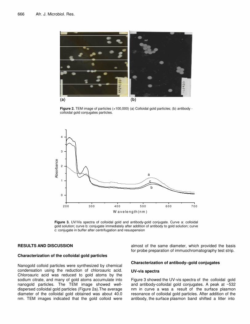
Figure 2. TEM image of particles (×100,000) (a) Colloidal gold particles; (b) antibody - colloidal gold conjugates particles.
2 0 0 3 0 0 4 0 0 5 0 0 6 0 0 7 0 0
0
1
2
3
4
Abs
orba
nce
W a v e le n g th (n m )
a
c
b
Figure 3. UV/Vis spectra of colloidal gold and antibody-gold conjugate. Curve a: colloidal gold solution; curve b: conjugate immediately after addition of antibody to gold solution; curve c: conjugate in buffer after centrifugation and resuspension
RESULTS AND DISCUSSION Characterization of the colloidal gold particles Nanogold colloid particles were synthesized by chemical condensation using the reduction of chloroauric acid. Chloroauric acid was reduced to gold atoms by the sodium citrate, and many of gold atoms accumulate into nanogold particles. The TEM image showed well-dispersed colloidal gold particles (Figure 2a).The average diameter of the colloidal gold obtained was about 40.0 nm. TEM images indicated that the gold colloid were
almost of the same diameter, which provided the basis for probe preparation of immuochromatography test strip. Characterization of antibody–gold conjugates UV-vis spectra Figure 3 showed the UV-vis spectra of the colloidal gold and antibody-colloidal gold conjugates. A peak at ~532 nm in curve a was a result of the surface plasmon resonance of colloidal gold particles. After addition of the antibody, the surface plasmon band shifted a litter into

the red direction which caused by the interaction of the antibody with colloidal gold particles. A peak at ~280 nm (curve b) in the conjugate system arose due to the tryptophan and tyrosine residues in the protein of monoclonal antibody (Gole et al., 2002). After centrifu-gation and resuspension of the conjugate (curve c), the intensity of the surface resonance band and protein absorption at 280 nm decreased because of reduced colloidal gold concentration and removal of the excess unconjugated monoclonal antibody molecules in solution. TEM images Figure 2b showed the TEM images of the antibody–colloidal gold conjugates formed on a carbon-coated copper grid. It was observed that the gold particles were still monodisperse and ordered as the colloidal gold. The mean size of the particles was 42 nm, which was propitious for strong signal generation in immuochromatography test strip. Immunochromatographic test The lateral flow assay result was determined visually by the degree of intensity of the gold color of the test line and the control line on which the SA and goat anti-mouse IgG were separately immobilized, and the result was immediately observable. If E. coli O157 is present in the sample, it binds to the biotinylated antibody in the sample pad first to form biotinylated antibody-antigen complex. Then the complex reacts with colloidal gold-labeled anti-E. coli O157 Mab to form biotinylated-colloidal gold complex (biotinylated antibody-antigen-colloidal gold body complex), then migrated by capillary action through the membrane on the detection zone. A red band appeared within 5 min as biotinylated-colloidal gold complex capturing by SA as the high-affinity biotin-streptavidin interaction in test line. However, the excess colloidal gold conjugates moved farther until they reacted with goat anti-mouse IgG, showed another red line on the upper portion of the NC membrane. Therefore, two red lines on the IC strip indicated the presence of E. coli O157. In contrast, negative samples resulted in only one red line between colloidal gold conjugates and anti-mouse IgG at the upper portion of the NC membrane. If no line forms, the test is invalid. Strains Table 1a showed the results of the immunochroma-tographic test pure-cultured bacteria. All E. coli O157 strains reacted strongly with immunochromatographic strip, regardless of H serotype. While the other strains, representing 10 E. coli Non-O157 serotype, did not react
Zhao et al. 667 with it. However, one (Salmonella choleraesuis) of the 19 tests strains of non- E. coli yielded false-positive reactions. It was reported that some of the non-E. coli strains, for example Salmonella, Citrobacter freundii and Escherichia hermannii, shared common antigens with E. coli O157 (Borczyk et al., 1990; Bettelheim, 1993). However, the disadvantage could be conquered by following selective plating media; Salmonella was inhibited the growth on cefixime-tellurite sorbitol MacConkey agar as well as fermented sorbitol (Fratamico and Bagi, 2007; Jung, 1999).
Table 2b showed the summary of the E. coli O157 immuochromatography strip test of pure-cultured bacteria. All E. coli O157 strains were correctly identified, so the sensitivity of the immunochromatographic test was 100%. One yielded a false-positive reaction, so the specificities of the test was 98.5%. Therefore, the false-negative rate was 0% (0/28) and false-positive rate was 1.5% (1/65). Detection of E. coli O157 in foods In the present study, the sensitivity of the IC strip was evaluated using 10-fold diluted E. coli O157:H7 with a range of 2.3 × 10⁶ to 2.3 CFU/ml. E. coli O157 could be detected at a minimum of 2.3 CFU/ml after enrichment (Figure 4) and 2.3 × 10³ CFU/ml without enrichment. The results showed that the sensitivity of the IC strip enhanced highly with sample after enrichment. Because of small numbles of E.coli O157:H7 in samples and food matrix reasons, we suggest that combining the enrichment procedures and IC test step.
When we applied the IC strips using food samples in the field, 8 of 265 water samples, 5 of 340 beef samples, 10 of 208 milk samples, and 5 of 120 cake showed positive signals. In contrast, E. coli O157:H7 were isolated from 6 (2.3%) of 265 water samples, 5 (1.5%) of 340 beef samples, 5 (2.4%) of 208 milk samples, and 3 (2.5%) of 120 cake samples (Table 2). We found that results obtained with the IC strip exhibited 100% agree-ment with those of traditional cultured methods after selective enrichment, since E. coli O157:H7 was then iso-lated from all the samples with positive strip test results.
Prior to constructing the one-step sandwish immuno-chromatographic assay strip by using nanogold–antibody conjugate probe, we employed two-step immunochroma-tographic assay. In order to enhance the sensitivity of detection, we selected the biotin-streptavidin amplification system. The first step was that biotinylated antibody and colloidal gold labeled Mab captured the E. coli O157:H7 from samples, then formed the complex. The second step was that the complex was captured by SA for high-affinity biotin-streptavidin interaction. Results showed that the sensitivity was near 100 times higher than one-step sandwish immunochromatographic assay strip by Jung et al. (2005) with pre-cultured strains in food.

668 Afr. J. Microbiol. Res.
Table 1a. Specificity of the E. coli O157 immuochromatographic strip tested with different pure-cultured bacteria. Microorganism No. of strains tested No. of positive tested No. of negative tested E. coli O157 c E. coli O157:H7 32 32 0 E. coli O157:H19 2 2 0 E. coli O157:H43 1 1 0 E. coli O157:H45 1 1 0 E. coli Non-O157 E. coli O3 3 0 3 E. coli O26 2 0 2 E. coli O38 1 0 1 E. coli O111 3 0 3 E. coli O145 1 0 1 Non- E. coli Salmonella typhimurium 1 0 1 Salmonella choleraesuis 2 1 1 Salmonella paratyphi 1 0 1 Shigella flexner 1 0 1 �-Hemolytic streptococcus 1 0 1 Enterococcus faecalis 2 0 2 Vibrio parahaemolyticus 3 0 3 Staphyloccs epidermidis 1 0 1 Staphylococcus albus 2 0 2 Streptococcus faecalis 1 0 1 Staphylicoccus aureus 2 0 2 Pseudomonas aeruginosa 2 0 2
Table 1b. Summary of the E. coli O157 immuochromatographic strip test of different bacteria.
No. of strains tested Microorganism Total Positive Negative
Specificity (%)
Sensitivity (%)
E. coli O157 36 36 0 98.5 100 E.coli Non-O157 10 0 10 Non- E. coli 19 1 18
Table 2. Comparison detection E. coli O157:H7 of immunochromatographic strip test with traditional isolation
No. of positive/ negative samples by immunochromatographic strip test E. coli O157:H7 isolation Water
(n = 265) Beef
(n = 340) Milk
(n = 208) Cake
(n = 120) Positive 6/0 5/0 5/0 3/0 Negative 2/247 7/329 11/192 6/111 Sensitivity 100.0 100.0 100.0 100.0 Specificity 99.2 97.9 94.6 94.9
Conclusion Monodisperse nanogold colloid was prepared with a
reduction of chloroauric acid by sodium citrate. An antibody probe to detect E. coli O157 was prepared by addition of the monoclonal antibody to the colloidal gold

Zhao et al. 669
Figure 4. Sensitivity of E. coli O157 immunochromatographic strip with enrichment. E. coli O157:H7 was spiked and then 10-fold diluted from 2.3 × 10⁶ CFU/ml (strip a) to 2.3 CFU/ml (strip g). Strip h was a negative control. The strip could detect 2.3 × 10³CFU/ml before enrichment; it could detect 2.3 CFU/ml (strip g) after enrichment. Strip i was an unvalid control.
solution to form conjugates. Optimal conditions for conjugation between colloidal gold and antibody were also ascertained. UV-visible light absorption spectra and transmission electron microscopy images showed that the nanogold and nanoprobe gold particles were well defined and regularly shaped.
The proposed method has been used for the determination of E. coli O157 in food samples with satisfactory results. Compared with the traditionally colloidal gold immunochromatographic detection, this method based the biotin-streptavidin system showed greatly heightened sensitivity. The minimum limit of detection improved by near two orders of magnitude, which was very suitable for the conditions with extremely low concentration of analysis or very small volumes of sample on site. The method could be obtained within 10 min and that all needed reagents were included in the strip, also referenced in the development of immunochromatographic assay for the detection of other food-borne pathogen. REFERENCES Bai YL, Huang WC, Yang ST (2007). Enzyme-linked Immunosorbent
assay of Escherichia coli O157 : H7 in surface enhanced Poly(Methyl methacrylate) microchannels. Biotechnol. Bioengin. 98: 328-339.
Baudouin V, Mariani-Kurkdjian P, Macher MA, Ait-Ifrane S, Garnier A, Bingen E, Loirat C, Deschenes G (2008). Treatment of a relapsing HUS secondary to prolonged carriage of E.coli O157: H7. Pediatric nephrol. 23: 1599-1599.
Bettelheim KAEH (1993). Isolation of a Citrobacter freundii strain which carries the E. coli O157 antigen. J. Clin. Microbiol. 31: 760-761.
Borczyk AA, Harnett N, Iombos M, Iior H (1990). False positive identification of E. coli O157 by commercial latex agglutination test. Lancet. 336: 946-947.
Chapman PA, Malo A, Siddons CA, Harkin M (1997). Use of commercial enzyme immunoassays and immunomagnetic separation systems for detecting E. coli O157 in bovine fecal samples. Appl. Environ. Microbiol, 63: 2549-2553.
Crawford CG, Wijey C, Fratamico P, TU SI, Brewster J (2000). Immunomagnetic-electrochemiluminescent detection of E.coli O157 : H7 in ground beef. J. Rapid Methods and Automation Microbiol. 8: 249-264.
Elizaquivel P, Aznar R (2008). A multiplex RTi-PCR reaction for simultaneous detection of E. coli O157 : H7, Salmonella spp. and Staphylococcus aureus on fresh, minimally processed vegetables. Food Microbiol. 25: 705-713.
Erdogan H, Erdogan A, Levent B, Kayali R, Arslan H (2008). Enterohemorrhagic E. coli O157:H7: case report. Turk. J. Pediatrics, 50: 488-491.
Feldsine PT, Kerr DE, Leung SC, Lienau AH, Miller SM, Mui LA (2002). Assurance (R) enzyme immunoassay eight hour method for detection of enterohemorrhagic E. coli O157 : H7 in raw and cooked beef (modification of AOAC official method 996.10): Collaborative study. J. Aoac Inter. 85: 1037-1044.
Fratamico PM, Bagi LK (2007). Comparison of methods for detection and isolation of cold- and freeze-stressed E. coli O157: H7 in raw ground beef. J. Food Prot. 70: 1663-1669.
Fratamico PM, Strobaugh TP (1998). Evaluation of an enzyme-linked immunosorbent assay, direct immunofluorescent filter technique, and multiplex polymerase chain reaction for detection of E. coli O157: H7 seeded in beef carcass wash water. J. Food Protection. 61: 934-938.
Gehring AG, Albin DM, Irwin PL, Reed SA, Tu SI (2006). Comparison of enzyme-linked immunomagnetic chemiluminescence with US Food and Drug Administration's Bacteriological Analytical Manual method for the detection of E. coli O157 : H7. J. Microbiol. Methods 67: 527-533.

670 Afr. J. Microbiol. Res. Gehring AG, Tu SI (2005a). Enzyme-linked immunomagnetic
electrochemical detection of live E. coli O157: H7 in apple juice. J. Food Protection 68: 146-149.
Gole A, Vyas S, Phadtare S, Lachke A, Sastry M (2002). Studies on the formation of bioconjugates of Endoglucanase with colloidal gold. Colloids and Surfaces B-Biointerfaces 25: 129-138.
Gooding CM, Choudary PV (1998). Detection of E. coli O157 : H7 in ground beef in eight hours. J. Microbiol. Methods 34: 89-98.
Hara-Kudo Y, Konishi N, Ohtsuka K, Hirramatsu R, Tanaka H, Konuma H, Takatori K (2008). Detection of Verotoxigenic E. coli O157 and O26 in food by plating methods and LAMP method: A collaborative study. Inter. J. Food Microbiol. 122: 156-161.
Hartley SFRA (1996). Biochemical Modification of Streptavidin and Avidin: In vitro and In vivo analysis. J. Nuclear Med. 37:1380-1384.
Huang SH, Wei HC, Lee YC (2007). One-step immunochromatographic assay for the detection of Staphylococcus aureus. Food Control. 18: 893-897.
Huo TM, Peng CF, Xu CL, Liu LQ (2006). Development of colloidal gold-based immunochromatographic assay for the rapid detection of medroxyprogesterone acetate residues. Food Agric. Immunol. 17: 183-190.
Ji XH, Song XN, Li J, Bai YB, Yang WS, Peng XG (2007). Size control of gold nanocrystals in citrate reduction: The third role of citrate. J. Am. Chem. Soc. 129: 13939-13948.
Jung BY, Jung SC, Kweon CH (2005). Development of a rapid immunochromatographic strip for detection of E. coli O157. J. Food Prot. 68: 2140-2143.
Jung BYSC (1999). Modified sorbitol MacConkey agar for the rapid isolation of E. coli O157:H7. Korean J.Vet. Res. 39: 765-771.
Laitinen OH, Nordlund HR, Hytonen VP, Kulomaa MS (2007). Brave new (strept)avidins in biotechnology. Trends In Biotechnol. 25: 269-277.
Lakner M, Schneider E, Usleber E, Dietrich R, Becker H, Martlbauer E (1998). Development and application of immunochromatographic tests for the detection of Staphylococcal enterotoxin E. Food Agric. Immunol. 10: 249-257.
Matalka K, El-Thaher T, Saleem M, Arafat T, Jehanli A, Badwan A (2001). Enzyme linked immunosorbent assay for determination of amlodipine in plasma. J. Clin. Lab. Anal. 15: 47-53.
Mcclure FD (1990). Design and analysis of qualitative collaborative studies: minimum collaborative program. J. Assoc. Official Anal. Chem. 73: 953-960.
Paffard SM, Miles RJ, Clark CR, Price RG (1997). Amplified enzyme-
linked-immunofilter assays enable detection of 50-10(5) bacterial cells within 1 h. Anal. Biochem. 248: 265-268.
Rambozzi L, Menzano A, Lavin S, Rossi L (2004). Biotin-avidin amplified ELISA for detection of antibodies to Sarcoptes scabiei in chamois (Rupicapra spp.). Vet. Res. 35: 701-708.
Reinstein S, Fox JT, Shi X, Alam MJ Nagaraja TG (2007). Prevalence of E. coli O157 : H7 in the American Bison (Bison bison). J. Food Prot. 70: 2555-2560.
Roth M, Jeltsch A (2000). Biotin-avidin microplate assay for the quantitative analysis of enzymatic methylation of DNA by DNA methyltransferases. Biol. Chemist. 381: 269-272.
Sanchez A, Navarro RB, Marquez RF, Quintana MF, Perez LS, Guilarte OD, Martinez HB, Bello LM (2008). Enterohemorrhagic E. coli O157: H7 Isolates From Children in Cuba. Pediatr. Infect. Dis. J. 27: 1122-1123.
Shelton DR, Karns JS (2001). Quantitative detection of E. coli O157 in surface waters by using immunomagnetic electrochemiluminescence. Appl. Environ. Microbiol. 67: 2908-2915.
Takeshi K, Ikeda T, Kubo A, Fujinaga Y, Makino S, Oguma K, Isogai E, Yoshida S, Sunagawa H, Ohyama T, Kimura H (1997). Direct detection by PCR of E. coli O157 and enteropathogens in patients with bloody diarrhea. Microbiol. Immunol. 41: 819-822.
Tu SI, Golden M, Fett WF, Gehring A, Irwin P (2003). Rapid detection of outbreak E. coli O157 and Salmonella on alfalfa sprouts by immunomagnetic capture and time-resolved fluorescence. J. Food Safety 23: 75-89.
Waswa J, Irudayaraj J, Debroy C (2007). Direct detection of E-Coli O157 : H7 in selected food systems by a surface plasmon resonance biosensor. Lwt-Food Sci. Technol. 40: 187-192.
Xihong Zhao YLLW (2010). Development and application of a loop-mediated isothermalamplification method on rapid detection E. coli O157 strains from food samples. Mol. Biol. Rep. DOI: 10.1007/s11033-009-9700-6.
Yu A, Geng TT, Fu Q, Chen C, Cui YL (2007). Biotin-avidin amplified magnetic immunoassay for hepatitis B surface antigen detection using GoldMag nanoparticles. J. Magn. Magn. Mater. 311: 421-424.