development of a scaffold incorporating zinc … · pembebasan ion daripada perancah zno-poc...
TRANSCRIPT

DEVELOPMENT OF A SCAFFOLD INCORPORATING
ZINC-OXIDE NANO-PARTICLES FOR CARTILAGE TISSUE
ENGINEERING UNDER PHYSIOLOGICAL CONDITIONS
ERAJ HUMAYUN MIRZA
FACULTY OF ENGINEERING
UNIVERSITY OF MALAYA
KUALA LUMPUR
2016
DEVELOPMENT OF A SCAFFOLD INCORPORATING
ZINC-OXIDE NANO-PARTICLES FOR CARTILAGE
TISSUE ENGINEERING UNDER PHYSIOLOGICAL
CONDITIONS
ERAJ HUMAYUN MIRZA
THESIS SUBMITTED IN FULFILMENT OF THE
REQUIREMENTS FOR THE DEGREE OF DOCTOR OF
PHILOSOPHY
FACULTY OF ENGINEERING
UNIVERSITY OF MALAYA
KUALA LUMPUR
2016
ii
UNIVERSITY OF MALAYA
ORIGINAL LITERARY WORK DECLARATION
Name of Candidate: Eraj Humayun Mirza (Passport No: AV0914812)
Matric No: KHA110058
Name of Degree: Doctor of Philosophy
Title of Thesis: The Development of a Scaffold incorporating Zinc-Oxide Nano-particles
for Cartilage Tissue Engineering under Physiological Conditions
Field of Study: Biomaterials and Tissue Engineering
I do solemnly and sincerely declare that:
(1) I am the sole author/writer of this Work;
(2) This Work is original;
(3) Any use of any work in which copyright exists was done by way of fair dealing and
for permitted purposes and any excerpt or extract from, or reference to or reproduction of
any copyright work has been disclosed expressly and sufficiently and the title of the Work
and its authorship have been acknowledged in this Work;
(4) I do not have any actual knowledge nor do I ought reasonably to know that the making
of this work constitutes an infringement of any copyright work;
(5) I hereby assign all and every rights in the copyright to this Work to the University of
Malaya (“UM”), who henceforth shall be owner of the copyright in this Work and that
any reproduction or use in any form or by any means whatsoever is prohibited without
the written consent of UM having been first had and obtained;
(6) I am fully aware that if in the course of making this Work I have infringed any
copyright whether intentionally or otherwise, I may be subject to legal action or any other
action as may be determined by UM.
Candidate’s Signature Date: 22nd August 2016
Subscribed and solemnly declared before,
Witness’s Signature Date: 22nd August 2016
Name:
Designation:

iii
Abstract
Cartilage tissue is among the most complex of all in the human body. Degeneration of
cartilage, lack of repair and various traumatic and pathological conditions among
individuals increase joint pain and disability. Researchers have been trying to repair
cartilage damage for decades, but have failed to achieve an optimal repair strategy.
Cartilage tissue is adapted to low oxygen environments and this condition appears to be
a key factor in the growth, regulation, and survival of chondrocytes; which are the only
cells present in cartilage tissue. The present thesis defines the fabrication and
characterisation of an antibacterial biomaterial for cartilage tissue repair. Furthermore,
this thesis promotes the use of Zinc Oxide (ZnO) nanoparticles (NP) for better growth of
cartilage cells and survival of cartilage tissue as a whole.
Composite scaffolds and thin films of polyoctanediol citrate (POC) polyester elastomer,
with varying concentrations (1wt%, 3wt% and 5wt%) of ZnO were fabricated by a
solvent-casting/particulate-leaching and mould casting technique respectively. It was
observed that material properties can be successfully controlled by simple variation of
NP concentration within the composite. The ion release kinetics from ZnO-POC scaffolds
are strongly dependent on NP concentration and degradation of pure POC matrix. All the
composite scaffolds showed strong antibacterial characteristics. However, cell culture
studies demonstrated that 1% ZnO incorporation in POC polymer is the optimal
concentration for chondrocyte cells.
Moreover, the effect of 1% ZnONP on chondrocyte proliferation and matrix synthesis
cultured under normoxia (21% O2) and hypoxia (5% O2) demonstrated upregulation of
chondrocyte proliferation and sulphated glycosaminoglycan (S-GAG) in hypoxic culture.
A synergistic effect of oxygen concentration and 1% ZnONP in up-regulation of anabolic
gene expression (Type II collagen (COL2A1) and aggrecan (ACAN)), and a down
regulation of catabolic (MMP-13) gene expression was observed. Furthermore,
iv
production of transcription factor hypoxia-inducible factor 1A (HIF-1A) in response to
hypoxic condition to regulate chondrocyte survival under hypoxia was not affected by the
presence of 1% ZnONP. Lastly this thesis discusses the physiological adaptations of
cartilage tissue in a dynamic mechanical loading and hypoxic condition to yield benefits
of combined bio-factors for cartilage tissue engineering applications. The results indicate
that the combination of dynamic loading is compatible with the nanoparticle addition.
Furthermore, dynamic loading suppresses MMP-13, and increase the expression of
COL2A1 and ACAN with an increase in cell viability, and promotion of rounded cell
morphology (a phenotypic marker).
It was concluded that POC-ZnONP scaffolds are of major importance in the development
of multifunctional scaffolds based on biodegradable polyesters for cartilage tissue
engineering and presence of 1% ZnONP appears to preserve homeostasis of cartilage in
its hypoxic environment. While 1% ZnONP should be considered for beneficial
incorporation into 3D hypoxic culture systems in the presence of mechanical stimulation.
Further studies must focus on determining the use of 1% ZnONP in different polymers
for use in various tissue engineering applications.
v
Abstrak
Tisu rawan adalah antara tisu yang paling kompleks di dalam badan manusia.
Kemerosotan rawan, kurang pembaikan dan pelbagai trauma dan keadaan patologi di
kalangan individu meningkatkan kesakitan pada sendi dan kecacatan. Penyelidik telah
berusaha untuk memperbaiki kerosakan rawan selama bertahun-tahun, tetapi telah gagal
mencapai strategi optimum pembaikan. Tisu rawan disuaidirikan kepada persekitaran
rendah oksigen dan keadaan ini menjadi satu faktor kepada pertumbuhan, peraturan dan
kebertahanan kondrosit; iaitu satu-satunya jenis sel yang ada di dalam tisu rawan. Tesis
ini menentukan fabrikasi dan pencirian biobahan antibakteria untuk pembaikan tisu
rawan. Tambahan pula, tesis ini menggalakkan penggunaan nano-partikel (NP) Zink
Oksida (ZnO) untuk pertumbuhan yang lebih baik bagi sel-sel rawan dan kebertahanan
tisu rawan secara keseluruhannya.
Perancah komposit dan filem nipis daripada elastomer polyester polyoctanediol sitrat
(POC), dengan kepekatan (1wt%, 3wt% and 5wt%) ZnO yang berbeza telah
difabrikasikan masing –masing dengan menggunakan teknik pelarut-pemutus/larutresap-
zarahan dan pemutus acuan. Daripada pemerhatian, didapati sifat-sifat material telah
berjaya terkawal oleh perubahan mudah kepekatan NP di dalam komposit itu. Kinetik
pembebasan ion daripada perancah ZnO-POC bergantung secara kuat kepada kepekatan
NP dan degradasi matriks tulen POC. Semua perancah komposit telah menunjukkan sifat-
sifat antibakteria yang kuat. Walau bagaimanapun, kajian sel kultur mendapati gabungan
1% ZnO di dalam polimer POC adalah kepekatan yang optimum untuk sel-sel kondrosit.
Tambahan lagi, kesan 1% ZnONP terhadap pertumbuhan kondrosit dan sintesis matriks
yang dikultur di bawah keadaan normoksia (21% O2) dan hipoksia (5% O2) telah
menunjukkan pengawalan-atas pertumbuhan kondrosit dan sulfat-glycosaminoglycan (S-
GAG) di dalam kultur hipoksia. Satu kesan sinergi kepekatan oksigen dan 1% ZnONP di
dalam penyataan gen pengawalan-atas anabolic (Kolagen Type II (COL2A1) dan

vi
aggrecan (ACAN)), dan penyataan gen pengawalan-bawah katabolic (MMP-13) telah
diperhatikan. Tambahan pula, pengeluaran hipoksia transkripsi faktor-dorongan factor
1A (HIF-1A) di dalam tindakbalasnya terhadap keadaan hipoksia untuk mengawal
kewujudan kondrosit tidak dipengaruhi dengan kehadiran 1% ZnONP. Akhir sekali, tesis
ini membincangkan tentang penyesuaian fisiologi tisu rawan di dalam pemuatan
mekanikal dinamik dan keadaan hipoksia untuk menghasilkan manfaat biofaktor yang
digabungkan untuk penggunaan kejuruteraan tisu rawan. Keputusan menunjukkan
gabungan pemuatan dinamik adalah serasi dengan penambahan nano-partikel. Tambahan
pula, pemuatan dinamik menyekat MMP-13 dan meningkatkan penyataan COL2A1 dan
ACAN dengan peningkatan pertumbuhan sel dan morfologi sel bulat (satu penanda
fenotip).
Ini dapat disimpulkan bahawa perancah POC-ZnONP sangat penting di dalam
penambahbaikan pelbagai fungsi perancah berasaskan polyester biodegradasi untuk
kejuruteraan tisu rawan dan kehadiran 1% ZnONP telah memelihara homestasis rawan di
dalam persekitaran hipoksia. Manakala 1% ZnONP perlu dipertimbangkan untuk
gabungan bermanfaat kepada system 3D kultur hipoksia dengan kehadiran rangsangan
mekanikal. Kajian lanjut fokus kepada penentuan penggunaan 1% ZnONP di dalam
polimer yang berbeza untuk kegunaan pelbagai aplikasi kejuruteraan tisu.

vii
I dedicate this thesis to Ammi and Baba

viii
ACKNOWLEDGEMENT
First of all I would like to praise ALLAH who blessed me with the strength to accomplish
my dream.
I would like to explicitly thank my supervisors Dr. Belinda Pingguan – Murphy and Dr.
Wan Mohd Azhar bin Wan Ibrahim, for their unconditional support, vast knowledge,
precious time and critical feedback. I am especially grateful to Dr. Belinda Pingguan–
Murphy for her patience with me and her critical review which allowed me to enhance
the quality of my articles and my thesis. Moreover, I sincerely acknowledge my ustaad,
Dr. Ali Moradi, from whom I have learnt not only cell culture techniques and scaffold
fabrication but also to help everyone. I would like to extend my appreciation to my
friends, my lab mates and my colleagues; Adel Al-Halawani, Yasser Al-Saffar, Haris
Akram, Salfarina Ezrina, Kar Wey, Poon Chi-Tat, Janu Ru, Dr. Chong Pan-Pan, Dr.
Eleanor Parker, Syed Shahabuddin, Shahid Mehmood, Kashan Pirzada, Forough
Ataollahi, Adel Dalilottojari and all those who supported me knowing or unknowingly.
I like to thank Dr. Wan Safwani and Dr. Farina to provide me with their valuable
suggestions during candidature defence.
I admire the support from Dr. Adib, Dr. Ahmad Khairi, Liyana bint Abu, Kakak Enas
naeem, Mr. Hanafi, Mr Adhli and all the staff at biomedical engineering administration.
I would also like to thank all the laboratory technical staff throughout University of
Malaya.
I would like to acknowledge the scholarship from MOHE without which I would have
never had enough financial support to continue my PhD. Furthermore, I would like to
thank University of Malaya for providing IPPP and HIR grant to fulfil my research needs.
I would like to thank Ammi and Baba for their unconditional love, support and long
conversations to calm down my anxiety. A sincere thanks to my brothers. My gratitude
and love to my wife who loves me and care for me.

ix
TABLE OF CONTENTS
LIST OF FIGURES ……………………………………………………… xv
LIST OF TABLES ………………………………………………………. xx
LIST OF ABBREVIATIONS …………………………………………… xxi
CHAPTER 1: INTRODUCTION ………………………………………..
1.1 Thesis Overview ………………………………………………………
1.1 Background ……………………………………………………………
1.2 Problem Statement …………………………………………………….
1.3 Objectives ……………………………………………………………...
1.4 Hypotheses …………………………………………………………….
1
1
1
3
4
5
CHAPTER 2: LITERATURE REVIEW ……………………………….
2.1 Introduction ……………………………………………………………
2.2 Cartilage ……………………………………………………………….
2.2.1 Composition of articular cartilage ………………………….
2.2.1.1 Chondrocytes …………………………………………
2.2.1.2 Extracellular Matrix ………………………………….
2.2.1.3 Water …………………………………………………
2.2.1.4 Collagens ……………………………………………..
2.2.1.5 Proteoglycans ………………………………………...
2.2.1.6 Non-Collagenous proteins and Glycoproteins ……….
2.2.2 Zonal arrangement in articular cartilage ……………………….
2.3 Chondrogenesis and chondrocyte differentiation ……………………...
2.4 Articular Cartilage Disease and Injury ………………………………...
2.5 Treatment Options for Cartilage Injury ………………………………..
2.5.1 Marrow Stimulation Technique ………………………………..
2.5.2 Osteochondral Grafting ………………………………………...
6
6
6
7
7
8
8
9
9
10
10
12
12
14
14
15

x
2.5.3 Autologous Chondrocyte Transplantation ……………………..
2.6 Cartilage Tissue Engineering ………………………………………….
2.6.1 Cell Sources ……………………………………………………
2.6.2 Cell Signalling Strategies ………………………………………
2.6.2.1 Mechanical stimulus ………………………………….
2.6.2.2 Oxygen Tension/Hypoxia ……………………………
2.6.3 Tissue Engineering Scaffolds ………………………………….
2.7 Poly Octanediol Citrate (POC) ………………………………………..
2.7.1 POC, its structure, synthesis, and form ………………………...
2.7.2 POC as a biomaterial …………………………………………..
2.7.3 POC in cartilage tissue engineering ……………………………
2.8 Infection and Anti-bacterial scaffolds …………………………………
2.9 Antibacterial activity of ZnO ………………………………………….
2.10 Motivation for current study …………………………………………..
16
17
18
19
20
22
23
25
25
26
27
28
32
33
CHAPTER 3: MATERIALS AND METHODS ………………………...
3.1 Introduction ……………………………………………………………
3.2 Preparation of POC pre-polymer ……………………………………...
3.3 Fabrication of POC and ZnO-POC composites ……………………….
3.4 Chondrocyte Isolation …………………………………………………
3.4.1 Chondrocyte Culture Medium …………………………………
3.4.2 Cartilage ECM digestion ………………………………………
3.4.2.1 Protease preparation ………………………………….
3.4.2.2 Collagenous preparation ……………………………...
3.4.3 Cell Viability …………………………………………………...
3.5 Cell proliferation assay ………………………………………………..
3.6 DNA Biochemical Assay ……………………………………………...
36
36
36
38
40
42
42
43
43
43
44
44

xi
3.6.1 Lysis of BACs ………………………………………………….
3.6.2 DNA quantification …………………………………………….
3.6.2.1 DNA Standard ………………………………………..
3.7 Sulphated Glycosaminoglycan (S-GAG) quantification assays ………
3.8 Quantitative Polymerase Chain Reaction (q-PCR) ……………………
3.8.1 RNA Isolation ………………………………………………….
3.8.2 cDNA synthesis ………………………………………………..
3.8.3 Gene Expression ……………………………………………….
3.9 Scanning Electron Microscopy (SEM) for cell adhesion ……………...
3.10 Live/Dead Assay ……………………………………………………..
3.11 Statistical Analysis …………………………………………………
45
45
46
47
48
48
49
49
50
51
51
CHAPTER 4: CHARACTERISATION OF POLYOCTANEDIOL
CITRATE-ZINC OXIDE NANO-COMPOSITE SCAFFOLD ………...
4.1 Introduction …………………………………………………………....
4.2 Materials and Methods ………………………………………………...
4.2.1 Surface and elemental properties ………………………………
4.2.2 Zinc oxide nanoparticles distribution within POC …………….
4.2.3 Surface wettability test …………………………………………
4.2.4 Mechanical property and porosity ……………………………..
4.2.5 Swelling test ……………………………………………………
4.2.6 Fourier transform infrared spectroscopy (FTIR) analysis ……..
4.2.7 Thermal analysis ……………………………………………….
4.2.8 Degradation analysis …………………………………………...
4.2.9 ZnO NPs release kinetics in physiological conditions …………
4.2.10 Anti-bacterial properties ……………………………………….
4.2.11 In vitro tests with chondrocyte cell culture …………………….
52
52
54
54
54
55
55
56
56
56
57
57
58
58
xii
4.3 Results …………………………………………………………………
4.3.1 Surface morphology ……………………………………………
4.3.2 Influence of ZnO-NPs on relative hydrophilicity ……………...
4.3.3 Physical properties of scaffolds ………………………………..
4.3.4 Swelling and cross-linking density …………………………….
4.3.5 Chemical composition …………………………………………
4.3.6 Thermal stability ……………………………………………….
4.3.7 Degradation …………………………………………………….
4.3.8 ZnO release kinetics ……………………………………………
4.3.9 Anti-bacterial properties of ZnO-POC scaffolds ………………
4.3.10 Cytotoxic effects of ZnO NPs on primary chondrocytes ………
4.4 Discussion ……………………………………………………………..
4.4.1 Surface morphology ……………………………………………
4.4.2 Influence of ZnO-NPs on relative hydrophilicity ……………...
4.4.3 Physical properties of ZnO-POC scaffolds: porosity and
elasticity ……………………………………………………......
4.4.4 Swelling and cross-linking density …………………………….
4.4.5 Chemical composition …………………………………………
4.4.6 Thermal stability ……………………………………………….
4.4.7 Degradation …………………………………………………….
4.4.8 ZnO release kinetics ……………………………………………
4.4.9 Anti-bacterial properties of ZnO-POC composite scaffolds …...
4.4.10 Cytotoxic effects of ZnO NPs on primary chondrocytes ………
59
59
60
62
63
63
64
65
66
68
70
71
71
71
72
73
73
74
75
75
76
77

xiii
CHAPTER 5: EFFECT OF HYPOXIA ON BOVINE ARTICULAR
CHONDROCYTES ……………………………………………………….
5.1 Introduction ……………………………………………………………
5.2 Materials and Methods ………………………………………………...
5.2.1 Experimental Design …………………………………………...
5.2.2 Cell Proliferation Assays ………………………………………
5.2.3 Sulphated glycosaminoglycan (S-GAG) quantification
assays …………………………………………………………..
5.2.4 Quantitative Polymerase Chain Reaction (q-PCR) …………….
5.2.5 Statistical Analysis ……………………………………………..
5.3 Results …………………………………………………………………
5.3.1 Cell proliferation ……………………………………………….
5.3.2 Proteoglycan Synthesis ………………………………………...
5.3.3 Gene expression ………………………………………………..
5.4 Discussion …………………………………………………………......
5.4.1 Cell proliferation ……………………………………………….
5.4.2 Proteoglycan Synthesis ………………………………………...
5.4.3 Gene expression ………………………………………………..
79
79
80
81
81
81
81
82
82
82
83
87
88
89
90
91
91
CHAPTER 6: EFFECT OF DYNAMIC MECHANICAL
COMPRESSION ON BOVINE ARTICULAR CHONDROCYTES ….
6.1 Introduction ……………………………………………………………
6.2 Materials and Methods ………………………………………………...
6.2.1 Experimental Design …………………………………………...
6.2.2 Application of dynamic compression ………………………….
6.2.3 Cell Proliferation Assay ………………………………………..
95
95
97
97
98
99

xiv
6.2.4 Sulphated glycosaminoglycan (S-GAG) quantification
assays ……………………………………………………………...
6.2.5 Quantitative Polymerase Chain Reaction (q-PCR) …………….
6.3 Results …………………………………………………………………
6.3.1 Cell proliferation and morphology …………………………….
6.3.2 Proteoglycan synthesis …………………………………………
6.3.3 Gene Expression ……………………………………………….
6.4 Discussion ……………………………………………………………..
6.4.1 Cell proliferation and morphology …………………………….
6.4.2 Proteoglycan synthesis …………………………………………
6.4.3 Gene Expression ……………………………………………….
100
100
100
100
105
106
107
108
109
109
CHAPTER 7: CONCLUSIONS AND FUTURE DIRECTIONS ………
7.1 Conclusions ………………………………………………………........
7.2 Future directions ……………………………………………………….
111
111
115
REFERENCES …………………………………...………………………. 116
PUBLICATIONS ……………………...…………………………………..
APPENDIX I ………………………………………………………………
APPENDIX II ……………………………………………………………...
149
153
154

xv
LIST OF FIGURES
Figure 2.1: Zones of Articular cartilage, modified from (Bhosale & Richardson,
2008…………...………………………………………………………..........................11
Figure 2.2: Wutizite structure of ZnO crystal ………………………………………….32
Figure 2.3: Chondrocytes cultured in a T-75 flask losing their morphology and turning
flat only after 24 hours of culture ………………………………………………………34
Figure 3.1: (A) The reaction scheme of POC pre-polymer fabrication via the
polycondensation reaction in a two necked round bottom flask with a nitrogen purge to
remove excess water content. (B) Synthesis scheme of monomers reacting to form POC
polymer ……………………………………………………..………………………….37
Figure 3.2: Steps involved in fabrication of (A) Pure-POC and (B) POC-ZnONP
scaffolds, films and coatings prior to curing ……………………………....……………39
Figure 3.3: A custom-designed rack placed under water to categorise Pure-POC scaffolds
from ZnO-POC scaffolds and to leach out the salt from scaffolds ……….......…………40
Figure 3.4: Procedure of isolation of primary chondrocytes from cow legs. (A) Removal
of hide from cow leg insert shows completely removed hide, (B) Opening of joint by
removing ligament, (C) Cutting of cartilage and (D) Complete shaved cartilage joint and
explants placed in sterile container ……………………………………………......……41
Figure 4.1: FESEM images of POC (control) and 1%, 3% and 5% ZnO-POC composite
scaffolds; (A) POC, (B) 1% ZnO-POC, (C) 3% ZnO-POC and (D) 5% ZnO-POC; inserts
show representative magnified surfaces of pore walls within scaffolds (inserts: A and B
bars = 1 µm; C and D bars = 2 µm) ……………………………………………...………60

xvi
Figure 4.2: Optical microscopy images of Pure-POC and ZnO-POC surfaces developed
by spin-coating technique at a magnification of 100X; (A) POC, (B) 1% ZnO-POC, (C)
3% ZnO-POC and (D) 5% ZnO-POC …………………………………………..………61
Figure 4.3: Water-in-air contact angle results measured for thin films of Pure-POC and
1%, 3% and 5% ZnO-POC produced by spin-coating technique ……………………….62
Figure 4.4: Percentage swelling in water for Pure-POC (control) and 1%, 3% and 5%
ZnO-POC composite films ……………………………………………………………..63
Figure 4.5: Representative ATR-FTIR spectra of POC and 5% ZnO-POC scaffolds …64
Figure 4.6: Thermal degradation curves of Pure-POC and (1%, 3% & 5%) ZnO-POC
scaffolds measured by TGA ……………………………………………………………65
Figure 4.7: Weight loss of Pure-POC, 1%, 3% and 5% ZnO-POC scaffolds over the
period of 20 weeks in Phosphate Buffered Saline (PBS) ………………………………66
Figure 4.8: In-vitro Release kinetics profile of ZnO (Zn2+) from ZnO-POC scaffolds in
PBS at 37°C; Mt = mass of ZnO released at time intervals; M∞ = total mass of ZnO within
the scaffold (all Mt /M∞ ratios are the mean values for five samples measured with
AAS)………………………………………………………........………………………67
Figure 4.9: Zn-ions released over the period of 28 days calculated in ppm via AAS.
Where * shows a significant difference between the number of days, p<0.05…………..68
Figure 4.10: Growth rates of E. coli and S. Aureus in LB broth inoculated with 50 µl of
104 – 5 CFU/ml for Pure-POC (positive control), LB Broth (negative control) and 1%, 3%
and 5% ZnO-POC scaffolds (optical density at 595 nm was measured in microplate
reader and is proportional to bacterial growth) ……………..…………………………69

xvii
Figure 4.11: (A) Resazurin reduction (%) for Pure-POC (control) and 1%, 3% and 5%
ZnO-POC scaffolds after 24 and after 72 hours. FESEM images of scaffolds after 72
hours in chondrocyte culture: (B) Pure-POC; and (C) 1% ZnO-POC (bar = 10
µm)……………………………………………………………………….…………….70
Figure 5.1: Summary of experimental design ……………………………...…………81
Figure 5.2: Cell vitality determined via resazurin reduction assay, of chondrocytes,
seeded on Pure-POC and 1% ZnO-POC in normoxic (21%O2) & hypoxic (5%O2)
conditions. Where * shows a significant difference between O2 tension. 5≤ N ≤ 6,
p<0.05…………………………………………………………………..………………83
Figure 5.3 (A – L): Live/Dead assay of Chondrocytes seeded on the scaffold at day 1, 7
and 14 under normoxic and hypoxic conditions. Outer images are at 10X magnification
with a scale bar of 300 µm and their respective inserts are at 40X magnification with a
scale bar of 100 µm ……………………………………………………………………..84
Figure 5.4: Cell quantification through DNA Hoechst fluorescence assay method, of
chondrocytes, seeded on Pure-POC and 1% ZnO-POC in normoxic (21%O2) & hypoxic
(5%O2) conditions. Where + shows significant differences between groups and * shows
a significant difference between O2 tension. 5≤ N ≤ 6, p<0.05 ………….…………….85
Figure 5.5 (A – L): SEM images showing the morphologies of Chondrocytes seeded on
the scaffold at day 1, 7 and 14 under normoxic and hypoxic conditions. Outer images are
at 1000X magnification with a scale bar of 10 µm and their respective inserts are at 3000X
magnification with a scale bar of 2 µm …………………………………………………86
Figure 5.6: (A) Total S-GAG, measured in digested scaffolds and (B) S-GAG measured
in medium via DMMB assay for chondrocytes seeded on Pure-POC and 1% ZnO-POC
in normoxic (21%O2) & hypoxic (5%O2) conditions. Where + shows significant

xviii
differences between groups and * shows a significant difference between O2 tension. 5≤
N ≤ 6, p<0.05……………………………………………………………...…………….87
Figure 5.7 (A, B, C & D): mRNA expression of COL2A1, ACAN, MMP – 13 and HIF –
1A respectively normalized to GAPDH with respect to Pure-POC on day 1. Where +
shows significant differences between groups and * shows a significant difference
between O2 tension. 3≤ N ≤ 5, p<0.05………………………………………………….89
Figure 5.8: Schematic of divalent ion involvement in binding COMP with collagens with
a selective preference of Zn+2 binding phenomenon within synovial fluid………..……92
Figure 5.9: FESEM image at 5K magnification of 1% ZnO-POC in hypoxia at day 14.
Set of red arrows points to visible fibrils in ECM………………………………………93
Figure 6.1: Illustration summarizing all experimental groups with respect to culture
conditions and oxygen tensions ………………………………………………..……….98
Figure 6.2: Bose ElectroForce® 5500 USA. (A) Bose Bioreactor setup with 24-well plate
fixture in a 5% CO2 incubator. (B) 24-well plate with compression platens in a box
chamber …………………………………………………….…………………………..98
Figure 6.3: Schematic illustration of the intermittent dynamic compression profile of cell
culture regimes………………………………………………………………………….99
Figure 6.4: Cell quantification through DNA Hoechst fluorescence assay for
chondrocytes seeded on pure-POC and 1% ZnO-POC in normoxic (21%O2) & hypoxic
(5%O2) conditions, under static and dynamic culture regimes. Where ^ shows a
significant difference between due to the presence of ZnONP, * shows a significant
difference between O2 tension and + shows a significant difference between static and
dynamic culture. p<0.05…………………………………………………...…………..101
xix
Figure 6.5: Confocal imaging of Chondrocytes (A-H) seeded on Pure-POC and 1% ZnO-
POC scaffolds in normoxic (21%O2) & hypoxic (5%O2) conditions, under static and
dynamic culture regimes. Outer images are at 10X with a scale bar of 300 μm and their
respective inserts are at 40X magnification with a scale bar of 100 μm……………….102
Figure 6.6: SEM imaging of Chondrocytes (A-H) seeded on Pure-POC and 1% ZnO-
POC scaffolds in normoxic (21%O2) & hypoxic (5%O2) conditions, under static and
dynamic culture regimes. Outer images are taken at 1000X magnification, it has a scale
bar of 10 μm. Corresponding images in inserts were taken at 3000X magnification having
a scale bar of 2 μm……………………………………………………………………104
Figure 6.7: DMMB assay for glycosaminoglycans (A) Total S-GAG, measured in
digested scaffolds and (B) S-GAG measured in medium via DMMB assay for
chondrocytes seeded on pure-POC and 1% ZnO-POC in normoxic (21%O2) & hypoxic
(5%O2) conditions, under static and dynamic culture regimes. Where ^ shows a
significant difference between due to the presence of ZnONP, * shows a significant
difference between O2 tension and + shows a significant difference between static and
dynamic culture. p<0.05………………………………………...……………………105
Figure 6.8: (A) COL2A1, (B) ACAN, (C) MMP–13 and (D) HIF–1A, for chondrocytes
seeded on pure-POC and 1% ZnO-POC in normoxic (21%O2) & hypoxic (5%O2)
conditions, under static and dynamic culture regimes. Where ^ shows a significant
difference between due to the presence of ZnONP, * shows a significant difference
between O2 tension and + shows a significant difference between static and dynamic
culture. p<0.05……………………………………..………………………………….107
Figure 6.9: Schematic of the possible pathway by which hypoxia inducible factor
controls cartilage destruction with the help of its 2 major genes that are up-regulated in
hypoxic conditions. …………………….……………………………………………..110
xx
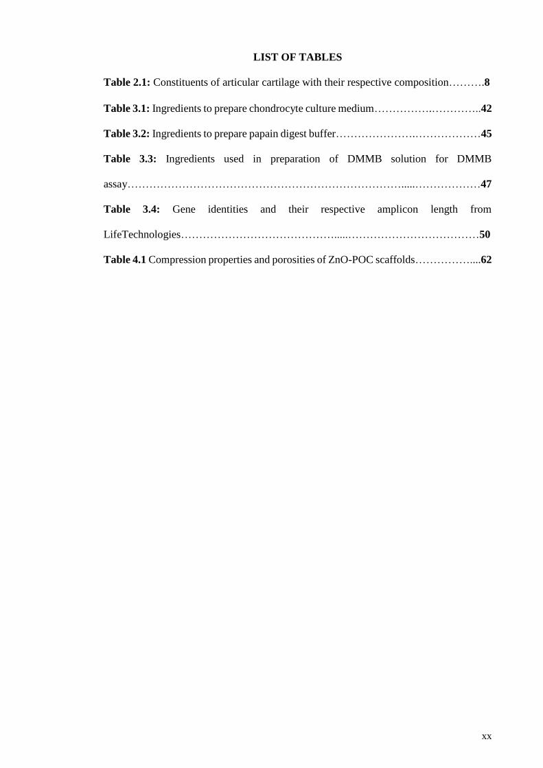
LIST OF TABLES
Table 2.1: Constituents of articular cartilage with their respective composition……….8
Table 3.1: Ingredients to prepare chondrocyte culture medium…………….…………..42
Table 3.2: Ingredients to prepare papain digest buffer………………….………………45
Table 3.3: Ingredients used in preparation of DMMB solution for DMMB
assay………………………………………………………………….....………………47
Table 3.4: Gene identities and their respective amplicon length from
LifeTechnologies…………………………………….....………………………………50
Table 4.1 Compression properties and porosities of ZnO-POC scaffolds……………....62
xxi
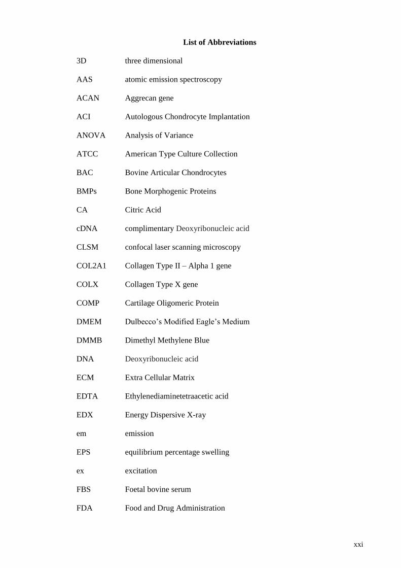
List of Abbreviations
3D three dimensional
AAS atomic emission spectroscopy
ACAN Aggrecan gene
ACI Autologous Chondrocyte Implantation
ANOVA Analysis of Variance
ATCC American Type Culture Collection
BAC Bovine Articular Chondrocytes
BMPs Bone Morphogenic Proteins
CA Citric Acid
cDNA complimentary Deoxyribonucleic acid
CLSM confocal laser scanning microscopy
COL2A1 Collagen Type II – Alpha 1 gene
COLX Collagen Type X gene
COMP Cartilage Oligomeric Protein
DMEM Dulbecco’s Modified Eagle’s Medium
DMMB Dimethyl Methylene Blue
DNA Deoxyribonucleic acid
ECM Extra Cellular Matrix
EDTA Ethylenediaminetetraacetic acid
EDX Energy Dispersive X-ray
em emission
EPS equilibrium percentage swelling
ex excitation
FBS Foetal bovine serum
FDA Food and Drug Administration

xxii
FESEM Field Emission Scanning Electron microscope
FTIR Fourier transform infrared spectroscopy
GAGs Glycosaminoglycans
GAPDH Glyceraldehyde-3-phosphate dehydrogenase gene
GRAS Generally Regarded as Safe
HA Hydroxyapatite
HCl Hydrochloride
HEPES 4-(2-hydroxyethyl)-1-piperazineethanesulfonic acid
HIF-1A Hypoxia Inducible Factor-1 alpha gene
HNMR Hydrogen nuclear magnetic resonance
LB-Broth Luria-Bertani broth
MALDI-TOF Matrix-assisted laser desorption ionization- Time of Flight
min minutes
MMP-13 Matrix Metalloproteinases – 13 gene
MP-AES microwave plasma-atomic emission spectrophotometer
N2 Nitrogen
NaCl Sodium Chloride
NaOH Sodium Hydroxide
NP Nanoparticles
O2 Oxygen
OD 1, 8 Octanediol
PBS Phosphate Buffered Saline
PCL Polycaprolactone
PEG Poly Ethylene Glycol
PGA Poly Glycolic Acid
PGS Poly (glycerol sebacate)

xxiii
PLA Poly D, L – Lactic acid
PLGA Poly D, L – Lactic acid-co-glycolic acid
PLLA Poly-L- Lactic acid
POC Poly 1, 8 Octanediol Citrate
PPLG Poly γ-propargyl-l-glutamate
Ppm parts per million
PVP polyvinylpyrolidone
q-PCR Quantitative Polymerase Chain Reaction
RA Rheumatoid Arthritis
RNA ribonucleic acid
ROS reactive oxygen species
SEM Scanning Electron Microscopy
S-GAG Sulphated Glycosaminoglycan
SOX9 SRY-box 9
SRTR Scientific Registry of Transplant Recipients
SSC Saline Sodium Citrate
TCP Tricalcium Phosphate
TE Tissue Engineeirng
TGA thermo gravimetric analysis
UNOS United Network for Organ Sharing
w/v weight by volume ratio
w/w weight by weight ratio
WACA Water-in-air contact angle
ZnO Zinc Oxide
ZnONP Zinc Oxide Nanoparticles
β-TCP Beta Tricalcium Phosphate

1
CHAPTER 1: INTRODUCTION
1.1 Thesis Overview
This thesis comprises 7 chapters. Chapter 1 describes the background of the research and
the research problem, followed by objectives and hypothesis. The second chapter
discusses the related literature in depth, and lastly, the motivation to perform the research
is included. A complete methodology and list of materials used is provided in chapter 3
so the reader can repeat the experiment if they want to. Finally, chapter 4, 5 and 6 are
divided according to the objectives, and each chapter includes the results and discussion
for the defined objectives. Chapter 7 is the last chapter in this thesis and provides the
essence of the thesis in the form of a conclusion, and future direction for related research.
1.2 Background
As per 17 May 2012, data provided by Organ Procurement and Transplantation
Network/Scientific Registry of Transplant Recipients (SRTR)/ glyco (UNOS) shows,
there are more than 114,000 people in the queue for organ transplants. Every 10 minutes
a new patient is added to the queue and 18 people die each day due to organ shortages
(Network, 2012). Each year, more than one million people are in need of heart valves,
corneas, skin cardiovascular tissue, and bone tissue (Network, 2012).
The facts and figures mentioned above only represent the United States of America. The
rest of the world population, especially the third world countries where quality of life is
poor and organ donations are scarce, are also affected. There is an urgent need for
biocompatible, efficacious, and cost-effective engineered tissues and organs, and this
need only increases with advances in quality of life and life expectancy.
Articular cartilage is by far one of the most essential and yet functionally complex tissues
that scientists are trying to repair, yet minimal success has been for decades. A major
cause of disability is the joint pain that cartilage disease produces, which is not only

2
limited to older people but found in middle aged persons as well. The cause of joint pain
is degeneration of articular cartilage due to primary osteoarthritis or trauma (Temenoff &
Mikos, 2000). Cartilage is a tissue with limited self-repair capabilities, and hence any
kind of injury to the cartilage tissue which is sustained for a substantial time eventually
leads to further detrimental effects (O'Driscoll, 1998; Steinert et al., 2007; Temenoff &
Mikos, 2000). Different approaches are adopted by clinicians to address the issue of
cartilage repair depending upon the nature of cartilage damage. However current
treatment options fail to secure a permanent solution. Furthermore, infection and
inflammation may lead to treatment failing. One of the major causes of biomaterial
infections is peri-operative contamination and research has shown that tissue engineered
implants also pose a risk similar to biomaterial associated infections (Kuijer et al., 2007).
Amongst total knee arthroplasty patients, a risk of peri-operative infection of up to 38%
has been reported recently (Janz et al., 2015). Matrix-induced Autologous Chondrocyte
Implantation has also been reported to cause superficial infection among patients (Bartlett
et al., 2005). Additionally, a recent study demonstrated that only the presence of nano
surface topography can help reduce bacterial adhesion while increasing cell proliferation
due to nanoscale roughness (Liu et al., 2015). This finding adds to the use of
nanomaterials, which are already employed in the delivery of drugs (Pi et al., 2015; Shi
et al., 2015), fighting of infection (Kalashnikova et al., 2015), enhancing mechanical
properties of scaffolds (Pooyan et al., 2015) and increasing cell proliferation (Holmes et
al., 2016).
Chondrocytes are the only cell type that is present in cartilage. These cells flourish in a
hypoxic environment, while the Extra Cellular Matrix (ECM) in which these cells are
embedded undergoes dynamic compression. Current research lack these native
physiologic stimuli and other biofactors as required for chondrocytes to flourish.

3
Tissue engineering provides an alternative approach to address existing problems in an
effective and efficient way. Even though cartilage tissue engineering have been adopted
by scientist to cure the dilemma of cartilage damage. However, till date there are many
problems that are associated directly and indirectly that impede implementation of
cartilage repair strategy via tissue engineering approach.
1.3 Problem Statement
Various solutions have been put forward by scientists to successfully implement cartilage
tissue engineering clinically. However, the limitations of their proposed techniques
exceed their benefits. the use of hydrogels, natural polymers, synthetic polymers, and
metal implants has been suggested but they have generally proved unsuitable for clinical
use, due to their inferior mechanical properties (Johnstone et al., 2013; Spiller et al., 2011)
and lack of recovery of the implanted material to its original level due to creep effect (Shi
et al., 2016). Moreover, current cartilage repair strategies have raised a multitude of
concerns, including infection (Lichstein et al., 2015; Yeo et al., 2015) implant failure
(Hazelwood et al., 2015; Vahdati & Wagner, 2013), lack of native tissue architecture
(Chung & Burdick, 2008; Doran, 2015) and absent cell morphology (Hardingham et al.,
2002). In order to fight any bacterial infection many antibacterial agents have been
studied (Aksoy et al., 2010; Davies, 2003). However, Zinc Oxide stands out as favourable
because it also plays a significant role in cartilage synthesis(Kirsch et al., 2000). Current
clinical methods include minimally invasive surgery to resurface articular cartilage,
arthoscopic lavage, or marrow stimulation techniques. Unfortunately all the
aforementioned clinical methods currently used are palliative and for temporary relief
only. The most frequently used treatment options for cartilage tissue repair have
limitations that hinder their long-term clinical implementation (Marlovits et al., 2006;
Portocarrero et al., 2013).

4
Furthermore, only a handful of studies are present that implement all three components
of cartilage tissue engineering: (a) cells, (b) scaffold, and (c) bio factors, together. More
recent findings signify the importance of using a three-dimensional environment with
dynamic loading for cartilage tissue engineering (Panadero et al., 2016).
A cartilage tissue engineering approach must benefit from a suitable biocompatible and
biodegradable scaffold that will provide a micro structure for the chondrocytes to settle
and proliferate, while being porous enough to allow cell-cell interaction and exchange of
nutrients for uniform tissue formation. Moreover, the newly fabricated scaffold must also
be flexible enough to be implanted via minimally invasive techniques, where required
(Johnstone et al., 2013).
A successful intervention for a cartilage repair strategy must involve external stimuli that
will deliver a physiologic environment to cultured chondrocytes under a 3D structure.
Furthermore, to tackle inflammation and infection, a suitable anti-inflammatory and
antibacterial mediator must be integrated to assist with diminishing these effects while
having no adverse effects on the cells.
In essence, the combination of biocompatible scaffold, physiologic stimuli and
facilitation of antibacterial and anti-inflammatory properties bears promise for more
effective treatment of cartilage disease.
1.4 Objectives
The objectives of this research are:
1. To develop a biodegradable scaffold with controlled pore size and porosity
mimicking the properties of native articular cartilage.
2. To select and optimise the concentration of nano particles that must be
biocompatible and having antibacterial effect while showing no toxicity towards
chondrocytes.

5
3. To study the independent and combined effect of Hypoxia and ZnONP on bovine
articular chondrocytes seeded on polymer scaffolds.
4. To establish a relationship between dynamic mechanical stimuli, ZnONP and
oxygen tension in chondrocyte seeded polymer scaffolds.
1.5 Hypotheses
1. It is hypothesised that chondrocytes will proliferate, secrete more extracellular
matrix, and show higher gene expression under a hypoxic environment.
Furthermore the incorporation of nanoparticles will deliver an anti-bacterial
mechanism to the fabricated scaffold.
2. Furthermore it is hypothesized that scaffolds that will go under dynamic
compression with a combination of hypoxia and nanoparticles, will eventually
display a more chondrogenic morphology and will help in elevating cartilage
specific matrix gene while lowering catabolic gene expression.

6
CHAPTER 2: LITERATURE REVIEW
2.1 Introduction
In the present study, a novel composite biomaterial for use within cartilage tissue
engineering is developed, optimised, and evaluated. The literature which underpins this
is therefore reviewed, starting with the current understanding of cartilage as a tissue,
followed by cartilage injury, and a review of cartilage repair options. From here, the focus
moves to the three components of a tissue engineering strategy as applied to cartilage
tissue engineering; first, cell source; second, cell signalling strategy (covering
biochemical and biomechanical signalling); and finally, the scaffold material (including
a focus on the target material for the present study).
2.2 Cartilage
Cartilage is a tissue that lacks blood vessels, nerves and lymph vessels, and is composed
of sparingly distributed chondrocytes (only 1% in humans) (Poole, 1997). There are 3
types of cartilage in the human body, depending upon matrix composition; elastic
cartilage, fibrocartilage and hyaline cartilage (Jung, 2014). Elastic cartilage makes up the
flexible cartilaginous structures of the nose and ear. It contains elastin as an additional
component to ECM. Fibrocartilage contains a higher content of collagen in ECM than in
hyaline cartilage and is present in ligaments and tendons that are located in close
proximity to bone (Temenoff & Mikos, 2000). Hyaline cartilage is mainly found in the
larynx, trachea, ribs, knee and bronchus (Jung, 2014).
Long bones have a covering at their articulating ends known as hyaline cartilage. This
type of cartilage is more commonly referred to as articular cartilage. This hyaline cartilage
provides a low-friction surface with wear resistant properties (Bhosale & Richardson,
2008).

7
Hyaline cartilage is made up of water and ECM. Within this ECM, type II collagen is
most abundant, followed by proteoglycans that suffice for mechanical properties in vivo
(Bhosale & Richardson, 2008). For an articular joint to function optimally, it is critical
that engineered cartilage must comply with the properties of native articular cartilage. It
must qualify to withstand load-bearing forces, have low friction a coefficient, and have
other necessary biological and mechanical properties (Doran, 2015).
2.2.1 Composition of articular cartilage
2.2.1.1 Chondrocytes
Among all the tissues of the human body, articular cartilage has the lowest cellular
density, according to volume. Humans have only 1% cellular material in their articular
cartilage, namely cells called chondrocytes (Temenoff & Mikos, 2000). Chondrocytes are
sparse but extremely important for replacement of degraded matrix molecules in order to
maintain homeostasis. At week 5 of gestational, a blastema is formed when few
mesenchyme cells form an aggregate. This blastema starts secreting a cartilage matrix
which forms the foundation of chondrocytes (Bhosale & Richardson, 2008). It is reported
that a cilia extends from surface of some chondrocytes into the ECM that contributes to
modifying ECM properties in response to mechanical stimulus (Buckwalter & Mankin,
1997). Chondrocytes pass through various lineages, producing proteins vital for ECM.
Chondrocytes in the periphery secrete collagen to form a hyaline cartilage. When
chondrocytes mature, they tend to halt their activity, appear circular and are enclosed in
a matrix (Buckwalter & Mankin, 1997; Hall, 1994). Table 2.1 summarises the
constituents of articular cartilage.

8
Table 2.1: Constituents of articular cartilage with their respective composition
(Jung, 2014; Poole, 1997; Temenoff & Mikos, 2000).
Constituent Wet weight
(%) Dry weight
(%)
Cellular
Material Chondrocytes ----- 1
Extracellular
Matrix Water 80 -----
Macromolecules
Collagen Proteoglycans Non-collagenous
proteins
20-40 10-20 10-20 -----
----- 50-60 25-35 15-20
2.2.1.2 Extracellular Matrix
Apart from chondrocytes, articular cartilage comprises a complex mix of water and
structural macromolecules in a framework that provides stability to the articular cartilage.
A combination of water and macromolecules is called ECM. The majority constituent in
ECM of articular cartilage is water, approximately 80% of the weight. However in
different regions of cartilage the percentage of water varies from 60% to 80%. The
macromolecules comprise collagens, proteoglycans, and non-collagenous proteins.
Macromolecules (when combined) account for 20 – 40 % wet weight of articular cartilage
(Jung, 2014).
2.2.1.3 Water
In articular cartilage water accounts for majority of the wet weight of the tissue; around
80%. Apart from water tissue fluid contains bulk of cations to neutralise negatively
charged GAG’s in ECM. Tissue fluid also contains metabolites and gases. The presence
of tissue fluid is of vital importance to avascular cartilage as it helps the exchange of
nutrients and oxygen (O2) with synovial fluid. Furthermore, recovery of cartilage and
resistance to compression is achieved via the presence of tissue fluid in ECM (Buckwalter
& Mankin, 1998b).

9
2.2.1.4 Collagens
The second-most abundant constituent of ECM is collagen, which accounts for 10% to
20% of the wet weight of articular cartilage (Bhosale & Richardson, 2008) while it
equates to 50% to 60% of the dry weight of articular cartilage (Jung, 2014). Collagen
types II, VI, IX, X and XI are found in articular cartilage. However, the major role player
is type II collagen, which represents 90 – 95% of all the collagens present. Collagen type
II also forms the foundation of interlinked fibrils in articular cartilage (Buckwalter &
Mankin, 1997). The interlinked fibrils formed by type II collagen not only provide
physical strength to the cartilage but also trap other macromolecules (Cohen et al., 1998;
Temenoff & Mikos, 2000).
2.2.1.5 Proteoglycans
The third constituent of ECM are the proteoglycans, proteins that give compressive
capabilities to the cartilage. However they are only 10 – 20 % of the total wet weight
(Bhosale & Richardson, 2008), and they account for 25% to 35% in dry form (Buckwalter
& Mankin, 1997). The proteoglycans are composed of polysaccharides and proteins in
the ratio of 19 to 1 respectively (Wirth & Rudert, 1996). The protein forms the core, with
attachment to one or more glycosaminoglycans (GAGs) chains (Buckwalter & Mankin,
1997; Temenoff & Mikos, 2000). GAGs are from a family of polysaccharides which have
a repeating disaccharide unit (Afratis et al., 2012; Anower-E-Khuda & Kimata, 2015).
Chondroitin sulfate, dermatan sulfate, keratin sulfate and hyaluronic acid are all GAGs
that are present in articular cartilage (Brézillon et al., 2014; Hall, 2012). Proteoglycans
are sub-divided into 2 groups: (a) large aggregating proteoglycans and (b) small
aggregating proteoglycans. Large aggregating proteoglycans are more commonly
referred as Aggrecan (Buckwalter & Mankin, 1997). The Aggrecans are responsible for
resilience and distribution of stress in articular cartilage (Temenoff & Mikos, 2000).

10
2.2.1.6 Non-Collagenous proteins and Glycoproteins
Non-Collagenous proteins and Glycoproteins account for about 15% to 20% of articular
cartilage in dry weight. In an articular cartilage there exists wide variety of these
molecules. They possess minute quantities of oligosaccharide attached to a protein core.
Contrary to proteoglycans and collagens, these non-collagenous proteins and
glycoproteins have not been studied extensively. However, they tend to help in
maintenance and organization of macromolecular network of ECM(Buckwalter et al.,
2005; Gallagher et al., 1983). Among them, cartilage oligomeric protein (COMP) is most
abundant. It is concentrated in the matrix of chondrocyte cells and has a binding affinity
chondrocytes (Buckwalter & Mankin, 1997; Temenoff & Mikos, 2000); COMP is
involved in maintaining the chondrogenic phenotype (Aigner & Fan, 2003).
2.2.2 Zonal arrangement in articular cartilage
Articular cartilage is divided in to 4 zones (figure 2.1) according to their structure and
function.
1. Superficial zone
2. Transitional zone
3. Deep zone
4. Calcified zone
1. Superficial zone
The superficial zone comprises of flat cells and is the thinnest layer. It is laminated by a
specialised layer of synovial fluid called the lamina splendens (Instructional Course
Lectures, The American Academy of Orthopaedic Surgeons - Articular Cartilage. Part I:
Tissue Design and Chondrocyte-Matrix Interactions*†, 1997). The lamina splendens is a
protein that enhances the low – friction properties of the surface of articular cartilage
(Buckwalter & Mankin, 1997; Temenoff & Mikos, 2000).

11
2. Transitional zone
This zone is dominated by spheroid-shaped cells, although the cell density is lower.
Collagen fibres with a higher diameter can be found in this zone, which also has a high
content of the proteoglycan aggrecan (Bhosale & Richardson, 2008; Buckwalter &
Mankin, 1997).
3. Deep zone
This zone has the lowest of cells densities, with spherical cells distributed throughout the
zone. The proteoglycan concentration is the highest in this region (Bhosale & Richardson,
2008; Buckwalter & Mankin, 1997).
4. Calcified zone
Mineralisation is higher in this region of articular cartilage, meaning that cells are
embedded in a matrix. The cells in calcified zone synthesise Type X collagen, which is
mainly responsible for providing mechanical stability to cartilage, and also assists in
absorbing shocks(Buckwalter & Mankin, 1997; Temenoff & Mikos, 2000).
Figure 2.1: Zones of Articular cartilage, modified from (Bhosale & Richardson,
2008).
12
2.3 Chondrogenesis and chondrocyte differentiation
The process by which cartilage forms is called chondrogenesis. This is a natural process
and chondroprogenitor cells are responsible for its initiation. Chondroprogneitor cells are
basically mesenchymal cells. Mesenchymal cells come together and form an aggregate
and start condensing in one area; this marks the beginning of chondrogenesis. Bone
Morphogenic Proteins (BMPs) are responsible for contributing to initiating
chondrogenisis by helping in aggregation of loose mesenchymal cells. In the early stages
of chondrogenesis, the mesenchymal cells secrete ECM and cell adhesion molecules
(Goldring et al., 2006). Among the secreted ECM there are large amounts of type II
collagen (Ng et al., 1993), N-cadherin (Oberlender & Tuan, 1994), N-cam (Widelitz et
al., 1993) and tenascin (Mackie et al., 1987). Apart from the ECM and cell adhesion
molecules, SRY-box 9 (SOX9); a transcription factor, is highly expressed. SOX9 plays a
vital role in chondrogenesis and differentiation of chondrocytes.
Differentiation of mesenchymal cells into chondrocytes causes the cells to secrete the
ECM that contains large quantities of type II collagen and Aggrecan. After early
chondrocyte differentiation, this is the next development stage of cartilage. In this stage
the cells proliferate rapidly providing a template for the cartilage. After a certain time old
cells draw away from the cell proliferation cycle and enter in to a phase of hypertrophic
differentiation. In this process the chondrocytes grow in size, terminally differentiate,
mineralize, and eventually undergo apoptosis (Freed et al., 1998; Zuscik et al., 2008).
After the death of chondrocytes, they degrade and leave a matrix of their cells that serves
as a scaffold for deposition of minerals and formation of bone (Zuscik et al., 2008).
2.4 Articular Cartilage Disease and Injury
Articular cartilage diseases represent the most common cause of joint pain in middle aged
and older people (Buckwalter & Mankin, 1998b), and hospital treatment cost for total

13
knee replacement was reported to be US$ 28.5 billion in 2009 in the US alone (Murphy
& Helmick, 2012). Together with acute injury, such cartilage disorders also result in a
substantial reduction in quality of life (Minas, 1999; Murray et al., 2015).
Depending upon the nature of injury, cartilage injury can be subdivided in to three major
categories; Full thickness defect, partial defect and matrix disruption. A full thickness
defect is the most painful and it usually affects entire cartilage thickness while also
penetrating deep into the subchondral bone (Temenoff & Mikos, 2000). In this type of
injury fibrin clot formation occurs at the site of defect. Blood and bone marrow cells bind
to this site and form patches of fibrin clots (Aigner & Fan, 2003). The newly formed
tissue lacks the strength of native articular cartilage and has a higher permeability. Lack
of strength and excess permeability eventually contribute to degradation and permanent
damage to articular cartilage and subchondral bone. An injury of as little as 3mm depth
is considered to be a full thickness defect. (Coutts et al., 1997; Fortier et al., 2011;
Temenoff & Mikos, 2000).
A secondary injury is a partial thickness defect, resulting in disruption to the surface of
cartilage. This damage, unlike a full thickness defect, does not extend to the subchondral
bone. In this type of injury the surrounding cells do proliferate but not to an extent which
can repair the defect completely (Temenoff & Mikos, 2000). Research over the years has
provided evidence against the healing of tissue (Ghadially et al., 1977) or progressive
degeneration (Grande et al., 1989) in the case of partial thickness defects. Not only is
regeneration limited at the cellular level of cartilage, but also negative charges amongst
proteoglycans during injury contribute to the lack of cellular adhesion (Hendrich et al.,
2003).
A blunt trauma to cartilage causes matrix disruption injury. This type of injury may occur
from a sporting activity or accident. If the extent of injury is not extreme the damage is

14
reversed by the viable chondrocytes present at the site of injury (Temenoff & Mikos,
2000).
Thus, despite it being such a serious and significant issue, treatment of cartilage problems
is severely restricted due to the limited ability of cartilage to self-repair (Lam et al., 2014).
This often means that degeneration occurs at a higher rate than repair, causing increasing
failure of the degenerative cartilage surface (O'Driscoll, 1998).
2.5 Treatment Options for Cartilage Injury
To provide a sustainable solution to the problem of cartilage damage the primary
objective of every clinician is to yield normal function of the joint(Temenoff & Mikos,
2000). Generally there exists three major interventions to restore joint functionality.
2.5.1 Marrow Stimulation Technique
The marrow stimulation technique involves micro-fracture, abrasion arthroplasty, or
drilling, and is usually adopted as a primary treatment for cartilage defects up to 2 to 4
cm2. This technique involves creating defects that extend to the sub-chondral bone that
eventually result in clotting at the site of defect and a natural repair response being
induced. This response leads to the formation of fibrocartilage repair tissue.
Fibrocartilage tissue is undesirable as it is less resilient than articular cartilage, and
mechanically speaking the properties fail to match that of the native cartilage (Buckwalter
& Mankin, 1998a; Caplan et al., 1997). Such marrow stimulation techniques are
considered to be effective only in younger patients without any prior surgical intervention
(Demange et al., 2014; Mithoefer et al., 2009), and the size of the defect that can be treated
by marrow stimulation technique is limited to 2 to 4 cm2. Further, this technique is
ineffective for the femoral condyle region (Gudas et al., 2005; Kreuz et al., 2006).
15
2.5.2 Osteochondral Grafting
Tissue transplantation of periosteum, perichondrium or osteochondral grafts is used for
regeneration of hyaline cartilage (Bouwmeester et al., 1997; Buckwalter & Mankin,
1998a; Hunziker, 2002). The most common approach is to harvest osteochondral
cylinders from the donor site and then fit them into holes already drilled in areas of defect.
The drilled areas are filled via one or more grafts that range from 5 to 11 mm in diameter
while their lengths are 13 to 15 mm (Hangody, 1997). Studies have shown mixed results
in cases of osteochondral transplants. Reduced pain and improved function was reported
after 6.5 years by Outerbridge et al. (Outerbridge et al., 1995). In two separate studies by
Jakob et al. (Jakob et al., 2002), and Hangody et al. (Hangody et al., 2001) improved knee
function and good results were reported in more than 90% of patients. In contrast, Laprell
and Petersen reported a normal knee in fewer than 50% of patients after a follow up of 6
to 12 years (Laprell & Petersen, 2001). A major limitation of this approach is the
availability of tissue where there are large cartilage defects. Further, long term follow up
is missing and this technique is reported to be most appropriate for patients less than 35
years of age (Buckwalter & Mankin, 1998a; Caplan et al., 1997; Hunziker, 2002; Insall,
2001). In addition, osteochondral transplantation is associated with some post-operative
complications. The accumulation of water (joint effusion), bleeding in joint spaces
(hemarthrosis), and persistent swelling have all been reported after osteochondral
transplantation (Bobic, 1999; Bös et al., 2000; Hangody et al., 2001; Hunziker, 2002).
Researchers have also reported loose bodies, donor site pain, and avascular necrosis as
other post-operative complications that emanate due to osteochondral transplantation
(Aigner & Fan, 2003).

16
2.5.3 Autologous Chondrocyte Transplantation
Due to the failure of osteochondral transplantation, autologous chondrocyte
transplantation was developed and used since 1994 by Brittberg and coworkers (Brittberg
et al., 1994). Autologous chondrocyte implantation (ACI) is considered to be used as the
last line of defence when other techniques fail. ACI is considered able to repair full-
thickness lesions in cartilage up to 16 cm2. This technique involves isolation of a patient’s
own cells grown in-vitro then implanted at the site of the defect. The follow up results of
ACI after 2 to 9 years have reported a failure in more than 30% patients after treatment
of multiple lesions. Furthermore, patients with patella lesion treatment demonstrated
more than 30% failure, but isolated cartilage lesion treatment displayed less than 10%
failure (Peterson et al., 2000). Another long-term follow up study by Harris J. D. et al.
sought to determine if current literature supports ACI over other interventions. They
observed that ACI can only be helpful over other interventions if the size of defect is
greater than 4 cm2. ACI or micro fracture or osteochondral transplants, in summary, only
provide short term success.(Harris et al., 2010) A study by Niemeyer P and co-workers
reported that complications like graft failure, delamination, chondromalacia (softening of
cartilage) and hypertrophy can arise after ACI (Niemeyer et al., 2008). More recent
reports by Harris J. D et al. have reported decreased failure rates (up to 7.5 %) for ACI,
however there are other reports that continue to demonstrate inconsistent outcomes of
ACI without any clear recommendation. Furthermore, it was also reported that the
differences between various treatments were so small that their clinical significance is
questionable, beyond mere statistical significance (Vavken & Samartzis, 2010). Lately it
was reported that ACI improves movement significantly after at least 24 months (von
Keudell et al., 2016).
17
2.6 Cartilage Tissue Engineering
Current treatment options, like ACI, Osteochondral transplants and marrow stimulation
have demonstrated variable success rates, however more long-term results are not
satisfactory (Kreuz et al., 2006; Niemeyer et al., 2008; Redman et al., 2005; Vavken &
Samartzis, 2010). The major limitation that arises due to the use of the aforementioned
therapeutic techniques for cartilage repair is the lack of mechanical properties of newly
formed tissue compared to native articular cartilage tissue. Due to these inferior
mechanical properties, the newly formed tissue is prone to failure (Hunziker, 2009).
Cartilage tissue engineering strategies contribute to a durable and functional replacement
option, beyond any other surgical intervention or technique (Kock et al., 2012).
Tissue engineering is a 3 tier based approach, involving the combination of living cells,
a cell signalling strategy, and a scaffold (Hendrich et al., 2003). Cartilage tissue
engineering allows researchers a great deal of flexibility in combining various techniques
independently and in combination to address the issue of cartilage repair. Cartilage tissue
engineering also allows the selection of various cell types to be implemented in-vitro for
a better understanding of cartilage repair. Moreover, researchers have shown positive
effect of signalling molecules in enhancing anabolic effects and increased ECM synthesis
(Elder & Athanasiou, 2009; Seifarth et al., 2009). Further, a widely researched area for
better fabrication of cartilage tissue is external mechanical stimuli. Direct dynamic
compression has been demonstrated to increase ECM production while improving the
compressive properties of the engineered tissue (Bian et al., 2010; Chen et al., 2015; Luo
et al., 2015). Another leverage given by a cartilage tissue engineering strategy to the
researchers is modification of the properties of scaffolds. Scaffolds provide a temporary
frame for the cells to attach to and proliferate while the tissue is being formed and the
scaffold is being degraded. Recent publications have shown enhanced mechanical and
biological properties of newly fabricated tissue using polymeric scaffolds (Camarero-
18
Espinosa et al., 2016; Schulze-Tanzil, 2015). Various cartilage tissue engineering studies
have been undertaken to engineer a tissue with native sGAG content. However, they have
failed to achieve the collagen content present in native articular cartilage, and which gives
the cartilage its tensile properties (Eyrich et al., 2007; Kock et al., 2012). Though there
are many advantages to cartilage tissue engineering, nevertheless finding optimal cell
source is the first issue, followed by choice of scaffold material, the role and the effect of
biochemical and mechanical stimulus and lastly tackling any infection that might occur
due to surgical intervention, amongst others are pitfalls that need to be addressed
(Bhattacharjee et al., 2015; Kock et al., 2012). Gaut C. and Sugaya K. recently suggested
that in order to obtain a clinically effective solution for cartilage tissue engineering; a
combination of factors is required, such as; cell signalling, scaffold material, mechanical
and biological stimulus must be considered (Gaut & Sugaya, 2015).
Given the clear advantages of Cartilage Tissue Engineering, but yet the continued
inability to fully restore cartilage functionality in the long term, the present study seeks
to further advance this technique toward an effective and lasting repair.
2.6.1 Cell Sources
A source of cells is one of the key components of a Tissue Engineering strategy. There is
no current consensus on the optimal source of cell for cartilage tissue engineering, with
division between the use of stem cells, fibroblasts, or chondrocytes – each with their
relative merits and disadvantages.
Stem cells are particularly attractive due to their differentiation potential and the fact they
may be harvested from various tissues. Though stem cells overcome the problem of
limited supply of cell as they can be easily harvested from the patient’s body itself (from
bone marrow (Boeuf & Richter, 2010) and adipose tissue (Gir et al., 2012)), bone marrow
stem cells have lower mechanical properties and matrix synthesis when compared to
chondrocyte seeded scaffolds (Thorpe et al., 2010) and adipose stem cells demonstrate a

19
lesser potential than bone marrow stem cells despite being capable of being differentiated
into chondrocytes (Kock et al., 2012). Furthermore, embryonic stem cells tend to
differentiate hypertrophically and moreover, there exists many ethical and legal issues to
overcome before considering embryonic stem cell use (Jukes et al., 2008). Mesenchymal
stem cells are another potential source, however they have demonstrated high cartilage
hypertrophy markers such as Collagen type X (COL X) & Matrix (MMP-13 (Mueller &
Tuan, 2008).
Chondrocytes are well suited to cartilage tissue engineering because they are the native
cells of cartilage tissue (Nicoll et al., 2001). However, they have limited availability and
generally need to be harvested from a monolayer to get an high number of cells, meaning
they eventually lose their cell phenotype (Cournil-Henrionnet et al., 2008).
Another potential source of cells for cartilage tissue engineering are the articular cartilage
progenitor cells that are useful in producing the amount required for cell culture but do
not have the capability to produce cartilage matrix (Zhou et al., 2014). It is reported that
they may also cause donor site morbidity (Mathur et al., 2012).
A new approach should be using chondrocytes in an in-vitro culture systems that mimics
the native environment to overcome the limitation of limited cell numbers and allows
maintenance of cell phenotype.
2.6.2 Cell Signalling Strategies
A key aspect of Tissue Engineering is the integration of a cell signalling strategy
alongside the cellular and scaffold components. This may include aspects such as
biochemical cues, facilitation or stimulation of cell-cell interactions, biomechanical
signalling, or manipulation of the environmental factors involved.
Human body tissues have various associated stimuli that help regulate normal function of
the tissue. Whether it is electrical, mechanical, or chemical, body tissues undergo
different kinds of stimulus; heart tissues undergo electrical stimulation (Cannizzaro et al.,

20
2007; Godier-Furnémont et al., 2015), bone tissues are subjected to tension and
compression (Nyman et al., 2009; Romanos et al., 2015), and similarly, cartilage tissue
undergoes compression, hydrostatic pressure, and shear strain (Ryan et al., 2009; Schätti
et al., 2015). Cartilage tissue also experiences a unique stimulus, which is tension in
oxygen (Portron et al., 2015). This is mainly due to the avascular nature of cartilage tissue
(Hansen et al., 2001). When considering cartilage tissue, in particular, successful tissue
engineering will be incomplete without a mechanical stimulus and low oxygen tension.
2.6.2.1 Mechanical stimulus
In the last decade, researchers have increasingly realised the importance of external
mechanical stimuli to enhance the quality of produced cartilage (Temenoff & Mikos,
2000). However, much conflicting information has been reported regarding how
chondrocytes behave in response to mechanical stimulation (Lee et al., 2005).
Physiologically, articular cartilage primarily undergoes compression at joint level (Lee et
al., 2005). In an average human pressure of up to 1MPa during standing, 0.4MPa while
walking, and up to 20MPa while standing up from a chair have been reported (Gooch &
Tennant, 1997; Lee et al., 2005; Urban, 1994). A range of factors are affected by the
compressive loading of articular cartilage. These include biochemical and physical
gradients of nutrients, ion concentrations, electrical charge, and pH (Gray et al., 1988;
Guilak et al., 1995). Different studies have shown that a mechanical stimulus positively
influences cartilage development (Heath & Magari, 1996). The way ECM is composed
and organised provides the cartilage with its biomechanical properties. The presence of
type II collagen helps strengthen the cartilage while proteoglycans build up compressive
resistance in the cartilage (McMahon et al., 2008). Grodzinsky A.J. and co-workers
reported that mechanical stimuli on chondrocytes is essential in order to maintain the
integrity of cartilage (Grodzinsky et al., 2000).

21
In its native environment cartilage is usually compressed to about 10% (Sanchez-Adams
et al., 2014). Various studies showcase the importance of dynamic compression.
Researchers have studied the effects of compressing the cartilage from about 5% to 50%
with frequencies ranging from 0.001 Hz to 1Hz using various polymers and gel constructs
(Albro et al., 2013; Appelman et al., 2011; Chen et al., 2015; Chowdhury et al., 2006;
Chowdhury et al., 2003; Davisson, Twana et al., 2002; Démarteau et al., 2003; Di
Federico et al., 2014; Hunter et al., 2002; Jeon et al., 2012; Kisiday et al., 2004; Li et al.,
2013; Morel et al., 2005; Piscoya et al., 2005). The majority of the researchers have
favoured dynamic compression and have demonstrated a higher GAG content with better
cell proliferation and increased cartilage matrix specific gene expression of Type II
collage (COL2A1) and Aggrecan (ACAN) (Appelman et al., 2011; Chen et al., 2015;
Davisson, Twana et al., 2002; Jeon et al., 2012; Kisiday et al., 2004). However,
contradicting results are present for the amount of compression. Some studies support
dynamic compression in the range of 0.001% to 31% (Appelman et al., 2011; Chen et al.,
2015; Davisson, Twana et al., 2002; Démarteau et al., 2003; Jeon et al., 2012) while others
have reported detrimental effect of dynamic compression ranging from 15% and up to
50% (Davisson, Twana et al., 2002; Di Federico et al., 2014; Hunter et al., 2002; Li et al.,
2013; Piscoya et al., 2005). From the current literature it is evident that a dynamic
compression beyond physiologic levels of 10% has proved to be detrimental for cartilage.
One thing is noteworthy here: most of the studies executed to study the behaviour of
cartilage tissue or cartilage cells are performed on agarose, hydrogels, alginate and type I
collagen gels. All the aforementioned constructs/ gels can only be used in laboratory
settings and lack the physical capabilities to be implanted clinically. Limited groups have
reported dynamic compression on polymer/ polymer composite scaffolds which they
deem to be physically compatible. Another important factor that seems neglected in the
current study is the physiologic oxygen level. The majority of the studies of dynamic

22
compression in the domain of cartilage tissue engineering were carried out under normal
oxygen settings without comparing the effect of atmospheric and physiologic oxygen
concentration on dynamic compression.
2.6.2.2 Oxygen Tension/Hypoxia
Research suggests that the nature of articular cartilage is avascular; hence it receives its
required nutrients and oxygen by synovial fluid following a passive diffusion mechanism
(Malda et al., 2003). In relation to rest of the body tissues, articular cartilage experiences
oxygen concentrations in the range of 1% to 10% as compared to 21% for other tissues
(Co et al., 2014). Chondrocytes are reported to thrive in oxygen concentration range of
5% to 10% (Grimshaw & Mason, 2000), while losing their activity in anoxic conditions
because 1% of Oxygen concentration is reported to exist in an abnormal or due to any
pathological condition (Grimshaw & Mason, 2000; Zhou et al., 2004). Hypoxia Inducible
Factors are responsible for chondrocytes adapting to low oxygen tension (Fedele et al.,
2002). Various researchers have reported that hypoxia in chondrocytes has resulted in
increased synthesis of ECM proteins in in-vitro conditions (Domm et al., 2002; Madeira
et al., 2015; Yodmuang, Marolt, et al., 2015). Inhibition of collagen type X had also been
demonstrated by inducing hypoxic conditions. The Collagen type X is major marker of
hypertrophy in chondrocytes during the chondrogenesis of adipose-derived mesenchymal
stem cells (Betre et al., 2006; Portron et al., 2015) and of epiphyseal chondrocytes (Chen,
X. C. et al., 2006). A transcription factor, Hypoxia inducible factor 1 (HIF-1) is mainly
responsible for regulation of hypoxic response in the cartilage tissue (Schipani et al.,
2001). HIF-1α is said to be responsible for angiogenesis and glycolysis while at the same
time causing cells to proliferate (Denko et al., 2003; Goda et al., 2003). Hypoxia further
plays a role in formation of collagen fibrils with the help of transcription factor HIF-1 α
(Takahashi et al., 2000). Current research has reported an individual role of hypoxia for
better engineered cartilage, however, not enough details are present in the current

23
literature as to how hypoxia will behave in combination with other bio-factors for
cartilage tissue engineering.
However, many studies have highlighted the importance of hypoxia as a success for tissue
engineering of cartilage (Meretoja et al., 2013; Munir et al., 2014; Schipani et al., 2001).
Researchers have shown that hypoxia induces ECM synthesis in in-vitro chondrocyte
culture (Domm et al., 2002) and also helps chondrocytes to proliferate (Choi et al., 2014).
Another important research by Schipani E. et al. demonstrated that lack of hypoxia causes
chondrocytes to terminally differentiate (Schipani et al., 2001). A study by Hansen U. et
al. reported a combination of hypoxia and hydrostatic pressure to enhance collagen type
II production while delaying expression of Collagen type I for in-vitro cartilage tissue
engineering (Hansen et al., 2001). Further research has also displayed the potential of low
oxygen tension to enhance Aggrecan and Collagen type II gene expression while
decreasing Matrix Metalloproteinase – 13 (a pro catabolic gene) expression (Parker et al.,
2013). Most of the studies were executed on chondrocytes on Tri Calcium Phosphate
(TCP) plates (Hansen et al., 2001), under agarose gel (Chowdhury et al., 2006; Parker et
al., 2013) or Alginate (Jeon et al., 2012). Others have used PEG (Appelman et al., 2011),
PGA (Davisson, Twana et al., 2002), or articular cartilage explants (Piscoya et al., 2005).
However, the above mentioned studies did not used scaffolds able to be used clinically as
they either lack the requisite mechanical properties or do not mimicked native cartilage
structure in-vivo.
2.6.3 Tissue Engineering Scaffolds
Different natural and synthetic materials have been tested as scaffolds. Scaffolds have
been developed by natural polymers (Van de Putte & Urist, 1965; Vandeputte & Urist,
1965), synthetic polymers (Elisseeff et al., 1999; Meinig et al., 1997), composites (Peter
et al., 1998; Zhang & Ma, 1999) and ceramics (Friedman et al., 1998; Hollinger &
Battistone, 1986). Synthetic scaffolds have copious benefits such as a greater availability,

24
decreased risk of disease transfer, fabrication as per requirement, and the ability to be
shaped into various shapes and sizes (Jeuken et al., 2016).
Just saying that a material should be biocompatible does not solve all the complications.
A tissue engineered scaffold should be readily available to the surgeon; it should promote
cell ingrowth as well as it must degrade in correct ratio with formation of new tissue
(Holmes, 1979; Levine et al., 1997). Also it should be able to fit into irregular places and
must be soft enough to mould and hard enough to withstand the mechanical loads in daily
routine activities (Lichte et al., 2011), meaning that the scaffold should possess
mechanical strengths of site where it needs to be implanted. The architectural properties
of the scaffold; which includes pore size and porosity, should be in relation to the actual
tissue (Peter et al., 1998). The scaffold should also promote cell adhesion and
proliferation (Lichte et al., 2011). Additionally the design of a scaffold should be
biomimetic, implicating it must imitate all the characteristics of the tissue where it is
going to be implanted; whether they are physical chemical or biological properties.
For the aforementioned reasons the polymeric scaffolds are a promising new approach
towards tissue engineering for repairing cartilage defects. However, use of polymeric
materials alone or with composites as cartilage tissue engineering scaffolds remains
largely unexplored.
Various materials have showed their ability to be used as engineered tissues; this includes
metals, ceramics, polymers and their combinations (Burg et al., 2000; Hidalgo-Bastida et
al., 2007; Hollinger & Battistone, 1986; Liu & Ma, 2004). Among these only using
materials and ceramics independently results in lack of degradability and processability.
On the contrary, if polymers are used with metals and ceramics they allow tailored
properties and proper biodegradability. Various scaffolds based on polymers and polymer
composites are being developed that demonstrate a promising future (Burg et al., 2000;
Liu & Ma, 2004; Peter et al., 1998).

25
Particularly for cartilage tissue engineering, various polymers and polymer composites
have been used. Polymers such as Poly (ε-caprolactone) (Li, W.-J. et al., 2005), poly (D,
L – Lactic acid-co-glycolic acid) [PLGA] and PLGA with Hyaluronic based polymers
(Yoo et al., 2005), polyurethane based polymers (Grad et al., 2003), poly (ethylene
glycol)-terephthalate and poly (butylene terephthalate) based polymers (Woodfield et al.,
2004), poly (D, L – Lactic acid) [PLA] based polymers (Camarero-Espinosa et al., 2016),
collagen/PLA and Chitosan/PLA polymers (Haaparanta et al., 2014), poly(γ-propargyl-l-
glutamate) (PPLG) polymers (Ren et al., 2015), Poly (glycerol sebacate) [PGS]
(Kemppainen & Hollister, 2010), and Polyoctanediol citrate polymers (Jeong et al.,
2011).
Among several polymers and polymer composites, Poly 1, 8 Octanediol Citrate (POC)
stands as a potential candidate due to its mechanical and chemical properties that suffice
for cartilage tissue engineering applications. POC has proved to enhance cell attachment
and helps in proliferation of cells, furthermore POC has been shown to enhance matrix
production when seeded with chondrocytes (Kang et al., 2006). Studies have also reported
POC polymer to be more effective than other polymers (PGS & polycaprolactone (PCL))
in enhancing chondrogenesis (Jeong & Hollister, 2010). Moreover, its physical properties
can be tailored to those of native articular cartilage (Kang et al., 2006; Moutos et al.,
2007).
2.7 Poly Octanediol Citrate (POC)
2.7.1 POC, its structure, synthesis, and form
Poly 1, 8 Octanediol Citrate (POC) elastomer is a synthetic polymer that is synthesised
from cost efficient and non-toxic monomers; 1, 8 Octane diol and citric acid (CA). POC
polymer was first fabricated in 2004 (Yang et al., 2004). Since then it has gained
popularity in the field of tissue engineering. Among its advantages; non-toxic monomer,

26
catalyst free polymerisation and good cell adherence are most notable (Yang et al., 2004).
Other properties include; simple synthesis, controllable mechanical and degradation
properties and easy processing. In case of POC, CA which is a polyfunctional monomer
reacts with a difunctional 1, 8 Octanediol monomer to form a crosslinked polyester
network. CA is a non-toxic polyfunctional monomer because it is a by-product of kerbs
cycle in body. Furthermore, it is readily available, inexpensive and it is Food and Drug
Administration (FDA) approved to be used in humans (Shi et al., 2007). Moreover, CA
can participate in hydrogen bonding in a polyester network (Yang et al., 2004; Zhang et
al., 2004). The presence of –COOH groups due to CA helps in covalent attachment of
ECM proteins and growth factors (Goddard & Hotchkiss, 2007). Presence of –COOH is
a feature that is important in polymers for biomaterial applications (Li, B. et al., 2005;
Sun et al., 2004). The second constituent of POC is a difunctional monomer; 1, 8
octanediol which is the largest water soluble aliphatic diol that is soluble in water without
any reported toxicity (Webb et al., 2004). This means 1, 8 Octanediol does not leave any
insoluble complexes when dissolved, a property beneficial for degradation.
2.7.2 POC as a biomaterial
Since its inception, POC has been used for cardiac (Hidalgo-Bastida et al., 2007), vessels
(Jiang et al., 2015), bone (Guo et al., 2015), bladder (Sharma et al., 2010), cartilage,
calvarial (Sun et al., 2014) and other biomedical applications (Hege & Schiller, 2015;
Zhao et al., 2014). POC is attractive as its mechanical and degradation properties can be
tailored (Ji et al., 2014; Yang et al., 2004). In a study by Hidalgo-Bastida L. A. et al., it
was reported that POC demonstrated a good support for cardiac muscle engineering by
exceeding the required mechanical properties in terms of elongation at break which
complies with that at physiological level. Furthermore, it has been reported that POC
scaffolds displays preference for HL-1 cells (Hidalgo-Bastida et al., 2007). POC is also
used for large bone defects by Guo Y. and co-workers. They reported bone tissue

27
ingrowth and efficacy of POC for osteoinductive and osteoconductive behaviour. This
potential of POC was due to citrate, as it allows better bone formation (Guo et al., 2015).
Other studies have also reported the advantage of citrate based materials to enhance cell
proliferation, excellent biocompatibility and oseointegration (Gyawali et al., 2013; Qiu et
al., 2006; Tran, R. T. et al., 2014). Another desired feature of POC is its hydrophilicity.
Higher hydrophilicity in POC is attributed to better water absorption capabilities that may
result in better cell attachment (Zeimaran et al., 2015). More recently pure-POC was used
in association with other monomers (POC-co-glycidyloxypropyl-trimethoxysilane and
POC-co-aminipropyl-triethoxysilane) to study the effect of silica incorporation. The
resulting elastomer was tested for thermal stability, mechanical properties, in-vitro
cellular compatibility, degradation and cell proliferation and metabolic activity to be used
in various biomedical applications. It was found that mechanical properties, thermal
stability and degradation can be controlled easily by tuning monomer concentration.
However, this was achieved at the loss of hydrophilicity (Du et al., 2015).
2.7.3 POC in cartilage tissue engineering
The potential of POC in the domain of cartilage tissue engineering was evaluated first by
Kang Y. et al (Kang et al., 2006). They reported an excellent recovery of 98.7% of pure-
POC scaffolds. Furthermore, they compared pure-POC scaffolds with Alginate, Agarose,
Poly (glycolic acid) (PGA) and Poly-L- Lactic acid (PLLA) scaffolds. Agaose was
reported to be fragile while Alginate constructs deformed without good recovery time as
in case of POC scaffolds. Moreover, PGA and PLLA scaffolds never returned to their
original state after compression. In the same study GAG and Type II collagen content
was reported to increase along with increase in cell number (Kang et al., 2006). In a
different study, Jeong C.G. et al. and co-workers compared pure-POC scaffold with poly
(glycerol sebacate) (PGS) and well-known and FDA approved polycaprolactone (PCL)
for cartilage tissue engineering purpose (Jeong & Hollister, 2010). They kept pore seize,
28
porosity, pore shape and surface area the same for all the scaffold materials. The results
were in favour of POC as higher Deoxyribonucleic acid (DNA) content, increased cell
proliferation, enhanced cartilaginous matrix production, and cartilage specific gene
expression was observed when compared to PCL or PGS scaffolds. Moreover, POC
demonstrated decreased MMP-13 expression when compared to PGS scaffolds (Jeong &
Hollister, 2010). It is noteworthy because MMP-13 takes part in cartilage matrix
destruction and aggrecan degradation (Burrage et al., 2006; Wang et al., 2013). A more
recent study reported a preference for clickable POC-hydroxyapatite (HA) polymer over
other renowned polymers like β-tricalcium phosphate (β-TCP), PLLA, PGA and their co-
polymers due to their brittleness, poor degradation, low biocompatibility, and diminished
mechanical properties (Stewart et al., 2004; Yang, W. et al., 2014). Furthermore, they
reported excellent cartilage formation for spinal fusion; a surgical intervention to join two
or more vertebrae in order to reduce movement between them. Histological evidence
demonstrated that POC supported cartilage formation within the material pores (Tang et
al., 2015).
Though POC is gaining fame in various areas of tissue engineering applications (Guo et
al., 2014; Guo et al., 2015; Hege & Schiller, 2015; Tran et al., 2015; Zeimaran et al.,
2015), nevertheless its use in the domain of cartilage tissue engineering has a great
potential to be further explored. Being a synthetic biomaterial POC holds great promise
as the most interesting part of this polymer is that its mechanical and chemical properties
can be tailored as per need to adapt for a wide range of applications.
2.8 Infection and Anti-bacterial scaffolds
Thousands of surgeries are performed daily round the world, and more and more people
are benefitting from these procedures but, at the same time they are being placed at risk
of microorganisms that may create a problem later via formation of biofilms at the implant
site, tissue implant, or at the site of sutures.

29
A biofilm is a layer made up of microorganisms that tends to develop at the site of an
implant or surgery or tissue that has been operated upon. It is a community of bacteria
living in layers. These layers can be formed on biotic, abiotic and even on human tissues
(Hoffman et al., 2005). The gravity of this problem can be so high at times that there is a
need for removal of implants (Pawlowski et al., 2005). As the number of biomaterial
implants increases, there is a requirement of more inert and more biocompatible
biomaterials. This is due to the fact that whenever an implanted biomaterial gets infected,
the infection is not just limited to the biomaterial itself; rather it affects the adjacent tissue
or bone. Ultimately the outcome may be devastating as there may be a need for
reoperation, amputation, and at times death (Gristina et al., 1989).
Biofilms can cause the bulk of microbial infections in the body. Extensive literature is
available that communicates clearly the areas affected by biofilm formation; this includes
urinary tract infections (Hoffman et al., 2005), middle-ear infections (Pawlowski et al.,
2005), oral implant infections (Heuer et al., 2007), infections after surgery (Singhal et al.,
2010) and on polymeric implants as well (Pavithra & Doble, 2008).
Many scientists around the world are working on how to cope with the formation of
biofilm. Many techniques have been devised to treat the problem of biofilm formation.
The scientists have tried numerous techniques for the prevention of biofilms; but using
antibiotics (Burns et al., 1993; Costerton et al., 1999; Hoiby, 1993) and coatings
incorporated with different antibacterial materials (Bakhshi et al., 2012; Paladini et al.,
2012; Regina et al., 2012) remains most promising against the bacterial biofilms.
Using antibiotics may be helpful in short term efficacy to prevent bacterial infections and
it may be used for catheters, sutures and surgical instruments. However, for long term
applications like cardiac assist devices, bone implants and tissue implants, a material is
needed with no degradation at all or a long degradation time. For tissue engineering

30
applications different scaffolds are now being developed that have showed successful
results for cell compatibility but there is a void space in relation to antibacterial scaffolds.
Things began to change in the 90’s and much work was undertaken on incorporation of
different anti-bacterial materials to be used in dental (Guigand et al., 1997; Imazato et al.,
1995; Morrier et al., 1998; Torabinejad et al., 1995) and wound dressing applications
(Blaker et al., 2004; Kudi et al., 1999; Stashak et al., 2004) and their antibacterial activity
was measured.
It is not known when the first research for antibacterial scaffolds started but the idea of
incorporating antibacterial materials flourished in late 90’s and in past decade when
antibacterial materials were being suggested to be used as coatings for implantable
medical devices (Fu et al., 2005) and dental materials (Guigand et al., 1997; Imazato et
al., 1995; Morrier et al., 1998; Torabinejad et al., 1995).
Not much is known about the antibacterial scaffolds as it is a relatively new field that is
being explored. However, the antibacterial properties of various materials have been
studied in different situations. Many materials exhibit antibacterial properties; but for
tissue engineering applications there is a need for a material that not only possesses
antibacterial properties but also enhances material integrity. The antibacterial effect of
metal oxides is much explored and has a promising future, similarly it is also responsible
for the mechanical integrity of a material (Fielding et al., 2012; Salinas et al., 2011;
Vitale-Brovarone et al., 2008).
More recent studies have reported a selective killing effect of ZnONP by killing all the
cancer cells while bearing no harm to healthy human cells (Akhtar et al., 2012; Taccola
et al., 2011).
An investigation into the effect of ZnONP on fetal development in rats resulted in no
significant toxicity in rats due to excess ZnONP (Hong et al., 2014). Furthermore, recent
reports in favour of ZnONP have arisen that demonstrate enhanced angiogenesis due to

31
the presence of ZnONP (Augustine, Dominic, et al., 2014). The beneficial effects of ZnO
for tissue engineering applications are also reported for bone tissue engineering (Feng et
al., 2014), vessel tissue engineering (Augustine, Dominic, et al., 2014), dentistry
(Toledano et al., 2012) and hip implants (Chang et al., 2014). Zinc is a trace element that
is essential for normal growth (Haase & Rink, 2014b). It also plays a significant role in
the proliferation of cells, their differentiation and their survival (Haase & Rink, 2014a).
Furthermore, Zn deficiency in the diet is reported to result in rheumatoid arthritis
(Mierzecki et al., 2011), cartilage defects (Westmoreland & Hoekstra, 1969) and growth
retardation (Prasad et al., 1961). More specifically Zn deficiency is reported to inhibit
chondrocyte proliferation and cause apoptosis (MacDonald, 2000). In a study by Seo H-
J. and co-workers, it was reported that Zn stimulates cell proliferation and synthesis of
collagen in osteoblastic cells (Seo et al., 2010). In a comparative study by Helliwell et al.,
Zn levels were monitored in femoral heads of individuals having fractures of femoral
neck and degenerated joints. The study concluded that fractured femoral head (154 µg/g)
and degenerated joints (167 µg/g) had lower levels of Zn content than in control group
(205 µg/g) (Helliwell et al., 1996). In a separate study it was reported that resting and
proliferative regions of growth plate contains high levels of Zn2+ ions while they are
significantly lower in hypertrophic cartilage zones. They also related chondrocyte
apoptosis with diminished Zn levels (Sauer et al., 2003). Addition of Zn also supports
chondrocyte proliferation as reported by Koyano Y. and their group (Koyano et al., 1996).
Recent studies have advised the use of nano Zn in biomedical applications (Varaprasad
et al., 2016). However, to date there are no studies that relate the role of Zinc Oxide
nanoparticles (ZnONP) at macro level with chondrocyte proliferation, morphology and
its ECM synthesis.

32
2.9 Antibacterial Activity of ZnO
Zinc Oxide mostly used in industry, including rubber, medicine, cream, lotions, pigments,
and food items (Kołodziejczak-Radzimska & Jesionowski, 2014). It is available in the
form of oxide in 2 major forms; hexagonal, and cubic (known as wurtizite (Figure 2.2)
and zincblende respectively). The former is the most stable and readily available.
Figure 2.2: Wutizite structure of ZnO crystal.
Testing of antibacterial properties of oxides dates back to as early as 1978. A team of
researchers (Cox Jr et al., 1978) explored antibacterial properties of zinc oxide for dental
treatment. They tested zinc oxide against 3 bacterial strains but they failed to deliver.
Inhibition of several species was confirmed in 1982 by endodontic gutta-percha cones
(Moorer & Genet, 1982). These cones released Zn2+ ions slowly and it was concluded
that zinc oxide should be used in medicine and dentistry for its antibacterial effect and
biocompatible nature, but this research lacked biocompatible testing. Later in 1990, the
root canal filling material known as gutta-percha was challenged for its toxicity due to
the presence of zinc oxide (Pascon & Spangberg, 1990). It was proved in the same study
that zinc oxide displays toxicity, and this was attributed to the leeching of zinc ions into
the fluid. In another study, both in vivo and in vitro studies were carried out and zinc
oxide was used in root filling cements (Nielsen et al., 1993). The results were successful
against streptococcus sanguis, staphylococcus aureus and Prevotella intermedia. Research

33
was carried out to cater for zinc oxide as an antifungal material (Odell & Pertl, 1995). In
this research, the hypothesis that zinc is a growth factor for Aspergillus species was tested.
Five different strains of aspergillus were tested by zone of inhibition method. The results
indicated that the incorporation of zinc as dental fillings can inhibit the growth of
Aspergillus. Other bacterial inhibitory effects in favour of zinc oxide has been tested, few
similar successful results for inhibitory effects of zinc oxide were produced by (Gordon
et al., 2011; Liedtke & Vahjen, 2012; Skoog et al., 2012) against Escherichia coli, by
(Gordon et al., 2011; Skoog et al., 2012) against staphylococcus aureus, by (Liedtke &
Vahjen, 2012) against Enterococcus faecalis, and by (Liedtke & Vahjen, 2012; Skoog et
al., 2012) against bacillus subtilis.
2.10 Motivation for current study
Previously discussed limitations for cartilage tissue engineering strategies suffice us to
research degradable, biocompatible and physically strong scaffolds that mimic native
articular cartilage properties. Cartilage cells grown in a monolayer culture gradually lose
their phenotype by converting to a fibroblastic morphology (Figure 2.3), hence they need
to be grown in a three dimensional (3D) culture system that mimics its native
environment.

34
Figure 2.3: Chondrocytes cultured in a T-75 flask losing their morphology and
turning flat only after 24 hours of culture.
Furthermore, the problems that arise due to biofilm formation and bacterial infection
reinforce the rationale to develop an anti-bacterial scaffold for cartilage tissue engineering
without compromising biocompatibility. Zinc Oxide (ZnO) is a potential candidate that
has shown anti-bacterial effects on bacteria while demonstrating no confrontational effect
on cells. ZnO has also been demonstrated to play a vital role in bone and cartilage
generation in early stages, while lower Zn levels are reported to be associated with
rheumatoid arthritis.
The majority of the research executed in the domain of cartilage tissue engineering
neglects the native environmental factors of mechanical stimulation and low oxygen
tension.
The cells in cartilage reside in an environment that is profoundly influenced by
mechanical forces. In its native environment cartilage undergoes around 10%
compression during routine activities. Biomechanical stimulation plays a major role in
upregulation of cartilage matrix specific genes. However conflicting results are present
for the type of genes that upregulate during dynamic mechanical compression of cartilage.
35
Cartilage comprises only one cell type (Chondrocyte) that too is avascular. Another bio
factor that researchers neglect is low oxygen tension for cartilage tissue engineering. Low
oxygen has been demonstrated to prevent hypertrophy in chondrocytes.
The current thesis describes the detailed fabrication process of a porous scaffold that
possesses native articular cartilage properties. Furthermore, in the present thesis the
optimised percentage of ZnO nanoparticles (ZnONP) is reported as having an
antibacterial effect while demonstrating no adverse effect on chondrocytes. Later in the
thesis, the synergistic effect of hypoxia in combination with dynamic mechanical
compression is studied. The combination of hypoxia and dynamic mechanical
compression gives an insight into the fundamental behaviour of chondrocytes under
native 3D environment.

36
CHAPTER 3: MATERIALS AND METHODS
3.1 Introduction
This chapter provides all the procured materials used and the methods applied in this
research. This chapter describes all the procedures and protocols necessary to repeat the
experiment. Section 3.2 describes the preparation of POC pre-polymer, while Section 3.3
describes preparation process of POC scaffolds with varying percentages of ZnONP.
Furthermore, section 3.4 provides the protocol for preparation of Chondrocyte specific
medium and isolation of Bovine Articular Chondrocyte (BAC) cells from cow legs.
Section 3.5 elaborates cell viability assay to assess the number of viable cells after
isolation from cow legs.
The protocol to perform biochemical assays for DNA quantification and production of S-
GAGs are presented in sections 3.6 and 3.7 respectively.
Finally this chapter describes the protocol to assess the regulation of cartilage specific
genes (COL2A1and ACAN), hypoxia inducible gene (HIF1A), and genes for catabolic
destruction of cartilage (MMP-13), in section 3.8.
3.2 Preparation of POC pre-polymer
To fabricate scaffolds from POC, it needs to be derived from a viscous pre-polymer
solution by heating Citric Acid (CA) and 1, 8 Octanediol (OD) in calculated ratios. A
pre-polymer solution of POC was synthesized by a polycondensation reaction. A
polycondensation reaction is one in which water between alcohol and acid is eliminated,
forming a polyester. In the current study monomers 1, 8 Octane diol (OD) (Sigma
#O3303) and Citric acid (CA) (Sigma #C0759) were reacted in the ratio of 1:1 molecular
weight.
Equal ratios, according to the molecular weights of the OD and CA monomers, were
placed in a 250 ml two-neck round bottom flask (Fisher #12418996) and heated in a
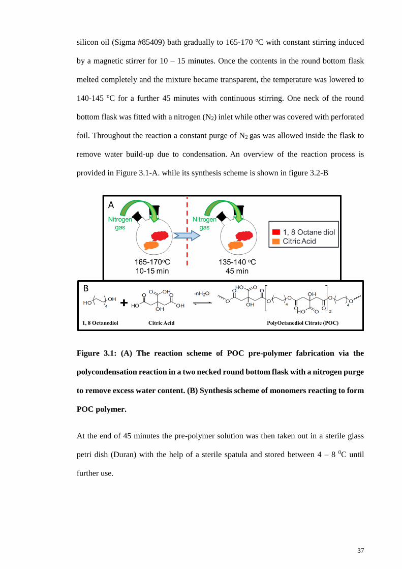
37
silicon oil (Sigma #85409) bath gradually to 165-170 oC with constant stirring induced
by a magnetic stirrer for 10 – 15 minutes. Once the contents in the round bottom flask
melted completely and the mixture became transparent, the temperature was lowered to
140-145 oC for a further 45 minutes with continuous stirring. One neck of the round
bottom flask was fitted with a nitrogen (N2) inlet while other was covered with perforated
foil. Throughout the reaction a constant purge of N2 gas was allowed inside the flask to
remove water build-up due to condensation. An overview of the reaction process is
provided in Figure 3.1-A. while its synthesis scheme is shown in figure 3.2-B
Figure 3.1: (A) The reaction scheme of POC pre-polymer fabrication via the
polycondensation reaction in a two necked round bottom flask with a nitrogen purge
to remove excess water content. (B) Synthesis scheme of monomers reacting to form
POC polymer.
At the end of 45 minutes the pre-polymer solution was then taken out in a sterile glass
petri dish (Duran) with the help of a sterile spatula and stored between 4 – 8 0C until
further use.

38
3.3 Fabrication of POC and ZnO-POC composites
Pure-POC scaffold and 3 different ZnO-POC composite scaffolds with varying w/w
percentages of ZnO NPs (1%, 3% and 5%) were prepared. ZnONP were obtained from
Sigma-Aldrich (Sigma #544906). The particle size for ZnONP was less than 100 nm.
Their specifications and raman FTIR are attached in appendix I. All scaffolds were
prepared by dissolving POC pre-polymer in 1, 4 Dioxane (50% w/v) (Sigma #360481);
for fabricating ZnO-POC scaffolds, an additional step was involved by adding pre-
calculated amounts of ZnO (Sigma #544906) (1%, 3% and 5% w/w with respect to weight
of the pure pre-polymer) to Pure-POC pre polymer solution. The POC-ZnO-dioxane
mixtures were sonicated (Sono Swiss SW12H, Sonoswiss AG, Ramsen, Switzerland.) for
10 minutes for homogenous distribution of ZnONP (Pure-POC-dioxane mixture was also
sonicated to follow uniform procedure). This was followed by adding sieved Sodium
Chloride (NaCl) (Sigma #S7653) crystals of size 200 – 300µm the ZnO-POC mixture
(NaCl/ZnO-POC = 9:1). The composite ZnO-POC-NaCl-dioxane slurry was poured in
custom designed TeflonTM moulds and placed for solvent evaporation and curing at 80oC
for 1 week in an oven (Memmert GmbH, Schwabach, Germany). During the curing
process, the slurry in Teflon moulds was mixed via spatula every 2 hours for 24 hours to
enhance elimination of trapped Dioxane. Steps involved in pre-polymer fabrication are
illustrated in Figure 3.2.

39
Figure 3.2: Steps involved in fabrication of (A) Pure-POC and (B) POC-ZnONP
scaffolds, films and coatings prior to curing.
Following the curing period, the solidified blocks of each of four samples (Pure-POC,
1%, 3% & 5% ZnO-POC) were removed from TeflonTM moulds and placed in a custom
designed rack as shown in Figure 3.3, to separate Pure-POC scaffold from ZnO-POC
scaffolds and to leach out the salt with sequential progressions in distilled water for 5
days at room temperature.

40
Figure 3.3: A custom-designed rack placed under water to categorise Pure-POC
scaffolds from ZnO-POC scaffolds and to leach out the salt from scaffolds.
Scaffolds were frozen in -80oC (Thermo Scientific, Waltham, MA, USA) for 1 hour and
sliced (~ 6 mm thickness) using a stainless steel blade. The slices were further cut using
a cork borer to produce cylinder-shape samples 6 mm in diameter. All samples were
freeze-dried (FreeZone 2.5, Labconco, Kansas, USA) for 24 hours and were kept in a
desiccator until further use. Pure-POC Scaffold was used as control in all measurements.
3.4 Chondrocyte Isolation
Chondrocytes were isolated from bovine metacarpal-phalangeal joints of cattle less than
2 years old. Cow legs were obtained within 4 hours of slaughter. The legs were thoroughly
washed with hot running water and washing detergent. After the wash, legs were soaked
for 45 minutes in 70% ethanol. Joints were opened using scalpel handle number 4 and
full thickness cartilage explants from articular cartilage were sliced using scalpel handle
number 3. A brief illustration of the procedure is provided in Figure 3.4.

41
Figure 3.4: Procedure of isolation of primary chondrocytes from cow legs. (A)
Removal of hide from cow leg insert shows completely removed hide, (B) Opening
of joint by removing ligament, (C) Cutting of cartilage and (D) Complete shaved
cartilage joint and explants placed in sterile container.
Sliced cartilage explants were immediately immersed in chondrocyte culture medium.
The protocol to make chondrocyte culture medium is provided in the subsequent section.
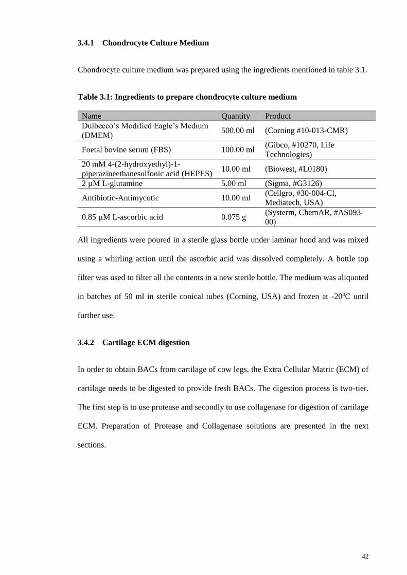
42
3.4.1 Chondrocyte Culture Medium
Chondrocyte culture medium was prepared using the ingredients mentioned in table 3.1.
Table 3.1: Ingredients to prepare chondrocyte culture medium
Name Quantity Product
Dulbecco’s Modified Eagle’s Medium
(DMEM) 500.00 ml (Corning #10-013-CMR)
Foetal bovine serum (FBS) 100.00 ml (Gibco, #10270, Life
Technologies)
20 mM 4-(2-hydroxyethyl)-1-
piperazineethanesulfonic acid (HEPES) 10.00 ml (Biowest, #L0180)
2 µM L-glutamine 5.00 ml (Sigma, #G3126)
Antibiotic-Antimycotic 10.00 ml (Cellgro, #30-004-Cl,
Mediatech, USA)
0.85 µM L-ascorbic acid 0.075 g (Systerm, ChemAR, #AS093-
00)
All ingredients were poured in a sterile glass bottle under laminar hood and was mixed
using a whirling action until the ascorbic acid was dissolved completely. A bottle top
filter was used to filter all the contents in a new sterile bottle. The medium was aliquoted
in batches of 50 ml in sterile conical tubes (Corning, USA) and frozen at -20oC until
further use.
3.4.2 Cartilage ECM digestion
In order to obtain BACs from cartilage of cow legs, the Extra Cellular Matric (ECM) of
cartilage needs to be digested to provide fresh BACs. The digestion process is two-tier.
The first step is to use protease and secondly to use collagenase for digestion of cartilage
ECM. Preparation of Protease and Collagenase solutions are presented in the next
sections.

43
3.4.2.1 Protease preparation
Protease was prepared by weightng 1g of protease (Sigma, #P8811) via an analytical
weighing balance (Shimadzu, AY220) and dissolving it in 175.00 ml of DMEM+20%
FBS. This solution was then filtered using a 0.20 µm syringe filter.
3.4.2.2 Collagenous preparation
Collagenase was prepared by weighing 0.021 g of collagenase type XI (Sigma, #C6885)
via an analytical weighing balance (Shimadzu, AY220) and dissolving it in 300.00 ml of
DMEM+20% FBS. This solution was then filtered using a 0.20 µm syringe filter.
After the entire joint was shaved, the medium was aspirated using a Pasteur pipette, and
10.00 ml of protease solution 700 unit/ml (Sigma #P8811) was added to digest the ECM
of BAC explants for 1 hour with gentle rotation in a hybridisation oven (Problot, Labnet,
NJ, USA). After 1 hour of protease digestion the medium was removed with a sterile
Pasteur pipette and 30.00 ml of fresh medium supplemented with 100 unit/ml of
collagenase type XI (Sigma #C6885) was added for a further digestion of 16 hours with
gentle rotation in hybridisation oven (Problot, Labnet, NJ, USA).
After 16 hours of collagenase digestion, the cell suspension was filtered with a 70 µm
nylon cell filter (BD Falcon, NJ, USA). The cell suspension was centrifuged at 1000 RPM
and the supernatant was carefully removed. The process was repeated twice to remove
any impurities of Protease or Collagenase.
3.4.3 Cell Viability
Cell viability was calculated using trypan blue exclusion assay. Trypan blue solution was
prepared by dissolving 0.2 g of Trypan Blue (Sigma #T6146) in 99.8 ml of ultra-pure
water which was filtered through 0.45 µm hydrophobic membrane syringe filter (Thermo
Scientific, #9057, Thermo Scientific, Waltham, MA, USA ). A working solution of trypan

44
blue was made by mixing 2ml of trypan blue stock solution to a solution of 9.58 ml Ultra-
pure water and 0.43 g NaCl.
20 µl of filtered cell concentrate and same amount of trypan blue working solution was
taken in a micro centrifuge tube. A 20 µl mixture of cell and trypan blue was placed in
Heamacytometer covered with a cover slip and read under a microscope. Viable
(unstained) cells and non-viable (stained blue) cells were counted using a cell counter in
each chamber. The total number of viable cells was divided by the total number of cells
(Viable + Non-Viable) and the resulting percentage was calculated for viable cells. Only
passage zero cells were used, with ≥ 95% viability.
3.5 Cell proliferation assay
Cell proliferation was determined via Resazurin reduction assay (Ataollahi et al., 2014).
Briefly, 140 mg of Resazurin powder (Sigma #R7017) was dissolved in 1000 ml of phosphate
buffered saline (PBS) (Sigma #P4417) to make the stock solution. It was then diluted with PBS
to 10% volume working solution. 1 ml of resazurin working solution was added into each well at
different time points. Prior to reading the plate via microplate reader (FLUOstar OPTIMA, BMG
labtech, GmbH, Ortenberg, Germany), at every time point each plate was incubated for 4 hours
at 37oC and 5% CO2 (Thermo Scientific, Waltham, MA, USA). After the incubation, the
plate was wrapped in aluminum foil and was shaken at 30 RPM on a bench top shaker for
1 minute. From each well, 100 µl of resazurin solution was placed in wells of a 96-well
plate, and resazurin solution from unseeded scaffold was taken as blank. Absorbance was
read by a microplate reader for wavelengths of 570 nm and 595nm.
3.6 DNA Biochemical Assay
The Hoechst binding dye method was used to quantify DNA within the scaffolds at each
time point. For this method, the cells needs to be lysed followed by their quantification.

45
Subsection 3.6.1 details the process of cell lysis and 3.6.2 provides the steps involved in
quantifying the DNA.
3.6.1 Lysis of BACs
To quantify the number of cells present in a particular scaffold, the cells first need to be
separated from the scaffold and lysed to obtain DNA. In order to obtain cell lysate, papain
is used as primary agent that digest protein substrates while leaving behind the DNA
intact and unharmed. To dissociate the cells from the scaffold, the scaffold laden with
cells was placed in a 1.5 ml micro-centrifuge tube and papain digest buffer topped with
papin was added to it. A manual tissue homogeniser was used to tear the scaffold apart to
expose BACs to papain. Ingredients presented in Table 3.2 were used to prepare papain
digest buffer.
Table 3.2: Ingredients to prepare papain digest buffer
Name Quantity Product
L-Cystein HCl monohydrate 0.788g (Sigma #C7880)
Ethylenediaminetetraacetic acid (EDTA) 0.403g (Sigma #E6758)
PBS 500ml (Sigma #P4417)
All ingredients were dissolved at a pH of 6.0; the pH was adjusted using 1M Sodium
Hydroxide (NaOH). Furthermore 3 µl of Papain (Sigma #P3125) per construct was added.
Cells were lysed overnight at 65oC because papain has the highest activity in the range of
60 – 70oC. After overnight activity, the cell-scaffold lysate was stored at -20oC until
further analysis. At -20oC the DNA can be kept safe for several months.
3.6.2 DNA quantification
For DNA quantification, a Hoechst binding assay was used (Pingguan-Murphy & Nawi,
2012). Hoechst is a dye that specifically binds to DNA to produce fluorescence at a wavelength
of 460 nm. Stock solution of Hoechst was prepared by dissolving 50.0 mg of Hoechst (Sigma

46
#861405) in 25.0 ml of ultra-pure water in an amber bottle wrapped with aluminum foil to prevent
degeneration of Hoechst.
3.6.2.1 DNA Standard
To calculate the quantity of DNA, a standard needs to be set for the microplate reader in
order to assess the quantity in the sample presented. A DNA standard was prepared by
dissolving 5 mg of Deoxyribonucleic acid sodium salt from calf thymus (Sigma #D1501)
in 5 ml of EDTA/PBS solution. The EDTA/PBS solution acts as a buffer for DNA from
calf thymus. EDTA/PBS solution was prepared by weighing EDTA (0.02922g) using an
analytical balance (Shimadzu, AY220) and dissolving it in 100 ml PBS (Sigma #P4417)
(pH 7.8), followed by autoclaving.
The solution of DNA from calf thymus and EDTA/PBS was left overnight in a 0-4oC
fridge. The DNA formed a gel which was then sheared by pipetting using a glass pipette
vigorously several times. A syringe filter of 0.45 µm (minisart SRP15, Sartorius,
Germany) was used to filter out the DNA. The DNA concentration was checked via
Nanodrop ND 2000 (Thermo Scientific, Waltham, MA, USA). Aliquots of DNA standard
were stored at -20oC in sterile glass bijou tubes to prevent absorbance of DNA in plastic.
After Papain digestion, the DNA standard was diluted to 20 µg/ml with 2xSSC/Papain
digest buffer. A 20x stock Saline Sodium Citrate (SSC) buffer was prepared by dissolving
87.65 g of Sodium Chloride (NaCl) (Sigma # S7653) and 44.1 g of CA (Sigma # C0759)
in 500 ml of distilled water. The pH was adjusted to 7.0 using 1M NaOH/1M HCl.
A serial double dilution was prepared at 20, 10, 5, 2.5, 1.25, 0.625 and 0.3124 µg/ml. For
0 µg/ml of DNA, 2xSSC/Papain digest buffer was used.
Cell-scaffold lysate (100µl each) in triplicates were pipetted into wells of black flat-
bottomed 96-well plates (Falcon, Corning #3916) and 100 µl of Hoechst working reagent
(20µl Hoechst stock added to 20ml of 2xSSC/Papain digest buffer) was added to each
well. All these processes were carefully carried out in a dark room. Aluminium foil was

47
used to wrap the plate before transferring it to the microplate reader. The plate was read
for fluorescence at wavelengths of excitation (ex) 355 nm and emission (em) 460 nm
using FLUOstar Optima, BMG Labtech, Ofdenberg, Germany.
3.7 Sulphated Glycosaminoglycan (S-GAG) quantification assays
Glycosaminoglycans (GAGs) are polysaccharides that are present in abundance in a
healthy cartilage. Sulphated-GAGs (S-GAGs) are the types that can be measured via a 1,
9-dimethylmethylene blue assay.
Total S – GAG was measured from Cell-scaffold lysate at each time point. The same cell-
scaffold lysate is used for S-GAG quantification as the ones used for DNA quantification
because papain digestion does not affect the S-GAGs. Released S – GAG was measured
by collecting the culture medium at every medium change from the wells. Total S – GAG
and released S – GAG were analyzed by Dimethyl Methylene Blue (DMMB) assay (Lee
& Bader, 1997; Pingguan-Murphy & Nawi, 2012). DMMB was prepared by using the
ingredients presented in Table 3.3.
Table 3.3: Ingredients used in preparation of DMMB solution for DMMB assay
Name Quantity Product
1, 9-dimethylmethylene blue 0.016g (Sigma, #341088)
Ethanol 5.0 ml (Merck)
Sodium Formate 2.0 g (Sigma, #247596)
Formic Acid 2.0 ml (Sigma, #F0507)
DMMB was dissolved in ethanol, followed by the addition of 950.00 ml of distilled water
and sodium formate. The final pH was adjusted by adding formic acid drop by drop. The
solution was then transferred to an amber bottle wrapped in aluminium foil.
The S-GAG standard stock solution was prepared by dissolving 1mg of Chondroitin
Sulfate-A sodium salt from Bovine trachea (Sigma, #C9819) in 1ml of distilled water. A

48
working solution for S-GAG assay was prepared by diluting to 100 µg/ml in distilled
water. A series of standard dilutions were made and distilled water was used as 0 µg/ml.
Briefly, 40 µl of cell-scaffold lysate for total S – GAG, 40 µl culture medium for released
S – GAG and serially diluted standards of Chondroitin sulfate salt in triplicates were
mixed with 250 µl of DMMB reagent in a transparent 96-well plate and absorbance at a
wavelength of 595nm was read via microplate reader. All the procedures were performed
in a dark room.
3.8 Quantitative Polymerase Chain Reaction (q-PCR)
To observe the behaviour of BACs at gene level Cartilage matrix specific genes; Collagen
type II (COL2A1) and Aggrecan (ACAN) were observed. COL2A1 is responsible for
producing type II collagen in the cartilage, while ACAN is responsible for producing
Aggrecan proteoglycan in the cartilage. The matrix degradation gene (MMP-13) and
hypoxia inducible factor gene (HIF1A) were also observed. MMP-13 provides an insight
in to the catabolic activity that the cartilage is undergoing, while HIF1A demonstrates the
effect of oxygen concentration on the BACs. Glyceraldehyde-3-phosphate
dehydrogenase gene (GAPDH) expression was determined and used as a house keeping
gene. Gene expressions were determined by the TaqMan gene expression assay using
StepOnePlusTM Real-Time PCR system (Applied Biosystems, Foster City, USA).
3.8.1 RNA Isolation
At each time point, culture medium was removed and scaffolds were washed with PBS
(Sigma #P4417) and placed in TRIzol reagent (#AM9738, Lifetechnologies™, Carlsbad,
CA, USA). Total ribonucleic acid (RNA) was extracted according to manufacturer’s
guidelines using TRI reagent (Hummon et al., 2007). Briefly, samples were homogenized
using a tissue homogenizer, followed by phase separation by adding 0.2ml of chloroform (Fisher
Scientific, UK, #BP1145-1) per 1ml of TRIzol reagent. Samples were then centrifuged at 12000

49
RCF for 15 minutes (CYT12, Centurion Scientific, West Sussex, UK) and upper aqueous layer
was carefully transferred to a new tube. RNA was precipitated by adding 0.5ml of 100%
isopropanol (Sigma, #I9516) and centrifuging at 12000 RCF for 10 minutes at 4oC. Supernatant
was removed leaving precipitated RNA. Visible RNA palette was washed by 1.0 ml of
75% ethanol (prepared from absolute ethanol from Merck and with Ultra-Pure distilled
RNAse/DNAse free water from Invitrogen) and left to air dry. 20 µl of RNase free water
(Invitrogen, #10977) was added to dissolve the palette and its purity was read via
NanoDrop (ND2000, Thermo Scientific, Waltham, MA, USA) at absorbance ratio of
260/280 nm. Only RNA samples with absorbance ratio greater or equal to 2 were further
used for analysis.
3.8.2 cDNA synthesis
Conversion of RNA to complimentary Deoxyribonucleic acid (cDNA) was performed
using a high capacity RNA-to-cDNA (Applied Biosystems, #4387406, Foster City, USA)
kit. Briefly, for every 2 µg of total RNA (2000 ng/ µl) 20 µl reaction mixture was used to
reverse transcribe RNA into cDNA by incubating the reaction plate for 60 minutes at
37oC followed by heating it to 95oC for 5 minutes, followed by holding the reaction plate
at 4oC for half an hour using MyGene series Peltier thermal cycler (MG96G).
3.8.3 Gene Expression
Quantitative gene expression of all the genes listed in Table 3.4 was identified using 1 µl
of cDNA sample in triplicates for each gene. 1 µl of cDNA sample was transferred to a
96-well plate (MicroAmp® InduraPlateTM, Applied Biosystems®), topped up with 10 µl
TaqMan master mix, 8 µl of RNAse/DNAse free Ultra-Pure water, and 1 µl of
target/housekeeping gene (TaqMan assay). The plate was sealed with a transparent
adhesive film and QPCR was performed using StepOnePlusTM Real-Time PCR system
(LifeTechnologies).
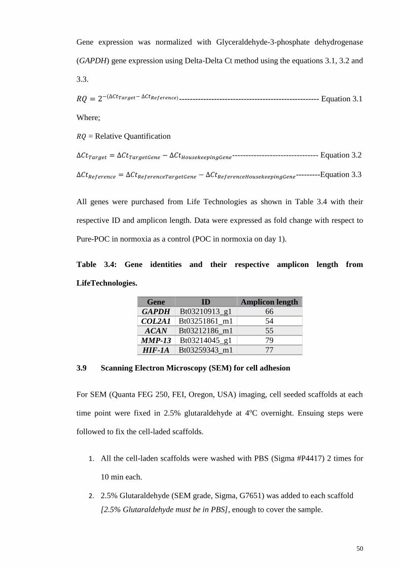
50
Gene expression was normalized with Glyceraldehyde-3-phosphate dehydrogenase
(GAPDH) gene expression using Delta-Delta Ct method using the equations 3.1, 3.2 and
3.3.
𝑅𝑄 = 2−(∆𝐶𝑡𝑇𝑎𝑟𝑔𝑒𝑡− ∆𝐶𝑡𝑅𝑒𝑓𝑒𝑟𝑒𝑛𝑐𝑒)---------------------------------------------------- Equation 3.1
Where;
𝑅𝑄 = Relative Quantification
∆𝐶𝑡𝑇𝑎𝑟𝑔𝑒𝑡 = ∆𝐶𝑡𝑇𝑎𝑟𝑔𝑒𝑡𝐺𝑒𝑛𝑒 − ∆𝐶𝑡𝐻𝑜𝑢𝑠𝑒𝑘𝑒𝑒𝑝𝑖𝑛𝑔𝐺𝑒𝑛𝑒-------------------------------- Equation 3.2
∆𝐶𝑡𝑅𝑒𝑓𝑒𝑟𝑒𝑛𝑐𝑒 = ∆𝐶𝑡𝑅𝑒𝑓𝑒𝑟𝑒𝑛𝑐𝑒𝑇𝑎𝑟𝑔𝑒𝑡𝐺𝑒𝑛𝑒 − ∆𝐶𝑡𝑅𝑒𝑓𝑒𝑟𝑒𝑛𝑐𝑒𝐻𝑜𝑢𝑠𝑒𝑘𝑒𝑒𝑝𝑖𝑛𝑔𝐺𝑒𝑛𝑒---------Equation 3.3
All genes were purchased from Life Technologies as shown in Table 3.4 with their
respective ID and amplicon length. Data were expressed as fold change with respect to
Pure-POC in normoxia as a control (POC in normoxia on day 1).
Table 3.4: Gene identities and their respective amplicon length from
LifeTechnologies.
Gene ID Amplicon length
GAPDH Bt03210913_g1 66
COL2A1 Bt03251861_m1 54
ACAN Bt03212186_m1 55
MMP-13 Bt03214045_g1 79
HIF-1A Bt03259343_m1 77
3.9 Scanning Electron Microscopy (SEM) for cell adhesion
For SEM (Quanta FEG 250, FEI, Oregon, USA) imaging, cell seeded scaffolds at each
time point were fixed in 2.5% glutaraldehyde at 4oC overnight. Ensuing steps were
followed to fix the cell-laded scaffolds.
1. All the cell-laden scaffolds were washed with PBS (Sigma #P4417) 2 times for
10 min each.
2. 2.5% Glutaraldehyde (SEM grade, Sigma, G7651) was added to each scaffold
[2.5% Glutaraldehyde must be in PBS], enough to cover the sample.

51
3. All the samples were kept overnight at 4oC. (2.5% Glutaraldehyde penetrates
only 1mm per hour).
4. Graded Alcohol was used to dehydrate the sample.
5. Starting from 25% followed by 35%, 50%, 70% and 90%. Each 10-15 minutes.
6. Finally 100% ethanol was placed in the well that contain cell-laden scaffolds
twice for 20 minutes each.
7. Finally the samples were dried in freeze dryer (FreeZone 2.5, Labconco, Kansas,
USA) for 2 hours.
n.b: samples can be kept in 2.5% glutaraldehyde at 4oC for up to one month.
3.10 Live/Dead Assay
The qualitative cell viability was determined using a LIVE/DEAD® viability kit
(Molecular Probes® LifeTechnologiesTM, Carlsbad, CA, USA). Briefly, a working
solution was made by adding 1µl Calcein and 2.5 µl Ethidium homodimer-1 in 1ml of
PBS (Sigma #P4417). Each scaffold was placed in 200 µl of working solution for 30
minutes before imaging. Imaging was performed with green fluorescence at ex/em
~495nm/~515nm for live cells and red fluorescence at ex/em ~495 nm/~635 nm for dead
cells using confocal laser scanning microscopy (CLSM) (Leica TCS SP5 II, Leica
Microsystems CMS GmbH, Mannheim, Germany).
3.11 Statistical Analysis
Data are represented as average ± standard deviation at each time point. Statistical
analysis was performed using IBM SPSS v21, with a two-way analysis of variance
(ANOVA) to determine significance between groups, and a post-hoc Tukey’s test was
applied to test significance between oxygen tension and within the days. In all conditions,
a p value of less than 0.05 (P<0.05) was considered to be significant. The number of
biological replicates in all the studies ranges from 5 to 12.
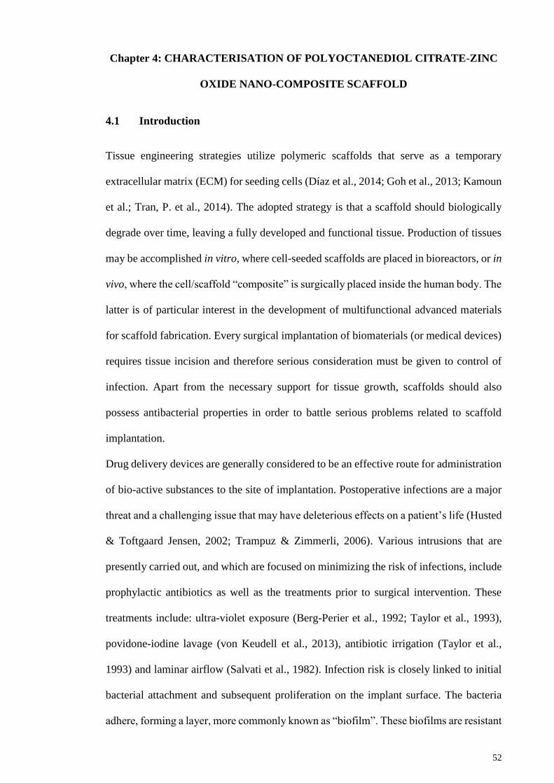
52
Chapter 4: CHARACTERISATION OF POLYOCTANEDIOL CITRATE-ZINC
OXIDE NANO-COMPOSITE SCAFFOLD
4.1 Introduction
Tissue engineering strategies utilize polymeric scaffolds that serve as a temporary
extracellular matrix (ECM) for seeding cells (Díaz et al., 2014; Goh et al., 2013; Kamoun
et al.; Tran, P. et al., 2014). The adopted strategy is that a scaffold should biologically
degrade over time, leaving a fully developed and functional tissue. Production of tissues
may be accomplished in vitro, where cell-seeded scaffolds are placed in bioreactors, or in
vivo, where the cell/scaffold “composite” is surgically placed inside the human body. The
latter is of particular interest in the development of multifunctional advanced materials
for scaffold fabrication. Every surgical implantation of biomaterials (or medical devices)
requires tissue incision and therefore serious consideration must be given to control of
infection. Apart from the necessary support for tissue growth, scaffolds should also
possess antibacterial properties in order to battle serious problems related to scaffold
implantation.
Drug delivery devices are generally considered to be an effective route for administration
of bio-active substances to the site of implantation. Postoperative infections are a major
threat and a challenging issue that may have deleterious effects on a patient’s life (Husted
& Toftgaard Jensen, 2002; Trampuz & Zimmerli, 2006). Various intrusions that are
presently carried out, and which are focused on minimizing the risk of infections, include
prophylactic antibiotics as well as the treatments prior to surgical intervention. These
treatments include: ultra-violet exposure (Berg-Perier et al., 1992; Taylor et al., 1993),
povidone-iodine lavage (von Keudell et al., 2013), antibiotic irrigation (Taylor et al.,
1993) and laminar airflow (Salvati et al., 1982). Infection risk is closely linked to initial
bacterial attachment and subsequent proliferation on the implant surface. The bacteria
adhere, forming a layer, more commonly known as “biofilm”. These biofilms are resistant

53
to antibiotics and the host immune system, and therefore treatment requires more
measures (Costerton et al., 1999). For that reason it is essential to prevent initial bacterial
attachment on the implant surface.
One of the materials that has proven to be effective against bacteria in general is zinc
oxide (ZnO) (Sirelkhatim et al., 2015). This substance is currently considered a Generally
Recognized as Safe (GRAS) material, approved by the FDA (Espitia et al., 2013).
Furthermore, ZnO can be fabricated into nanoparticles (NPs) (Khalil et al.; Wallace et al.,
2013; Zak et al., 2011), that can be embedded inside a biodegradable polymer matrix and
subsequently released in a highly controlled manner. Polyoctanediol citrate (POC)
elastomer is one of the prominent new materials for scaffold fabrication (Djordjevic,
Choudhury, et al., 2010; Djordjevic, Szili, et al., 2010; Serrano et al., 2010). POC and
other types of citric acid (CA)-based elastomers have shown excellent performance in
various biomaterial applications, including scaffolds for soft tissue engineering,
biodegradable bone fixation devices and drug delivery devices (Qiu et al., 2006; Serrano
et al., 2011; Zhang et al., 2009).
In the current chapter, a newly-developed ZnO-POC scaffold produced by a well-
established solvent-casting / particulate-leaching technique is reported. Tissue
engineering applications require three-dimensional structures with high level of porosity
and pore sizes in the range of 100-200µm. In many cases such porous structures alter the
material properties that were originally recorded for sample films (Djordjevic et al., 2009;
Kompany et al., 2014). Apart from antibacterial properties of the ZnO-POC composite
scaffolds, the scaffolds must be investigated for their potential support of cellular growth,
proliferation and toxicity. Similar to other polymer/solid composite scaffolds, the
presence of ZnO NPs physically, strengthens the scaffold thus providing necessary
toughness for engineering of hard tissues. In particular, the target was to explore the
possibility of using ZnO-POC nano-composite scaffolds for engineering of cartilage

54
tissue. ZnO-POC nano-composite scaffolds with variation of ZnO concentration and a
series of experiments was conducted in order to: (i) investigate the influence of ZnO
concentration on material characteristics; (ii) establish bactericidal properties by
measuring the proliferation rate of Escherichia coli and Staphylococcus aureus; and (iii)
examine the bioactivity, biocompatibility and potential of ZnO-POC scaffolds for
cartilage tissue engineering by in vitro experiments with primary bovine chondrocyte
cells
4.2 Materials and Methods
4.2.1 Surface and elemental properties
Pure-POC scaffolds and 3 different ZnO-POC composite scaffolds with varying w/w
percentages of ZnO NPs (1%, 3% and 5%) were prepared, the details of the fabrication
methods can be found in Section 3.3. The final dimension of the scaffold is 6mm (Ø) x
6mm height. The scaffold morphology and element composition analysis were assessed
using Quanta FEG 250 Field Emission Scanning Electron microscope (FESEM) equipped
with Energy Dispersive X-ray (EDX) analysis.
4.2.2 Zinc oxide nanoparticles distribution within POC
Optical microscopy (OLYMPUS, BX51TRF, Japan) was used to view zinc oxide
nanoparticles distribution within POC Before the experiment, POC and 1%, 3% and 5%
ZnO-POC (50% w/v in dioxane) solutions were prepared and spin-coated on microscope
coverslips, using Laurell (WS-650MZ-23NPP) spin coater. Briefly, the microscope
coverslip was placed inside the spin coater followed by turning on the vacuum suction to
prevent the microscope coverslip from moving. The spin coater was rotating at 3000 RPM
for 10 seconds. During rotation, 200 µl of polymer solution was dropped at the centre of
the coverslip and the process was repeated thrice to make sure the coverslip is uniformly

55
covered with polymer. Subsequently, the coated coverslips were placed flat in the oven
at 800C for another week for curing and solvent evaporation. The coated coverslip was
placed on the microscope and the distribution on ZnO was captured by camera.
4.2.3 Surface wettability test
Good wettability of a surface will ensure good cell bonding with polymer (Arima & Iwata,
2007). Water-in-air contact angle (WACA) experiments were performed on both ZnO-
POC and POC samples. POC and 1%, 3% and 5% ZnO-POC (50% w/v in dioxane)
solutions were coated on the microscope coverslip. The sample preparation method is
similar to the one described earlier in Section 4.2.2. The WACA experiments were carried
out at room temperature by a sessile drop method using an OCA 20 optical contact angle
measuring instrument (DataPhysics Instruments GmbH, Filderstadt, Germany) After the
droplet (2µl) was dispensed on the prepared sample, the image was captured within 5
seconds and analysed. Each measurement was repeated 5 times (5 new samples were
prepared for each condition). The water angles were measured at 3 different spots on each
sample and the results were averaged (n=5).
4.2.4 Mechanical property and porosity
The compression tests were performed on POC and 1%, 3% and 5% ZnO-POC scaffolds
using Instron mechanical tester (Instron Microtester 5848, Instron Corp., Norwood, MA,
USA) equipped with a 2kN load cell. Scaffolds were cut into cylindrical shapes (6 mm-
diameter / 6 mm-height) and subjected to 50 % compression at a rate of 1 mm/min. For
percentage recovery, the height of each cylindrical scaffold was measured 2 min after the
compression test was completed (n= 5). The porosity of the scaffolds was measured based
on the Archimedes principle (Zhu et al., 2008).

56
4.2.5 Swelling test
For Swelling test, 10ml of each POC and 1%, 3% and 5% ZnO-POC (50% w/v in
dioxane) solutions were poured into custom designed (5cm diameter X 2 mm thickness)
TeflonTM moulds. The polymer filled moulds were placed flat in the oven at 800C for
curing and solvent evaporation for 1 week. After that, the samples were removed from
the moulds and cut into disk shape (6mm diameter X 1 mm thickness) using a cork borer.
Each sample was placed separately in a conical tube containing 10 ml of distilled water
which was taken out at different time points (For first hour every 10 min then every hour
for 6 hours). The weight was measured after lightly cleaning the surface with lint-free
paper. Percentage swelling was calculated using equation 4.1.
𝑆𝑤𝑒𝑙𝑙𝑖𝑛𝑔(%) =𝑊𝑠−𝑊𝑜
𝑊𝑜∗ 100 ------------------------------------------------------- Equation 4.1
Where; 𝑊𝑠 is the weight of the swollen polymer disk at each time point and 𝑊𝑜 is samples’
dry weight.
4.2.6 Fourier transform infrared spectroscopy (FTIR) analysis
Fourier transform infrared spectra were obtained for the POC and 1%, 3% and 5% ZnO-
POC scaffolds using spectrophotometer-Thermo Scientific Nicolet iS10 (Thermo
Scientific, Waltham, MA, USA) operating in attenuated total reflectance mode (ATR-
FTIR) at room temperature, within a wavelength range of 450 – 4000 cm-1.
4.2.7 Thermal analysis
For assessing thermal properties, thermo gravimetric analysis (TGA) was performed on
POC and 1%, 3% and 5% ZnO-POC scaffolds within a temperature range of 30-900 oC
at a rate of 10 oC/min using PerkinElmer, TGA 4000 (PerkinElmer, Waltham, MA, USA).
The onset of degradation temperature was measured after 10 % of degradation.

57
4.2.8 Degradation analysis
For degradation analysis, POC and 1%, 3% and 5% ZnO-POC scaffolds were immersed
in a conical tube containing 10 ml of Phosphate Buffered Saline (PBS) The samples were
incubated at 370C and at each time point the samples were taken out and freeze dried
overnight, and the mass loss was measured by equation 4.2. Each condition was repeated
5 times.
𝑀𝑎𝑠𝑠𝑙𝑜𝑠𝑠(%) =𝑊𝑜−𝑊𝑡1
𝑊𝑜∗ 100 ----------------------------------------------------- Equation 4.2
Where, 𝑊𝑜 is the initial dry weight of the scaffold, 𝑊𝑡1 is the weight of the scaffold at
each time point after freeze-drying.
4.2.9 ZnO NPs release kinetics in physiological conditions
To observe the release of ZnO NPs, POC and 1%, 3% and 5% ZnO-POC scaffolds were
placed in 10 ml of PBS in conical tubes and kept at 37oC throughout the whole
experiment. The scaffolds prepared for this experiment have a thickness of 2mm,
elsewhere in the thesis, when scaffold is mentioned the thickness should be 6mm. Two
milliliter of solution from each conical tube was withdrawn at each time point (day 1, 7,
14 & 28) and was kept at 4 – 8oC until further analysis. The concentration of Zn2+ (ppm)
was detected by atomic emission spectroscopy (AAS) using Agilent 4100 (Agilent
Technologies, Inc., Santa Clara, CA, USA) microwave plasma-atomic emission
spectrophotometer (MP-AES). The standard for Zn2+ was purchased from Sigma-Aldrich
Malaysia (Sigma, #18827).

58
4.2.10 Anti-bacterial properties
The antibacterial properties of POC and its composites were tested. POC and 1%, 3% and
5% ZnO-POC scaffolds were placed in direct contact with the bacteria, a technique
known as the liquid culture method (Raffi et al., 2008). Briefly, rectangular strips (30 mm
x 10 mm x 5 mm) of POC and 1%, 3% and 5% ZnO-POC scaffolds were placed in 15 ml
conical tubes with 10 ml of Luria-Bertani (LB) broth inoculated with 50 µl of 104-5
CFU/ml E. Coli (ATCC 15597). Samples were placed in a shaking incubator at 370C with
a speed of 50 RPM. Pure-POC scaffold and LB broth (without any scaffold) were
inoculated with similar bacterial densities and were used as positive and negative controls
respectively. The optical density was measured at 595 nm every 2 hours (for 8 hours)
with a micro plate reader (BMG LABTECH, Offenburg, Germany).
4.2.11 In vitro tests with chondrocyte cell culture
POC and 1%, 3% and 5% ZnO-POC scaffolds were tested for their cytotoxicity towards
bovine chondrocytes. Bovine legs of post-slaughtered cows were obtained up to carpal
joint only. Bovine chondrocytes were isolated from metacarpalphalangeal joints as
described in section 3.4. Cells were used when only greater than 95% viability was
obtained (refer to section 3.4.3). Toxicity was measured by resazurin reduction cell
viability test as described earlier in section 3.5. Scaffolds were autoclaved before cell
seeding and then were allowed to neutralise in chondrocyte medium overnight. Scaffolds
were then placed in 24 well plates and dried under laminar flow for an hour. A volume of
20 µl cell suspension with a concentration of 3 x 10⁷cells/ml was spotted on top of the
scaffold. Cells were allowed to attach for 2 hours, after which 2ml of medium/well was
added. Reduction in resazurin was determined after 24 and 72 hours as described
previously in section 3.5 (Larson et al., 1997). Briefly, medium was removed from the
wells containing cell seeded scaffolds and then washed with PBS. 1ml of resazurin was

59
added to each well and then kept in the incubator for 4 hours at 37oC and 5% CO2. After
the incubation time the plate was wrapped in aluminium foil and was shaken at 30 RPM
on a bench top shaker for 1 minute. From each cell-scaffold well, 100 µl of resazurin
solution was placed in 96-well plate and resazurin solution from unseeded scaffold was
taken as blank. Absorbance was read by a microplate reader. Scaffolds with cells were
taken out after 24 hours and 72 hours and the morphology of cells was observed by
FESEM by using a standard protocol of cell fixation (refer to section 3.9 for complete
protocol).
4.3 Results
The analysis of this section has been previously published in Digest journal of
nanomaterials and biostructures volume 10, issue 2, reproduced by permission of the
publisher.
4.3.1 Surface morphology
FESEM images (Figure 4.1 (A-D)) of Pure-POC (control) and 1%, 3% and 5% ZnO-POC
scaffolds demonstrate that the pores were formed into semi-rectangular shapes after
leaching out the salt crystals. In comparison to Pure-POC (control) (Figure 4.1A),
addition of ZnO NPs did not have any significant impact on the pore size or pore structure
that could be observed through FESEM images. Inserts in Figure 4.1 (A-D) are
representative images of surfaces recorded within the pores of produced scaffolds.
Furthermore, EDX analysis confirmed the presence of Zn on the outermost surface layer
and the detected amount of Zn on ZnO-POC surfaces was 0.49%, 1.64%, and 2.02 % for
1% ZnO-POC, 3% ZnO-POC and 5% ZnO-POC respectively. It is important to note that
the increase in surface concentration of Zn corresponds to the pre-determined
concentrations of ZnO NPs in composite scaffolds.
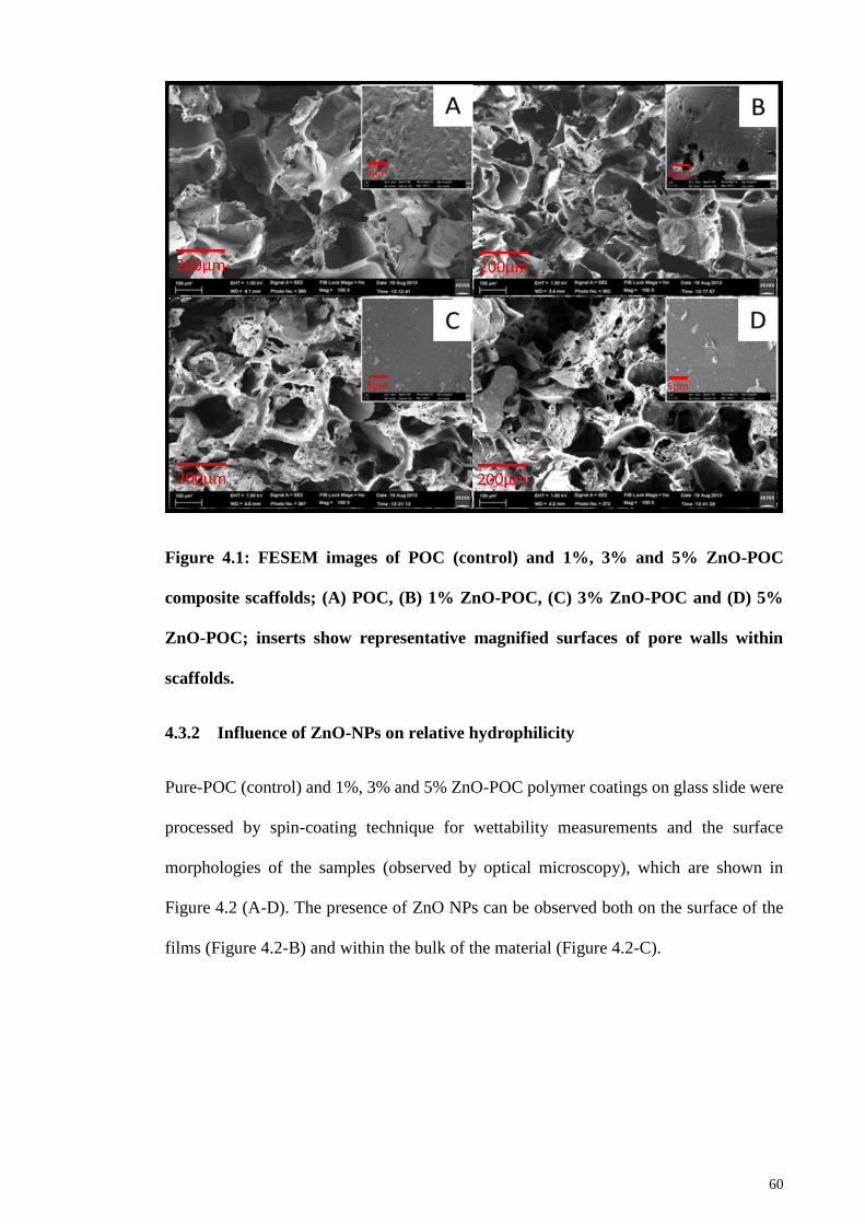
60
Figure 4.1: FESEM images of POC (control) and 1%, 3% and 5% ZnO-POC
composite scaffolds; (A) POC, (B) 1% ZnO-POC, (C) 3% ZnO-POC and (D) 5%
ZnO-POC; inserts show representative magnified surfaces of pore walls within
scaffolds.
4.3.2 Influence of ZnO-NPs on relative hydrophilicity
Pure-POC (control) and 1%, 3% and 5% ZnO-POC polymer coatings on glass slide were
processed by spin-coating technique for wettability measurements and the surface
morphologies of the samples (observed by optical microscopy), which are shown in
Figure 4.2 (A-D). The presence of ZnO NPs can be observed both on the surface of the
films (Figure 4.2-B) and within the bulk of the material (Figure 4.2-C).

61
Figure 4.2: Optical microscopy images of Pure-POC and ZnO-POC surfaces
developed by spin-coating technique at a magnification of 100X; (A) POC, (B) 1%
ZnO-POC, (C) 3% ZnO-POC and (D) 5% ZnO-POC.
Water-in-air contact angle values on polymer coated glass slides were measured to
determine the effect of surface ZnO concentration on hydrophilicity and the results are
shown in Figure 4.3. Evidently, an increase in ZnO content results in more hydrophobic
surfaces. There is no significant difference between contact angles measured for 3% and
5% ZnO-POC, although all ZnO-POC composite coatings displayed a consistently higher
contact angle values in comparison to hydrophilic POC.

62
Figure 4.3: Water-in-air contact angle results measured for thin films of Pure-POC
and 1%, 3% and 5% ZnO-POC produced by spin-coating technique.
4.3.3 Physical properties of scaffolds
Table 4.1 represents the porosities of Pure-POC (control) and 1%, 3% and 5% ZnO-POC
scaffolds. The porosities for all the scaffolds are ranging from 82 to 86%.
Table 4.1 Compression properties and porosities of ZnO-POC scaffolds
Scaffold Compressive
Modulus (MPa) Recovery (%) Porosity (%)
Pure-POC 0.243 ± 0.09 94.55 ± 2.52 82.91 ± 3.49
1% Zno-POC 0.31 ± 0.10 91.94 ± 4.5 85.92 ± 2.41
3% ZnO-POC 0.537 ± 0.33 76.81 ± 4.82 86.01 ± 1.92
5% ZnO-POC 0.442 ± 0.23 60.66 ± 4.49 82.57 ± 7.33
The compression moduli increase with increasing concentration of ZnO, as expected for
a contribution of the filler within the composite scaffold (Table 4.1).

63
4.3.4 Swelling and cross-linking density
Swelling percentages for Pure-POC (control) and 1%, 3% and 5% ZnO-POC films in
distilled water were found. The results from swelling experiments are shown in Figure
4.4. The initial swelling rate in water was the same for all the tested films. After 1 h, a
lower swelling rate was observed for POC and 1% ZnO-POC while other two composite
films (3% and 5% ZnO-POC) showed a constant equilibrium percentage swelling (EPS).
An obvious decrease in EPS after 6 hours is a result of increasing cross-linking density
with ZnO concentration (Figure 4.4).
Figure 4.4: Percentage swelling in water for Pure-POC (control) and 1%, 3% and
5% ZnO-POC composite films.
4.3.5 Chemical composition
The ATR-FTIR spectra of Pure-POC and 5% ZnO-POC are shown in Figure 4.5. The
FTIR spectrum of POC that shows peaks at 1178 CM-1 which expresses both functional

64
groups i.e. alcohols and carboxylic acids of OD and CA respectively. A peak at 1723 CM-
1 shows the C=O stretch that confirms the presence of carboxylic acids, while a peak at
3468 CM-1 confirms the alcohols of 1,8 Octanediol (OD). It is to be noted that peaks at
1178 CM-1, 1723 CM-1 and at 3468 CM-1 are similarly present in the POC-1%, 3% and
5% ZnO scaffolds. Peaks at 1630 cm-1 and 610 cm-1 indicate the presence of ZnO NPs
within the POC matrix at the depth of several microns, detectable with ATR-FTIR. A
broad peak at ~1720 cm-1 (present in all samples) indicates successful polyesterification
between CA and OD monomers (Qiu et al., 2006). Other ZnO-POC scaffolds (1% and
3%) did not show any significant difference when compared to 5% ZnO-POC (Shirazi et
al., 2014; Yang et al., 2006)
Figure 4.5: Representative ATR-FTIR spectra of POC and 5% ZnO-POC scaffolds.
4.3.6 Thermal stability
To investigate thermal stability of the developed scaffolds, Thermograms of Pure-POC
(control) and 1%, 3% and 5% ZnO-POC scaffolds were recorded in TGA experiment.
Figure 4.6 indicates a high stability and a high degree of polymerization without detection
of low molecular weight residues. TGA results show a two-step thermal degradation
65
process, recorded for all the examined samples. The onset of degradation was detected in
the range of 200-220 oC, where POC has the lowest and 5% ZnO-POC the highest onset
of degradation temperature, corresponding to the pre-determined concentrations of ZnO
(Moradi et al.).
Figure 4.6: Thermal degradation curves of Pure-POC and (1%, 3% & 5%) ZnO-
POC scaffolds measured by TGA.
4.3.7 Degradation
The degradation rates of Pure-POC and 1%, 3% & 5% ZnO-POC scaffolds are shown in
Figure 4.7. It is observed that there is a gradual weight loss. However, for the first couple
of weeks no significant change can be seen in the weight loss. After week 2 a weight loss
of about 2% was seen in all the scaffolds. This fell to 24%, 22%, 15% and 13% for Pure-
POC, 1%, 3% & 5% ZnO-POC respectively in the tenth week. At week 10 there was no
significant difference between Pure-POC & 1% ZnO-POC and 3% & 5% ZnO-POC,
however, a highly significant difference was seen when Pure-POC was compared with

66
3% & 5% ZnO-POC. At the end of week 20 there was no significant difference between
Pure-POC and 1% Zno-POC, whereas on the other hand a significant difference was seen
on comparing Pure-POC with 3% & 5% ZnO-POC. There was no significant difference
between 3% and 5% ZnO-POC and between Pure-POC and 1% ZnO-POC at all phases.
Figure 4.7: Weight loss of Pure-POC, 1%, 3% and 5% ZnO-POC scaffolds over the
period of 20 weeks in Phosphate Buffered Saline (PBS).
4.3.8 ZnO release kinetics
The release studies were performed in order to evaluate the relative concentration of Zn+2
ions liberated from 1%, 3% and 5% ZnO-POC scaffolds. Ion concentration was
reasonably considered as directly proportional to the release of ZnO NPs into an aqueous
medium. This simulation of physiological conditions (in vitro: 37 °C; PBS) is important
in order to assess the scaffolds for future experiments with cell seeding and subsequent
implantation in vivo. The release kinetics curves after 24 h are presented in Figure 4.8.
Only after 7 days relatively small difference in the amount of ZnO released from ZnO-
POC scaffolds was observed. The relative concentration of Zn2+ after days 14 and 28,
measured for 5% ZnO-POC was much higher in comparison to other two scaffolds (1%
and 3% ZnO-POC) (Figure 4.8). The high concentration of ZnO inside the POC matrix

67
(5%) caused a “burst release” between days 14 and 28, where the Zn2+ concentration
doubled between two time points. As expected the lowest concentration of Zn2+ was
detected for 1% ZnO-POC after 28 days. Unlike 5% ZnO-POC, both 1% and 3% ZnO-
POC scaffolds maintained “zero order release kinetics” in the period of three weeks
(between day 1 and day 28 in Figure 4.8) (Hahn et al., 2011). There are two possible
mechanisms of ZnO NPs release from the POC matrix: (1) particle erosion after polymer
swelling; and (2) polymer degradation and subsequent particle release.
Figure 4.8: In-vitro Release kinetics profile of ZnO (Zn2+) from ZnO-POC scaffolds
in PBS at 37°C; Mt = mass of ZnO released at time intervals; M∞ = total mass of ZnO
within the scaffold (all Mt /M∞ ratios are the mean values for five samples measured
with AAS).
The actual concentration of ZnO released in the medium over the period of 28 days
revealed that highest release of Zn ions was 4.61 ppm from scaffold containing 5%
ZnONP while 1% and 3% ZnONP-POC demonstrated a significantly lower amount of Zn
ions released with values of 2.7 and 2.68 respectively (Figure 4.9).

68
Figure 4.9: Zn-ions released over the period of 28 days calculated in ppm via AAS.
Where * shows a significant difference between the number of days, p<0.05.
4.3.9 Anti-bacterial properties of ZnO-POC scaffolds
Bacterial growth of E. Coli and S. Aureus in the presence of both Pure-POC (control) and
1%, 3% and 5% ZnO-POC scaffolds over the period of 8 h is shown in Figure 4.10. There
is a possibility that small amounts of ZnO had been released into the surrounding medium
due to the polymer swelling and thus caused the depletion of bacteria. On the other hand,
the highest concentration of Zn was present on the surface of 5% ZnO-POC (Figure 4.2
and EDX results, section 4.3.2).

69
Figure 4.10: Growth rates of E. coli and S. Aureus in LB broth inoculated with 50
µl of 104 – 5 CFU/ml for Pure-POC (positive control), LB Broth (negative control)
and 1%, 3% and 5% ZnO-POC scaffolds (optical density at 595 nm was
measured in microplate reader and is proportional to bacterial growth). Where
* shows significant difference among scaffold types at various time intervals, p<0.05.

70
4.3.10 Cytotoxic effects of ZnO NPs on primary chondrocytes
Resazurin reduction results in (Figure 4.11-A) revealed that Pure-POC and 1% ZnO-POC
did not have any adverse effect on cell viability, however 3% and 5% ZnO-POC
demonstrated highly cytotoxic behaviour towards primary bovine chondrocytes even
after 24 hours. Another important aspect of biomaterials in general is the cellular
morphology of adhering cells. Images from FESEM (Figure 4.11-B and 4.10-C) reveal a
healthy morphology of seeded chondrocytes within the scaffolds.
Figure 4.11: (A) Resazurin reduction (%) for Pure-POC (control) and 1%, 3% and
5% ZnO-POC scaffolds after 24 and after 72 hours. Where * shows a significant
difference within the scaffold type at different times, p<0.05. FESEM images of
scaffolds after 72 hours in chondrocyte culture: (B) Pure-POC; and (C) 1% ZnO-
POC (bar = 20 µm).

71
4.4 Discussion
POC is an established elastomer that have long been characterised and its synthesis
scheme has been established (Yang et al., 2006). HNMR and MALDI-TOF have already
been performed to establish the confirmation of polymerisation (Ivan et al., 2009).
However, as in this research ZnONP are incorporated in POC hence related
characterisation of the polymeric scaffold is done. This section determines the potential
of POC scaffolds incorporated with ZnONPs for cartilage tissue engineering. Moreover,
this section discusses the evidence that indicates higher ZnONPs concentrations are
cytotoxic.
4.4.1 Surface morphology
FESM images in Figure 4.1 (A-D), revealed that the pore sizes were in the range of 100-
200 µm, (which is smaller than the dimensions of the sieved salt in the range of 200-300
µm), consistent with previously reported results (Moradi et al.). Most importantly,
FESEM images reveal a random distribution of pores with interconnecting channels,
which is a desirable feature for optimal cell distribution after the seeding (Hou et al.,
2003; Karageorgiou & Kaplan, 2005). Furthermore, presence of ZnO NPs on the scaffold
surface inside the pores were observed in the scaffold containing ZnO NPs (Figure. 4.1-
(A-D)). The presence of ZnO NPs would most likely have an influence on both
bactericidal properties and cell toxicity.
4.4.2 Influence of ZnO-NPs on relative hydrophilicity
In comparison to nano-composite coatings, Pure-POC showed polymer delamination
from the glass surface. Such irregular morphology could not be detected on composite
coatings (Figure 4.2 (A-D)). ZnO NP aggregates of 10-20 µm were observed that could
not be avoided in the current method (Gawish et al., 2012). However, in the case of 1%

72
ZnO-POC, ZnONP are visibly lower, when compared to the other coatings. This result
somehow opens a new possibility to develop nano-composite coatings with a high level
of control over surface composition and homogenous spreading of NPs.
Contact angle results in Figure 4.3 show an increasing trend in contact angles due to
higher ZnO NP content. Higher contact angles are due to increase in cohesive forces
associated with bulk water, more than that of the water with the surface (Vafaei et al.,
2006). Higher contact angles can also be attributed to increased surface roughness which
results in trapped air between liquid and solid to cause greater repellence (Athauda et al.,
2013). Such surface interactions with water are causing the more hydrophobic behaviour
of the ZnO-POC composite material in comparison to POC control.
4.4.3 Physical properties of ZnO-POC scaffolds: porosity and elasticity
Differences in calculated and measured porosity imply that after salt leaching the actual
porosities may drop 4-8% from expected value due to the recovery of the elastomeric
material (Thadavirul et al., 2014) (Table 4.1). Moreover, it was found that the scaffolds
can withstand stresses of up to 70 MPa at around 50% strain, which is far greater value
than what a human tissues experience during normal activity (Brand, 2005; Moradi et al.;
Thambyah et al., 2005). It is also reported that the average compressive modulus of
human articular cartilage is ranges from 0.2 to less than 0.5 MPa (Boschetti et al., 2004).
The results in Table 4.1 are close to what is required for clinical implementation. Judging
from compression test alone, ZnO-POC scaffolds possess great potential to be exploited
for load bearing tissues such as cartilage.
Overall, the analysed samples showed elastic nature which is an important feature in
biomaterials. If a degree of compressive elasticity can be judged from the recovery after
stress, ZnO-POC scaffolds showed substantial variation for recovery within the range of
94-60 %. As the concentration of ZnO increases, the scaffolds tend to recover at a slower
pace. This is most likely a result of agglomeration of ZnO NPs. Increased stiffness is
73
attributed to solid ZnO NPs and facilitated interaction between both phases of the
scaffold. On the other hand, the addition of ZnO NPs increased the ductility, as previously
described for other polymer nano-composites (Luo et al., 2009; Meng et al., 2011). Apart
from the expected influence on mechanical properties by the presence of “filler” in
polymer composite materials, ZnO is expected to show antibacterial, wound healing, and
cell proliferating capabilities, which are the major attributes in current research (Colon et
al., 2006; Moezzi et al., 2012). However, all tissues responded to the physical properties
of the biomaterial so the fundamental physical tests are necessary in order to establish
sensitive and highly complex interactions at cell-biomaterial interfaces.
4.4.4 Swelling and cross-linking density
According to the rubber elasticity theory, Young’s modulus is directly proportional to
cross-linking density of elastomers. Therefore, the lowest swelling rate is expected to
show the highest value for Young’s modulus (Table 4.1). Another important feature that
could influence swelling behaviour is a relative hydrophilicity/hydrophobicity of
developed composite materials. A significant increase in water contact angle (observed
after addition of ZnO; Figure 4.3) also influences the swelling behaviour (Figure 4.4).
The hydrophobic component of the composite material (ZnO NPs) does not allow water
to penetrate into the material and therefore causes decreased swelling (Djordjevic et al.,
2009). This is an important result which would most likely influence the initial release of
ZnO NPs from POC matrix and would subsequently influence different interactions with
biological systems.
4.4.5 Chemical composition
FTIR results show successful polyesterification between CA and OD monomers. In a
separate, previous work the films were examined that were produced with higher

74
concentrations of ZnO (2.5%, 5% and 10%) (Kompany et al., 2014). In case of the films
(thickness: 1 mm) the intensities of the peaks at 1630 cm-1 and 610 cm-1 were much more
pronounced even for 2.5% and 5% ZnO-POC. The peak at 3468 CM-1 refers to the
hydroxyl groups which hints at increased mechanical strength (Wang et al., 2002). The
peaks at 1723 CM-1 and at 3468 CM-1 indicates successful polymerisation along with the
formation of polyester groups (Moradi et al.; Yang et al., 2006). Moreover, at around
1630 CM-1 gradual decrease in transmittance can be seen as there is an increase in nano-
ZnO content, which might give rise to the fact that, the oxides of ZnO have partially
cross-linked during the curing process. Results presented in Figure 4.5 are of scaffolds
and it is most likely the consequence of porosity of scaffolds (in comparison to composite
films) when measured in ATR mode, because scaffolds are porous and they may allow
light to pass through them. More accurate results can be obtained by analysis of polymer
composite films with a flat surface.
4.4.6 Thermal stability
An important structural feature is that thermal stability increases with increasing
concentration of ZnO in the ZnO-POC scaffolds (Figure 4.6). Such a feature is likely to
influence other material properties such as swelling in water and release kinetics of ZnO
from a polymer matrix. Similar results were reported by Laachachi and co-workers. They
reported that increase in wt% of nanoparticles caused lowered heat release rate
(Laachachi et al., 2005). Moreover, the increase in concentration of nanoparticles cause
slowing of ablation of the surface (Laachachi et al., 2005) and ultimately reducing
degradation rate. In another study, Tang E. and co-workers reported that presence of
ZnONP in polymer matrix can cause and alteration in wettability (Tang et al., 2006) due
to change in hydrophobicity as can be seen in this research as well (Figure 4.3). The
experiments were repeated for different sections of the scaffolds and the solid residue
after complete degradation corresponded to ZnO concentrations within the scaffolds. This
75
result proves the efficiency of the method and the even distribution of ZnO NPs over the
entire volume of the scaffolds.
4.4.7 Degradation
Degradation studies imply that the increase in content of nano-ZnO particles stemmed in
sinking the degradation rate. At the end of week 20 the Pure-POC, 1%, 3% & 5% ZnO-
POC degraded 45%, 41% 29% and 26% respectively (Figure 4.7). These results are in
accordance with another study (Meng et al., 2011), where they showed that with the
increase in carbon nano-tube content the weight loss of the POC-nano composite was
lesser as compared to Pure-POC. An ideal tissue culture construct must be able to
maintain its mechanical integrity until the natural tissue grows and adapt to the
environment and produce a sufficient extracellular matrix prior to complete degradation
or absorption by the body (Wu & Ding, 2004).
4.4.8 ZnO release kinetics
The release kinetics of ZnO (Figure 4.8) indicate that the most likely release mechanism
is through the degradation process. Since there was a minimal concentration of Zn2+
detected after 1 day it is less likely that the swelling would have any influence on the
release of ZnO (Figure 4.8 and Figure 4.9). The degradation rate of POC can vary
depending on the processing conditions, in particular, curing temperature, pressure and
time have a strong influence on a cross-linking density and subsequent in vitro
degradation properties of the final product (Yang et al., 2006). POC reported in this paper
substantially degrades in physiological conditions within the period of one month (70 %
of degradation) (Djordjevic et al., 2009). For that reason it is expected for the amount of
released particles is proportional to the content of solids inside the scaffold. Since there
is evidence of a strong anti-bacterial property of ZnO, it is important to establish the
correlation between ZnO release kinetics and influence on surrounding bacteria.

76
4.4.9 Anti-bacterial properties of ZnO-POC composite scaffolds
All scaffolds demonstrated antibacterial properties which contained ZnO-NPs. The
results can be related to anti-bacterial activity, given that bacteria perish more rapidly in
the presence of increasing surface concentration of ZnO (Figure 4.10). As mentioned
before, ZnO is known to possess anti-bacterial properties. T. Jin et al. have tested ZnO
NPs against various bacteria and it was suggested that ZnO particles should contact or
penetrate the bacterial cell wall to demonstrate antibacterial behaviour (Jin et al., 2009).
For that reason ZnO NPs must come in contact with the bacteria in order to express their
anti-bacterial behaviour. ZnO containing scaffolds showed ZnO NPs on the outermost
surface layer (Figure 4.1 and Figure 4.2) which is a desirable feature that would help fight
infections and formation of biofilm. Other polymer matrices have also been mixed with
ZnO in order to produce the anti-bacterial scaffolds. Zhang et al. have used ZnO NPs that
were mixed with polyethylene glycol (PEG) and polyvinylpyrolidone (PVP) as
dispersants.
They reported that the antibacterial activity against E. Coli increases with the increase in
concentration of NPs (independent of particle size) (Zhang et al., 2007). Till submission
of current thesis, no attempt has been made to produce biodegradable tissue engineering
scaffolds from CA-based polyester elastomer and ZnO NPs that would have strong anti-
bacterial properties.
Biological systems are complex and ZnO-NPs (or Zn2+ ions) can directly influence some
processes. For example, Zn is attributed to cause a reduction in inflammation among cells
by lowering oxidative stresses (Prasad et al., 2007). It has also been reported that Zn-
deficient cells are more susceptible to DNA damage and development of cancer (Ho et
al., 2003; Prasad, 2008). In tissue engineering applications, different “bio-activity”
factors must be considered, such as: (i) influence of released particles on the growth and

77
proliferation of tissue-specific cells, (ii) particle influence on phenotype, (iii) blood
compatibility and inflammatory response, and (iv) ECM formation.
POC is known to have excellent biomaterial properties (Serrano et al., 2010; Yang et al.,
2004). The elastic nature and non-toxic components (used in the preparation process) are
the key features of POC and other CA-based elastomers. The addition of ZnO retains the
elastic nature (Table 4.1) and does not change the chemical composition of the
synthesised polymer (Figure 4.4). There are reasonable concerns about the influence of
ZnO on cellular growth since ZnO effectively inhibits the growth of bacterial cells (Figure
4.10) (Yang, S. et al., 2014). 1, 3 and 5% of ZnONP were selected based on the literature
available, as higher concentrations can cause severe toxicity to healthy cells(Liu et al.,
2009; Reddy et al., 2007). Moreover, the antibacterial activity of ZnONP is concentration
dependent (Jin et al., 2009) and if the concentration is too low the scaffolds might no
longer act as antibacterial scaffolds. All concentrations of ZnO-NPs have shown better
performance than Pure-POC in terms of bacterial inhibition (Figure 4.10). A key feature
of ZnO-POC composite scaffold is that these scaffolds can be produced in the same
fashion as POC but with significant advantages over POC. The presence of ZnO enables
anti-bacterial potential that would “shield” the implanted scaffold in both initial stages of
implantation and over periods of bio-degradation time in vivo.
4.4.10 Cytotoxic effects of ZnO NPs on primary chondrocytes
Cytotoxicity of NPs has long been discussed. Higher ZnO content exhibited cytotoxic
effects towards chondrocytes. Cytotoxic outcomes were also reported by Feng Pei et.al
where 3.5 wt% of ZnO in β-tricalcium phosphate scaffolds demonstrated cytotoxicity for
MG-63 cells (Feng et al., 2014). In the same study, 2.5 wt% of ZnO was reported to
enhance osteoblast cell proliferation and increase cell viability. The scaffold matrix must
be carefully considered in such system. Degradation rate, swelling and hydrophilicity of

78
the polymer scaffold (or ceramic) must have a strong influence on particle release and
therefore the influence on both bacterial and tissue-specific cells.
Cytotoxicity results for reduction of resazurin is presented in Figure 4.11-(A). Cells on
control scaffolds (POC) tend to be more fibroblastic which is a characteristic of
monolayer cultures (Figure 4.11-B) (Brodkin et al., 2004). In particular, nano-composite
1% ZnO-POC scaffold (Figure 4.11-C) is more uniformly covered by cells with spherical
morphology characteristic of chondrocyte cells (Dwivedi et al., 2014). Another reason
why cells on ZnO-POC scaffold showed more characteristic morphology is that NPs
could provide anchorage to the cells and a rough surface that prevents the chondrocytes
changing their morphology to fibroblasts (Gutwein & Webster, 2002; Na et al., 2007).
Findings reported in this section reveal that 1% ZnO-POC is a prospective candidate for
cartilage tissue engineering, while higher concentration of ZnO will compromise cellular
health. Better cell morphology preservation and no cytotoxicity of ZnO-NPs in low
concentrations can be attributed to the role of ZnO in reactive oxygen species (ROS),
anti-inflammatory and wound healing capabilities (Kumar et al., 2011; Lang et al., 2007;
Prasad, 2008). It is widely documented that ZnO is a producer of ROS, which may
damage cell health at high concentration, but at low concentrations is beneficial for cell
proliferation (Augustine, Malik, et al., 2014).

79
CHAPTER 5: EFFECT OF HYPOXIA ON BOVINE ARTICULAR
CHONDROCYTES
5.1 Introduction
The physiological environment of articular cartilage is typically avascular and aneural
(Park & Lee, 2014). Moreover, it is well established that the microenvironment of
cartilage is hypoxic, with oxygen tension ranging from <10% at the surface and <1% in
the deepest layer (Grimshaw & Mason, 2000).
However, although cartilage naturally resides in this specific low oxygen tension
microenvironment, articular chondrocytes are customarily cultured at normal
atmospheric oxygen tension (19–21 %) (Balasundaram et al., 2014; Chen et al., 2013).
Many studies have reported that hypoxia is nevertheless a key factor in the growth and
survival of chondrocytes (Ab-Rahim et al.; Genes et al., 2004; Meyer et al., 2010), a factor
which enhances the production of cartilage-like ECM, and which maintains cell
phenotype (Murphy & Polak, 2004; Murphy & Sambanis, 2001). How chondrocytes
make use of oxygen is not clear, however, researchers have recognized that 5% oxygen
tension up-regulates matrix synthesis (Hansen et al., 2001; Murphy & Polak, 2004;
Murphy & Sambanis, 2001; Saini & Wick, 2004). Due to the avascular nature of cartilage,
its complex biological characteristics and low cell concentration, articular cartilage have
a limited capacity for self-repair (Yodmuang et al., 2013). Tissue engineering (TE) offers
an alternative option for is cartilage treatment (Chen, F. H. et al., 2006; Hollister, 2005).
Many studies have focused on tissue-engineering methods to create cartilaginous tissue
in vitro, to enable cartilage transplantation (Peretti et al., 2000; Yodmuang, McNamara,
et al., 2015).
Zinc (Zn) is an essential trace element for human and animal growth (Haase & Rink,
2014b) and has a critical role in cell proliferation, differentiation, and survival (Haase &
Rink, 2014a). A Zn-deficient diet has been shown to result in inhibiting chondrocyte

80
proliferation and increased apoptosis (MacDonald, 2000). Zn deficiency has also been
related to rheumatoid arthritis (RA) (Mierzecki et al., 2011) defects in epiphyseal
cartilage (Westmoreland & Hoekstra, 1969) and growth retardation (Prasad et al., 1961).
Besides that, Zn is also known to possess anti-bacterial, anti-inflammatory, and anti-
oxidant properties (Azizi et al., 2014; Geilich, 2013; Kompany et al., 2014; Memarzadeh
et al., 2014; Prasad, 2014; Rink & Haase, 2007; Sunzel et al., 1997), hence the inclusion
of Zn in an in-vitro scaffold is attractive because the infection is a common problem for
cell culture. Previously, it was found that a 1wt% of Zinc Oxide nanoparticles (ZnONP)
yields a higher cell viability than does a higher dose, and yet still maintains its anti-
bacterial properties. It was reported that at higher doses of Zinc Oxide (ZnO) are
considered toxic {Reddy, 2007 #373}.
The full effect of ZnO nanoparticles, particularly in combination with a physiological
oxygen level (5% O2), remains poorly characterized and understood. In order to address
this, it was hypothesized that the combination of hypoxia and ZnONP would further
enhance cell proliferation and cartilage matrix production. In order to test this hypothesis,
chondrocytes were seeded in porous POC scaffolds, which have been previously shown
to be able to support chondrocyte attachment, proliferation, and differentiation in-vitro
(Kang et al., 2006). Furthermore, the production of transcription factor hypoxia-inducible
factor 1A (HIF-1A) in response to hypoxic condition was also evaluated, with and without
ZnONP. HIF-1A is a transcription factor that regulates genes related to cellular survival
under hypoxia, and so it is desirable that it not be disrupted by ZnONP.
5.2 Materials and Methods
Pure POC and 1% ZnO-POC scaffolds were immersed in 70% alcohol for half an hour
followed by autoclaving at 1210C for 15 minutes at 15 psi. Once removed from the
autoclave, the scaffolds were immersed in medium to neutralise overnight prior to cell
seeding. Isolated bovine articular chondrocytes (BAC) (refer to section 3.4 for details of

81
BAC isolation) were seeded on the scaffolds. The final cell concentration was 0.6X10⁶
cells per scaffold. The cells were allowed to attach for 4 hours and then the wells were
replenished with chondrocyte culture medium (refer to section 3.4.1 for formulation).
5.2.1 Experimental Design
Pure-POC and 1% ZnO-POC scaffolds were kept in 2 conditions; one in hypoxia (5% O2)
and the second one in normoxia (21% O2) for 1, 3, 7, or 14 days. The experimental design
is summarised in figure 5.1.
Figure 5.1: Summary of experimental design.
5.2.2 Cell Proliferation Assays
Cell viability was determined via Resazurin reduction assay (refer to section 3.5 for
detailed protocol). Moreover, the Hoechst binding dye method was used to quantify DNA
within the scaffolds at each time point (refer to section 3.6 for detailed protocol). The
qualitative cell viability was determined using a LIVE/DEAD® viability kit (Molecular
Probes® LifeTechnologiesTM, Carlsbad, CA, USA) (refer to section 3.10 for detailed
protocol). Scanning electron microscope (SEM) was to view cell-materials interactions
(refer to section 3.9 for experimental protocol).
5.2.3 Sulphated glycosaminoglycan (S-GAG) quantification assays
Glycosaminoglycans (GAGs) are polysaccharides that are present in abundance in
healthy cartilage. Sulphated-GAGs (S-GAGs) are the types that can be measured via a 1,

82
9-dimethylmethylene blue assay. Total S – GAG was measured from Cell-scaffold lysate
at each time point. Released S – GAG was measured by collecting the culture medium at
every medium change from the wells (refer to section 3.7 for detailed protocol).
5.2.4 Quantitative Polymerase Chain Reaction (q-PCR)
To observe the behaviour of BACs at gene level Cartilage matrix specific genes; Collagen
type II (COL2A1) and Aggrecan (ACAN) were observed. Moreover the matrix
degradation gene (MMP-13) and hypoxia inducible factor gene (HIF1A) were also
observed. Q-PCR gene expressions were determined by the TaqMan gene expression
assay using StepOnePlusTM Real-Time PCR system (refer to section 3.8 for detailed
protocol).
5.2.5 Statistical Analysis
Data are represented as average ± standard deviation for Day 1, 3, 7 and 14. Statistical
analysis was performed using IBM SPSS v21, with a two-way analysis of variance
(ANOVA) to determine significance between groups (Pure-POC vs 1% ZnO-POC), and
a post-hoc Tukey’s test was applied to test significance between oxygen tension and
within the days. In all conditions, a p value of less than 0.05 (P<0.05) was considered to
be significant. The number of biological replicates in all the studies ranges from 5 to 12.
5.3 Results
This section describes the proliferation behavior, ECM formation and regulation of ECM
specific genes for chondrocytes seeded on Pure-POC and 1% ZnO-POC scaffolds over
the period of 14 days under normoxia (21% O2) and hypoxia (5% O2).
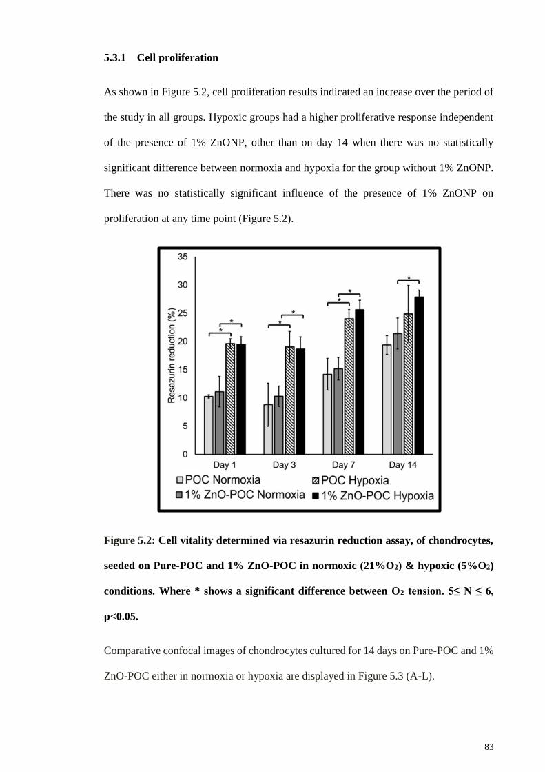
83
5.3.1 Cell proliferation
As shown in Figure 5.2, cell proliferation results indicated an increase over the period of
the study in all groups. Hypoxic groups had a higher proliferative response independent
of the presence of 1% ZnONP, other than on day 14 when there was no statistically
significant difference between normoxia and hypoxia for the group without 1% ZnONP.
There was no statistically significant influence of the presence of 1% ZnONP on
proliferation at any time point (Figure 5.2).
Figure 5.2: Cell vitality determined via resazurin reduction assay, of chondrocytes,
seeded on Pure-POC and 1% ZnO-POC in normoxic (21%O2) & hypoxic (5%O2)
conditions. Where * shows a significant difference between O2 tension. 5≤ N ≤ 6,
p<0.05.
Comparative confocal images of chondrocytes cultured for 14 days on Pure-POC and 1%
ZnO-POC either in normoxia or hypoxia are displayed in Figure 5.3 (A-L).

84
Figure 5.3 (A – L): Live/Dead assay of Chondrocytes seeded on the scaffold at day
1, 7 and 14 under normoxic and hypoxic conditions. Outer images are at 10X
magnification with a scale bar of 300 µm and their respective inserts are at 40X
magnification with a scale bar of 100 µm.
DNA quantification results (Figure 5.4) indicate a positive correlation with time in all
groups, and the amount of DNA had increased from day 1 for each group. The hypoxic

85
groups yielded a statistically significant increase in DNA at all points as compared to the
equivalent normoxic group. These results agree with the CLSM images (Figure 5.3 A-L.)
A live/dead assay based on Ethidium Homodimer and Calcein-AM was used to
differentiate between live cells and dead cells. There are clearly more live cells (more
green stain) in the hypoxic group as compared to the normoxic group. The presence of
1% ZnONP produced no statistically significant effect in normoxia (comparative to
CLSM images (Figure 5.3 (A-F)), but produced a statistically significant increase in DNA
content in hypoxia until day 7 (no statistically significant difference was found at day 14)
(Figure 5.4).
Figure 5.4: Cell quantification through DNA Hoechst fluorescence assay method, of
chondrocytes, seeded on Pure-POC and 1% ZnO-POC in normoxic (21%O2) &
hypoxic (5%O2) conditions. Where + shows significant differences between groups
and * shows a significant difference between O2 tension. 5≤ N ≤ 6, p<0.05.
Similarly, by day 14, both Pure-POC (Figure 5.3-I) and 1% ZnO-POC (Figure 5.3-L)
scaffolds in the hypoxic group show dense and packed live cells. The CLSM images also

86
confirm that both Pure-POC and 1% ZnO-POC scaffolds support chondrocytes as there
are hardly any red staining that shows dead cells. FESEM images in Figure 5.5 (A-L)
display an increase in cell population over time. It further shows that incorporation of
ZnO in polymer maintains the cell phenotype over 14 days (Figure 5.5-F & 5.5-L) and
improves ECM formation.
Figure 5.5 (A – L): SEM images showing the morphologies of Chondrocytes seeded
on the scaffold at day 1, 7 and 14 under normoxic and hypoxic conditions. Outer
images are at 1000X magnification with a scale bar of 20 µm and their respective
inserts are at 3000X magnification with a scale bar of 3 µm.

87
5.3.2 Proteoglycan Synthesis
S-GAG incorporation in the scaffolds was in all cases positively correlated over time,
with hypoxia producing a statistically significant increase over normoxia at every time
point (Figure 5.6-A). Whilst 1% ZnONP did not affect S-GAG incorporation in the
scaffold, it did produce a statistically significant increase in S-GAG release to the medium
by day 14 in the hypoxic group (Figure 5.6 (A & B)). No significant increase was
observed due to hypoxia in released S-GAG until day 3. However, on day 7 and day 14 a
significant difference was observed due to hypoxia in released S-GAG (Figure 5.6-B).
Figure 5.6: (A) Total S-GAG, measured in digested scaffolds and (B) S-GAG
measured in medium via DMMB assay for chondrocytes seeded on Pure-POC and
1% ZnO-POC in normoxic (21%O2) & hypoxic (5%O2) conditions. Where + shows
significant differences between groups and * shows a significant difference between
O2 tension. 5≤ N ≤ 6, p<0.05.

88
5.3.3 Gene expression
The effect of hypoxia and normoxia, with and without ZnO on anabolic (type II collagen
and aggrecan) and catabolic (MMP-13) gene expression, is presented in Figure 5.7 (A-
C). COL2A1 gene expression results indicate an increase over the period of the study in
all groups (Figure 5.7-A). Only at day 3, there is a statistical difference between hypoxia
and normoxia without Zn, otherwise, at every time point, the COL2A1 gene expression
was higher in hypoxia and 1% ZnONP conditions (all groups, p<0.05; Figure 5.7-A). A
highly significant difference was observed for COL2A1 gene expression, however for
ACAN gene expression the results indicate an increase over the period of the study in all
groups (Figure 5.7-B). ACAN gene expression results demonstrated a similar trend as of
COL2A1 gene expression results except on day 14, when there was no difference between
hypoxia and normoxia for Pure-POC. At every time point, scaffold containing 1%
ZnONP had a higher reading of ACAN gene expression irrespective of hypoxic or
normoxic condition (p<0.05). A highly significant difference was observed for COL2A1,
ACAN and MMP-13 at every time point when Pure-POC scaffold in Normoxia was
compared to scaffolds with ZnONP in hypoxia. The presence of 1% ZnONP in POC
greatly suppressed the expression of a catabolic gene such as MMP-13 from day 3 to day
14 (p<0.05, compared to Pure-POC (figure 5.7-C). HIF-1A is a well-known regulator of
the hypoxic response. In all hypoxia cases, the up-regulation of HIF-1A gene expression
(figure 5.7-D) from day 3 to day 14 is shown (p<0.05, compared to the normoxic group).
The up-regulation of HIF-1A in the hypoxic group is not affected by the presence of 1%
ZnONP. No effect of ZnONP incorporation in POC polymer was seen for HIF-1A gene
expression throughout the study.

89
Figure 5.7 (A, B, C & D): mRNA expression of COL2A1, ACAN, MMP – 13 and HIF
– 1A respectively normalized to GAPDH with respect to Pure-POC on day 1. Where
+ shows significant differences between groups and * shows a significant difference
between O2 tension. 3≤ N ≤ 5, p<0.05.
5.4 Discussion
In this study, the effect of native oxygen tension in cartilage (hypoxia, 5% O2) was
compared with normoxia (21% O2) on 3D Poly 1, 8 Octane diol Citrate scaffolds fused
with 1% ZnONP. Scaffolds were seeded with bovine chondrocytes and their cell behavior
and protein synthesis was reported. Further, gene expression analysis of ECM specific
genes (COL2A1 and ACAN) and catabolic gene expression MMP-13 was performed. If
the combination of 1% ZnONP and hypoxia promotes cartilage synthesis through
maintaining chondrocyte phenotype and morphology, then the following observations
would be expected, compared to control, to just 1% ZnONP, or to hypoxia:

90
a) Increased cell viability and proliferation
b) Increased synthesis of cartilage protein such as sulfated GAG (S-GAG)
c) Up-regulation of characteristic cartilage matrix genes (such as COL2A1 and ACAN)
d) Downregulation of catabolic genes (MMP-13)
e) The role of HIF-1A in hypoxic environment
Discussion of each aforementioned observations is presented in turn, with reference to
the results presented in section 5.3 of current thesis.
5.4.1 Cell proliferation
Several investigators have shown that hypoxia can influence cell proliferation in-vitro
(Hubka et al., 2014; Schipani et al., 2001; Thoms et al., 2013) and results in Figure 5.2,
agreeably demonstrated cell proliferation increasing considerably in a hypoxic
environment. Previously, it was reported that hypoxia contributed to a better cell viability
for chondrocytes cultured in the hypoxic environment (Tsuchida et al., 2014). An oxygen
tension of 5% was selected because it is well documented that 5% O2 tension mimics
physiological O2 tension (Domm et al., 2002). Whereas lower O2 tensions may lead to
reduced cell viability and chondrocyte health is compromised (Collins et al., 2013;
Grimshaw & Mason, 2000). To further quantify cell proliferation, a nucleic acid binding
dye, Hoechst DNA Assay was used, a more sensitive test for cell proliferation.
Statistically the results demonstrated that there was a synergistic effect of hypoxia and
ZnONP on cell proliferation for every time point, except day 14. Although at day 14 both
hypoxic groups (with or without 1% ZnONP) showed the highest reading of DNA, they
did not show a differential effect of ZnONP on cell proliferation (Figure 5.4). However,
under a closer examination of the FESEM micrographs (Figure 5.5-K & 5.5-L), it is clear
that all the surface area of scaffolds on the day 14 was covered by cells, suggesting the
presence of contact inhibition (Abercrombie, 1979; Takai et al., 2008).

91
5.4.2 Proteoglycan Synthesis
Besides promoting cell proliferation and DNA production, cells cultured in a hypoxic
environment showed a higher production of sulfated GAG production in both scaffolds
(Figure 5.6-A). Present findings are consistent with previous studies by various
researchers who studied the effect of oxygen levels on bovine chondrocytes matrix
synthesis (Domm et al., 2002; Hansen et al., 2001). Furthermore, current findings are
supported by a separate study by Malda J. et al (Malda et al., 2004), where human nasal
chondrocytes were subjected to 21% O2, 5% O2, and 1% O2. Their results demonstrated
a higher S – GAG production after just 7 days under 5% O2 concentration. It is also
notable that GAG released into the medium was higher than the GAG within the scaffold
(Figure 5.6-B). This finding can be explained by the fact that cells were covering all the
polymer and the cells were in direct contact with the culture medium as the material was
porous. Also, the size of the scaffold (6x6 mm) was substantially smaller than the volume
of culture medium in each well (2ml). similar findings were reported by Bassleer C. et al
(Bassleer et al., 1998).
5.4.3 Gene expression
In previous studies, POC (Kang et al., 2006) and oxygen tension (Saini & Wick, 2004)
have been employed individually in order to support chondrocyte proliferation,
attachment, and ECM production. However, this is the first time that hypoxia in
combination with 1% ZnONP producing a stimulatory effect on COL2A1 and ACAN
synthesis has been reported (Figure 5.7-(A & B)). Rosenberg K. et al has previously
shown that Zinc ions (Zn+2) have a preference for binding between cartilage oligomeric
matrix proteins (COMP) and collagen type I and II. Moreover, they also are responsible
for promoting binding of collagen to COMP (Figure 5.8) (Rosenberg et al., 1998).

92
N.B: The idea is modified from (Rosenberg et al., 1998).
Figure 5.8: Schematic of divalent ion involvement in binding COMP with collagens
with a selective preference of Zn+2 binding phenomenon within synovial fluid.
Zn+2 ions have been demonstrated as playing a functional and biological role in
organizing cartilage matrix by modulating link proteins in bovine articular cartilage
(Rosenberg et al., 1991). A closer examination of the SEM micrographs demonstrates the
difference between all the groups in terms of cell morphologies and matrix synthesis. The
greatest contrasts were shown on scaffolds incorporating ZnONP and cultured in a
hypoxic environment. Similarly, the SEM image in Figure 5.9 shows a denser fibril
formation in 1% ZnO-POC scaffold under hypoxia at day 14. The cells in these groups
have round and spherical morphologies and produced a dense matrix, which is agreeable
to phenotypic stability (Benya & Shaffer, 1982). A possible mechanism is the release of
Zn+2 ions from the polymer. It has also been reported that Zn+2 ions take an active
functional and biological role in organizing cartilage matrix by modulating link proteins
in bovine articular cartilage (Rosenberg et al., 1991).

93
Figure 5.9: FESEM image at 5000X magnification of 1% ZnO-POC in hypoxia at
day 14. Set of red arrows points to visible fibrils in ECM.
A highly significant difference was observed for COL2A1, ACAN and MMP-13 at every
time point when Pure-POC scaffold in normoxia was compared to scaffolds with ZnONP
in hypoxia. The presence of 1% ZnONP in POC greatly suppressed the expression of
catabolic gene MMP-13 from day 3 to day 14 (p<0.05, compared to Pure-POC (Figure
5.7-C), but conversely yielded no statistically significant effect under normoxia from day
3 onwards. HIF-1A is a well-known regulator of the hypoxic response. Present findings
elaborated the role of hypoxia in the regulation of cartilage destruction gene, more
commonly known as MMP-13 (Fosang et al., 1996; Wang et al., 2013). The up-regulation
of MMP-13 was suppressed by the 1% ZnONP present in the scaffolds but hypoxia alone
did not prevent the up-regulation of MMP-13. These results suggest ZnONP has a
chondroprotective role. Previously, it was reported that serum Zn levels are found to be
substantially decreased in rheumatoid arthritis (RA) (Mierzecki et al., 2011; Zoli et al.,
94
1998) and that the reduced level of serum Zn in RA is associated with immune
inflammatory processes (Honkanen et al., 1989; Milanino et al., 1993).
In all hypoxia cases, up-regulation of HIF-1A gene expression (Figure 5.7-D) from day 3
to day 14 is shown (p<0.05, compared to the normoxic group). The up-regulation of HIF-
1A in the hypoxic group is not affected by the presence of 1% ZnONP. It was reported by
Schipani et al that HIF-1A is a major regulator of the hypoxic response in chondrocytes
(Schipani et al., 2001). Current results show that in hypoxic condition there was a higher
level of cartilage matrix and gene up-regulation. While other studies have also implicated
the role of HIF-1A in the regulation of cartilage-specific genes (Dahlin et al., 2013;
Robins et al., 2005; Thoms et al., 2013).

95
CHAPTER 6: EFFECT OF DYNAMIC MECHANICAL COMPRESSION ON
BOVINE ARTICULAR CHONDROCYTES
6.1 Introduction
Trauma or disease of articular cartilage leads to articular cartilage lesions, which have a
very limited ability to repair themselves, and gradually progress to osteoarthritic
conditions. The incidence of the cartilage-related disease is increasing throughout the
world (Humphreys et al., 2013; Lawrence et al., 2008; Theis et al., 2007). Various
treatments are used for individuals with cartilage-based problems (Filardo et al., 2013;
Marcacci et al., 2013), however, the drawbacks associated with these treatments are
many, including inconsistency in treatment outcomes (Farr et al., 2011) and diminutive
mechanical properties of replaced tissue resulting due to fibrocartilage formation (Gomoll
& Minas, 2014). Therefore, new options are required to deal with the current situation. A
key area for such new solutions is the application of Tissue Engineering to cartilage.
Cartilage comprises only one type of cell, the chondrocyte (Palukuru et al., 2014), and
are currently used for autologous chondrocyte implantation (Chilelli et al.) as well as
being the mainstay of many other therapies. However, the use of chondrocytes entails
numerous constraints (Harris et al., 2011) that hamper their use. The foremost is difficulty
in obtaining chondrocyte-bearing tissues for isolation of cells and in vitro expansion
(Minas & Nehrer, 1997). Such a method can result in donor site morbidity (Harris et al.,
2011; Hjelle et al., 2002; Pelttari et al., 2008), and a consequent increased long-term risk
of the development of osteoarthritis (Hunziker, 2002; Messner & Gillquist, 1996).
Further, due to the scarcity of chondrocytes even in healthy articular cartilage, there is
typically a lack of sufficient numbers of chondrocytes available (Dahlin et al., 2013;
Palukuru et al., 2014). Even where sufficient cells can be obtained for expansion, typical
in vitro methods can easily produce dedifferentiation of chondrocytes (Schulze-Tanzil et
al., 2004).

96
Given this resultant need to maximise expansion of what chondrocytes are available, and
to maintain a useful phenotype, a number of factors have been identified which promote
more effective chondrocyte culture.
First, the use of three-dimensional culture models has been introduced, in view of the
identification of a two-dimensional culture with the tendency of chondrocytes to
dedifferentiate toward a fibroblastic morphology (Yoon et al., 2002). Second, research
into replicating a physiological mechanical environment within culture systems has
shown the beneficial effect of dynamic mechanical loading on metabolic activity and the
promotion of cartilage formation (Jeon et al., 2012; Li et al., 2013). Chondrocytes are
highly mechanosensitive (Madej et al., 2014), and different studies have supported the
role of mechanical stimulation on chondrocytes to enhance extracellular matrix (ECM)
production and deposition (Ikenoue et al., 2003; Millward-Sadler et al., 2000). More
recently, this research direction has included the addition of hypoxic conditions,
mimicking the low oxygen tension found within diarthroidal joints (Li et al., 2013; Madej
et al., 2014). In the native environment, chondrocytes are continuously under a gradient
of oxygen tension that ranges from 10% O2 in the superficial layers to around 1% O2 in
the deepest layers (Grimshaw & Mason, 2000). Chondrocytes adapt well to decreased O2
concentrations and hypoxia has been proved to play a major role in chondrocyte
proliferation and their function in vivo (Foldager et al., 2011; Schipani et al., 2001).
More recently, research has shown that combining three-dimensional culture with
hypoxic conditions was enhanced through the incorporation of Zinc Oxide nanoparticles
(ZnONP), with marked chondroprotective benefits, including the promotion of cartilage
synthesis, upregulation of matrix gene expression, downregulation of catabolic genes, and
the preservation of cell morphology (Mirza, Pan-Pan, et al., 2015). Zinc (Zn) is an
essential element for humans and animals, particularly in cell proliferation,
differentiation, and survival (Beyersmann & Haase, 2001; Haase & Rink, 2014b). In

97
addition, it is known to possess antibacterial properties (Mirza, Wan Ibrahim, et al., 2015).
In light of this, it is important that the further interaction of ZnONP with mechanical
loading be evaluated to establish whether such findings can be extended to such models
for a further advancement in chondrocyte culture.
6.2 Materials and Methods
Pure POC and 1% ZnO-POC scaffolds were placed in sterile bijou tubes and immersed
in 70% alcohol for 30 minutes. Alcohol was then aspirated and scaffolds were left to air
dry under a laminar flow, followed by autoclaving at 121oC for 15 minutes at 15 psi.
Once removed from the autoclave, the scaffolds were immersed in chondrocyte medium
to neutralise (neutralisation helps better cell attachment) overnight prior to cell seeding.
Isolated bovine articular chondrocytes (BAC) (refer to section 3.4 for details of BAC
isolation) were seeded on the scaffolds, with a concentration of 1X10⁶ cells per scaffold
(higher cell seeding density prevent dislodging of cells under dynamic mechanical
compression). The cells were allowed to attach for 4 hours and then the wells were
replenished with chondrocyte culture medium (refer to section 3.4.1 for formulation).
6.2.1 Experimental Design
Pure-POC and 1% ZnO-POC Scaffolds were kept in under 2 different oxygen
concentrations; one in hypoxia (5% O2) and second in normoxia (21% O2). Furthermore,
scaffolds under each oxygen concentration were subjected to either; (a) static culture or
(b) dynamic mechanical compression, for 72 hours. The experimental design is
summarised in figure 6.1.

98
Figure 6.1: Illustration summarizing all experimental groups with respect to culture
conditions and oxygen tensions.
6.2.2 Application of dynamic compression
An incubator-housed ex-vivo bioreactor (Bose, ElectroForce® 5500 USA) was used to
apply dynamic compression to cell-seeded scaffolds at 5% or 21% oxygen tension (Figure
6.2-A). The scaffolds were transferred into individual wells of a 24-well plate fixture
chamber (Figure 6.2-B) for compressive loading.
Figure 6.2: Bose ElectroForce® 5500 USA. (A) Bose Bioreactor setup with 24-well
plate fixture in a 5% CO2 incubator. (B) 24-well plate with compression platens in a
box chamber.

99
The scaffolds were left in the bioreactor for a 24-hour free swelling period, followed
by intermittent compression under unconfined conditions, with a profile of 10 minutes
(min) compression followed by a 5-hour 50-min unstrained period for 48 hours (Figure
6.3).
Figure 6.3: Schematic illustration of the intermittent dynamic compression profile
of cell culture regimes.
The bioreactor was programmed for 10% compression at a frequency of 1Hz via WinTest
7 software. Control scaffolds were unstrained and were left inside the ex-vivo system.
6.2.3 Cell Proliferation Assay
The Hoechst binding dye method was used to quantify DNA within the scaffolds at each
time point (refer to section 3.6 for detailed protocol).

100
6.2.4 Sulphated glycosaminoglycan (S-GAG) quantification assays
Glycosaminoglycans (GAGs) are polysaccharides that are present in abundance in a
healthy cartilage. Sulphated-GAGs (S-GAGs) are the types that can be measured via a 1,
9-dimethylmethylene blue assay. Total S – GAG was measured from Cell-scaffold lysate
at each time point. Released S – GAG was measured by collecting the culture medium at
every medium change from the wells (refer to section 3.7 for detailed protocol).
6.2.5 Quantitative Polymerase Chain Reaction (q-PCR)
To observe the behaviour of BACs at gene level Cartilage matrix specific genes; Collagen
type II (COL2A1) and Aggrecan (ACAN) were observed. Moreover, the matrix
degradation gene (MMP-13) and hypoxia inducible factor gene (HIF1A) were also
observed. Q-PCR gene expressions were determined by the TaqMan gene expression
assay using StepOnePlusTM Real-Time PCR system (refer to section 3.8 for detailed
protocol).
6.3 Results
This section elaborates proliferation behavior, ECM formation and regulation of ECM
specific genes for chondrocytes seeded on Pure-POC and 1% POC-ZnO scaffolds over
the period of 48 Hours under normoxia (21% O2) and hypoxia (5% O2) when subjected
to dynamic compression.
6.3.1 Cell proliferation and morphology
Cell quantification results in Figure 6.4 show the effect of dynamic compression on DNA
production in presence and absence of 1% ZnONP in normoxic and hypoxic conditions
for 48 hours. DNA production was greatly enhanced under the influence of mechanical
stimulation, particularly for 1% ZnO-POC (hypoxia), Pure-POC (hypoxia), and Pure-

101
POC (normoxia). However, this was not true in the case of 1% ZnO-POC (normoxia).
Both strained and unstrained scaffolds showed that hypoxia-enhanced DNA production
and with the combination of dynamic compression demonstrated its beneficial effects on
cellular proliferation.
Figure 6.4: Cell quantification through DNA Hoechst fluorescence assay for
chondrocytes seeded on pure-POC and 1% ZnO-POC in normoxic (21%O2) &
hypoxic (5%O2) conditions, under static and dynamic culture regimes. Where ^
shows a significant difference between due to the presence of ZnONP, * shows a
significant difference between O2 tension and + shows a significant difference
between static and dynamic culture. p<0.05.
Further qualitative assessment of cell proliferation was carried out using live (green)/dead
(red) stains. The number of live and dead cells can be viewed via confocal microscopic
images. Figure 6.5 (A-L) indicated that in general, dynamic culture produces more live
cells compared to static culture. Closer examination of the insert (Figure 6.5 (B, D, F &
H)) of confocal images for cells which were previously cultured in a hypoxic environment
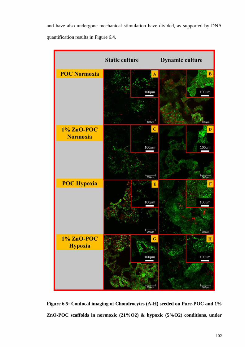
102
and have also undergone mechanical stimulation have divided, as supported by DNA
quantification results in Figure 6.4.
Figure 6.5: Confocal imaging of Chondrocytes (A-H) seeded on Pure-POC and 1%
ZnO-POC scaffolds in normoxic (21%O2) & hypoxic (5%O2) conditions, under

103
static and dynamic culture regimes. Outer images are at 10X with a scale bar of 300
μm and their respective inserts are at 40X magnification with a scale bar of 100 μm.
FESEM images (Figure 6.6 (A-H)) also showed that there were more cells in scaffolds
which were previously cultured in dynamic environments. Notably, the cells which had
undergone dynamic compression tended to keep a rounded morphology, which is typical
morphology of chondrocytes. In static culture, cells in scaffolds which contained ZnONP
were more rounded and bulged as compared to cells cultured in pure POC.
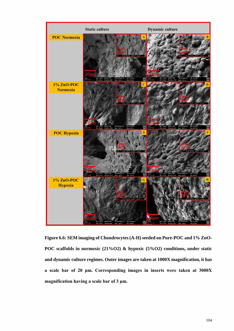
104
Figure 6.6: SEM imaging of Chondrocytes (A-H) seeded on Pure-POC and 1% ZnO-
POC scaffolds in normoxic (21%O2) & hypoxic (5%O2) conditions, under static
and dynamic culture regimes. Outer images are taken at 1000X magnification, it has
a scale bar of 20 μm. Corresponding images in inserts were taken at 3000X
magnification having a scale bar of 3 μm.

105
6.3.2 Proteoglycan synthesis
S-GAG production in the scaffolds was not affected by the presence of dynamic
compression, however, the effect of O2 tension and the incorporation on ZnONP
influenced the higher synthesis of S-GAG (Figure 6.7-A). In dynamic culture, there was
a higher production of S-GAG in all hypoxic groups. In dynamic culture, when pure POC
was compared with 1%ZnO-POC, both in hypoxic condition, the presence of 1%ZnO
further enhanced the production of S-GAG. A similar trend is observed in the normoxic
groups in a dynamic culture. Static cultures also exhibited similar trend to dynamic
culture but did not show a statistically significant effect of ZnONP in the hypoxic group.
Dynamic compression showed more distinctive effects of S-GAG released in the medium
than in all other groups, except in pure POC culture in normoxic conditions (Figure 6.7-
B). Again, hypoxia and ZnONP together or separately have shown a higher production of
S-GAG released in the medium in a dynamic culture. The trend of S-GAG release in the
medium is identical to S-GAG release from scaffolds in static culture.
Figure 6.7: DMMB assay for glycosaminoglycans (A) Total S-GAG, measured in
digested scaffolds and (B) S-GAG measured in medium via DMMB assay for
chondrocytes seeded on pure-POC and 1% ZnO-POC in normoxic (21%O2) &
hypoxic (5%O2) conditions, under static and dynamic culture regimes. Where ^
shows a significant difference between due to the presence of ZnONP, * shows a

106
significant difference between O2 tension and + shows a significant difference
between static and dynamic culture. p<0.05.
6.3.3 Gene Expression
The effect of oxygen tension and ZnONP on anabolic (type II collagen and aggrecan) and
catabolic (MMP-13) gene expression is presented in Figure 6.8-(A, B & C). In static
culture, the effect of hypoxia in increased COL2A1 (p<0.05) can be seen in the 1% ZnO-
POC group only, whereas, in a dynamic culture, the effect of hypoxia and ZnONP can be
seen in all groups in the up-regulation of COL2A1 (p<0.05). Compared to static culture,
dynamic culture, in general, has resulted in an up-regulation of COL2A1 in all groups
(p<0.5), except for the pure POC group under normoxia.
The up-regulation of aggrecan expression is greatly affected by O2 tension and ZnONP
in static culture (all groups, p<0.05), and dynamic culture can also further improve the
up-regulation of aggrecan (all groups, p<0.05); Figure 6.8-B.
MMP-13 is a well-known cartilage-destructive gene. Hypoxia had an inhibitory effect on
MMP-13 activity both in static and dynamic culture (Figure 6.8-C). Dynamic
compression helped in further reducing MMP-13 activity in normoxia for both Pure-POC
and 1%ZnO-POC samples, excluding the result from the 1%ZnO-POC group under
dynamic culture, in which the level was low both with and without hypoxia. The results
demonstrated that hypoxia and dynamic compression both act to reduce the pro-catabolic
response. ZnONP also helped to reduce MMP-13 up-regulation for dynamic culture in
normoxic conditions.
The HIF-1A gene, which is thought to be one of the major regulators of the hypoxic
response, was only present in scaffolds cultured in hypoxic conditions (Figure 6.8-D). It
was not affected by the incorporation of ZnONP or mechanical stimulation.

107
Figure 6.8: (A) COL2A1, (B) ACAN, (C) MMP–13 and (D) HIF–1A, for chondrocytes
seeded on pure-POC and 1% ZnO-POC in normoxic (21%O2) & hypoxic (5%O2)
conditions, under static and dynamic culture regimes. Where ^ shows a significant
difference between due to the presence of ZnONP, * shows a significant difference
between O2 tension and + shows a significant difference between static and dynamic
culture. p<0.05.
6.4 Discussion
This study set out to evaluate the feasibility of adding ZnONP to a 3D culture system
incorporating mechanical stimulation and hypoxia for chondrocytes. The mechanical
stimulation is part of biomimicry, as it is found in the native environment of chondrocytes
(Jeon et al., 2012), and has been shown to produce a significant modulation in a matrix
formation, cell proliferation, and S-GAG formation by chondrocytes (Grodzinsky et al.,

108
2000). The use of hypoxia, a further physiological feature of native cartilage (Grimshaw
& Mason, 2000), and likewise plays a major role towards in-vivo chondrocyte metabolism
(Foldager et al., 2011; Schipani et al., 2001).
Whilst the combination of ZnONP and hypoxia has been previously reported (Mirza, Pan-
Pan, et al., 2015), it has not been formerly tested in conjunction with mechanical
stimulation. Therefore, in the present study, the focus was upon the addition of 1%
ZnONP (0% ZnONP = control) to a system comprising native oxygen tension in cartilage
(hypoxia, 5% O2) (normoxia at 21% O2 = control), with (static culture = control) dynamic
compression.
6.4.1 Cell proliferation and morphology
The DNA content demonstrated a highly significant increase due to the hypoxic
condition. Furthermore, scaffolds that were dynamically compressed displayed a higher
DNA content (Figure 6.4). Upon closer examination of (Figure 6.5-(B, D, F & H)) many
‘twinned’ chondrocytes can be seen in scaffolds which were previously cultured in
dynamic conditions, this indicating that there is an increase in cell proliferation, providing
qualitative data in support of the DNA results. Similar results were reported by (Dahlin
et al., 2013) where it was concluded that a combination of hypoxia and flow perfusion
cultures displayed enhanced DNA content per scaffold. Irrespective of oxygen
concentration and the presence of ZnONP, dynamically compressed scaffolds display a
more rounded morphology (Figure 6.6-(B, D, F & H)), which is agreeable to phenotypic
stability (Villar-Suárez et al., 2004). In separate studies, it was reported that low oxygen
tension and flow perfusion is required to maintain cell phenotype and enhance ECM
production (Davisson, T. et al., 2002; Murphy & Polak, 2004).

109
6.4.2 Proteoglycan synthesis
Higher S-GAG formation was observed for scaffolds which incorporate ZnONP when
compared to pure-POC in normoxia and hypoxia in a dynamic culture. However, the total
S-GAG remained significantly higher among scaffolds that were present in the hypoxic
environment (Figure 6.7-A).
Where the S-GAG level in the medium was measured, a similar trend of S-GAG levels
in the digested samples was observed. However, the samples which underwent dynamic
compression recorded significantly higher levels than those in static conditions,
irrespective of oxygen tension or ZnONP incorporation (Figure 6.7-B).
It is postulated that the mechanical loading itself may have triggered leakage of S-GAG
from the highly porous POC scaffold. Moreover, the presented results are coherent with
another study by Wernike E. et.al, which reported that dynamic compression enhances
GAG synthesis (Wernike et al., 2008).
6.4.3 Gene Expression
The gene expression profiles of COL2A1 and ACAN demonstrated enhanced expression
due to the incorporation of ZnONP, but this enhanced expression became significant due
to dynamic compression (Figure 6.8-(A & B)). Gene expression results suggest that
incorporation of ZnONP had a stimulatory effect on COL2A1 and ACAN synthesis, which
are the major contributors to extracellular matrix (ECM) (Chen et al., 2014). The increase
in gene expression profiles of COL2A1 and ACAN due to dynamic forces was also
reported in a separate study which suggested loading duration and strain percentage affect
gene expression (Jeon et al., 2012).Overall, the greatest influence on the MMP-13 level
was the change in oxygen tension, however, the presence of ZnONP did yield a
statistically significant difference in one case under dynamic culture, suggesting a
contributory role that may be further clarified in further research (Figure 6.8-C).

110
Regarding the levels of HIF-1A, a key regulator of hypoxic response, whilst as expected;
the HIF-1A levels increased dramatically under hypoxia. Neither the addition of
mechanical loading nor ZnONP disrupted the expression of this gene, suggesting that
normal hypoxic response is retained in the culture model (Figure 6.8-D).
The mechanism of the Hypoxia Inducible Factor (HIF) protein is illustrated in Figure 6.9.
It is understood that the HIF protein is continuously being produced. However,
researchers suggest that in the presence of oxygen, this protein is degraded as its
hydroxylation takes place by HIF-specific polyl hydroxylases which target HIF-alpha
subunits (Thoms et al., 2013). In hypoxia, the oxygen concentration is roughly 5%. As
hydroxylation requires oxygen to proceed, a lack of oxygen causes the mechanism to
diminish substantially, which eventually slows down the degradation process in tissue
(Thoms et al., 2013). Previously it had been reported that the cartilage destruction that
was induced by interleukin 1α (IL – 1α) – an inflammatory cytokine (Kobayashi et al.,
2005; Pujol & Loyau, 1987), was inhibited by hypoxia (Thoms et al., 2013).
Figure 6.9: Schematic of the possible pathway by which hypoxia inducible factor
controls cartilage destruction with the help of its 2 major genes that are up-regulated
in hypoxic conditions, based on the work by Thoms et al. The schematic is modified
from (Thoms et al., 2013).

111
CHAPTER 7: CONCLUSIONS AND FUTURE DIRECTIONS
7.1 Conclusions
Articular cartilage tissue is made up of a complex network of ECM but only a single cell
type, the chondrocyte. Cartilage tissue has a very low capability of self-healing. Cartilage
in articulating joints is a vulnerable tissue that is subjected to injuries and degenerative
conditions. More than 2 decades have passed since interdisciplinary teams of biologists,
engineers, clinicians and materials scientists first studied cartilage behaviour in-vitro and
in-vivo. However, till now, there is no single treatment method that outweighs the rest as
assured technique to successfully address the cartilage repair dilemma.
The goal of cartilage tissue engineering is to restore smooth movement and load bearing
activities as in native articular cartilage. Numerous clinicians, researchers, and scientists
have put forward their potential solutions but none have succeeded to be implemented as
a single favoured choice all round the world because the limitations of the proposed
techniques outweigh their long term benefits.
As discussed in chapter 2, there are several issues that need to be addressed for a
successful cartilage tissue engineering strategy. The most important criteria to consider
for cartilage tissue engineering are the combination of; (a) cells, (b) scaffold and (c) bio-
factors. Together, these are assembled into a tissue construct, which is then implanted to
the defect site surgically. This surgical implantation bears the additional risk of infection.
The fundamental aim of this thesis was to enhance the understanding of chondrocyte
behaviour under a native physical and chemical environment; and to establish concrete
evidence for functional antibacterial scaffolds. As highlighted in chapter 1; section 1.4,
there were 4 basic objectives of this thesis.
The first objective was to develop a biodegradable scaffold with controlled pore size and
porosity mimicking the properties of native articular cartilage. Next objective was to

112
select and optimise the concentration of nano particles that must be biocompatible and
having antibacterial effect while showing no toxicity towards chondrocytes.
A new type of nano-composite scaffold has been produced from citric acid-based
polyester and zinc oxide nanoparticles. The scaffolds have been fabricated in two steps:
(1) polyesterification synthesis of pre-polymer and mixing with nanoparticles; and (2)
production of porous matrix by solvent-casting/particulate- leaching technique. The
scaffolds demonstrate elastomeric nature and optimal porosity to be used in cartilage
tissue engineering. The current method provides an effective route for production of
scaffolds with controlled release of bio-active nanoparticles. The ZnONP have
demonstrated strong anti-bacterial properties as evident from the results presented in
chapter 4. The antibacterial activity of the reported scaffolds may enhance the clinical
outcomes by: (i) reduction of post-operative complications by eliminating the risk of
infection from donor; and (ii) reduction of the risk of infection caused by biofilm
formation. A multifunctional scaffolds also show a strong potential for cartilage tissue
engineering. The concentration of 1 wt% of ZnONP was found to have better performance
with chondrocyte proliferation than pure POC, used as control. The 1wt% ZnO-POC
nano-composite scaffold has proven to be optimal for seeding with chondrocytes while
simultaneously depleting both the gram positive and gram negative bacteria.
The third objective of current thesis was to study the independent and combined effect of
Hypoxia and ZnONP on bovine articular chondrocytes (BAC) seeded on POC polymer
scaffolds and cultured under 2 different oxygen concentrations; (a) normoxia (21% O2)
and (b) hypoxia (5% O2). 5% O2 concentration was selected, as a review of the literature
in chapter 2. Section 2.6.2.2 reveals that at a 5% O2 concentration chondrocytes thrive
while lower O2 concentrations are not feasible and may only be present in abnormal or
pathological conditions.

113
This study highlights the potential for combination of ZnONP and hypoxia, which has
demonstrated the ability to promote cartilage synthesis through maintaining chondrocyte
morphology when used with POC scaffolds. Incorporation of small proportion of ZnONP
have significantly enhanced the chondroprotective behavior of scaffolds. This has been
demonstrated particularly in terms of promoting cell proliferation, preserving cell
morphology, up-regulating cartilage matrix genes and down-regulating cartilage
destructive gene. Further, the presence of ZnONP did not modulate the expression of HIF-
1A, indicating a preservation of cartilage homeostasis. This provides an important new
route for in-vitro cartilage tissue engineering.
Finally, the thesis aims at establishing a relationship between dynamic mechanical
stimuli, nanoparticles, and oxygen tension, in chondrocyte seeded on POC polymer
scaffolds. In order to understand the underlying mechanism of combining hypoxia,
mechanical stimulus and ZnONP, the BAC were seeded on Pure-POC and 1% ZnONP
scaffolds and subjected to (a) normoxia (21% O2) and (b) hypoxia (5% O2), while
simultaneously undergoing a 10% dynamic mechanical compression for 72 hours.
Specifically, catabolic gene expression of MMP-13 was observed to be curtailed in
normoxic, and the cartilage matrix specific (COL2A1 and ACAN) genes were increased.
Differential comparison of groups and controls within the study confirms the benefit of
moving from a model without dynamic loading to one including such, yielding a marked
increase in cell viability, roundedness of cell morphology, and gene expression even with
the presence of ZnONP. ZnONP neither limited nor decreased the previously reported
advantages of the use of hypoxia or of mechanical loading in the present model.
In conclusion, all the objectives mentioned in chapter 1, section 1.4 were effectively and
efficiently achieved. Furthermore, the hypothesis in chapter 1, section 1.5 stands true, as
this thesis successfully demonstrates that chondrocytes proliferated, secreted more
extracellular matrix, and showed higher gene expression under a hypoxic environment,
114
while also showing a strong antibacterial inhibition. The SEM images in figure 5.5 and
figure 6.6 reveals that scaffolds incorporated with 1% ZnONP displayed a packed
network of interconnecting cells while simultaneously having more rounded morphology.
An absence of ZnONP from scaffolds revealed that cells tend to be spindle shaped or
elongated like fibroblasts. The combination of hypoxia and the presence of ZnONP in
scaffolds was fruitful as it retained cell morphology, which is one of the long awaited
predicament to be addressed. Moreover, the inclusion of dynamic mechanical
compression enhanced the rounded morphology and elevated cartilage specific matrix
genes, while lowering catabolic gene expression.

115
7.2 Future Direction
In order to support the current data, future work must target in-vivo studies, as the current
thesis is based upon in-vitro models only. Moreover, the current thesis provides the data
for one kind of polymer i.e. Poly 1, 8 – Octanediol Citrate. It is suggested that future
research must be directed towards establishing non-cytotoxic percentages of ZnONP in
other polymers because each polymer has its own degradation and cross-linking
properties. Although the current thesis provides a comprehensive account for hypoxic
studies, however, only short-term results are provided for dynamic mechanical
compression. The reason for this is the limitation of the current bioreactor, which does
not provide a sterile environment for long term studies. Hence it is suggested that a new
bioreactor should be designed that allows long-term application of dynamic mechanical
culture, while maintaining the integrity of culture system.
To obtain qualitative data regarding the performance of scaffolds under hypoxia seeded
with BAC, it is suggested that staining and western blotting should also be undertaken to
support the existing data.
Lastly, it is suggested that an animal in-vivo model be created for long-term investigation
of the chondroprotective response to ZnONP, and its further beneficial effects in wound
healing after surgical implantation.
116
References
Ab-Rahim, S., Masjudin, T., Selvaratnam, L., & Kamarul, T. Evaluation of Human
Autologous Chondrocyte Transplantation Effectiveness in Relation to Rabbit
Assessment as an Animal Model.
Abercrombie, M. (1979). Contact inhibition and malignancy. Nature, 281(5729): 259-
262.
Afratis, N., Gialeli, C., Nikitovic, D., Tsegenidis, T., Karousou, E., Theocharis, A. D.,
Pavão, M. S., Tzanakakis, G. N., & Karamanos, N. K. (2012).
Glycosaminoglycans: key players in cancer cell biology and treatment. Febs
Journal, 279(7): 1177-1197.
Aigner, T., & Fan, Z. (2003). Anatomy and Biochemistry of Articular Cartilage. In C.
Hendrich, U. Nöth & J. Eulert (Eds.), Cartilage Surgery and Future Perspectives
(pp. 3-7): Springer Berlin Heidelberg.
Akhtar, M. J., Ahamed, M., Kumar, S., Khan, M. A. M., Ahmad, J., & Alrokayan, S. A.
(2012). Zinc oxide nanoparticles selectively induce apoptosis in human cancer
cells through reactive oxygen species. Int J Nanomedicine, 7: 845-857.
Aksoy, B., Atakan, N., Aksoy, H. M., Tezel, G. G., Renda, N., Özkara, H. A., & Önder,
E. (2010). Effectiveness of topical zinc oxide application on hypertrophic scar
development in rabbits. Burns, 36(7): 1027-1035.
Albro, M. B., Nims, R. J., Cigan, A. D., Yeroushalmi, K. J., Shim, J. J., Hung, C. T., &
Ateshian, G. A. (2013). Dynamic mechanical compression of devitalized articular
cartilage does not activate latent TGF-β. Journal of Biomechanics, 46(8): 1433-
1439.
Anower-E-Khuda, M. F., & Kimata, K. (2015). Human Blood Glycosaminoglycans:
Isolation and Analysis Glycosaminoglycans (pp. 95-103): Springer.
Appelman, T. P., Mizrahi, J., Elisseeff, J. H., & Seliktar, D. (2011). The influence of
biological motifs and dynamic mechanical stimulation in hydrogel scaffold
systems on the phenotype of chondrocytes. Biomaterials, 32(6): 1508-1516.
Arima, Y., & Iwata, H. (2007). Effect of wettability and surface functional groups on
protein adsorption and cell adhesion using well-defined mixed self-assembled
monolayers. Biomaterials, 28(20): 3074-3082.
Ataollahi, F., Pramanik, S., Moradi, A., Dalilottojari, A., Pingguan-Murphy, B., Wan
Abas, W. A. B., & Abu Osman, N. A. (2014). Endothelial cell responses in terms
of adhesion, proliferation, and morphology to stiffness of polydimethylsiloxane
elastomer substrates. Journal of Biomedical Materials Research Part A: n/a-n/a.

117
Athauda, T., Williams, W., Roberts, K., & Ozer, R. (2013). On the surface roughness and
hydrophobicity of dual-size double-layer silica nanoparticles. Journal of
Materials Science, 48(18): 6115-6120.
Augustine, R., Dominic, E. A., Reju, I., Kaimal, B., Kalarikkal, N., & Thomas, S. (2014).
Investigation of angiogenesis and its mechanism using zinc oxide nanoparticle-
loaded electrospun tissue engineering scaffolds. RSC Advances, 4(93): 51528-
51536.
Augustine, R., Malik, H., Singhal, D., Mukherjee, A., Malakar, D., Kalarikkal, N., &
Thomas, S. (2014). Electrospun polycaprolactone/ZnO nanocomposite
membranes as biomaterials with antibacterial and cell adhesion properties.
Journal of Polymer Research, 21(3): 1-17.
Azizi, S., Ahmad, M. B., Hussein, M. Z., Ibrahim, N. A., & Namvar, F. (2014).
Preparation and properties of poly (vinyl alcohol)/chitosan blend
bionanocomposites reinforced with cellulose nanocrystals/ZnO-Ag
multifunctional nanosized filler. Int J Nanomedicine, 9: 1909.
Bakhshi, H., Yeganeh, H., & Mehdipour-Ataei, S. (2012). Synthesis and evaluation of
antibacterial polyurethane coatings made from soybean oil functionalized with
dimethylphenylammonium iodide and hydroxyl groups. Journal of Biomedical
Materials Research Part A: n/a-n/a.
Balasundaram, G., Storey, D. M., & Webster, T. J. (2014). Novel nano-rough polymers
for cartilage tissue engineering. Int J Nanomedicine, 9: 1845-1853.
Bartlett, W., Skinner, J., Gooding, C., Carrington, R., Flanagan, A., Briggs, T., & Bentley,
G. (2005). Autologous chondrocyte implantation versus matrix-induced
autologous chondrocyte implantation for osteochondral defects of the knee A
PROSPECTIVE, RANDOMISED STUDY. Journal of Bone & Joint Surgery,
British Volume, 87(5): 640-645.
Bassleer, C., Rovati, L., & Franchimont, P. (1998). Stimulation of proteoglycan
production by glucosamine sulfate in chondrocytes isolated from human
osteoarthritic articular cartilage in vitro. Osteoarthritis and Cartilage, 6(6): 427-
434.
Benya, P. D., & Shaffer, J. D. (1982). Dedifferentiated chondrocytes reexpress the
differentiated collagen phenotype when cultured in agarose gels. Cell, 30(1): 215-
224.
Berg-Perier, M., Cederblad, A., & Persson, U. (1992). Ultraviolet radiation and ultra-
clean air enclosures in operating rooms. UV-protection, economy, and comfort. J
Arthroplasty, 7(4): 457-463.
118
Betre, H., Ong, S. R., Guilak, F., Chilkoti, A., Fermor, B., & Setton, L. A. (2006).
Chondrocytic differentiation of human adipose-derived adult stem cells in elastin-
like polypeptide. Biomaterials, 27(1): 91-99.
Beyersmann, D., & Haase, H. (2001). Functions of zinc in signaling, proliferation and
differentiation of mammalian cells. Biometals, 14(3-4): 331-341.
Bhattacharjee, M., Coburn, J., Centola, M., Murab, S., Barbero, A., Kaplan, D. L., Martin,
I., & Ghosh, S. (2015). Tissue engineering strategies to study cartilage
development, degeneration and regeneration. Advanced Drug Delivery Reviews,
84: 107-122.
Bhosale, A. M., & Richardson, J. B. (2008). Articular cartilage: structure, injuries and
review of management. British medical bulletin, 87(1): 77-95.
Bian, L., Fong, J. V., Lima, E. G., Stoker, A. M., Ateshian, G. A., Cook, J. L., & Hung,
C. T. (2010). Dynamic mechanical loading enhances functional properties of
tissue-engineered cartilage using mature canine chondrocytes. Tissue Engineering
Part A, 16(5): 1781-1790.
Blaker, J. J., Nazhat, S. N., & Boccaccini, A. R. (2004). Development and characterisation
of silver-doped bioactive glass-coated sutures for tissue engineering and wound
healing applications. Biomaterials, 25(7–8): 1319-1329.
Bobic, V. (1999). Die Verwendung von autologen Knochen-Knorpel-Transplantaten in
der Behandlung von Gelenkknorpellasionen. Der Orthopäde, 28(1): 19-25.
Boeuf, S., & Richter, W. (2010). Chondrogenesis of mesenchymal stem cells: role of
tissue source and inducing factors. Stem Cell Res Ther, 1(4): 31.
Bös, L., Ellermann, A., & aus dem Spring, E.-S. (2000). Ergebnisse und Komplikationen
mit dem OATS-Instrumentarium. Arthroskopie, 13(3): 99-102.
Boschetti, F., Pennati, G., Gervaso, F., Peretti, G. M., & Dubini, G. (2004).
Biomechanical properties of human articular cartilage under compressive loads.
Biorheology, 41(3-4): 159-166.
Bouwmeester, S. J., Beckers, J. M., Kuijer, R., van der Linden, A. J., & Bulstra, S. K.
(1997). Long-term results of rib perichondrial grafts for repair of cartilage defects
in the human knee. Int Orthop, 21(5): 313-317.
Brand, R. A. (2005). Joint contact stress: a reasonable surrogate for biological processes?
Iowa Orthop J, 25: 82-94.
Brézillon, S., Untereiner, V., Lovergne, L., Tadeo, I., Noguera, R., Maquart, F.-X.,
Wegrowski, Y., & Sockalingum, G. (2014). Glycosaminoglycan profiling in
different cell types using infrared spectroscopy and imaging. Analytical and
Bioanalytical Chemistry, 406(24): 5795-5803.
119
Brittberg, M., Lindahl, A., Nilsson, A., Ohlsson, C., Isaksson, O., & Peterson, L. (1994).
Treatment of deep cartilage defects in the knee with autologous chondrocyte
transplantation. New England Journal of Medicine, 331(14): 889-895.
Brodkin, K. R., Garcı́a, A. J., & Levenston, M. E. (2004). Chondrocyte phenotypes on
different extracellular matrix monolayers. Biomaterials, 25(28): 5929-5938.
Buckwalter, J., & Mankin, H. (1997). Articular cartilage: tissue design and chondrocyte-
matrix interactions. Instructional course lectures, 47: 477-486.
Buckwalter, J. A., & Mankin, H. J. (1998a). Articular cartilage repair and transplantation.
Arthritis & Rheumatism, 41(8): 1331-1342.
Buckwalter, J. A., & Mankin, H. J. (1998b). Articular cartilage: degeneration and
osteoarthritis, repair, regeneration, and transplantation. Instructional course
lectures, 47: 487-504.
Buckwalter, J. A., Mankin, H. J., & Grodzinsky, A. J. (2005). Articular cartilage and
osteoarthritis. Instructional Course Lectures-American Academy of Orthopaedic
Surgeons, 54: 465.
Burg, K. J. L., Porter, S., & Kellam, J. F. (2000). Biomaterial developments for bone
tissue engineering. Biomaterials, 21(23): 2347-2359.
Burns, J. L., Ramsey, B. W., & Smith, A. L. (1993). Clinical manifestations and treatment
of pulmonary infections in cystic fibrosis. Adv Pediatr Infect Dis, 8: 53-66.
Burrage, P. S., Mix, K. S., & Brinckerhoff, C. E. (2006). Matrix metalloproteinases: role
in arthritis. Front Biosci, 11(1): 529-543.
Camarero-Espinosa, S., Rothen-Rutishauser, B., Weder, C., & Foster, E. J. (2016).
Directed cell growth in multi-zonal scaffolds for cartilage tissue engineering.
Biomaterials, 74: 42-52.
Cannizzaro, C., Tandon, N., Figallo, E., Park, H., Gerecht, S., Radisic, M., Elvassore, N.,
& Vunjak-Novakovic, G. (2007). Practical aspects of cardiac tissue engineering
with electrical stimulation. Methods Mol Med, 140: 291-307.
Caplan, A. I., Elyaderani, M., Mochizuki, Y., Wakitani, S., & Goldberg, V. M. (1997).
Principles of cartilage repair and regeneration. Clin Orthop Relat Res(342): 254-
269.
Chang, B. P., Akil, H. M., Nasir, R. B. M., Bandara, I., & Rajapakse, S. (2014). The effect
of ZnO nanoparticles on the mechanical, tribological and antibacterial properties
of ultra-high molecular weight polyethylene. Journal of Reinforced Plastics and
Composites, 33(7): 674-686.

120
Chen, F. H., Rousche, K. T., & Tuan, R. S. (2006). Technology Insight: adult stem cells
in cartilage regeneration and tissue engineering. Nature clinical practice
rheumatology, 2(7): 373-382.
Chen, J.-L., Duan, L., Zhu, W., Xiong, J., & Wang, D. (2014). Extracellular matrix
production in vitro in cartilage tissue engineering. Journal of Translational
Medicine, 12(1): 88.
Chen, X., Li, J., Wang, E., Zhao, Q., Kong, Z., & Yuan, X. (2015). Dynamic compression
combined with SOX-9 overexpression in rabbit adipose-derived mesenchymal
stem cells cultured in a three-dimensional gradual porous PLGA composite
scaffold upregulates HIF-1α expression. Journal of Biomedical Materials
Research Part A: n/a-n/a.
Chen, X. C., Wang, R., Zhao, X., Wei, Y. Q., Hu, M., Wang, Y. S., Zhang, X. W., Zhang,
R., Zhang, L., Yao, B., Wang, L., Jia, Y. Q., Zeng, T. T., Yang, J. L., Tian, L.,
Kan, B., Lin, X. J., Lei, S., Deng, H. X., Wen, Y. J., Mao, Y. Q., & Li, J. (2006).
Prophylaxis against carcinogenesis in three kinds of unestablished tumor models
via IL12-gene-engineered MSCs. Carcinogenesis, 27(12): 2434-2441.
Chen, Z., Chen, J., Wu, L., Li, W., Chen, J., Cheng, H., Pan, J., & Cai, B. (2013).
Hyaluronic acid-coated bovine serum albumin nanoparticles loaded with brucine
as selective nanovectors for intra-articular injection. Int J Nanomedicine, 8: 3843-
3853.
Chilelli, B. J., Mastrocola, M. R., & Gomoll, A. H. Autologous Chondrocyte
Implantation: Surgical Technique and Outcomes. Operative Techniques in
Orthopaedics(0).
Choi, J. R., Pingguan-Murphy, B., Wan Abas, W. A. B., Noor Azmi, M. A., Omar, S. Z.,
Chua, K. H., & Wan Safwani, W. K. Z. (2014). Impact of low oxygen tension on
stemness, proliferation and differentiation potential of human adipose-derived
stem cells. Biochemical and Biophysical Research Communications, 448(2): 218-
224.
Chowdhury, T., Bader, D., & Lee, D. (2006). Anti-inflammatory effects of IL-4 and
dynamic compression in IL-1β stimulated chondrocytes. Biochemical and
biophysical research communications, 339(1): 241-247.
Chowdhury, T. T., Bader, D. L., & Lee, D. A. (2003). Dynamic compression counteracts
IL-1β-induced release of nitric oxide and PGE2by superficial zone chondrocytes
cultured in agarose constructs. Osteoarthritis and Cartilage, 11(9): 688-696.
Chung, C., & Burdick, J. A. (2008). Engineering cartilage tissue. Advanced Drug
Delivery Reviews, 60(2): 243-262.
121
Co, C., Vickaryous, M. K., & Koch, T. G. (2014). Membrane culture and reduced oxygen
tension enhances cartilage matrix formation from equine cord blood mesenchymal
stromal cells in vitro. Osteoarthritis and Cartilage, 22(3): 472-480.
Cohen, N. P., Foster, R. J., & Mow, V. C. (1998). Composition and dynamics of articular
cartilage: structure, function, and maintaining healthy state. Journal of
Orthopaedic & Sports Physical Therapy, 28(4): 203-215.
Collins, J. A., Moots, R. J., Winstanley, R., Clegg, P. D., & Milner, P. I. (2013). Oxygen
and pH-sensitivity of human osteoarthritic chondrocytes in 3-D alginate bead
culture system. Osteoarthritis and Cartilage, 21(11): 1790-1798.
Colon, G., Ward, B. C., & Webster, T. J. (2006). Increased osteoblast and decreased
Staphylococcus epidermidis functions on nanophase ZnO and TiO2. J Biomed
Mater Res A, 78(3): 595-604.
Costerton, J. W., Stewart, P. S., & Greenberg, E. P. (1999). Bacterial Biofilms: A
Common Cause of Persistent Infections. Science, 284(5418): 1318-1322.
Cournil-Henrionnet, C., Huselstein, C., Wang, Y., Galois, L., Mainard, D., Decot, V.,
Netter, P., Stoltz, J.-F., Muller, S., & Gillet, P. (2008). Phenotypic analysis of cell
surface markers and gene expression of human mesenchymal stem cells and
chondrocytes during monolayer expansion. Biorheology, 45(3): 513.
Coutts, R. D., Sah, R. L., & Amiel, D. (1997). Effects of growth factors on cartilage
repair. Instructional course lectures, 46: 487-494.
Cox Jr, S. T., Hembree Jr, J. H., & McKnight, J. P. (1978). The bactericidal potential of
various endodontic materials for primary teeth. Oral Surgery, Oral Medicine,
Oral Pathology, 45(6): 947-954.
Dahlin, R. L., Meretoja, V. V., Ni, M., Kasper, F. K., & Mikos, A. G. (2013). Hypoxia
and flow perfusion modulate proliferation and gene expression of articular
chondrocytes on porous scaffolds. AIChE Journal, 59(9): 3158-3166.
Davies, D. (2003). Understanding biofilm resistance to antibacterial agents. Nature
reviews Drug discovery, 2(2): 114-122.
Davisson, T., Kunig, S., Chen, A., Sah, R., & Ratcliffe, A. (2002). Static and dynamic
compression modulate matrix metabolism in tissue engineered cartilage. Journal
of Orthopaedic Research, 20(4): 842-848.
Davisson, T., Sah, R. L., & Ratcliffe, A. (2002). Perfusion increases cell content and
matrix synthesis in chondrocyte three-dimensional cultures. Tissue Eng, 8(5):
807-816.
Demange, M., Minas, T., & Gomoll, A. (2014). Autologous Chondrocyte Implantation
After Previous Treatment with Marrow Stimulation Techniques. In P. J. Emans &

122
L. Peterson (Eds.), Developing Insights in Cartilage Repair (pp. 213-225):
Springer London.
Démarteau, O., Wendt, D., Braccini, A., Jakob, M., Schäfer, D., Heberer, M., & Martin,
I. (2003). Dynamic compression of cartilage constructs engineered from expanded
human articular chondrocytes. Biochem Biophys Res Commun, 310(2): 580-588.
Denko, N. C., Fontana, L. A., Hudson, K. M., Sutphin, P. D., Raychaudhuri, S., Altman,
R., & Giaccia, A. J. (2003). Investigating hypoxic tumor physiology through gene
expression patterns. Oncogene, 22(37): 5907-5914.
Di Federico, E., Bader, D. L., & Shelton, J. C. (2014). Design and validation of an in vitro
loading system for the combined application of cyclic compression and shear to
3D chondrocytes-seeded agarose constructs. Med Eng Phys, 36(4): 534-540.
Díaz, E., Puerto, I., & Sandonis, I. (2014). The Effects of Bioactive Nanoparticles on the
Degradation of DLGA. International Journal of Polymeric Materials and
Polymeric Biomaterials, 64(1): 38-46.
Djordjevic, I., Choudhury, N. R., Dutta, N. K., & Kumar, S. (2009). Synthesis and
characterization of novel citric acid-based polyester elastomers. Polymer, 50(7):
1682-1691.
Djordjevic, I., Choudhury, N. R., Dutta, N. K., Kumar, S., Szili, E. J., & Steele, D. A.
(2010). Polyoctanediol citrate/sebacate bioelastomer films: surface morphology,
chemistry and functionality. J Biomater Sci Polym Ed, 21(2): 237-251.
Djordjevic, I., Szili, E. J., Choudhury, N. R., Dutta, N., Steele, D. A., & Kumar, S. (2010).
Osteoblast biocompatibility on poly(octanediol citrate)/sebacate elastomers with
controlled wettability. J Biomater Sci Polym Ed, 21(8-9): 1039-1050.
Domm, C., Schünke, M., Christesen, K., & Kurz, B. (2002). Redifferentiation of
dedifferentiated bovine articular chondrocytes in alginate culture under low
oxygen tension. Osteoarthritis and Cartilage, 10(1): 13-22.
Doran, P. (2015). Cartilage Tissue Engineering: What Have We Learned in Practice? In
P. M. Doran (Ed.), Cartilage Tissue Engineering (Vol. 1340, pp. 3-21): Springer
New York.
Du, Y., Ge, J., Shao, Y., Ma, P. X., Chen, X., & Lei, B. (2015). Development of silica
grafted poly (1, 8-octanediol-co-citrates) hybrid elastomers with highly tunable
mechanical properties and biocompatibility. Journal of Materials Chemistry B,
3(15): 2986-3000.
Dwivedi, P., Bhat, S., Nayak, V., & Kumar, A. (2014). Study of Different Delivery Modes
of Chondroitin Sulfate Using Microspheres and Cryogel Scaffold for Application
in Cartilage Tissue Engineering. International Journal of Polymeric Materials
and Polymeric Biomaterials, 63(16): 859-872.

123
Elder, B. D., & Athanasiou, K. A. (2009). Systematic assessment of growth factor
treatment on biochemical and biomechanical properties of engineered articular
cartilage constructs. Osteoarthritis and Cartilage, 17(1): 114-123.
Elisseeff, J., Anseth, K., Sims, D., McIntosh, W., Randolph, M., & Langer, R. (1999).
Transdermal photopolymerization for minimally invasive implantation. Proc Natl
Acad Sci U S A, 96(6): 3104-3107.
Espitia, P. J., Soares Nde, F., Teofilo, R. F., Coimbra, J. S., Vitor, D. M., Batista, R. A.,
Ferreira, S. O., de Andrade, N. J., & Medeiros, E. A. (2013). Physical-mechanical
and antimicrobial properties of nanocomposite films with pediocin and ZnO
nanoparticles. Carbohydr Polym, 94(1): 199-208.
Eyrich, D., Wiese, H., Maier, G., Skodacek, D., Appel, B., Sarhan, H., Tessmar, J.,
Staudenmaier, R., Wenzel, M. M., & Goepferich, A. (2007). In vitro and in vivo
cartilage engineering using a combination of chondrocyte-seeded long-term stable
fibrin gels and polycaprolactone-based polyurethane scaffolds. Tissue Eng, 13(9):
2207-2218.
Farr, J., Cole, B., Dhawan, A., Kercher, J., & Sherman, S. (2011). Clinical Cartilage
Restoration: Evolution and Overview. Clinical Orthopaedics and Related
Research®, 469(10): 2696-2705.
Fedele, A. O., Whitelaw, M. L., & Peet, D. J. (2002). Regulation of gene expression by
the hypoxia-inducible factors. Mol Interv, 2(4): 229-243.
Feng, P., Wei, P., Shuai, C., & Peng, S. (2014). Characterization of Mechanical and
Biological Properties of 3-D Scaffolds Reinforced with Zinc Oxide for Bone
Tissue Engineering. PLoS ONE, 9(1): e87755.
Fielding, G. A., Bandyopadhyay, A., & Bose, S. (2012). Effects of silica and zinc oxide
doping on mechanical and biological properties of 3D printed tricalcium
phosphate tissue engineering scaffolds. Dental Materials, 28(2): 113-122.
Filardo, G., Madry, H., Jelic, M., Roffi, A., Cucchiarini, M., & Kon, E. (2013).
Mesenchymal stem cells for the treatment of cartilage lesions: from preclinical
findings to clinical application in orthopaedics. Knee Surgery, Sports
Traumatology, Arthroscopy, 21(8): 1717-1729.
Foldager, C. B., Nielsen, A. B., Munir, S., Ulrich-Vinther, M., Søballe, K., Bünger, C.,
& Lind, M. (2011). Combined 3D and hypoxic culture improves cartilage-specific
gene expression in human chondrocytes. Acta Orthopaedica, 82(2): 234-240.
Fortier, L. A., Barker, J. U., Strauss, E. J., McCarrel, T. M., & Cole, B. J. (2011). The
role of growth factors in cartilage repair. Clinical Orthopaedics and Related
Research®, 469(10): 2706-2715.

124
Fosang, A. J., Last, K., Knäuper, V., Murphy, G., & Neame, P. J. (1996). Degradation of
cartilage aggrecan by collagenase-3 (MMP-13). FEBS Letters, 380(1–2): 17-20.
Freed, L., Hollander, A., Martin, I., Barry, J., Langer, R., & Vunjak-Novakovic, G.
(1998). Chondrogenesis in a cell-polymer-bioreactor system. Exp Cell Res,
240(1): 58-65.
Friedman, C. D., Costantino, P. D., Takagi, S., & Chow, L. C. (1998). BoneSource
hydroxyapatite cement: a novel biomaterial for craniofacial skeletal tissue
engineering and reconstruction. J Biomed Mater Res, 43(4): 428-432.
Fu, J., Ji, J., Yuan, W., & Shen, J. (2005). Construction of anti-adhesive and antibacterial
multilayer films via layer-by-layer assembly of heparin and chitosan.
Biomaterials, 26(33): 6684-6692.
Gallagher, J. T., Spooncer, E., & Dexter, T. (1983). Role of the cellular matrix in
haemopoiesis. I. Synthesis of glycosaminoglycans by mouse bone marrow cell
cultures. Journal of cell science, 63(1): 155-171.
Gaut, C., & Sugaya, K. (2015). Critical review on the physical and mechanical factors
involved in tissue engineering of cartilage. Regenerative Medicine(0): 1-15.
Gawish, S. M., Avci, H., Ramadan, A. M., Mosleh, S., Monticello, R., Breidt, F., &
Kotek, R. (2012). Properties of Antibacterial Polypropylene/Nanometal
Composite Fibers. Journal of Biomaterials Science, Polymer Edition, 23(1-4): 43-
61.
Geilich, B. M. (2013). Reduced adhesion of Staphylococcus aureus to ZnO/PVC
nanocomposites. Int J Nanomedicine, 8: 1177-1184.
Genes, N. G., Rowley, J. A., Mooney, D. J., & Bonassar, L. J. (2004). Effect of substrate
mechanics on chondrocyte adhesion to modified alginate surfaces. Archives of
biochemistry and biophysics, 422(2): 161-167.
Ghadially, F., Thomas, I., Oryschak, A., & Lalonde, J. M. (1977). Long‐term results of
superficial defects in articular cartilage: A scanning electronmicroscope study.
The Journal of pathology, 121(4): 213-217.
Gir, P., Oni, G., Brown, S. A., Mojallal, A., & Rohrich, R. J. (2012). Human adipose stem
cells: current clinical applications. Plastic and reconstructive surgery, 129(6):
1277-1290.
Goda, N., Ryan, H. E., Khadivi, B., McNulty, W., Rickert, R. C., & Johnson, R. S. (2003).
Hypoxia-inducible factor 1α is essential for cell cycle arrest during hypoxia.
Molecular and cellular biology, 23(1): 359-369.
Goddard, J. M., & Hotchkiss, J. (2007). Polymer surface modification for the attachment
of bioactive compounds. Progress in polymer science, 32(7): 698-725.

125
Godier-Furnémont, A. F., Tiburcy, M., Wagner, E., Dewenter, M., Lämmle, S., El-
Armouche, A., Lehnart, S. E., Vunjak-Novakovic, G., & Zimmermann, W.-H.
(2015). Physiologic force-frequency response in engineered heart muscle by
electromechanical stimulation. Biomaterials, 60: 82-91.
Goh, Y.-F., Shakir, I., & Hussain, R. (2013). Electrospun fibers for tissue engineering,
drug delivery, and wound dressing. Journal of Materials Science, 48(8): 3027-
3054.
Goldring, M. B., Tsuchimochi, K., & Ijiri, K. (2006). The control of chondrogenesis.
Journal of Cellular Biochemistry, 97(1): 33-44.
Gomoll, A. H., & Minas, T. (2014). The quality of healing: Articular cartilage. Wound
Repair and Regeneration, 22(S1): 30-38.
Gooch, K., & Tennant, C. (1997). Chondrocytes. In K. Gooch & C. Tennant (Eds.),
Mechanical Forces: Their Effects on Cells and Tissues (pp. 79-100): Springer
Berlin Heidelberg.
Gordon, T., Perlstein, B., Houbara, O., Felner, I., Banin, E., & Margel, S. (2011).
Synthesis and characterization of zinc/iron oxide composite nanoparticles and
their antibacterial properties. Colloids and Surfaces A: Physicochemical and
Engineering Aspects, 374(1–3): 1-8.
Grad, S., Kupcsik, L., Gorna, K., Gogolewski, S., & Alini, M. (2003). The use of
biodegradable polyurethane scaffolds for cartilage tissue engineering: potential
and limitations. Biomaterials, 24(28): 5163-5171.
Grande, D. A., Pitman, M. I., Peterson, L., Menche, D., & Klein, M. (1989). The repair
of experimentally produced defects in rabbit articular cartilage by autologous
chondrocyte transplantation. Journal of Orthopaedic Research, 7(2): 208-218.
Gray, M. L., Pizzanelli, A. M., Grodzinsky, A. J., & Lee, R. C. (1988). Mechanical and
physiochemical determinants of the chondrocyte biosynthetic response. J Orthop
Res, 6(6): 777-792.
Grimshaw, M. J., & Mason, R. M. (2000). Bovine articular chondrocyte function in vitro
depends upon oxygen tension. Osteoarthritis and Cartilage, 8(5): 386-392.
Gristina, A., Naylor, P., & Myrvik, Q. (1989). The Race for the Surface: Microbes, Tissue
Cells, and Biomaterials. In L. Switalski, M. Höök & E. Beachey (Eds.), Molecular
Mechanisms of Microbial Adhesion (pp. 177-211): Springer New York.
Grodzinsky, A. J., Levenston, M. E., Jin, M., & Frank, E. H. (2000). Cartilage tissue
remodeling in response to mechanical forces. Annu Rev Biomed Eng, 2: 691-713.
Gudas, R., Kalesinskas, R. J., Kimtys, V., Stankevic̆ius, E., Tolius̆is, V., Bernotavic̆ius,
G., & Smailys, A. (2005). A prospective randomized clinical study of mosaic

126
osteochondral autologous transplantation versus microfracture for the treatment
of osteochondral defects in the knee joint in young athletes. Arthroscopy: The
Journal of Arthroscopic & Related Surgery, 21(9): 1066-1075.
Guigand, M., Vulcain, J.-M., Dautel-Morazin, A., & Bonnaure-Mallet, M. (1997). In vitro
study of intradentinal calcium diffusion induced by two endodontic biomaterials.
J Endod, 23(6): 387-390.
Guilak, F., Ratcliffe, A., & Mow, V. C. (1995). Chondrocyte deformation and local tissue
strain in articular cartilage: a confocal microscopy study. J Orthop Res, 13(3):
410-421.
Guo, J., Xie, Z., Tran, R. T., Xie, D., Jin, D., Bai, X., & Yang, J. (2014). Click Chemistry
Plays a Dual Role in Biodegradable Polymer Design. Advanced Materials, 26(12):
1906-1911.
Guo, Y., Tran, R. T., Xie, D., Wang, Y., Nguyen, D. Y., Gerhard, E., Guo, J., Tang, J.,
Zhang, Z., Bai, X., & Yang, J. (2015). Citrate-based biphasic scaffolds for the
repair of large segmental bone defects. Journal of Biomedical Materials Research
Part A, 103(2): 772-781.
Gutwein, L., & Webster, T. (2002). Osteoblast and Chrondrocyte Proliferation in the
Presence of Alumina And Titania Nanoparticles. Journal of Nanoparticle
Research, 4(3): 231-238.
Gyawali, D., Nair, P., Kim, H. K., & Yang, J. (2013). Citrate-based biodegradable
injectable hydrogel composites for orthopedic applications. Biomaterials science,
1(1): 52-64.
Haaparanta, A.-M., Järvinen, E., Cengiz, I., Ellä, V., Kokkonen, H., Kiviranta, I., &
Kellomäki, M. (2014). Preparation and characterization of collagen/PLA,
chitosan/PLA, and collagen/chitosan/PLA hybrid scaffolds for cartilage tissue
engineering. Journal of Materials Science: Materials in Medicine, 25(4): 1129-
1136.
Haase, H., & Rink, L. (2014a). Multiple impacts of zinc on immune function.
Metallomics, 6(7): 1175-1180.
Haase, H., & Rink, L. (2014b). Zinc signals and immune function. Biofactors, 40(1): 27-
40.
Hahn, A., Brandes, G., Wagener, P., & Barcikowski, S. (2011). Metal ion release kinetics
from nanoparticle silicone composites. J Control Release, 154(2): 164-170.
Hall, B. K. (1994). Mechanisms of Bone Development and Growth: CRC Press.
Hall, B. K. (2012). Cartilage V1: Structure, Function, and Biochemistry: Elsevier
Science.

127
Hangody, L. (1997). Autogenous osteochondral graft technique for replacing knee
cartilage defects in dogs. Orthopedics, 5: 175-181.
Hangody, L., Feczkó, P., Bartha, L., Bodó, G., & Kish, G. (2001). Mosaicplasty for the
treatment of articular defects of the knee and ankle. Clinical orthopaedics and
related research, 391: S328-S336.
Hansen, U., Schunke, M., Domm, C., Ioannidis, N., Hassenpflug, J., Gehrke, T., & Kurz,
B. (2001). Combination of reduced oxygen tension and intermittent hydrostatic
pressure: a useful tool in articular cartilage tissue engineering. J Biomech, 34(7):
941-949.
Hardingham, T., Tew, S., & Murdoch, A. (2002). Tissue engineering: chondrocytes and
cartilage. Arthritis res, 4(Suppl 3): S63-S68.
Harris, J. D., Siston, R. A., Brophy, R. H., Lattermann, C., Carey, J. L., & Flanigan, D.
C. (2011). Failures, re-operations, and complications after autologous
chondrocyte implantation – a systematic review. Osteoarthritis and Cartilage,
19(7): 779-791.
Harris, J. D., Siston, R. A., Pan, X., & Flanigan, D. C. (2010). Autologous Chondrocyte
Implantation. The Journal of Bone & Joint Surgery, 92(12): 2220-2233.
Hazelwood, K. J., O’Rourke, M., Stamos, V. P., McMillan, R. D., Beigler, D., & Robb,
W. J. (2015). Case series report: Early cement–implant interface fixation failure
in total knee replacement. The Knee.
Heath, C. A., & Magari, S. R. (1996). Mini-review: Mechanical factors affecting cartilage
regeneration in vitro. Biotechnol Bioeng, 50(4): 430-437.
Hege, C., & Schiller, S. (2015). New Bioinspired Materials for Regenerative Medicine.
Current Molecular Biology Reports, 1(2): 77-86.
Helliwell, T., Kelly, S., Walsh, H., Klenerman, L., Haines, J., Clark, R., & Roberts, N.
(1996). Elemental analysis of femoral bone from patients with fractured neck of
femur or osteoarthrosis. Bone, 18(2): 151-157.
Hendrich, C., Schütze, N., Barthel, T., Nöth, U., & Eulert, J. (2003). Cartilage injury and
repair Cartilage Surgery and Future Perspectives (pp. 9-15): Springer.
Heuer, W., Elter, C., Demling, A., Neumann, A., Suerbaum, S., Hannig, M., Heidenblut,
T., Bach, F. W., & Stiesch-Scholz, M. (2007). Analysis of early biofilm formation
on oral implants in man. Journal of Oral Rehabilitation, 34(5): 377-382.
Hidalgo-Bastida, L., Barry, J., Everitt, N., Rose, F., Buttery, L., Hall, I., Claycomb, W.,
& Shakesheff, K. (2007). Cell adhesion and mechanical properties of a flexible
scaffold for cardiac tissue engineering. Acta biomaterialia, 3(4): 457-462.

128
Hjelle, K., Solheim, E., Strand, T., Muri, R., & Brittberg, M. (2002). Articular cartilage
defects in 1,000 knee arthroscopies. Arthroscopy: The Journal of Arthroscopic &
Related Surgery, 18(7): 730-734.
Ho, E., Courtemanche, C., & Ames, B. N. (2003). Zinc deficiency induces oxidative DNA
damage and increases P53 expression in human lung fibroblasts. Journal of
Nutrition, 133(8): 2543-2548.
Hoffman, L. R., D'Argenio, D. A., MacCoss, M. J., Zhang, Z., Jones, R. A., & Miller, S.
I. (2005). Aminoglycoside antibiotics induce bacterial biofilm formation. Nature,
436(7054): 1171-1175.
Hoiby, N. (1993). Antibiotic therapy for chronic infection of pseudomonas in the lung.
Annu Rev Med, 44: 1-10.
Hollinger, J. O., & Battistone, G. C. (1986). Biodegradable bone repair materials.
Synthetic polymers and ceramics. Clin Orthop Relat Res(207): 290-305.
Hollister, S. J. (2005). Porous scaffold design for tissue engineering. Nature materials,
4(7): 518-524.
Holmes, B., Fang, X., Zarate, A., Keidar, M., & Zhang, L. G. (2016). Enhanced human
bone marrow mesenchymal stem cell chondrogenic differentiation in electrospun
constructs with carbon nanomaterials. Carbon, 97: 1-13.
Holmes, R. E. (1979). Bone regeneration within a coralline hydroxyapatite implant. Plast
Reconstr Surg, 63(5): 626-633.
Hong, J.-S., Park, M.-K., Kim, M.-S., Lim, J.-H., Park, G.-J., Maeng, E.-H., Shin, J.-H.,
Kim, Y.-R., Kim, M.-K., Lee, J.-K., Park, J.-A., Kim, J.-C., & Shin, H.-C. (2014).
Effect of zinc oxide nanoparticles on dams and embryo–fetal development in rats.
Int J Nanomedicine, 9(Suppl 2): 145-157.
Honkanen, V., Pelkonen, P., Mussalo-Rauhamaa, H., Lehto, J., & Westermarck, T.
(1989). Serum trace elements in juvenile chronic arthritis. Clinical Rheumatology,
8(1): 64-70.
Hou, Q., Grijpma, D. W., & Feijen, J. (2003). Porous polymeric structures for tissue
engineering prepared by a coagulation, compression moulding and salt leaching
technique. Biomaterials, 24(11): 1937-1947.
Hubka, K. M., Dahlin, R. L., Meretoja, V. V., Kasper, F. K., & Mikos, A. G. (2014).
Enhancing Chondrogenic Phenotype for Cartilage Tissue Engineering:
Monoculture and Co-culture of Articular Chondrocytes and Mesenchymal Stem
Cells. Tissue Eng(ja).

129
Hummon, A. B., Lim, S. R., Difilippantonio, M. J., & Ried, T. (2007). Isolation and
solubilization of proteins after TRIzol extraction of RNA and DNA from patient
material following prolonged storage. Biotechniques, 42(4): 467-470, 472.
Humphreys, J. H., Verstappen, S. M. M., Hyrich, K. L., Chipping, J. R., Marshall, T., &
Symmons, D. P. M. (2013). The incidence of rheumatoid arthritis in the UK:
comparisons using the 2010 ACR/EULAR classification criteria and the 1987
ACR classification criteria. Results from the Norfolk Arthritis Register. Annals of
the Rheumatic Diseases, 72(8): 1315-1320.
Hunter, C. J., Imler, S. M., Malaviya, P., Nerem, R. M., & Levenston, M. E. (2002).
Mechanical compression alters gene expression and extracellular matrix synthesis
by chondrocytes cultured in collagen I gels. Biomaterials, 23(4): 1249-1259.
Hunziker, E. B. (2002). Articular cartilage repair: basic science and clinical progress. A
review of the current status and prospects. Osteoarthritis Cartilage, 10(6): 432-
463.
Hunziker, E. B. (2009). The elusive path to cartilage regeneration. Advanced Materials,
21(32‐33): 3419-3424.
Husted, H., & Toftgaard Jensen, T. (2002). Clinical outcome after treatment of infected
primary total knee arthroplasty. Acta Orthop Belg, 68(5): 500-507.
Ikenoue, T., Trindade, M. C. D., Lee, M. S., Lin, E. Y., Schurman, D. J., Goodman, S.
B., & Smith, R. L. (2003). Mechanoregulation of human articular chondrocyte
aggrecan and type II collagen expression by intermittent hydrostatic pressure in
vitro. Journal of Orthopaedic Research, 21(1): 110-116.
Imazato, S., Russell, R. R. B., & McCabe, J. F. (1995). Antibacterial activity of MDPB
polymer incorporated in dental resin. Journal of Dentistry, 23(3): 177-181.
Insall, J. N. (2001). Surgery of the Knee: Churchill Livingstone.
Instructional Course Lectures, The American Academy of Orthopaedic Surgeons -
Articular Cartilage. Part I: Tissue Design and Chondrocyte-Matrix
Interactions*†. (1997). (Vol. 79).
Ivan, D., Choudhury, N. R., Dutta, N. K., & Kumar, S. (2009). Synthesis and
characterization of novel citric acid-based polyester elastomers. Polymer, 50(7):
1682-1691.
Jakob, R. P., Franz, T., Gautier, E., & Mainil-Varlet, P. (2002). Autologous osteochondral
grafting in the knee: indication, results, and reflections. Clinical orthopaedics and
related research, 401: 170-184.
Janz, V., Wassilew, G. I., Kribus, M., Trampuz, A., & Perka, C. (2015). Improved
identification of polymicrobial infection in total knee arthroplasty through

130
sonicate fluid cultures. Archives of Orthopaedic and Trauma Surgery, 135(10):
1453-1457.
Jeon, J. E., Schrobback, K., Hutmacher, D. W., & Klein, T. J. (2012). Dynamic
compression improves biosynthesis of human zonal chondrocytes from
osteoarthritis patients. Osteoarthritis and Cartilage, 20(8): 906-915.
Jeong, C. G., & Hollister, S. J. (2010). A comparison of the influence of material on in
vitro cartilage tissue engineering with PCL, PGS, and POC 3D scaffold
architecture seeded with chondrocytes. Biomaterials, 31(15): 4304-4312.
Jeong, C. G., Zhang, H., & Hollister, S. J. (2011). Three-dimensional poly (1, 8-
octanediol–co-citrate) scaffold pore shape and permeability effects on sub-
cutaneous in vivo chondrogenesis using primary chondrocytes. Acta
biomaterialia, 7(2): 505-514.
Jeuken, R., Roth, A., Peters, R., van Donkelaar, C., Thies, J., van Rhijn, L., & Emans, P.
(2016). Polymers in cartilage defect repair of the knee.
Ji, Y., Wang, X., & Liang, K. (2014). Regulating the mechanical properties of poly(1,8-
octanediol citrate) bioelastomer via loading of chitin nanocrystals. RSC Advances,
4(78): 41357-41363.
Jiang, B., Akgun, B., Lam, R. C., Ameer, G. A., & Wertheim, J. A. (2015). A polymer–
extracellular matrix composite with improved thromboresistance and
recellularization properties. Acta Biomaterialia, 18: 50-58.
Jin, T., Sun, D., Su, J. Y., Zhang, H., & Sue, H. J. (2009). Antimicrobial efficacy of zinc
oxide quantum dots against Listeria monocytogenes, Salmonella Enteritidis, and
Escherichia coli O157:H7. J Food Sci, 74(1): M46-52.
Johnstone, B., Alini, M., Cucchiarini, M., Dodge, G. R., Eglin, D., Guilak, F., Madry, H.,
Mata, A., Mauck, R. L., & Semino, C. E. (2013). Tissue engineering for articular
cartilage repair--the state of the art. Eur Cell Mater, 25(248): e67.
Jukes, J. M., Both, S. K., Leusink, A., Lotus, M. T., Van Blitterswijk, C. A., & De Boer,
J. (2008). Endochondral bone tissue engineering using embryonic stem cells.
Proceedings of the National Academy of Sciences, 105(19): 6840-6845.
Jung, C. (2014). Articular Cartilage: Histology and Physiology. In A. A. Shetty, S.-J.
Kim, N. Nakamura & M. Brittberg (Eds.), Techniques in Cartilage Repair
Surgery (pp. 17-21): Springer Berlin Heidelberg.
Kalashnikova, I., Das, S., & Seal, S. (2015). Nanomaterials for wound healing: scope and
advancement. Nanomedicine, 10(16): 2593-2612.
131
Kamoun, E. A., Chen, X., Mohy Eldin, M. S., & Kenawy, E.-R. S. Crosslinked poly(vinyl
alcohol) hydrogels for wound dressing applications: A review of remarkably
blended polymers. Arabian Journal of Chemistry(0).
Kang, Y., Yang, J., Khan, S., Anissian, L., & Ameer, G. A. (2006). A new biodegradable
polyester elastomer for cartilage tissue engineering. Journal of Biomedical
Materials Research Part A, 77(2): 331-339.
Karageorgiou, V., & Kaplan, D. (2005). Porosity of 3D biomaterial scaffolds and
osteogenesis. Biomaterials, 26(27): 5474-5491.
Kemppainen, J. M., & Hollister, S. J. (2010). Tailoring the mechanical properties of 3D-
designed poly(glycerol sebacate) scaffolds for cartilage applications. Journal of
Biomedical Materials Research - Part A, 94(1): 9-18.
Khalil, M. I., Al-Qunaibit, M. M., Al-zahem, A. M., & Labis, J. P. Synthesis and
characterization of ZnO nanoparticles by thermal decomposition of a curcumin
zinc complex. Arabian Journal of Chemistry(0).
Kirsch, T., Harrison, G., Worch, K. P., & Golub, E. E. (2000). Regulatory Roles of Zinc
in Matrix Vesicle‐Mediated Mineralization of Growth Plate Cartilage. Journal of
Bone and Mineral Research, 15(2): 261-270.
Kisiday, J. D., Jin, M., DiMicco, M. A., Kurz, B., & Grodzinsky, A. J. (2004). Effects of
dynamic compressive loading on chondrocyte biosynthesis in self-assembling
peptide scaffolds. Journal of Biomechanics, 37(5): 595-604.
Kobayashi, M., Squires, G. R., Mousa, A., Tanzer, M., Zukor, D. J., Antoniou, J., Feige,
U., & Poole, A. R. (2005). Role of interleukin-1 and tumor necrosis factor alpha
in matrix degradation of human osteoarthritic cartilage. Arthritis Rheum, 52(1):
128-135.
Kock, L., van Donkelaar, C. C., & Ito, K. (2012). Tissue engineering of functional
articular cartilage: the current status. Cell and Tissue Research, 347(3): 613-627.
Kołodziejczak-Radzimska, A., & Jesionowski, T. (2014). Zinc oxide—from synthesis to
application: a review. Materials, 7(4): 2833-2881.
Kompany, K., Mirza, E. H., Hosseini, S., Pingguan-Murphy, B., & Djordjevic, I. (2014).
Polyoctanediol citrate–ZnO composite films: Preparation, characterization and
release kinetics of nanoparticles from polymer matrix. Materials Letters, 126:
165-168.
Koyano, Y., Hejna, M., Flechtenmacher, J., Schmid, T. M., Thonar, E. J. M. A., &
Mollenhauer, J. (1996). Collagen and Proteoglycan Production by Bovine Fetal
and Adult Chondrocytes Under Low Levels of Calcium and Zinc Ions. Connective
Tissue Research, 34(3): 213-225.

132
Kreuz, P., Steinwachs, M., Erggelet, C., Krause, S., Konrad, G., Uhl, M., & Südkamp, N.
(2006). Results after microfracture of full-thickness chondral defects in different
compartments in the knee. Osteoarthritis and Cartilage, 14(11): 1119-1125.
Kudi, A. C., Umoh, J. U., Eduvie, L. O., & Gefu, J. (1999). Screening of some Nigerian
medicinal plants for antibacterial activity. Journal of Ethnopharmacology, 67(2):
225-228.
Kuijer, R., Jansen, E. J. P., Emans, P. J., Bulstra, S. K., Riesle, J., Pieper, J., Grainger, D.
W., & Busscher, H. J. (2007). Assessing infection risk in implanted tissue-
engineered devices. Biomaterials, 28(34): 5148-5154.
Kumar, A., Pandey, A. K., Singh, S. S., Shanker, R., & Dhawan, A. (2011). Engineered
ZnO and TiO2 nanoparticles induce oxidative stress and DNA damage leading to
reduced viability of Escherichia coli. Free Radical Biology and Medicine, 51(10):
1872-1881.
Laachachi, A., Leroy, E., Cochez, M., Ferriol, M., & Cuesta, J. L. (2005). Use of oxide
nanoparticles and organoclays to improve thermal stability and fire retardancy of
poly (methyl methacrylate). Polymer Degradation and Stability, 89(2): 344-352.
Lam, D., Wong, K. L., Law, P., & Hui, J. H. (2014). Injectable Autologous Bone-
Marrow-Derived Mesenchymal Stem Cells for Cartilage Repair: The Singapore
Technique Techniques in Cartilage Repair Surgery (pp. 205-214): Springer.
Lang, C., Murgia, C., Leong, M., Tan, L.-W., Perozzi, G., Knight, D., Ruffin, R., &
Zalewski, P. (2007). Anti-inflammatory effects of zinc and alterations in zinc
transporter mRNA in mouse models of allergic inflammation. American Journal
of Physiology-Lung Cellular and Molecular Physiology, 292(2): L577-L584.
Laprell, H., & Petersen, W. (2001). Autologous osteochondral transplantation using the
diamond bone-cutting system (DBCS): 6–12 years’ follow-up of 35 patients with
osteochondral defects at the knee joint. Archives of Orthopaedic and Trauma
Surgery, 121(5): 248-253.
Larson, E. M., Doughman, D. J., Gregerson, D. S., & Obritsch, W. F. (1997). A new,
simple, nonradioactive, nontoxic in vitro assay to monitor corneal endothelial cell
viability. Invest Ophthalmol Vis Sci, 38(10): 1929-1933.
Lawrence, R. C., Felson, D. T., Helmick, C. G., Arnold, L. M., Choi, H., Deyo, R. A.,
Gabriel, S., Hirsch, R., Hochberg, M. C., Hunder, G. G., Jordan, J. M., Katz, J.
N., Kremers, H. M., Wolfe, F., & National Arthritis Data, W. (2008). Estimates
of the prevalence of arthritis and other rheumatic conditions in the United States:
Part II. Arthritis & Rheumatism, 58(1): 26-35.
Lee, C., Grad, S., Wimmer, M., & Alini, M. (2005). The influence of mechanical stimuli
on articular cartilage tissue engineering. Topics in tissue engineering, 2: 1-32.
133
Lee, D. A., & Bader, D. L. (1997). Compressive strains at physiological frequencies
influence the metabolism of chondrocytes seeded in agarose. Journal of
Orthopaedic Research, 15(2): 181-188.
Levine, J. P., Bradley, J., Turk, A. E., Ricci, J. L., Benedict, J. J., Steiner, G., Longaker,
M. T., & McCarthy, J. G. (1997). Bone morphogenetic protein promotes
vascularization and osteoinduction in preformed hydroxyapatite in the rabbit. Ann
Plast Surg, 39(2): 158-168.
Li, B., Ma, Y., Wang, S., & Moran, P. M. (2005). Influence of carboxyl group density on
neuron cell attachment and differentiation behavior: gradient-guided neurite
outgrowth. Biomaterials, 26(24): 4956-4963.
Li, W.-J., Tuli, R., Okafor, C., Derfoul, A., Danielson, K. G., Hall, D. J., & Tuan, R. S.
(2005). A three-dimensional nanofibrous scaffold for cartilage tissue engineering
using human mesenchymal stem cells. Biomaterials, 26(6): 599-609.
Li, Y., Frank, E. H., Wang, Y., Chubinskaya, S., Huang, H. H., & Grodzinsky, A. J.
(2013). Moderate dynamic compression inhibits pro-catabolic response of
cartilage to mechanical injury, tumor necrosis factor-α and interleukin-6, but
accentuates degradation above a strain threshold. Osteoarthritis and Cartilage,
21(12): 1933-1941.
Lichstein, P., Su, S., Hedlund, H., Suh, G., Maloney, W. J., Goodman, S. B., &
Huddleston III, J. I. (2015). Treatment of Periprosthetic Knee Infection With a
Two-stage Protocol Using Static Spacers. Clinical Orthopaedics and Related
Research®: 1-6.
Lichte, P., Pape, H. C., Pufe, T., Kobbe, P., & Fischer, H. (2011). Scaffolds for bone
healing: Concepts, materials and evidence. Injury, 42(6): 569-573.
Liedtke, J., & Vahjen, W. (2012). In vitro antibacterial activity of zinc oxide on a broad
range of reference strains of intestinal origin. Veterinary Microbiology, 160(1–2):
251-255.
Liu, L., Ercan, B., Sun, L., Ziemer, K. S., & Webster, T. J. (2015). Understanding the
Role of Polymer Surface Nanoscale Topography on Inhibiting Bacteria Adhesion
and Growth. ACS Biomaterials Science & Engineering.
Liu, X., & Ma, P. (2004). Polymeric Scaffolds for Bone Tissue Engineering. Annals of
Biomedical Engineering, 32(3): 477-486.
Liu, Y., He, L., Mustapha, A., Li, H., Hu, Z., & Lin, M. (2009). Antibacterial activities
of zinc oxide nanoparticles against Escherichia coli O157: H7. Journal of applied
microbiology, 107(4): 1193-1201.
Luo, L., Thorpe, S. D., Buckley, C. T., & Kelly, D. J. (2015). The effects of dynamic
compression on the development of cartilage grafts engineered using bone

134
marrow and infrapatellar fat pad derived stem cells. Biomedical Materials, 10(5):
055011.
Luo, Y.-B., Li, W.-D., Wang, X.-L., Xu, D.-Y., & Wang, Y.-Z. (2009). Preparation and
properties of nanocomposites based on poly(lactic acid) and functionalized TiO2.
Acta Materialia, 57(11): 3182-3191.
MacDonald, R. S. (2000). The Role of Zinc in Growth and Cell Proliferation. The Journal
of Nutrition, 130(5): 1500S-1508S.
Mackie, E. J., Thesleff, I., & Chiquet-Ehrismann, R. (1987). Tenascin is associated with
chondrogenic and osteogenic differentiation in vivo and promotes chondrogenesis
in vitro. The Journal of cell biology, 105(6): 2569-2579.
Madeira, C., Santhagunam, A., Salgueiro, J. B., & Cabral, J. M. (2015). Advanced cell
therapies for articular cartilage regeneration. Trends in Biotechnology, 33(1): 35-
42.
Madej, W., van Caam, A., Blaney Davidson, E. N., van der Kraan, P. M., & Buma, P.
(2014). Physiological and excessive mechanical compression of articular cartilage
activates Smad2/3P signaling. Osteoarthritis and Cartilage, 22(7): 1018-1025.
Malda, J., Martens, D. E., Tramper, J., van Blitterswijk, C. A., & Riesle, J. (2003).
Cartilage tissue engineering: controversy in the effect of oxygen. Crit Rev
Biotechnol, 23(3): 175-194.
Malda, J., van Blitterswijk, C. A., van Geffen, M., Martens, D. E., Tramper, J., & Riesle,
J. (2004). Low oxygen tension stimulates the redifferentiation of dedifferentiated
adult human nasal chondrocytes. Osteoarthritis Cartilage, 12(4): 306-313.
Marcacci, M., Filardo, G., & Kon, E. (2013). Treatment of cartilage lesions: What works
and why? Injury, 44: S11-S15.
Marlovits, S., Zeller, P., Singer, P., Resinger, C., & Vecsei, V. (2006). Cartilage repair:
generations of autologous chondrocyte transplantation. Eur J Radiol, 57(1): 24-
31.
Mathur, D., Pereira, W. C., & Anand, A. (2012). Emergence of chondrogenic progenitor
stem cells in transplantation biology—prospects and drawbacks. Journal of
Cellular Biochemistry, 113(2): 397-403.
McMahon, L., O'Brien, F., & Prendergast, P. (2008). Biomechanics and mechanobiology
in osteochondral tissues.
Meinig, R. P., Buesing, C. M., Helm, J., & Gogolewski, S. (1997). Regeneration of
diaphyseal bone defects using resorbable poly(L/DL-lactide) and poly(D-lactide)
membranes in the Yucatan pig model. J Orthop Trauma, 11(8): 551-558.

135
Memarzadeh, K., Sharili, A. S., Huang, J., Rawlinson, S. C. F., & Allaker, R. P. (2014).
Nanoparticulate zinc oxide as a coating material for orthopedic and dental
implants. Journal of Biomedical Materials Research Part A: n/a-n/a.
Meng, Z. X., Zheng, W., Ding, M. H., Zhou, H. M., Chen, X. Q., Chen, J. C., Liu, M. K.,
& Zheng, Y. F. (2011). Fabrication and characterization of elastomeric
polyester/carbon nanotubes nanocomposites for biomedical application. J
Nanosci Nanotechnol, 11(4): 3126-3133.
Meretoja, V. V., Dahlin, R. L., Wright, S., Kasper, F. K., & Mikos, A. G. (2013). The
effect of hypoxia on the chondrogenic differentiation of co-cultured articular
chondrocytes and mesenchymal stem cells in scaffolds. Biomaterials, 34(17):
4266-4273.
Messner, K., & Gillquist, J. (1996). Cartilage repair: a critical review. Acta Orthopaedica,
67(5): 523-529.
Meyer, E. G., Buckley, C. T., Thorpe, S. D., & Kelly, D. J. (2010). Low oxygen tension
is a more potent promoter of chondrogenic differentiation than dynamic
compression. J Biomech, 43(13): 2516-2523.
Mierzecki, A., Strecker, D., & Radomska, K. (2011). A pilot study on zinc levels in
patients with rheumatoid arthritis. Biol Trace Elem Res, 143(2): 854-862.
Milanino, R., Frigo, A., Bambara, L. M., Marrella, M., Moretti, U., Pasqualicchio, M.,
Biasi, D., Gasperini, R., Mainenti, L., & Velo, G. P. (1993). Copper and zinc status
in rheumatoid arthritis: studies of plasma, erythrocytes, and urine, and their
relationship to disease activity markers and pharmacological treatment. Clin Exp
Rheumatol, 11(3): 271-281.
Millward-Sadler, S. J., Wright, M. O., Davies, L. W., Nuki, G., & Salter, D. M. (2000).
Mechanotransduction via integrins and interleukin-4 results in altered aggrecan
and matrix metalloproteinase 3 gene expression in normal, but not osteoarthritic,
human articular chondrocytes. Arthritis & Rheumatism, 43(9): 2091-2099.
Minas, T. (1999). The role of cartilage repair techniques, including chondrocyte
transplantation, in focal chondral knee damage. Instructional course lectures, 48:
629-643.
Minas, T., & Nehrer, S. (1997). Current concepts in the treatment of articular cartilage
defects. Orthopedics, 20(6): 525-538.
Mirza, E. H., Pan-Pan, C., Wan Ibrahim, W. M. A. B., Djordjevic, I., & Pingguan-
Murphy, B. (2015). Chondroprotective effect of zinc oxide nanoparticles in
conjunction with hypoxia on bovine cartilage-matrix synthesis. Journal of
Biomedical Materials Research Part A, 103(11): 3554 - 3563.

136
Mirza, E. H., Wan Ibrahim, W. M. A. B., Pingguan-Murphy, B., & Djordjevic, I. (2015).
Polyoctanediol Citrate-Zinc Oxide Nano-Composite Multifunctional Tissue
Engineering Scaffolds with Anti-Bacterial Properties. Digest Journal of
Nanomaterials and Biostructures, 10(2): 415-428.
Mithoefer, K., McAdams, T., Williams, R. J., Kreuz, P. C., & Mandelbaum, B. R. (2009).
Clinical efficacy of the microfracture technique for articular cartilage repair in the
knee an evidence-based systematic analysis. The American journal of sports
medicine, 37(10): 2053-2063.
Moezzi, A., McDonagh, A. M., & Cortie, M. B. (2012). Zinc oxide particles: Synthesis,
properties and applications. Chemical Engineering Journal, 185–186(0): 1-22.
Moorer, W. R., & Genet, J. M. (1982). Antibacterial activity of gutta-percha cones
attributed to the zinc oxide component. Oral Surg Oral Med Oral Pathol, 53(5):
508-517.
Moradi, A., Dalilottojari, A., Pingguan-Murphy, B., & Djordjevic, I. Fabrication and
characterization of elastomeric scaffolds comprised of a citric acid-based
polyester / hydroxyapatite microcomposite. Materials & Design, 50(0): 446-450.
Morel, V., Merçay, A., & Quinn, T. M. (2005). Prestrain decreases cartilage susceptibility
to injury by ramp compression in vitro. Osteoarthritis and Cartilage, 13(11): 964-
970.
Morrier, J. J., Suchett-Kaye, G., Nguyen, D., Rocca, J. P., Blanc-Benon, J., & Barsotti,
O. (1998). Antimicrobial activity of amalgams, alloys and their elements and
phases. Dental Materials, 14(2): 150-157.
Moutos, F. T., Freed, L. E., & Guilak, F. (2007). A biomimetic three-dimensional woven
composite scaffold for functional tissue engineering of cartilage. Nature
materials, 6(2): 162-167.
Mueller, M. B., & Tuan, R. S. (2008). Functional characterization of hypertrophy in
chondrogenesis of human mesenchymal stem cells. Arthritis & Rheumatism,
58(5): 1377-1388.
Munir, S., Foldager, C., Lind, M., Zachar, V., Søballe, K., & Koch, T. (2014). Hypoxia
enhances chondrogenic differentiation of human adipose tissue-derived stromal
cells in scaffold-free and scaffold systems. Cell and Tissue Research, 355(1): 89-
102.
Murphy, C. L., & Polak, J. M. (2004). Control of human articular chondrocyte
differentiation by reduced oxygen tension. J Cell Physiol, 199(3): 451-459.
Murphy, C. L., & Sambanis, A. (2001). Effect of oxygen tension and alginate
encapsulation on restoration of the differentiated phenotype of passaged
chondrocytes. Tissue Eng, 7(6): 791-803.

137
Murphy, L., & Helmick, C. G. (2012). The impact of osteoarthritis in the United States:
a population-health perspective. AJN The American Journal of Nursing, 112(3):
S13-S19.
Murray, I. R., Benke, M. T., & Mandelbaum, B. R. (2015). Management of knee articular
cartilage injuries in athletes: chondroprotection, chondrofacilitation, and
resurfacing. Knee Surgery, Sports Traumatology, Arthroscopy: 1-10.
Na, K., Kim, S., Sun, B., Woo, D., Chung, H.-M., & Park, K.-H. (2007). Blended
construct consisting of thermo-reversible hydrogels and heparinized nanoparticles
for increasing the proliferation activity of the rabbit chondrocyte in vivo test.
Biotechnology Letters, 29(10): 1447-1452.
Network, N. Y. O. D. (2012). from http://www.donatelifeny.org/about-
donation/data/#Data US1
Ng, L. J., Tam, P. P., & Cheah, K. S. (1993). Preferential expression of alternatively
spliced mRNAs encoding type II procollagen with a cysteine-rich amino-
propeptide in differentiating cartilage and nonchondrogenic tissues during early
mouse development. Developmental biology, 159(2): 403-417.
Nicoll, S. B., Wedrychowska, A., Smith, N. R., & Bhatnagar, R. S. (2001). Modulation
of proteoglycan and collagen profiles in human dermal fibroblasts by high density
micromass culture and treatment with lactic acid suggests change to a
chondrogenic phenotype. Connect Tissue Res, 42(1): 59-69.
Nielsen, T. H., Arenholt-Bindslev, D., Kilian, M., & Philipsen, H. P. (1993). Chelate root
filling cements: biological properties. J Endod, 19(1): 17-21.
Niemeyer, P., Pestka, J. M., Kreuz, P. C., Erggelet, C., Schmal, H., Suedkamp, N. P., &
Steinwachs, M. (2008). Characteristic Complications After Autologous
Chondrocyte Implantation for Cartilage Defects of the Knee Joint. The American
journal of sports medicine, 36(11): 2091-2099.
Nyman, J. S., Leng, H., Neil Dong, X., & Wang, X. (2009). Differences in the mechanical
behavior of cortical bone between compression and tension when subjected to
progressive loading. Journal of the Mechanical Behavior of Biomedical
Materials, 2(6): 613-619.
O'Driscoll, S. W. (1998). The healing and regeneration of articular cartilage. J Bone Joint
Surg Am, 80(12): 1795-1812.
Oberlender, S. A., & Tuan, R. S. (1994). Expression and functional involvement of N-
cadherin in embryonic limb chondrogenesis. Development, 120(1): 177-187.
Odell, E., & Pertl, C. (1995). Zinc as a growth factor for Aspergillus sp. and the antifungal
effects of root canal sealants. Oral Surg Oral Med Oral Pathol Oral Radiol
Endod, 79(1): 82-87.

138
Outerbridge, H., Outerbridge, A., & Outerbridge, R. (1995). The use of a lateral patellar
autologous graft for the repair of a large osteochondral defect in the knee. The
Journal of Bone & Joint Surgery, 77(1): 65-72.
Paladini, F., Pollini, M., Talà, A., Alifano, P., & Sannino, A. (2012). Efficacy of silver
treated catheters for haemodialysis in preventing bacterial adhesion. Journal of
Materials Science: Materials in Medicine, 23(8): 1983-1990.
Palukuru, U. P., McGoverin, C. M., & Pleshko, N. (2014). Assessment of hyaline
cartilage matrix composition using near infrared spectroscopy. Matrix Biology,
38(0): 3-11.
Panadero, J. A., Lanceros-Mendez, S., & Ribelles, J. L. G. (2016). Differentiation of
mesenchymal stem cells for cartilage tissue engineering: Individual and
synergetic effects of three-dimensional environment and mechanical loading.
Acta Biomaterialia, 33: 1 - 12.
Park, H., & Lee, K. Y. (2014). Cartilage regeneration using biodegradable oxidized
alginate/hyaluronate hydrogels. Journal of Biomedical Materials Research Part
A, 102(12): 4519-4525.
Parker, E., Vessillier, S., Pingguan-Murphy, B., Abas, W., Bader, D. L., & Chowdhury,
T. T. (2013). Low oxygen tension increased fibronectin fragment induced
catabolic activities--response prevented with biomechanical signals. Arthritis Res
Ther, 15(5): R163.
Pascon, E. A., & Spangberg, L. S. (1990). In vitro cytotoxicity of root canal filling
materials: 1. Gutta-percha. J Endod, 16(9): 429-433.
Pavithra, D., & Doble, M. (2008). Biofilm formation, bacterial adhesion and host
response on polymeric implants—issues and prevention. BIOMEDICAL
MATERIALS, 3.
Pawlowski, K. S., Wawro, D., & Roland, P. S. (2005). Bacterial biofilm formation on a
human cochlear implant. Otol Neurotol, 26(5): 972-975.
Pelttari, K., Steck, E., & Richter, W. (2008). The use of mesenchymal stem cells for
chondrogenesis. Injury, 39(1, Supplement): 58-65.
Peretti, G. M., Randolph, M. A., Villa, M. T., Buragas, M. S., & Yaremchuk, M. J. (2000).
Cell-based tissue-engineered allogeneic implant for cartilage repair. Tissue Eng,
6(5): 567-576.
Peter, S. J., Miller, M. J., Yasko, A. W., Yaszemski, M. J., & Mikos, A. G. (1998).
Polymer concepts in tissue engineering. J Biomed Mater Res, 43(4): 422-427.
139
Peterson, L., Minas, T., Brittberg, M., Nilsson, A., Sjögren-Jansson, E., & Lindahl, A.
(2000). Two-to 9-year outcome after autologous chondrocyte transplantation of
the knee. Clinical orthopaedics and related research, 374: 212-234.
Pi, Y., Zhang, X., Shao, Z., Zhao, F., Hu, X., & Ao, Y. (2015). Intra-articular delivery of
anti-Hif-2α siRNA by chondrocyte-homing nanoparticles to prevent cartilage
degeneration in arthritic mice. Gene therapy.
Pingguan-Murphy, B., & Nawi, I. (2012). Upregulation of matrix synthesis in
chondrocyte-seeded agarose following sustained bi-axial cyclic loading. Clinics,
67(8): 939-944.
Piscoya, J. L., Fermor, B., Kraus, V. B., Stabler, T. V., & Guilak, F. (2005). The influence
of mechanical compression on the induction of osteoarthritis-related biomarkers
in articular cartilage explants. Osteoarthritis and Cartilage, 13(12): 1092-1099.
Poole, C. A. (1997). Review. Articular cartilage chondrons: form, function and failure.
Journal of anatomy, 191(01): 1-13.
Pooyan, P., Brewster, L., Tannenbaum, R., & Garmestani, H. (2015). Design and
Integration of a Nanohybrid Functional Biomaterial with Enhanced Mechanical
and Thermal Properties Integrated Systems: Innovations and Applications (pp. 55-
67): Springer.
Portocarrero, G., Collins, G., & Livingston Arinzeh, T. (2013). Challenges in cartilage
tissue engineering. J Tissue Sci Eng, 4(1): e120.
Portron, S., Hivernaud, V., Merceron, C., Lesoeur, J., Masson, M., Gauthier, O., Vinatier,
C., Beck, L., & Guicheux, J. (2015). Inverse Regulation of Early and Late
Chondrogenic Differentiation by Oxygen Tension Provides Cues for Stem Cell-
Based Cartilage Tissue Engineering. Cellular Physiology and Biochemistry,
35(3): 841-857.
Prasad, A. S. (2008). Clinical, immunological, anti-inflammatory and antioxidant roles
of zinc. Experimental Gerontology, 43(5): 370-377.
Prasad, A. S. (2014). Zinc is an antioxidant and anti-inflammatory agent: Its role in human
health. Frontiers in Nutrition, 1.
Prasad, A. S., Beck, F. W. J., Bao, B., Fitzgerald, J. T., Snell, D. C., Steinberg, J. D., &
Cardozo, L. J. (2007). Zinc supplementation decreases incidence of infections in
the elderly: Effect of zinc on generation of cytokines and oxidative stress.
American Journal of Clinical Nutrition, 85(3): 837-844.
Prasad, A. S., Halsted, J. A., & Nadimi, M. (1961). Syndrome of iron deficiency anemia,
hepatosplenomegaly, hypogonadism, dwarfism and geophagia. The American
Journal of Medicine, 31(4): 532-546.
140
Pujol, J. P., & Loyau, G. (1987). Interleukin-1 and osteoarthritis. Life Sci, 41(10): 1187-
1198.
Qiu, H., Yang, J., Kodali, P., Koh, J., & Ameer, G. A. (2006). A citric acid-based
hydroxyapatite composite for orthopedic implants. Biomaterials, 27(34): 5845-
5854.
Raffi, M., Hussain, F., Bhatti, T., Akhter, J., Hameed, A., & Hasan, M. (2008).
Antibacterial characterization of silver nanoparticles against E. coli ATCC-15224.
Journal of Materials Science and Technology, 24(2): 192-196.
Reddy, K. M., Feris, K., Bell, J., Wingett, D. G., Hanley, C., & Punnoose, A. (2007).
Selective toxicity of zinc oxide nanoparticles to prokaryotic and eukaryotic
systems. Applied physics letters, 90(21): 213902.
Redman, S., Oldfield, S., & Archer, C. (2005). Current strategies for articular cartilage
repair. Eur Cell Mater, 9(23-32): 23-32.
Regina, V. R., Søhoel, H., Lokanathan, A. R., Bischoff, C., Kingshott, P., Revsbech, N.
P., & Meyer, R. L. (2012). Entrapment of Subtilisin in Ceramic Sol–Gel Coating
for Antifouling Applications. ACS Applied Materials & Interfaces, 4(11): 5915-
5921.
Ren, K., He, C., Xiao, C., Li, G., & Chen, X. (2015). Injectable glycopolypeptide
hydrogels as biomimetic scaffolds for cartilage tissue engineering. Biomaterials,
51: 238-249.
Rink, L., & Haase, H. (2007). Zinc homeostasis and immunity. Trends in Immunology,
28(1): 1-4.
Robins, J. C., Akeno, N., Mukherjee, A., Dalal, R. R., Aronow, B. J., Koopman, P., &
Clemens, T. L. (2005). Hypoxia induces chondrocyte-specific gene expression in
mesenchymal cells in association with transcriptional activation of Sox9. Bone,
37(3): 313-322.
Romanos, G. E., Delgado‐Ruiz, R. A., Gómez‐Moreno, G., López‐López, P. J., Mate
Sanchez de Val, J. E., & Calvo‐Guirado, J. L. (2015). Role of mechanical
compression on bone regeneration around a particulate bone graft material: an
experimental study in rabbit calvaria. Clinical oral implants research.
Rosenberg, K., Olsson, H., Mörgelin, M., & Heinegård, D. (1998). Cartilage Oligomeric
Matrix Protein Shows High Affinity Zinc-dependent Interaction with Triple
Helical Collagen. Journal of Biological Chemistry, 273(32): 20397-20403.
Rosenberg, L., Choi, H. U., Tang, L. H., Pal, S., Johnson, T., Lyons, D. A., & Laue, T.
M. (1991). Proteoglycans of bovine articular cartilage. The effects of divalent
cations on the biochemical properties of link protein. Journal of Biological
Chemistry, 266(11): 7016-7024.

141
Ryan, J. A., Eisner, E. A., DuRaine, G., You, Z., & Reddi, A. H. (2009). Mechanical
compression of articular cartilage induces chondrocyte proliferation and inhibits
proteoglycan synthesis by activation of the ERK pathway: implications for tissue
engineering and regenerative medicine. J Tissue Eng Regen Med, 3(2): 107-116.
Saini, S., & Wick, T. M. (2004). Effect of low oxygen tension on tissue-engineered
cartilage construct development in the concentric cylinder bioreactor. Tissue Eng,
10(5-6): 825-832.
Salinas, A. J., Shruti, S., Malavasi, G., Menabue, L., & Vallet-Regí, M. (2011).
Substitutions of cerium, gallium and zinc in ordered mesoporous bioactive
glasses. Acta Biomaterialia, 7(9): 3452-3458.
Salvati, E. A., Robinson, R. P., Zeno, S. M., Koslin, B. L., Brause, B. D., & Wilson, P.
D., Jr. (1982). Infection rates after 3175 total hip and total knee replacements
performed with and without a horizontal unidirectional filtered air-flow system. J
Bone Joint Surg Am, 64(4): 525-535.
Sanchez-Adams, J., Leddy, H., McNulty, A., O’Conor, C., & Guilak, F. (2014). The
Mechanobiology of Articular Cartilage: Bearing the Burden of Osteoarthritis.
Current Rheumatology Reports, 16(10): 1-9.
Sauer, G. R., Smith, D. M., Cahalane, M., Wu, L. N. Y., & Wuthier, R. E. (2003).
Intracellular zinc fluxes associated with apoptosis in growth plate chondrocytes.
Journal of Cellular Biochemistry, 88(5): 954-969.
Schätti, O., Gallo, L., & Torzilli, P. (2015). Mechanobiological response of articular
cartilage subjected to simultaneous compression and sliding. Osteoarthritis and
Cartilage, 23: A270-A271.
Schipani, E., Ryan, H. E., Didrickson, S., Kobayashi, T., Knight, M., & Johnson, R. S.
(2001). Hypoxia in cartilage: HIF-1α is essential for chondrocyte growth arrest
and survival. Genes & development, 15(21): 2865-2876.
Schulze-Tanzil, G. (2015). Polymer-Assisted Cartilage and Tendon Repair. In H. Zreiqat,
C. R. Dunstan & V. Rosen (Eds.), A Tissue Regeneration Approach to Bone and
Cartilage Repair (pp. 229-254): Springer International Publishing.
Schulze-Tanzil, G., Mobasheri, A., de Souza, P., John, T., & Shakibaei, M. (2004). Loss
of chondrogenic potential in dedifferentiated chondrocytes correlates with
deficient Shc–Erk interaction and apoptosis. Osteoarthritis and cartilage, 12(6):
448-458.
Seifarth, C., Csaki, C., & Shakibaei, M. (2009). Anabolic actions of IGF-I and TGF-ß1
on Interleukin-1ß-treated human articular chondrocytes: Evaluation in two and
three dimensional cultures. Histol Histopathol, 24: 1245-1262.
142
Seo, H.-J., Cho, Y.-E., Kim, T., Shin, H.-I., & Kwun, I.-S. (2010). Zinc may increase
bone formation through stimulating cell proliferation, alkaline phosphatase
activity and collagen synthesis in osteoblastic MC3T3-E1 cells. Nutrition
research and practice, 4(5): 356-361.
Serrano, M. C., Chung, E. J., & Ameer, G. A. (2010). Advances and Applications of
Biodegradable Elastomers in Regenerative Medicine. Advanced Functional
Materials, 20(2): 192-208.
Serrano, M. C., Vavra, A. K., Jen, M., Hogg, M. E., Murar, J., Martinez, J., Keefer, L. K.,
Ameer, G. A., & Kibbe, M. R. (2011). Poly(diol-co-citrate)s as Novel Elastomeric
Perivascular Wraps for the Reduction of Neointimal Hyperplasia.
Macromolecular Bioscience, 11(5): 700-709.
Sharma, A. K., Hota, P. V., Matoka, D. J., Fuller, N. J., Jandali, D., Thaker, H., Ameer,
G. A., & Cheng, E. Y. (2010). Urinary bladder smooth muscle regeneration
utilizing bone marrow derived mesenchymal stem cell seeded elastomeric poly (1,
8-octanediol-co-citrate) based thin films. Biomaterials, 31(24): 6207-6217.
Shi, R., Zhang, Z., Liu, Q., Han, Y., Zhang, L., Chen, D., & Tian, W. (2007).
Characterization of citric acid/glycerol co-plasticized thermoplastic starch
prepared by melt blending. Carbohydr Polym, 69(4): 748-755.
Shi, S., Jiang, W., Zhao, T., Aifantis, K. E., Wang, H., Lin, L., Fan, Y., Feng, Q., Cui, F.
z., & Li, X. (2015). The application of nanomaterials in controlled drug delivery
for bone regeneration. Journal of Biomedical Materials Research Part A, 103(12):
3978-3992.
Shi, Y., Xiong, D., Peng, Y., & Wang, N. (2016). Effects of polymerization degree on
recovery behavior of PVA/PVP hydrogels as potential articular cartilage
prosthesis after fatigue test. Express Polymer Letters, 10(2).
Shirazi, H. S., Forooshani, P. K., Pingguan-Murphy, B., & Djordjevic, I. (2014).
Processing and characterization of elastomeric polycaprolactone triol–citrate
coatings for biomedical applications. Progress in Organic Coatings, 77(4): 821-
829.
Singhal, D., Psaltis, A. J., Foreman, A., & Wormald, P. J. (2010). The impact of biofilms
on outcomes after endoscopic sinus surgery. Am J Rhinol Allergy, 24(3): 169-174.
Sirelkhatim, A., Mahmud, S., Seeni, A., Kaus, N. H. M., Ann, L. C., Bakhori, S. K. M.,
Hasan, H., & Mohamad, D. (2015). Review on zinc oxide nanoparticles:
antibacterial activity and toxicity mechanism. Nano-Micro Letters, 7(3): 219-242.
Skoog, S. A., Bayati, M. R., Petrochenko, P. E., Stafslien, S., Daniels, J., Cilz, N.,
Comstock, D. J., Elam, J. W., & Narayan, R. J. (2012). Antibacterial activity of
zinc oxide-coated nanoporous alumina. Materials Science and Engineering: B,
177(12): 992-998.

143
Spiller, K. L., Maher, S. A., & Lowman, A. M. (2011). Hydrogels for the repair of
articular cartilage defects. Tissue engineering part B: reviews, 17(4): 281-299.
Stashak, T. S., Farstvedt, E., & Othic, A. (2004). Update on wound dressings: Indications
and best use. Clinical Techniques in Equine Practice, 3(2): 148-163.
Steinert, A., Ghivizzani, S., Rethwilm, A., Tuan, R., Evans, C., & Nöth, U. (2007). Major
biological obstacles for persistent cell-based regeneration of articular cartilage.
Arthritis research & therapy, 9(3): 1-15.
Stewart, M., Welter, J. F., & Goldberg, V. M. (2004). Effect of hydroxyapatite/tricalcium‐
phosphate coating on osseointegration of plasma‐sprayed titanium alloy implants.
Journal of Biomedical Materials Research Part A, 69(1): 1-10.
Sun, D., Chen, Y., Tran, R. T., Xu, S., Xie, D., Jia, C., Wang, Y., Guo, Y., Zhang, Z.,
Guo, J., Yang, J., Jin, D., & Bai, X. (2014). Citric Acid-based Hydroxyapatite
Composite Scaffolds Enhance Calvarial Regeneration. Scientific Reports, 4: 6912.
Sun, T., Higham, M., Layton, C., Haycock, J., Short, R., & MacNeil, S. (2004).
Developments in xenobiotic‐free culture of human keratinocytes for clinical use.
Wound repair and regeneration, 12(6): 626-634.
Sunzel, B., Söderberg, T. A., Johansson, A., Hallmans, G., & Gref, R. (1997). The
protective effect of zinc on rosin and resin acid toxicity in human
polymorphonuclear leukocytes and human gingival fibroblasts in vitro. Journal
of Biomedical Materials Research, 37(1): 20-28.
Taccola, L., Raffa, V., Riggio, C., Vittorio, O., Iorio, M. C., Vanacore, R., Pietrabissa,
A., & Cuschieri, A. (2011). Zinc oxide nanoparticles as selective killers of
proliferating cells. Int. J. Nanomed, 6: 1129-1140.
Takahashi, Y., Takahashi, S., Shiga, Y., Yoshimi, T., & Miura, T. (2000). Hypoxic
induction of prolyl 4-hydroxylase α (I) in cultured cells. Journal of Biological
Chemistry, 275(19): 14139-14146.
Takai, Y., Miyoshi, J., Ikeda, W., & Ogita, H. (2008). Nectins and nectin-like molecules:
roles in contact inhibition of cell movement and proliferation. Nature Reviews
Molecular Cell Biology, 9(8): 603-615.
Tang, E., Cheng, G., & Ma, X. (2006). Preparation of nano-ZnO/PMMA composite
particles via grafting of the copolymer onto the surface of zinc oxide
nanoparticles. Powder Technology, 161(3): 209-214.
Tang, J., Guo, J., Li, Z., Yang, C., Xie, D., Chen, J., Li, S., Li, S., Kim, G. B., & Bai, X.
(2015). A fast degradable citrate-based bone scaffold promotes spinal fusion.
Journal of Materials Chemistry B, 3(27): 5569-5576.

144
Taylor, G. J., Leeming, J. P., & Bannister, G. C. (1993). Effect of antiseptics, ultraviolet
light and lavage on airborne bacteria in a model wound. J Bone Joint Surg Br,
75(5): 724-730.
Temenoff, J. S., & Mikos, A. G. (2000). Review: tissue engineering for regeneration of
articular cartilage. Biomaterials, 21(5): 431-440.
Thadavirul, N., Pavasant, P., & Supaphol, P. (2014). Improvement of dual-leached
polycaprolactone porous scaffolds by incorporating with hydroxyapatite for bone
tissue regeneration. Journal of Biomaterials Science, Polymer Edition: 1-23.
Thambyah, A., Goh, J. C., & De, S. D. (2005). Contact stresses in the knee joint in deep
flexion. Med Eng Phys, 27(4): 329-335.
Theis, K. A., Murphy, L., Hootman, J. M., Helmick, C. G., & Yelin, E. (2007). Prevalence
and correlates of arthritis-attributable work limitation in the US population among
persons ages 18–64: 2002 National Health Interview Survey Data. Arthritis Care
& Research, 57(3): 355-363.
Thoms, B. L., Dudek, K. A., Lafont, J. E., & Murphy, C. L. (2013). Hypoxia Promotes
the Production and Inhibits the Destruction of Human Articular Cartilage.
Arthritis & Rheumatism, 65(5): 1302-1312.
Thorpe, S. D., Buckley, C. T., Vinardell, T., O’Brien, F. J., Campbell, V. A., & Kelly, D.
J. (2010). The response of bone marrow-derived mesenchymal stem cells to
dynamic compression following TGF-β3 induced chondrogenic differentiation.
Annals of Biomedical Engineering, 38(9): 2896-2909.
Toledano, M., Yamauti, M., Osorio, E., & Osorio, R. (2012). Zinc-Inhibited MMP-
Mediated Collagen Degradation after Different Dentine Demineralization
Procedures. Caries Research, 46(3): 201-207.
Torabinejad, M., Hong, C. U., Ford, T. R. P., & Kettering, J. D. (1995). Antibacterial
effects of some root end filling materials. J Endod, 21(8): 403-406.
Trampuz, A., & Zimmerli, W. (2006). Diagnosis and treatment of infections associated
with fracture-fixation devices. Injury, 37(2, Supplement): S59-S66.
Tran, P., Biswas, D., & O’Connor, A. (2014). Simple one-step method to produce
titanium dioxide–polycaprolactone composite films with increased
hydrophilicity, enhanced cellular interaction and improved degradation for skin
tissue engineering. Journal of Materials Science, 49(18): 6373-6382.
Tran, R. T., Wang, L., Zhang, C., Huang, M., Tang, W., Zhang, C., Zhang, Z., Jin, D.,
Banik, B., Brown, J. L., Xie, Z., Bai, X., & Yang, J. (2014). Synthesis and
characterization of biomimetic citrate-based biodegradable composites. Journal
of Biomedical Materials Research Part A, 102(8): 2521-2532.

145
Tran, R. T., Yang, J., & Ameer, G. A. (2015). Citrate-Based Biomaterials and Their
Applications in Regenerative Engineering. Annual Review of Materials
Research(0).
Tsuchida, S., Arai, Y., Takahashi, K. A., Kishida, T., Terauchi, R., Honjo, K., Nakagawa,
S., Inoue, H., Ikoma, K., Ueshima, K., Matsuki, T., Mazda, O., & Kubo, T. (2014).
HIF-1α-induced HSP70 regulates anabolic responses in articular chondrocytes
under hypoxic conditions. Journal of Orthopaedic Research, 32(8): 975-980.
Urban, J. P. (1994). The chondrocyte: a cell under pressure. Br J Rheumatol, 33(10): 901-
908.
Vafaei, S., Borca-Tasciuc, T., Podowski, M. Z., Purkayastha, A., Ramanath, G., &
Ajayan, P. M. (2006). Effect of nanoparticles on sessile droplet contact angle.
Nanotechnology, 17(10): 2523-2527.
Vahdati, A., & Wagner, D. R. (2013). Implant size and mechanical properties influence
the failure of the adhesive bond between cartilage implants and native tissue in a
finite element analysis. J Biomech, 46(9): 1554-1560.
Van de Putte, K. A., & Urist, M. R. (1965). Osteogenesis in the interior of intramuscular
implants of decalcified bone matrix. Clin Orthop Relat Res, 43: 257-270.
Vandeputte, K. A., & Urist, M. R. (1965). EXPERIMENTAL MINERALIZATION OF
COLLAGEN SPONGE AND DECALCIFIED BONE. Clin Orthop Relat Res, 40:
48-56.
Varaprasad, K., Raghavendra, G. M., Jayaramudu, T., & Seo, J. (2016). Nano zinc oxide–
sodium alginate antibacterial cellulose fibres. Carbohydr Polym, 135: 349-355.
Vavken, P., & Samartzis, D. (2010). Effectiveness of autologous chondrocyte
implantation in cartilage repair of the knee: a systematic review of controlled
trials. Osteoarthritis and Cartilage, 18(6): 857-863.
Villar-Suárez, V., Calles-Venal, I., Bravo, I. G., Fernández-Álvarez, J. G., Fernández-
Caso, M., & Villar-Lacilla, J. M. (2004). Differential Behavior Between Isolated
and Aggregated Rabbit Auricular Chondrocytes on Plastic Surfaces. Journal of
Biomedicine and Biotechnology, 2004(2): 86-92.
Vitale-Brovarone, C., Miola, M., Balagna, C., & Verné, E. (2008). 3D-glass–ceramic
scaffolds with antibacterial properties for bone grafting. Chemical Engineering
Journal, 137(1): 129-136.
von Keudell, A., Canseco, J. A., & Gomoll, A. H. (2013). Deleterious Effects of Diluted
Povidone–Iodine on Articular Cartilage. The Journal of Arthroplasty, 28(6): 918-
921.

146
von Keudell, A., Han, R., Bryant, T., & Minas, T. (2016). Autologous Chondrocyte
Implantation to Isolated Patella Cartilage Defects Two-to 15-Year Follow-up.
Cartilage: 1947603516654944.
Wallace, R., Brown, A. P., Brydson, R., Wegner, K., & Milne, S. J. (2013). Synthesis of
ZnO nanoparticles by flame spray pyrolysis and characterisation protocol. Journal
of Materials Science, 48(18): 6393-6403.
Wang, M., Sampson, E., Jin, H., Li, J., Ke, Q., Im, H.-J., & Chen, D. (2013). MMP13 is
a critical target gene during the progression of osteoarthritis. Arthritis Res Ther,
15(1): R5.
Wang, Y., Ameer, G. A., Sheppard, B. J., & Langer, R. (2002). A tough biodegradable
elastomer. Nat Biotechnol, 20(6): 602-606.
Webb, A. R., Yang, J., & Ameer, G. A. (2004). Biodegradable polyester elastomers in
tissue engineering. Expert opinion on biological therapy, 4(6): 801-812.
Wernike, E., Li, Z., Alini, M., & Grad, S. (2008). Effect of reduced oxygen tension and
long-term mechanical stimulation on chondrocyte-polymer constructs. Cell and
tissue research, 331(2): 473-483.
Westmoreland, N., & Hoekstra, W. G. (1969). Pathological Defects in the Epiphyseal
Cartilage of Zinc-deficient Chicks. The Journal of Nutrition, 98(1): 76-82.
Widelitz, R. B., Jiang, T. X., Murray, B. A., & Chuong, C. M. (1993). Adhesion
molecules in skeletogenesis: II. Neural cell adhesion molecules mediate
precartilaginous mesenchymal condensations and enhance chondrogenesis. J Cell
Physiol, 156(2): 399-411.
Wirth, C. J., & Rudert, M. (1996). Techniques of cartilage growth enhancement: a review
of the literature. Arthroscopy: The Journal of Arthroscopic & Related Surgery,
12(3): 300-308.
Woodfield, T. B. F., Malda, J., de Wijn, J., Péters, F., Riesle, J., & van Blitterswijk, C.
A. (2004). Design of porous scaffolds for cartilage tissue engineering using a
three-dimensional fiber-deposition technique. Biomaterials, 25(18): 4149-4161.
Wu, L., & Ding, J. (2004). In vitro degradation of three-dimensional porous poly(D,L-
lactide-co-glycolide) scaffolds for tissue engineering. Biomaterials, 25(27): 5821-
5830.
Yang, J., Webb, A. R., & Ameer, G. A. (2004). Novel Citric Acid-Based Biodegradable
Elastomers for Tissue Engineering. Advanced Materials, 16(6): 511-516.
Yang, J., Webb, A. R., Pickerill, S. J., Hageman, G., & Ameer, G. A. (2006). Synthesis
and evaluation of poly(diol citrate) biodegradable elastomers. Biomaterials,
27(9): 1889-1898.
147
Yang, S., Zhang, Y., Yu, J., Zhen, Z., Huang, T., Tang, Q., Chu, P. K., Qi, L., & Lv, H.
(2014). Antibacterial and mechanical properties of honeycomb ceramic materials
incorporated with silver and zinc. Materials & Design, 59(0): 461-465.
Yang, W., Both, S. K., van Osch, G., Wang, Y., Jansen, J., & Yang, F. (2014).
Performance of different three-dimensional scaffolds for in vivo endochondral
bone generation. Bone Regeneration: From Intramembranous to Endochondral
Pathway: 81.
Yeo, Q. Y., Lye, D. C., & Sathappan Sathappan Sathappan, F. O. (2015). Can Intra-
Articular Therapies Prior to Total Knee Arthroplasty Increase the Risk of
Periprosthetic Infection? Med J Malaysia, 70(1): 55.
Yodmuang, S., Gadjanski, I., Chao, P.-h. G., & Vunjak-Novakovic, G. (2013). Transient
hypoxia improves matrix properties in tissue engineered cartilage. Journal of
Orthopaedic Research, 31(4): 544-553.
Yodmuang, S., Marolt, D., Marcos-Campos, I., Gadjanski, I., & Vunjak-Novakovic, G.
(2015). Synergistic Effects of Hypoxia and Morphogenetic Factors on Early
Chondrogenic Commitment of Human Embryonic Stem Cells in Embryoid Body
Culture. Stem Cell Reviews and Reports, 11(2): 228-241.
Yodmuang, S., McNamara, S. L., Nover, A. B., Mandal, B. B., Agarwal, M., Kelly, T.-
A. N., Chao, P.-h. G., Hung, C., Kaplan, D. L., & Vunjak-Novakovic, G. (2015).
Silk microfiber-reinforced silk hydrogel composites for functional cartilage tissue
repair. Acta biomaterialia, 11: 27-36.
Yoo, H. S., Lee, E. A., Yoon, J. J., & Park, T. G. (2005). Hyaluronic acid modified
biodegradable scaffolds for cartilage tissue engineering. Biomaterials, 26(14):
1925-1933.
Yoon, Y.-M., Kim, S.-J., Oh, C.-D., Ju, J.-W., Song, W. K., Yoo, Y. J., Huh, T.-L., &
Chun, J.-S. (2002). Maintenance of differentiated phenotype of articular
chondrocytes by protein kinase C and extracellular signal-regulated protein
kinase. Journal of Biological Chemistry, 277(10): 8412-8420.
Zak, A. K., Majid, W. H. A., Darroudi, M., & Yousefi, R. (2011). Synthesis and
characterization of ZnO nanoparticles prepared in gelatin media. Materials
Letters, 65(1): 70-73.
Zeimaran, E., Pourshahrestani, S., Pingguan-Murphy, B., Kadri, N., Rothan, H., Yusof,
R., Towler, M., & Djordjevic, I. (2015). Fabrication and characterization of
poly(octanediol citrate)/gallium-containing bioglass microcomposite scaffolds.
Journal of Materials Science, 50(5): 2189-2201.
Zhang, L., Jiang, Y., Ding, Y., Povey, M., & York, D. (2007). Investigation into the
antibacterial behaviour of suspensions of ZnO nanoparticles (ZnO nanofluids).
Journal of Nanoparticle Research, 9(3): 479-489.
148
Zhang, L., Long, Y., Chen, Z., & Wan, M. (2004). The Effect of Hydrogen Bonding on
Self‐Assembled Polyaniline Nanostructures. Advanced Functional Materials,
14(7): 693-698.
Zhang, R., & Ma, P. X. (1999). Poly(alpha-hydroxyl acids)/hydroxyapatite porous
composites for bone-tissue engineering. I. Preparation and morphology. J Biomed
Mater Res, 44(4): 446-455.
Zhang, X. Q., Tang, H., Hoshi, R., De Laporte, L., Qiu, H., Xu, X., Shea, L. D., & Ameer,
G. A. (2009). Sustained transgene expression via citric acid-based polyester
elastomers. Biomaterials, 30(13): 2632-2641.
Zhao, X.-Y., Sun, L., Wang, M.-Z., Sun, Z.-Y., & Xie, J. (2014). Review of crosslinked
and non-crosslinked copolyesters for tissue engineering and drug delivery.
Polymer International, 63(3): 393-401.
Zhou, C., Zheng, H., Seol, D., Yu, Y., & Martin, J. A. (2014). Gene expression profiles
reveal that chondrogenic progenitor cells and synovial cells are closely related.
Journal of Orthopaedic Research, 32(8): 981-988.
Zhou, S., Cui, Z., & Urban, J. P. G. (2004). Factors influencing the oxygen concentration
gradient from the synovial surface of articular cartilage to the cartilage–bone
interface: A modeling study. Arthritis & Rheumatism, 50(12): 3915-3924.
Zhu, X., Cui, W., Li, X., & Jin, Y. (2008). Electrospun Fibrous Mats with High Porosity
as Potential Scaffolds for Skin Tissue Engineering. Biomacromolecules, 9(7):
1795-1801.
Zoli, A., Altomonte, L., Caricchio, R., Galossi, A., Mirone, L., Ruffini, M. P., & Magaró,
M. (1998). Serum zinc and copper in active rheumatoid arthritis: Correlation with
interleukin 1β and tumour necrosis factor α. Clinical Rheumatology, 17(5): 378-
382.
Zuscik, M. J., Hilton, M. J., Zhang, X., Chen, D., & O’Keefe, R. J. (2008). Regulation of
chondrogenesis and chondrocyte differentiation by stress. The Journal of clinical
investigation, 118(2): 429.

149
List of Publications
Accepted Publications
1. Kompany, K., Mirza, E. H., Hosseini, S., Pingguan-Murphy, B., & Djordjevic, I.
(2014). Polyoctanediol citrate–ZnO composite films: Preparation,
characterization and release kinetics of nanoparticles from polymer matrix.
Materials Letters, 126, 165-168.
2. Mirza, E. H., Pan-Pan, C., Wan Ibrahim, W. M. A. B., Djordjevic, I., & Pingguan-
Murphy, B. (2015). Chondroprotective effect of zinc oxide nanoparticles in
conjunction with hypoxia on bovine cartilage-matrix synthesis. Journal of
Biomedical Materials Research Part A, 103(11), 3554 - 3563. doi:
10.1002/jbm.a.35495
3. Mirza, E. H., Wan Ibrahim, W. M. A. B., Pingguan-Murphy, B., & Djordjevic, I.
(2015). Polyoctanediol Citrate-Zinc Oxide Nano-Composite Multifunctional
Tissue Engineering Scaffolds with Anti-Bacterial Properties. Digest Journal of
Nanomaterials and Biostructures, 10(2), 415-428.
Under Review
1. The effect of Zinc Oxide on Bovine Chondrocytes in three-dimensional culture,
under mechanical loading in hypoxic conditions. Eraj Humayun Mirza, Wan
Mohd Azhar bin Wan Ibrahim and Belinda Pingguan-Murphy (Applied Sciences)
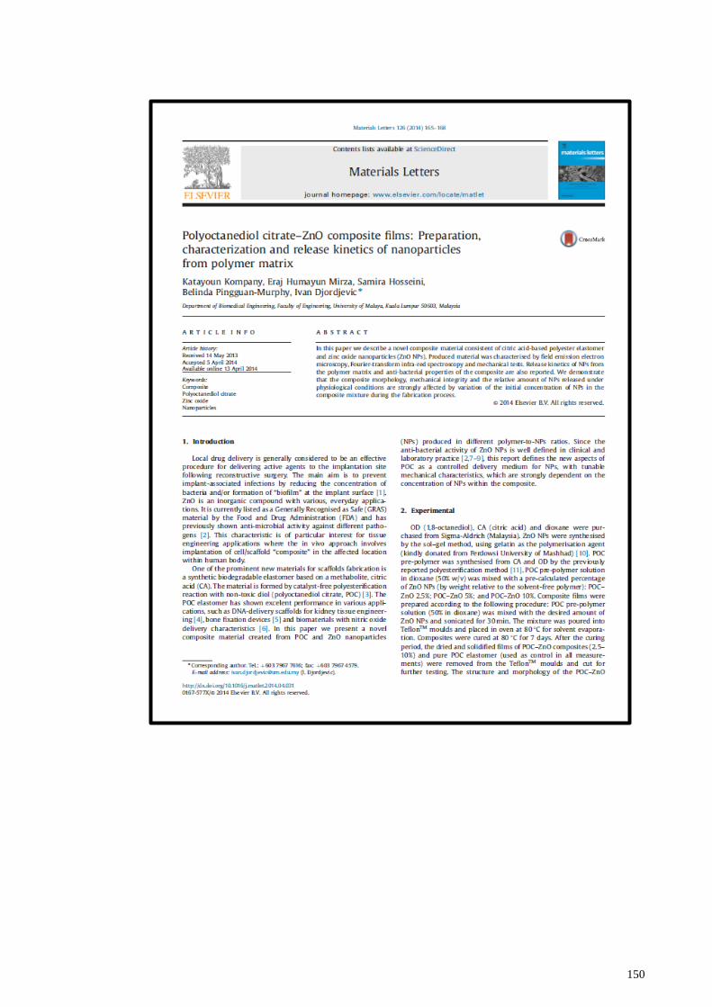
150

151

152

153
APPENDIX I
Product specifications of ZnONPs

154
APPENDIX II
Raman FTIR for ZnONPs