development of a unified dmpk and cnbp pcr … • myotonic dystrophy (dm) is the most common...
TRANSCRIPT
Summary• Myotonic dystrophy (DM) is the most
common adult-onset muscular dystrophy.Type 1 is caused by an expansion of CTGin DMPK while Type 2 is caused by anexpansion of CCTG in CNBP.
• Both diseases have similar presentationmaking differentiation important.
• The repeat regions of both DMPK and CNBPare extremely challenging to amplify andresolve using PCR.
• Here we demonstrate a unified workflowto genotype at least 200 CTG repeats inDMPK and flag expansions of >75 CCTGrepeats in CNBP.
Development of a unified DMPK and CNBP PCR workflow for determining repeat expansions relevant to myotonic dystrophiesJacob Wisotsky, Jon Kemppainen, Vivian Le, Gary J Latham, and Bradley HallAsuragen, Inc., Austin, Texas, USA
IntroductionMyotonic dystrophy type 1 and 2 (DM1 and DM2) are the most prevalent adult-onset muscular dystrophies. DM1 is caused by a trinucleotide expansion of CTG in the 3’ untranslated region (UTR) of myotonic dystrophy protein kinase (DMPK). Clinical phenotypes manifest in patients who have >50 repeats with classic disease occurring in those with >100 repeats. DM2 is caused by a quadruplet nucleotide expansion of CCTG in intron 1 of the cellular nucleic acid-binding protein (CNBP). Pathogenicity is observed in those with >75 repeats. Expansions in the DM2 disease statecan be between 75-11,000 repeats; however, theaverage is 5,000. In both cases, expansions ofteninclude hundreds to thousands of repeat units. DM1and DM2 molecular diagnosis relies on a complexcombination of PCR and Southern blot analysis.Due to a set of overlapping symptoms, DM1 andDM2 can be part of the same differential diagnosis.Current methods fail to identify expansions inDMPK and CNBP using a streamlined assay format.Thus we have proposed a workflow that unifies thedetermination of an expansion occurring in eitherDMPK or CNBP.
Materials and MethodsA streamlined DMPK/CNBP assay and workflow was developed with prototype AmplideX® PCR/CE reagents optimized for the repeat-primed amplification of both DMPK and CNBP repeats using distinct dye-tagged primers. Sample gDNAs were amplified in separate PCR reactions for DMPK and CNBP on the same PCR plate. Amplicons were resolved on a 3500xL Genetic Analyzer CE instrument (Thermo Fisher) using POP7 polymer, with 2.5 kV, 20 sec injection and 40 min run time. Samples for Figure 6 were multiplexed post PCR and run on the CE. Genotyping for DMPK was achieved following sizing analysis using a ROX 1000 size ladder (Asuragen) and a four-point calibrator. Sanger sequencing was accomplished by cloning purified gene-specific DMPK PCR product into a vector and then screening for inserts via colony PCR. Vectors containing inserts were then sequenced.
*Proof-of-concept data onlyPresented at ASHG 2017
References1. Kamsteeg, E. J., Kress, W., Catalli, C., Hertz, J. M., Witsch-Baumgartner, M., Buckley, M. F., et al. (2012). Best practice
guidelines and recommendations on the molecular diagnosis of myotonic dystrophy types 1 and 2. European Journal of Human Genetics: EJHG, 20(12), 1203–8. http://doi.org/10.1038/ejhg.2012.108
2. Bachinski, L. L., Czernuszewicz, T., Ramagli, L. S., Suominen, T., Shriver, M. D., Udd, B., et al. (2009). Premutation allele pool in myotonic dystrophy type 2. Neurology, 72(6), 490–497. http://doi.org/10.1212/01.wnl.0000333665.01888.33
3. Dalton, J. C., Ranum, L. P., & Day, J. W. (1993). Myotonic Dystrophy Type 2. GeneReviews®. University of Washington,Seattle. Retrieved from http://www.ncbi.nlm.nih.gov/pubmed/20301639
4. Prior T. W., American College of Medical Genetics (ACMG) Laboratory Quality Assurance Committee (2009) Technical standards and guidelines for myotonic dystrophy type 1 testing. Genet Med 11:552– 555. doi: 10.1097/GIM.0b013e3181abce0f
5. Savić Pavićević D., Miladinović J., Brkušanin M., et al. (2013) Molecular Genetics and Genetic Testing in Myotonic Dystrophy Type 1. Biomed Res Int 2013:1–13. doi: 10.1155/2013/391821
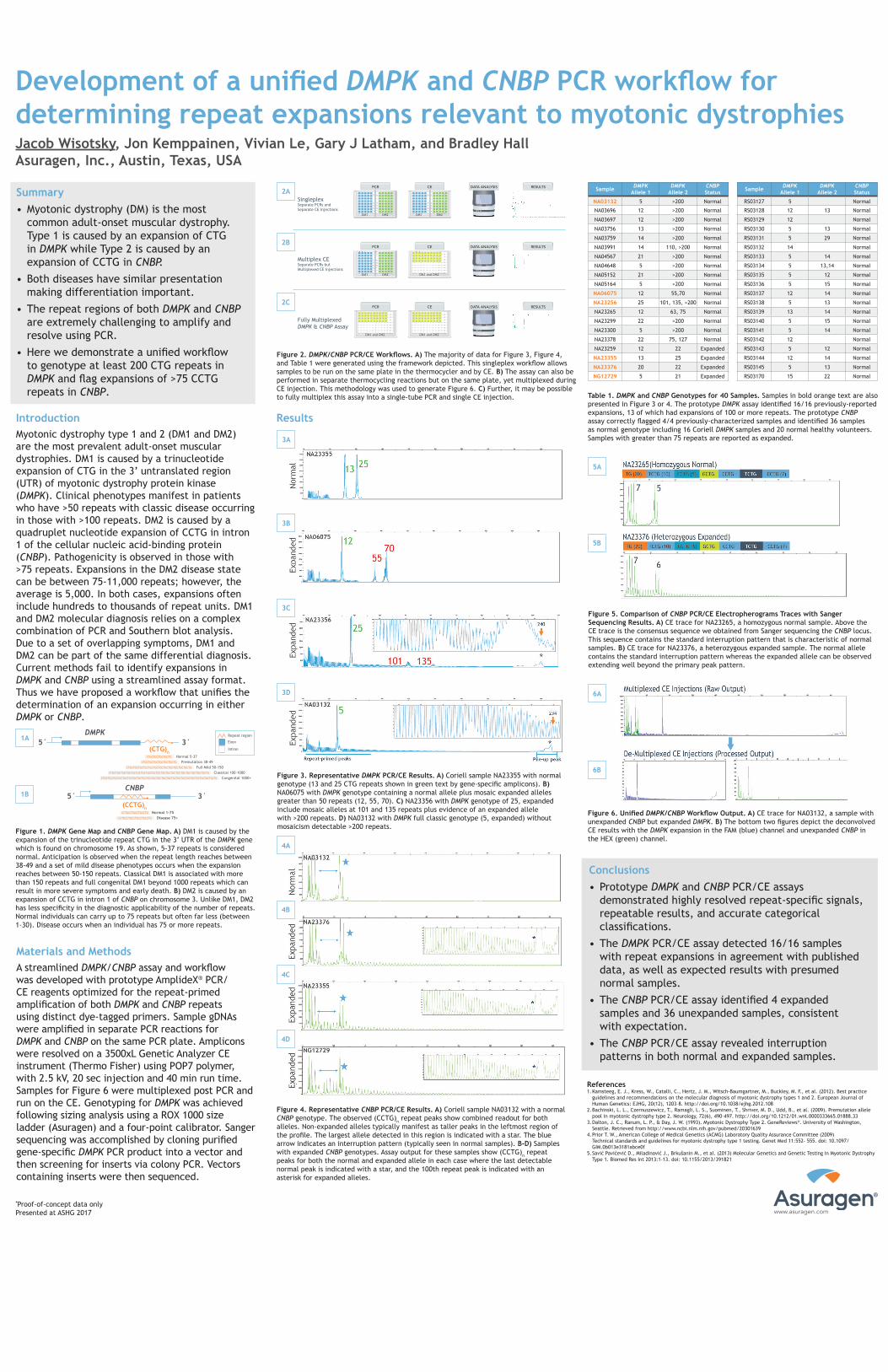
Figure 2. DMPK/CNBP PCR/CE Workflows. A) The majority of data for Figure 3, Figure 4, and Table 1 were generated using the framework depicted. This singleplex workflow allows samples to be run on the same plate in the thermocycler and by CE. B) The assay can also be performed in separate thermocycling reactions but on the same plate, yet multiplexed during CE injection. This methodology was used to generate Figure 6. C) Further, it may be possible to fully multiplex this assay into a single-tube PCR and single CE injection. Table 1. DMPK and CNBP Genotypes for 40 Samples. Samples in bold orange text are also
presented in Figure 3 or 4. The prototype DMPK assay identified 16/16 previously-reported expansions, 13 of which had expansions of 100 or more repeats. The prototype CNBP assay correctly flagged 4/4 previously-characterized samples and identified 36 samples as normal genotype including 16 Coriell DMPK samples and 20 normal healthy volunteers. Samples with greater than 75 repeats are reported as expanded.
Figure 1. DMPK Gene Map and CNBP Gene Map. A) DM1 is caused by the expansion of the trinucleotide repeat CTG in the 3’ UTR of the DMPK gene which is found on chromosome 19. As shown, 5-37 repeats is considered normal. Anticipation is observed when the repeat length reaches between 38-49 and a set of mild disease phenotypes occurs when the expansion reaches between 50-150 repeats. Classical DM1 is associated with more than 150 repeats and full congenital DM1 beyond 1000 repeats which can result in more severe symptoms and early death. B) DM2 is caused by an expansion of CCTG in intron 1 of CNBP on chromosome 3. Unlike DM1, DM2 has less specificity in the diagnostic applicability of the number of repeats. Normal individuals can carry up to 75 repeats but often far less (between 1-30). Disease occurs when an individual has 75 or more repeats.
DMPK
(CTG)n
(CCTG)n
5´ 3´Repeat region
Exon
Intron
CCTGCCTGCCTGCCTG Normal 1-75Disease 75+CCTGCCTGCCTGCCTGCCTG
CTGCTGCTGCTGCTG Normal 5-37Premutation 38-49CTGCTGCTGCTGCTGCTGCTG
Full Mild 50-150 CTGCTGCTGCTGCTGCTGCTGCTGCTGCTGCTGCTGCTG
CTGCTGCTGCTGCTGCTGCTGCTGCTGCTGCTGCTGCTGCTGCTGCTGCTGCTGCTGCTG
CTGCTGCTGCTGCTGCTGCTGCTGCTGCTGCTGCTGCTGCTGCTGCTGCTGCTGCTGCTGCTGCTGCTGCTG
Classical 100-1000 Congenital 1000+
CNBP5´ 3´
Singleplex Separate PCRs and Separate CE Injections
PCR1 2 3 4 5 6 7 8 9 10 11 12
DM1 DM2
CE1 2 3 4 5 6 7 8 9 10 11 12
DM1 DM2
DATA ANALYSIS RESULTS
Multiplex CE Separate PCRs butMultiplexed CE Injections
PCR1 2 3 4 5 6 7 8 9 10 11 12
DM1 DM2
DATA ANALYSIS RESULTS
Fully MultiplexedDMPK & CNBP Assay
DATA ANALYSIS RESULTS
CE1 2 3 4 5 6 7 8 9 10 11 12
DM1 and DM2
CE1 2 3 4 5 6 7 8 9 10 11 12
DM1 and DM2
PCR1 2 3 4 5 6 7 8 9 10 11 12
DM1 and DM2
Results
Figure 4. Representative CNBP PCR/CE Results. A) Coriell sample NA03132 with a normal CNBP genotype. The observed (CCTG)n repeat peaks show combined readout for both alleles. Non-expanded alleles typically manifest as taller peaks in the leftmost region of the profile. The largest allele detected in this region is indicated with a star. The blue arrow indicates an interruption pattern (typically seen in normal samples). B-D) Samples with expanded CNBP genotypes. Assay output for these samples show (CCTG)n repeat peaks for both the normal and expanded allele in each case where the last detectable normal peak is indicated with a star, and the 100th repeat peak is indicated with an asterisk for expanded alleles.
Sample DMPK Allele 1
DMPK Allele 2
CNBP Status
NA03132 5 >200 Normal
NA03696 12 >200 Normal
NA03697 12 >200 Normal
NA03756 13 >200 Normal
NA03759 14 >200 Normal
NA03991 14 110, >200 Normal
NA04567 21 >200 Normal
NA04648 5 >200 Normal
NA05152 21 >200 Normal
NA05164 5 >200 Normal
NA06075 12 55,70 Normal
NA23256 25 101, 135, >200 Normal
NA23265 12 63, 75 Normal
NA23299 22 >200 Normal
NA23300 5 >200 Normal
NA23378 22 75, 127 Normal
NA23259 12 22 Expanded
NA23355 13 25 Expanded
NA23376 20 22 Expanded
NG12729 5 21 Expanded
Sample DMPK Allele 1
DMPK Allele 2
CNBP Status
RS03127 5 Normal
RS03128 12 13 Normal
RS03129 12 Normal
RS03130 5 13 Normal
RS03131 5 29 Normal
RS03132 14 Normal
RS03133 5 14 Normal
RS03134 5 13,14 Normal
RS03135 5 12 Normal
RS03136 5 15 Normal
RS03137 12 14 Normal
RS03138 5 13 Normal
RS03139 13 14 Normal
RS03140 5 15 Normal
RS03141 5 14 Normal
RS03142 12 Normal
RS03143 5 12 Normal
RS03144 12 14 Normal
RS03145 5 13 Normal
RS03170 15 22 Normal
Conclusions• Prototype DMPK and CNBP PCR/CE assays
demonstrated highly resolved repeat-specific signals,repeatable results, and accurate categoricalclassifications.
• The DMPK PCR/CE assay detected 16/16 sampleswith repeat expansions in agreement with publisheddata, as well as expected results with presumednormal samples.
• The CNBP PCR/CE assay identified 4 expandedsamples and 36 unexpanded samples, consistentwith expectation.
• The CNBP PCR/CE assay revealed interruptionpatterns in both normal and expanded samples.
1A
1B
2A
2B
Figure 5. Comparison of CNBP PCR/CE Electropherograms Traces with Sanger Sequencing Results. A) CE trace for NA23265, a homozygous normal sample. Above the CE trace is the consensus sequence we obtained from Sanger sequencing the CNBP locus. This sequence contains the standard interruption pattern that is characteristic of normal samples. B) CE trace for NA23376, a heterozygous expanded sample. The normal allele contains the standard interruption pattern whereas the expanded allele can be observed extending well beyond the primary peak pattern.
5A
5B
Figure 6. Unified DMPK/CNBP Workflow Output. A) CE trace for NA03132, a sample with unexpanded CNBP but expanded DMPK. B) The bottom two figures depict the deconvolved CE results with the DMPK expansion in the FAM (blue) channel and unexpanded CNBP in the HEX (green) channel.
6A
6B
2C
Figure 3. Representative DMPK PCR/CE Results. A) Coriell sample NA23355 with normal genotype (13 and 25 CTG repeats shown in green text by gene-specific amplicons). B) NA06075 with DMPK genotype containing a normal allele plus mosaic expanded alleles greater than 50 repeats (12, 55, 70). C) NA23356 with DMPK genotype of 25, expanded include mosaic alleles at 101 and 135 repeats plus evidence of an expanded allele with >200 repeats. D) NA03132 with DMPK full classic genotype (5, expanded) without mosaicism detectable >200 repeats.
3A
3B
3C
3D
4A
4B
4C
4D