development of an insect cell factory for the production...
TRANSCRIPT

1
UNIVERSIDADE DE LISBOA
FACULDADE DE CIÊNCIAS
DEPARTAMENTO DE BIOLOGIA VEGETAL
Development of an insect cell factory for the production of complex
biopharmaceuticals using a synthetic biology approach
João Miguel Nunes Vidigal
MESTRADO EM BIOLOGIA CELULAR E BIOTECNOLOGIA
2011

2
UNIVERSIDADE DE LISBOA
FACULDADE DE CIÊNCIAS
DEPARTAMENTO DE BIOLOGIA VEGETAL
Development of an insect cell factory for the production of complex
biopharmaceuticals using a synthetic biology approach
Supervised by:
Dr. Paula Alves, Laboratório de Tecnologia de Células Animais from the Instituto de Biologia
Experimental e Tecnológica and Instituto de Tecnologia Química e Biologica, Universidade Nova de
Lisboa (IBET/ITQB-UNL).
Dr. Ana Margarida Fortes, Plant Systems Biology Lab, Department of Plant Biology
Center for Biodiversity, Functional & Integrative Genomics-BioFIG, Faculdade de Ciências da
Universidade de Lisboa (FCUL).
João Miguel Nunes Vidigal
MESTRADO EM BIOLOGIA CELULAR E BIOTECNOLOGIA
2011

3
Master's Thesis in Cell Biology and Biotechnology, Faculdade
de Ciências, Universidade de Lisboa, held at the Animal Cell
Technology Laboratory from the Instituto de Biologia
Experimental e Tecnológica and Instituto de Tecnologia
Química e Biologica, Universidade Nova de Lisboa
(IBET/ITQB-UNL) under the supervision of Dr. Paula Alves.

4
Acknowledgements
I am lucky to have many people to acknowledge for the help, support, and contributions that made this Master Thesis possible. To Dr. Paula Alves, for the supervising and the opportunity to perform this master thesis at the Laboratory of Animal Cell Technology, for the example of rigor, and leadership and the excellent working conditions. To Dr. Ana Teixeira for the pleasure and privilege to work with, for all the knowledge, confidence, patience, understanding and preseversence in me. For the hours of conversation and for being an example in science and in life. To Dr. Ana Sofia Coradinha for the availability and interest shown throughout my thesis, and revision. To Fabiana Fernandes, for the introduction into this new scientific world. For all the disponibility for help, for all the discussions and the relaxing funny moments. To Dr. Ana Margarida Fortes for accepting to be my supervisor, availability and revision of my thesis. To all the members of the Animal Cell Technology Unit for creating the healthy and the most enjoyable working environment, especially Hélio, Daniel, Marta, Francisca, Claudia C. ,Tiago and Paulo. To Raquel for all the help, support, patience and for being always there in this journey. To all my friends that made this journey possible, for being part of my life, by understanding, for all laugh and all the great moments shared. To my family, especially my parents and sister for the example and unconditional love and support at all times and circumstances and for making my dreams possible.
A thank you to all…

5
Index
Abbreviation list.................................................................................................................................... 7
1. Preface ......................................................................................................................................... 9
2. Abstract ...................................................................................................................................... 10
3. Resumo ...................................................................................................................................... 12
4. Introduction ................................................................................................................................. 15
4.1 Animal Cell Technology and Biopharmaceutical Production ..................................................... 15
4.2 Insect Cell Expression: Baculovirus vs Stable expression ........................................................ 16
4.3 Traditional Cell Line Development ............................................................................................ 18
4.4 Site-specific Recombination (SSR) ........................................................................................... 19
4.4.1 Flippase Site-specific Recombination (SSR) ...................................................................... 19
4.4.2 Flippase RMCE, Recombinant mediated cassette exchange systems ............................... 20
4.5. Inducible Gene Networks ......................................................................................................... 21
4.5.1. Tetracycline (Tet) regulated gene expression. ................................................................... 22
5. Aim of the thesis ......................................................................................................................... 25
6. Materials and Methods ................................................................................................................ 26
6.1 Molecular Biology ................................................................................................................ 26
6.1.1. Vector Design and Construction ........................................................................................ 26
Stable expression Study with Different Promoters ................................................................... 26
RMCE related vectors ............................................................................................................. 26
Tet–Off Inducible Circuit vectors .............................................................................................. 27
6.1.2. Techniques supporting vector construction ....................................................................... 27
General PCR-protocol ............................................................................................................. 27
Colony PCR-screening ............................................................................................................ 27
Digestion of DNA ..................................................................................................................... 27
Ligation with T4 ligase ............................................................................................................. 28
Preparation of competent E. coli cells...................................................................................... 28
Transformation of E. coli cells.................................................................................................. 28
Isolation of vector-DNA............................................................................................................ 29
Agarose gel electrophoresis .................................................................................................... 29
Determination of DNA-Conecntartions ................................................................................... 289
6.2 Cell Culture .......................................................................................................................... 29
6.2.1. Culture maintenance ......................................................................................................... 29
6.2.2. Freezing and thawing of insect cells .................................................................................. 29
6
6.2.3. Transfection of insect cells ................................................................................................ 30
Electroporation ........................................................................................................................ 30
Lipotranfection ......................................................................................................................... 30
6.2.4. Killing curves ..................................................................................................................... 30
6.2.5. Cloning by Limiting dilution................................................................................................ 30
6.2.6. Doxycycline Assay ............................................................................................................ 31
6.3 Analytical Techniques .......................................................................................................... 31
6.3.1. Cells counting ................................................................................................................... 31
6.3.2. Flow cytometry .................................................................................................................. 31
6.3.3. Western Blot to the Transactivactor .................................................................................. 31
6.3.4. Southern Blot .................................................................................................................... 31
6.3.5. RNA extraction and RT-PCR ............................................................................................. 32
6.3.6. Statistical analysis ............................................................................................................. 32
7. Results........................................................................................................................................ 33
7.1 Comparison of promoters strength ........................................................................................... 33
7.1.1 Metallothionein Promoter ................................................................................................... 34
7.1.2 OpIE2 and Hsp70 promoters .............................................................................................. 35
7.2 Tag and target cassettes .......................................................................................................... 36
7.3 Transfection of Sf9 cells with tagging cassette ......................................................................... 38
7.4 Limiting dilution and clone screening ........................................................................................ 41
7.5 Southern blot analysis of tagged clones ................................................................................... 42
7.6 Flippase-mediated cassette exchange ...................................................................................... 42
7.7 Tetracycline Inducible Circuits .................................................................................................. 44
8. Discussion .................................................................................................................................. 47
8.1 Comparision of Different promoters strength ............................................................................ 48
8.2 Flippase-Mediated Cassette Exchange .................................................................................... 48
8.3 Transcriptional regulatory circuits ............................................................................................. 49
9. Conclusion and Future Work Perspectives ................................................................................. 51
Bibliography ....................................................................................................................................... 52
Appendix ............................................................................................................................................ 57

7
Abbreviation list
AcIE1 promoter – Immediate-early promoter from Autographa californica (Ac)
BEVS - Baculovirus Expression Vector Systems
CHO - Chinese Hamster Ovary
DIG – Digoxigenin
DNA - Deoxyribonucleic acid
Dox - Doxycycline
G418 – Geneticin Antibiotic
dsRED – Red fluorescent protein
E. coli - Escherichia coli
ECL - Enhanced chemiluminescence
eGFP – Enhanced green fluorescent protein
ES - Embryonic stem
FLP - Flippase
Flp-RMCE - Flp-based recombinase-medicated cassette exchange
Fn - FRT variants
FRT - FLP recognition target sites
GOI - Gene of interest
HeLa cells - Cell line derived from the cervical cancer cells of patient Henrietta Lacks.
hESCs - Human Embryonic stem cells
iPS - Induced pluripotent stem cells
LB – Lysogeny broth
Mtn - Metallothionein
OpMNPV -Orgyia pseudotsugata multicapsid nuclear polyhedrosis virus
Ori - Origin of replication
PBS - Phosphate-buffered saline
PCR – Polymerase Chain Reaction
PhCMV - human Cytomegalovirus minimal promoter
RMCE - Recombinase mediated cassette exchange systems
RT - Recognition-target
rtTA – Tetracycline transactivator
SSR - Site-specific Recombination

8
Sf9 – Cell line derived from Spodoptera frugiperda ovarian cells.
TAFII40 – TATA binding protein-associated factor II 40
TBP – Tata Binding Protein
Tet - Tetracycline
TetO - Tet operator
Tet-Off system – Inducible system which needs tetracycline to shut off.
TetR - Tet repressor
TFIIB – Trascription Factor II B
TIRs - Translation initiation regions
tTA - Tetracycline transactivator
TRE – Tetracycline response element

9
1. Preface
This master thesis is within the scope of the project PTDC/EBB-EBI/102266/2008, entitled "Novel Cell
Factories for the production of secreted complex bioproducts: a syntetic biology approach for
improved product stoichiometries" funded by the Portuguese Fundação para a Ciência e Tecnologia
(FCT). The global aim of this project is to develop a flexible and re-usable insect cell line able to
produce different correctly assembled multi-component biopharmaceuticals at relevant yields. The
transcription rate of the different components will be stoichiometrically tuned by bridging synthetic
biology and molecular biology approaches, namely by combining transcriptional regulatory circuits and
the recombinase mediated cassette exchange (RMCE) technology.
The majority of the results described in this thesis were presented at several international
scientific meetings:
Fernandes F, Vidigal J, Teixeira AP, Coroadinha AS, Alves PM “Implementation of Recombinase Mediated cassette Exchange Systems in Sf9 cells for expression of multiple recombinant proteins”, 22nd ESACT Meeting 2011, Viena, Austria.
Fernandes F, Vidigal J, Coroadinha AS, Teixeira AP, Alves PM “Implementation of Synthetic Circuits in Sf9 Insect Cells at Tagged loci generated by Recombinase Mediated Cassette Exchange Systems”, 5th Annual Workshop on the Business-Government Interface: Systems and Synthetic Biology, June 2011, Braga, Portugal.
Fernandes F, Vidigal J, Tomás H, Teixeira AP, Coroadinha AS, Alves PM “Fast insect cell line development for production of complex biopharmaceuticals”, 11thCHEMPOR 2011, Sept 2011, Caparica, Portugal.
Fernandes F, Vidigal J, Tomás H, Teixeira AP, Coroadinha AS, Alves PM “Development of an insect cell factory for production of complex biopharmaceuticals using flipase-mediated cassette exchange”, 10th PEACe, Sept 2011, Cascais, Portugal.
Fernandes F, Vidigal J, Tomás H, Carrondo MJT, Coroadinha AS, Teixeira AP, Alves PM "Exploring novel approaches to produce virus-like particles", 2nd ENVVP, Oct 2011, Frankfurt, Germany.

10
2. Abstract
Insect cells, in particular the Spodoptera frugiperda Sf9 cell line, are a popular system for the
production of biologically active recombinant proteins. However, the current technology uses
baculovirus infection which has two main disadvantages: firstly, the recombinant gene is only
expressed transiently during the infection cycle, after which cells die; secondly, due to the lytic nature
of this system, the cellular protein processing machinery is severely compromised at the end of the
infection cycle, affecting the correct formation of recombinant proteins whose expression is usually
controlled by very-late baculovirus promoters.
Stably transformed insect cell lines represent an alternative system for continuous protein production.
However, their establishment is laborious, requiring the identification of cell clones that display the
right expression properties due to random integration of the gene-of-interest. Furthermore,
independently of the expression system, multimeric biopharmaceuticals continue to have very low
yields, mainly because the stoichiometry between components is not properly reached in the producer
cells.
To overcome these issues, this report shows work towards the development of a novel Sf9 cell factory
combining (i) recombinase mediated cassette exchange (RMCE) for targeted gene integration and (ii)
a tetracycline (Tet) inducible system for the modulation of gene expression levels.
We started by evaluating the strength of different promoters to drive GFP expression in Sf9 cells as
well as different transfection protocols. The baculovirus promoter OpIE2 allowed the strongest
expression when compared to others, and the efficiency of transfection was significantly higher using
the lipotransfection method. From the different transfection protocols, 4 cell populations with distinct
fluorescence distributions were obtained after 3 weeks under hygromycin selection. These populations
were independently subject to limiting dilution and isolated cell clones were analyzed by southern blot
to screen for single-copy integration of the tagging GFP construct. While the majority of clones were
single copy, cassette exchange was performed in a few clones by co-transfection with flippase and the
target plasmid containing dsRed. Cells were selected in the presence of neomycin and cassette
exchange was confirmed by PCR of genomic DNA, revealing the absence of tagging cassette and the
presence of the target cassette in the some locus.
In parallel, the functionality of a tetracycline inducible system in Sf9 cells was evaluated by substituting
mammalian promoters with insect cell specific promoters. However, transactivation was suboptiomal,
requiring additional changes to the original system.

11
In summary, this work reports for the first time the implementation of a RMCE system in Sf9 cells and
a preliminary assessment of Tet transactivation in these cells. This new cell line will be a major
breakthrough since it will combine the advantages inherent to Sf9 cell growth, continuous protein
production, the ability of re-using a well characterized locus for targeted DNA integration and inducible
gene expression.
Keywords: Sf9 cells, cell line development, multimeric biopharmaceuticals, RMCE systems, Tetracycline inducible systems.

12
3. Resumo
A cultura de células de insecto conjugada com a infecção por baculovirus representa um dos
sistemas biológicos de eleição para a produção de proteínas recombinantes biologicamente activas.
Há vários biofármacos em ensaios clínicos e alguns já no mercado, como é o caso da vacina contra o
vírus do papiloma humano (Cervarix, GSK), que utilizam este tipo tipo de sistema de expressão. No
entanto, o uso de baculovirus representa um sistema de expressão transiente / lítico inerente ao
processo de infecção.
Em contraste, quando estavelmente transformadas, as células de insecto podem ser usadas como
um sistema de expressão contínuo e não-lítico. Contudo, o desenvolvimento de linhas celulares
estáveis é significativamente demorado e laborioso, sendo necessário identificar e isolar clones que
exibam elevadas taxas de expressão. A grande variabilidade existente nos níveis de expressão entre
clones deve-se ao chamado efeito de posição, ou seja a aleatoriedade de integração no genoma da
célula.
Além disso, e independentemente do sistema de expressão, quando se pretende expressar
biofármacos multiméricos, como as particulas semelhantes a vírus, estes continuam a ter
rendimentos muito baixos. A estequiometria entre os componentes é um factor chave e, quando não
é alcançada adequadamente nas células produtoras, resulta na formação de uma grande
percentagem de partículas incorrectamente formadas.
De forma a superar estas limitações, neste trabalho é iniciado o desenvolvimento de uma linha celular
derivada de Spodoptera frugiperda, usando uma combinação de duas tecnologias: (i) sistema de
troca de cassete mediada por recombinase (RMCE) e (ii) circuito transcricional indutível. Os sistemas
RMCE utilizam um eficiente e avançado processo de troca génica localizada por intermédio de uma
reacção de recombinação. Anteriormente implementada em células de mamífero, este tipo de
tecnologia foi agora aplicada num cenário de expressão estável em células de insecto com o
objectivo de permitir o uso repetido de locais genómicos pré-caracterizados com elevada taxa de
expressão. Já os circuitos indutíveis sintéticos, como o sistema de indução por tetraciclina, permitem
ajustar a expressão de diferentes genes de interesse por intermédio de um indutor, facilitando a
produção de produtos multiméricos correctamente formados.
O trabalho desenvolvido nesta dissertação de mestrado começou por centrar-se na análise de
diferentes promotores no controlo da expressão de genes repórter na linha celular Sf9. Foram
estudados promotores com diferentes origens mas com uma base de funcionamento comum em

13
células de insecto, como os promotores de baculovirus OpIE2 e OpIE1, o promotor da metalotioneína
e o das proteínas de choque térmico (hsp70) de Drosophila. Em resultado, verificou-se que o
promotor OpIE2 superou consideravelmente os restantes na expressão de eGFP. Por outro lado, o
promotor da metalotioneína induzido por cobre revelou ser um sistema desadequado para expressão
de proteínas de interesse em células Sf9, uma vez que apenas para doses citotóxicas se verificou
uma expressão de eGFP mensurável.
Para o funcionamento do sistema RMCE são necessários dois vectores distintos e complementares
(Tagging / Target). Em ambos os vectores foi utilizado o promotor OpIE2 para a expressão das
proteínas repórter (diferentes nos dois vectores: Tagging – dsRed; Target - eGFP), e o promotor
OpIE1foi usado para a expressão do agente de selecção, necessário para garantir a expressão
estável dos dois vectores no genoma da célula (Tagging – higromicina; Target -neomicina). Enquanto
a integração do plasmídeo Tagging no genoma se dá através de um processo aleatório, a integração
do Target é o resultado da troca bem-sucedida por recombinação enzimática. A selecção pelo agente
neomicina assegura o sucesso da troca génica uma vez que o gene de resistência é activado pela
inserção de um códão de iniciação (ATG) presente apenas no Target.
Antes do processo de troca, é importante que apenas uma cópia da cassete Tagging seja integrada
no genoma da célula para que o sistema RMCE seja funcional. Utilizaram-se dois métodos em
paralelo para transfectar as células Sf9 com o plasmídeo Tagging: lipotransfecção usando cellfectin e
eletroporação, donde resultaram quatro populações celulares com distribuições de fluorescência
distintas. Para cada população de células foi posteriormente realizada uma diluição progressiva para
a obtenção de clones produtores, os quais foram analisados por citometria de fluxo para comparar o
nível de expressão de eGFP. Posteriomente, foi implementado um protocolo de Southern blot para
analisar o número de cópias integradas da cassete Tagging em cada clone., Dos nove clones
analisados, apenas um revelou ter mais do que uma cópia. Em alguns dos clones com apenas uma
cópia procedeu-se à co-transfecção da construção Target com a enzima recombinase (flippase). Foi
possível seleccionar células resistentes à neomicina sugerindo o sucesso da troca de cassetes, tendo
sido mais tarde confirmado por meio de PCR do DNA genómico e de RT-PCR do RNA dos genes
repórteres.
No que toca o circuito indutível de transcrição, o trabalho conduzido teve como objectivo fazer uma
avaliação preliminar da sua funcionalidade em células Sf9 antes da implementação directa na cassete
de recombinação. No entanto, os resultados revelaram uma fraca indução por parte do agente
tetraciclina, justificando a necessidade de uma optimização do sistema original.
Em conclusão, com este trabalho reportamos pela primeira vez a implementação bem sucedida de
um sistema de troca de cassete por intermédio de recombinação enzimática em células Sf9. Esta

14
tecnologia representa um grande avanço na produção de novas linhas celulares de insecto,
combinando as vantagens inerentes à cultura deste tipo de células com a possibilidade de atingir
elevados níveis de expressão contínua de diferentes proteínas recombinantes através da fácil
integração de genes de interesse no genoma celular.
Palavras-chave: cultura de células de insecto, biofármacos complexos, sistemas de troca de
cassetes mediada por recombinase, sistemas transcricionais indutiveis.

15
4. Introduction
4.1 Animal Cell technology and Biopharmaceutical Production
Biopharmaceutical compounds produced in living systems are continuously gaining importance in
medicine and are expected to help cure diseases not yet treatable today (Schmidt, 2004; Butler,
2005). Currently, these products represent approximately one in every four genuinely new
pharmaceuticals coming on the market and this ratio is expected to increase as seen by the increasing
number of new products enrolling in clinical trials (Hunt, 2005; Walsh, 2005). To this class of
compounds belong the nucleic acid-based medicinal products and recombinant therapeutic proteins,
including monoclonal antibodies. The majority of animal cell-derived biopharmaceuticals on the market
are monoclonal antibodies targeting mainly cancer therapy (Walsh, 2005).
Animal cell lines have been the preferred biological system to produce complex therapeutic proteins
due to their innate capacity to perform post-translational modifications. In particular, glycosylation is of
special interest since it influences functionality, serum half-life and immunogenicity of some proteins.
The ability to perform correct post-translational modifications has justified a rapid increase in the
number of approved therapeutics produced from animal cells over the last twenty years, which
surpassed the number of biopharmaceuticals produced in E. coli or yeast around 1996. Table 1
compares different expression systems in terms of their ability for recombinant protein production.
Animal cells have lower growth rates and reach lower maximum cell densities when compared to
bacteria or yeast systems, resulting in lower volumetric productivities. However, during the last years
these discrepancies have been progressively shortened through successive improvements in
expression technology, media composition and process operation.
Characterization of cellular metabolism and physiology allowed the design of efficient fed-batch and
perfusion bioreactor processes. In some monoclonal antibody processes, product yield is now over 5
g/L (Wurm 2004). However, some analysts indicate that this still may not be enough, especially due to
the large doses of mAB required; future manufacturing demand may exceed production capacity as
the number of approved biotherapeutics increases (Butler, 2005). Therefore, if in the past, the concept
of ‘‘time-to-market’’ dominated the industrial arena, nowadays, companies are willing to focus on
process optimization and cost reduction (Hunt, 2005).

16
Table 1- Main characteristics of the most important expression systems in terms of quality and yield, two major criteria for
recombinant pharmaceuticals (adapted from www.invitrogen.com and Beljelarskaya, 2002).
Host System
Advantages Challenges Protein yield, % dry weight
Bacterial expression
Low cost
Simple culture conditions
Higher growth rates and maximum cell densities
Protein solubility
Minimal posttranslational modifications
Difficult to express functional mammalian proteins
1-5%
Yeast Expression
Eukaryotic protein processing
Simple media requirements Fermentation required for very high yields 1%
Insect Expression
Posttranslational modifications similar
to mammalian systems
Higher yields than mammalian systems
More demanding culture conditions
Transient/lytic system (BEVS) 20%
Mammalian Expression
Improved levels of correct posttranslational modifications
Increased probability of obtaining fully functional human proteins
More demanding culture conditions <1%
4.2 Insect Cell Expression: Baculovirus vs Stable expression
Insect cell culture is a mature technology and is being applied in the routine production of recombinant
proteins. This technology have major advantages compared with mammalian systems: insect cells
are more stress-resistant, easier to culture, more productive and they have the capability to grow in
suspension in defined serum-free culture media (Ikonomou et al, 2003; Schmidt, 2004; Weber and
Fussenegger, 2009). Their efficiency to perform post-translational modifications is comparable to
mammalian cells. Furthermore, these cells tolerate higher levels of free amino acids and glucose
without switching to overflow metabolism, which enables the design of more nutrient-rich culture
media. Unlike mammalian cells, they support lower pH (6.2–6.9) which is commonly maintained by a
phosphate buffer, removing the need for carbon dioxide in the culture, as required for the open
bicarbonate buffer system in mammalian cell culture media. Growing at lower temperatures (27°C) is
also an advantage in terms of process (Ikonomou et al, 2003; Weber and Fussenegger, 2009).
Two of the most used insect cell lines (Sf21 and Sf9) were isolated from the ovarian tissue of
Spodoptera frugiperda pupae. The Sf21 cell line was established at the USDA Insect Pathology
Laboratory in 1970 (Vaughn et al, 1977) and Sf9 cells constitute a sub-clone of Sf21 cells, selected for
faster growth rate and higher cell densities than the Sf21 cells. Both cell lines are 1) robust and easy

17
to culture in mono-layer or suspension, 2) able to grow rapidly to high cell densities, 3) highly
susceptible to Baculovirus (BV), 4) able to produce high virus titers, and 5) readily adaptable to serum-
free medium and scale up culture (Granados et al, 2007). The Baculovirus expression vector systems
(BEVS) is the most used insect cell expression system, mainly because of the successful expression
of a large variety of proteins, either intracellularly expressed, surface bound, or secreted into the
culture medium (Geisse et al, 1996). For application in biotechnology, baculoviruses have been
engineered to express recombinant target genes under the control of the very late promoters p10 or
polyhedrin, while eliminating the genes that form occlusion bodies.
The baculovirus-insect cell technology is currently being used by numerous small to midsize startup
biotechnology companies in North America and Europe to produce custom recombinant proteins for
research and commercial applications (Ikonomou et al, 2003; Granados et al, 2007). Furthermore,
since it is an accepted technology for the production of viral antigens, several vaccine candidates are
now in clinical trials, such as FluBlock (a flu vaccine from Protein Sciences Corporation) or already in
the market, such as Cervarix (vaccine against the human papilloma virus, from Glaxo Smith Kline
Biologicals).
While existing insect cell lines provide good results in baculovirus expression vector systems, they can
still be engineered for high quantity and quality of recombinant protein. In this respect, (Jarvis et al
1997) engineered a Spodoptera frugiperda cell line to perform human-like glycosylation patterns in
recombinant therapeutic proteins. In addition, insect cell lines can be engineered for stable protein
expression (Granados et al, 2007). Indeed, the expression of proteins from an uninfected engineered
cell line can provide certain advantages in protein quality, when compared with protein expression
from a baculovirus expression vector. Major drawbacks due to baculovirus infection are: (1) an
impairment of the protein folding and secretion capacity of the cell (McCarroll and King, 1997), (2) a
high, in part baculovirus-encoded, protease activity implying the use of protease inhibitors in the
culture media or protease-deficient vectors (Ikonomou et al, 2003), (3) deviations of the
posttranslational modification pattern, which could act immunogenically and (4) probably the major
disadvantage of the system, is the impossibility of continuous protein production due to the lytic nature
of the viral infection process (Schmidt, 2004; Yamaji et al, 2008).
To overcome these problems, stable plasmid-based expression systems, capable of continuous
protein production in Spodoptera frugiperda cells started to be developed. There is a small number of
promoters normally used for stable protein expression in insect cells, including baculovirus-derived
immediate-early (IE) promoters, insect cells constitutive promoters, such as actin and Hsp70
promoters, and inducible promoters such as the Drosophila metallothionein promoter (McCarroll and
King, 1997) reported similar expression levels for the tissue–plasminogen activator expressed under

18
the control of the AcIE1 promoter (an IE promoter from Autographa californica (Ac) baculovirus) in
stably transfected Sf9 cells or in baculovirus-infected Sf9 cells. There are studies showing even better
protein yields in stable engineered insect cell lines compared with BEVS (Jarvis and Summers, 1989;
French and Roy, 1990; Park et al, 2004). Despite potential advantages, two major botlenecks still
needed to be adressed i) the long development timelines of stable cell lines and ii) the random
integration of the transgene into the host chromosome.
4.3 Traditional Cell Line Development
The first step in the development of a recombinant stable cell line is the transport of the foreign DNA
into the cells by physical, chemical or biological methods (e.g. microinjection, liposomes,
electroporation, gene-gun or viral vectors) (Wurm, 2004). Stable integration of the foreign DNA will
only occur in a small portion of the cells that have taken up the DNA. The integration is mediated by
cellular DNA repair enzymes and occurs at random sites of the host genome. The genome is
packaged into a unique pattern of heterochromatin and euchromatin, which is maintained after cell
division. This structure plays a major role in regulating gene transcription by allowing or not the access
of the transcriptional apparatus to genes (Narlikar et al. 2002), thus it determines which genes will be
active when integrated (Orphanides and Reinberg, 2002). A stringent selection for the generation of
stable transgenic cell lines proceeds by means of a cytotoxic antibiotic or depriving cells from an
essential metabolic enzyme (Wurm, 2004). The bacterial neomycin phosphotransferase enzyme
encoded by the neo gene is one of the most used antibiotic resistance marker in eukaryotic cells (Trill
et al, 1995; Li et al, 2007). In the presence of the drug G418 it confers selected ability to the cells that
have integrated the plasmid. A number of alternative antibiotic selection markers are available (e.g.,
hygromycin, zeocin).
The expression level of transfected cells surviving from selection is variable and unstable due to
different and unpredictable locations of the integrated copies. Furthermore, having in mind that high-
producers are only a minor portion of successfully transfected cells, and that non- and low-producers
tend to overgrow, an intensive screening is needed to find highproducing clones which have to divert
their metabolic resources to the expression of the gene-of-interest (GOI) (Kromenaker and Srienc,
1994). To isolate clonal cell lines stably expressing the recombinant protein at high levels, the most
commonly used method is limiting dilution. Although simple, it is a very laborious and time-consuming
process (Fig. 1). Finally, even if a transcriptional hotspot is hit by incidence, a random screening
process cannot guarantee the recurrence of such an event.

19
In this respect, reuse of an identified hotspot would radically change the time needed for the
development of a production cell line for various recombinant proteins. Site-specific recombination,
which enables the reuse of loci with the desired expression characteristics, is nowadays being
exploited to this end.
Figure 1 – Traditional cell line development step-by-step, being the transgene one fluorescent reporter protein.
4.4 Site-specific Recombination (SSR)
To integrate a transgene into a pre-tagged genomic site, a number of site-specific recombinases have
been of great help. The most widely used site-specific recombinases are the E. coli P1 phage-derived
Cre (Sternberg et al. 1986), the Saccharomyces cerevisiae-derived Flp (Buchholz et al, 1996; Schaft
et al, 2001) and the bacteriophage ΦC31-derived integrase (Thorpe and Smith 1998). These enzymes
can only mediate recombination between two copies of their target sequence, i.e. loxP site (for Cre),
Flp-recognition target site (FRT; for Flp) or a combination of attP/attB (for ΦC31), which enable to
excise (Cre, Flp), insert (Cre, Flp, ΦC31), or invert (Cre, Flp) DNA molecules. When two copies of the
recognition-target (RT) site are arranged as direct repeats, the corresponding enzyme excises the
DNA segment within the RTs and releases it as circular DNA, whereas the reverse reaction leads to
an insertion of a circular DNA.
4.4.1 Flippase Site-specific Recombination (SSR)
In this work we will focus on the flippase system. Flippase was originally isolated by Broach and Hicks
(1980) from the Saccharomyces cerevisiae 2-μm circle; it is a member of the integrase family of site-
specific recombinases. The FLP protein targets two FRT sites that are within the 599 bp inverted
repeats separating a small and a larger unique region in the 2-μm plasmid. A wild type FRT site
consists of 48 bp, composed of two inverted 13 bp repeats and an 8 bp spacer together with a third 13

20
bp direct repeat and a single additional base pair. FLP protein binds in a site-specific manner to the
13-bp symmetry elements. The 8 bp spacer is involved in DNA-DNA pairing during strand exchange.
Together with the extra repeat, its asymmetry determines the direction of site alignment in the
recombination event, which will consequently lead to either inversion or excision (Zhu & Sadowski,
1995; Turan et al., 2011). Mutations in the 8 bp spacer result in functional FRT variants (Fn), which will
recombine with a second site of the same composition but not with a wild type one (Schlake and
Bode, 1994). If F and Fn sites are in a strategically favorable position, flippase can mediate the
exchange of expression cassettes in a double reciprocal crossover event, which is called Flp-based
recombinase-medicated cassette exchange (Flp-RMCE).
4.4.2 Flippase RMCE, Recombinant mediated cassette exchange systems
The RMCE concept was first introduced by Schlake and Bode (1994) to enable multiple uses of pre-
characterized genomic sites; it is a process in which a tagging cassette (inserted in host genome)
flanked by a set of incompatible sites (for non-cross-interactions), is exchanged for a target cassette
(targets a tagged place) that is flanked by identical sites by means of site-specific recombinases as
illustrated by the Fig. 2 (Qiao et al, 2009; Turan et al., 2011). In the particular case of flippase-
mediated cassette exchange, two different FRT sites, for instance the wild type F and the mutant F3
(Schlake and Bode 1994), flank an expression cassette anchored in the genome which is then site-
specific recombined by an analogous cassette which is provided on an exchange plasmid.
RMCE technology has been successfully used for different purposes in cultured mammalian and
silkworm cells and in living organisms such as Drosophila and mice (Coroadinha et al. 2006;
Nakayama et al, 2006; and Cobellis et al, 2005). For instance, introduction of RMCE in mouse
embryonic stem (ES) cells by homologous recombination could be used repeatedly to generate
different knock-ins in the same locus in order to study gene function or towards the development of
models for human diseases (Roebroek et al, 2003). Also studies in human cell cultures, such as
embryonic stem cells (hESCs) and induced pluripotent stem (iPS) cells, with great interest in
transplant therapies for genetic diseases, have benefited from the application of RMCE. Indeed, much
of these medical applications benefit from continuous expression of therapeutic transgenes in this
case without random integration, preventing the possibility of mutagenesis and subsequent tumor
formation (Ramachandra et al, 2011).
Concerning cell line development for biopharmaceuticals production, Coroadinha et al (2006) reported
RMCE for the production of retroviral vectors for gene therapy, in which the target integration was
used for eventual exchange of the therapeutic gene. Targeted integration strategies have been

21
adopted also to express relevant therapeutic proteins, such as monoclonal antibodies and other
proteins in Chinese Hamster Ovary (CHO) cells (Groth et al, 2000; Thyagarajan et al, 2001; Kito et al,
2002; Wiberg et al, 2006; Huang et al, 2007). Recently, a flippase-mediated cassette exchange
system was exploited to generate 25 CHO cell lines to produce a human polyclonal anti-RhD antibody
mixture composed by equal amounts of 25 monoclonal antibodies (Frandsen et al, 2011). The
engineered polyclonal cell pool displayed highly consistent manufacturing yields of each antibody in
the final composition. This strategy was important to minimize genomic position effects allowing the
development of cell lines with similar growth and production characteristics (Frandsen et al, 2011).
Despite the required single copy integration of the gene of interest, the identification of a potent
integration site can allow to reach competitive productivity levels in RMCE systems (Wiberg et al,
2006).
Figure 2 – Recombinase mediated cassette exchange in tagged genomic loci. The tagging cassette is exchanged by a target cassette encoding the gene of interest. (Adapted from Turan et al, 2011)
4.5. Inducible Gene Networks
Indeed the integration site is very important but for complex multimeric proteins not enough to
guarantee their production at commercially relevant levels and costs. Genes must be expressed at
appropriately balanced levels to avoid the accumulation of intermediate products that result in
suboptimal yields of correctly assembled products (Pfleger et al, 2006). This is the case of the
production of a rotavirus-like particle, composed by three structural proteins of the virus, in which the
percentage of correctly assembled particles represent only about 15% of the total viral protein mass
produced (Vieira et al, 2005; Roldão et al, 2006).
Reversibility and tuning of expression rate can be achieved by drug inducible/repressible systems that
can work by two means: 1) molding activity pattern of the protein (post-transcriptional regulation) or by
2) directly control of the transgene’s transcription (transcriptional regulation) (Fussenegger 2001).
The main system now being used for regulating recombinant protein expression is based on
transcriptional regulation. Drug inducible transcriptional networks are composed by two major
components: 1) a transactivator/transrepressor which activates/blocks transcription in the presence or

22
presence of a specific molecule (e.g. an antibiotic) and 2) a responsive promoter driving expression of
the gene of interest (GOI) (May et al., 2006). This control can be done by dimerisation of the
transactivator homodimers or other conformational change determining the transactivator’s capacity to
bind to its target DNA sequence. Examples for this kind of regulation mechanism are the
streptogramin system (Pip/pristinamycin; Fussenegger et al., 2000), the macrolide based system
(E/erythromycin, clarithromycin, and roxithromycin; Weber et al, 2002) and the most common the
tetracycline (tet) regulated system (Gossen and Bujard, 1992) addressed in more detail below.
These systems have been combined into artificial regulatory networks allowing for sensitive tuning of
expression (Kramer et al, 2003). For bioprocessing applications, the ideal regulation system should
fulfill the following criteria (Fussenegger, 2001):
i) Specificity: In early attempts to regulate transgene expression endogenous regulatory elements
were utilized that were activated by exogenous or stress signals (e.g. heat, hypoxia, metal ions).
However, this strategy interfered with (stress) response mechanisms from the host cell leading to
unpredictable effects. Thus, the regulation system should respond to heterologous or modified
endogenous inducing/repressing molecules that are not toxic and do not crossreact with host
regulatory networks avoiding pleiotropic side effects.
ii) Inducibility: Low basal activity and high expression levels upon induction are favoured. The
extent of inducibility is measured by the ratio expression level after induction/basal expression
level.
iii) Reversibility: The system should allow repeated induction and repression of transgene
expression by addition or depletion of the regulating drug. The time that is required to reach
basal/maximal expression levels after aministration/removal of the respective drug is dependent on
the degradation rate of the chemical.
iv) Dose-dependence: Expression levels of the regulated transgene should proportionally correlate
to concentrations of the regulating agent.
4.5.1. Tetracycline (Tet) regulated gene expression.
Gossen and Bujard (1992) developed a bacterial promoter-based tetracycline-controlled gene
expression system that later became the most frequently used inducible expression system for
mammalian cells. Regulation in this system involves a highly specific interaction between the
transcription factor Tet repressor (TetR) and a Tet operator (TetO) DNA sequence (Tet response
element or TRE). In the original Tet-Off system, the DNA binding domain of TetR was fused with the

23
potent herpes simplex virus VP16 transactivation domain to form the tetracycline responsive
transactivator (tTA) (Kaern et al, 2003). Homodimers of this fusion protein bind to the TRE which
consists of 7 TetO repeats placed close to the minimal human Cytomegalovirus promoter (PhCMV)
(Boshart et al, 1985). Thereby, the VP16 domain is capable of initiating transcription of the
downstream transgene by recruitment of endogenous transcription factors like TFIIB (Lin et al, 1991),
TBP (Ingles et al., 1991), or TAFII40 (Goodrich et al., 1993).
In the Tet-Off system (Fig. 3), the transactivator tTA is constitutively expressed and in the absence of
tetracycline (Tet) or its analogous most used doxycycline (dox), it binds to the TetO to initiate
transcription; in the presence of dox, tTA cannot bind to the Tet operators in the hybrid promoter, and
thus transcription is inhibited. To control gene expression in cultured cells, dox concentrations of 1 to 5
ng/ml are required, which are far below the toxicity threshold reported for HeLa cells (5 to 10 g/ml)
(Gossen and Bujard, 2002).
Random mutagenesis of TetR generated a new transactivator (rtTA), which binds and transactivates
gene expression in the presence of dox – this is called the Tet-On system. Improved versions of rtTA
have been developed to give tighter gene expression, increased sensitivity towards the inducer,
enhanced stability and expression in mammalian cells, and more uniform transgene expression in the
induced cells (Gossen and Bujard, 2002; Kaern et al, 2003; Alexander et al, 2007).
Tetracycline systems have proven to be particularly valuable for inducible expression as the inducer
doxycycline is inexpensive, ii) they have a high dynamic range, iii) low background in un-induced state
and iv) can be used for in vivo and in vitro applications (Gossen and Bujard, 2002).
Figure 3 - Tetracycline inducible system, in this case the Tet-Off, in which the presence of doxycycline inhibits gene expression in a dose-dependent manner. Left part shows the mode of action of the Tc-controlled transactivator (tTA). In the absence of the effector molecule Dox, tTA binds to the tetO sequence within Ptet and activates transcription of gene x. Addition of Dox prevents tTA from binding and, thus, abolishes the initiation of transcription. Right part depicts the dose response curve for the effects of Dox on tTA-dependent gene expression. Gene activity is maximal in the absence of the antibiotic, but as effector concentrations increase, transcription gradually decreases to background levels at Dox concentration of 5 ng/ml (addapted from Gossen and Bujard, 2002).

24
As for RMCE, gene inducible networks have become essential tools for studies of gene function in
vivo, expression of protein or RNA molecules, and for the development of different approaches for
cancer and genetic disease, like gene therapy (Tigges et al, 2009). Indeed in gene therapy these
transcriptional regulatory systems have been encoded within several viral vectors to facilitate gene
expression and further improve the kinetics of gene regulation (Chtarto et al, 2003). The application of
inducible expression systems to achieve stoichiometric expression of the different proteins composing
a multimeric product, as intended in this work, has not been reported so far.

25
5. Aim of the thesis
The work developed in this master thesis is in the scope of a project which aims at developing a
flexible Sf9 insect cell platform to produce different complex products that require co-expression of 2
or 3 genes, such as triple-layered rotavirus like-particles, monoclonal antibodies or viral vectors for
gene therapy. The final goal is to implement in the same cell line two or three Recombinase Mediated
Cassette Exchange (RMCE) systems, each enconding one of the sub-units composing the target
product, and to be able control the expression rate of the subunit in relation to other sub-units by
implementing synthetic gene circuits inside those cassettes.
The work developed in this thesis will cover two main tasks of the global project: i) the implementation
of the first cassette system in Sf9 cells and also ii) the first steps in the development of an inducible
system for Sf9 cells. To accomplish these main objectives several sub-tasks were addressed, namely
the selection of the most suitable promoters to be used in this Sf9 cell line, the optimization of the
transfection protocols, the design and construction of the expression vectors, and the implementation
of several analytical techniques crucial to support the development of this cell line.

26
6. Materials and Methods
6.1 Molecular Biology
6.1.1. Vector Design and Construction
Stable expression Study with Different Promoters
pIZT/OpIE2 vector: The pIZT/OpIE2 vector was derived from two vectors: pIZT/V5-His (Invitrogen,
Carlsbad, USA) and pMDISGFP (provided by Dr. Dagmar Wirth, HZI Braunschweig, Germany). eGFP
from pMDISGFP was amplified by PCR (primers in Table 2, Appendix) and cloned into an EcoRI/NotI
excised pIZT/V5-His.
pIZT/hsp70 vector: The pIZT/hsp70 vector was derived from pIZT/OpIE2 and pUAST (provided by Dr.
Pedro Domingos, ITQB, Portugal) vectors. Hsp70 Drosophila promoter was amplified by PCR (primers
in Table 2, Appendix) and cloned into an BspHI/AgeI excised pIZT/OpIE2.
pIZT/mtn vector: The pIZT/hsp70 vector was derived from pIZT/OpIE2 and pRmHa (provided by
Dr.Pedro Domingos) vectors. Mtn Drosophila promoter was amplified by PCR (primers in Table 2,
Appendix) and cloned into an BspHI/AgeI excised pIZT/OpIE2.
RMCE related vectors
Tagging vector (Flp-tagging): The vectors pIZT/V5-His (Invitrogen), pCAG-dsRed (Addgene) and
pTagFwF5 were used to construct the tagging cassette. pTagFwF5 was designed by Dr. Ana
Coroadinha and synthetized by Geneart (Carlsbad, USA). OpIE2 and OpIE1 promoters were cloned
into pCAG-dsRed by excision with EcoRI/AgeI and HindIII/AvrII, respectively. From this construct a
fragment containing the OpIE2 promoter, dsRed, poly A terminator and OpIE1 was amplified by PCR
(primers in Table 2, Appendix). This amplicon was ligated by digestion of SacI/EcoRI into pTagFwF5
that contains a wild-type FRT site (FW) and a spacer mutant FRT site (F5) with and hygromycin
resistance gene followed by an ATG-deleted neomycin resistance gene.
Flippase vector (pOpIE2FLP): this vector results from pSVFLP (provided by Dr. Dagmar Wirth) which
was modified by cloning: SV40 promoter was replaced by OpIE2 promoter.
Target vector: Vectors pIZT/V5-His (Invitrogen), pMDISGFP (provided by Dr. Dagmar Wirth) and
pTAR-MCS (provided by Dr. Ana Coroadinha, ITQB, Portugal) were used for designing the target
cassette. eGFP from pMDISGFP was amplified by PCR and cloned into an EcoRI/NotI excised
pIZT/V5-His. OpIE2 promoter and eGFP which resulted from the previous ligation were amplified and
cloned into pTAR-MCS in SphI/XhoI unique recognition sites. Subsequently, using ECORI/ECORV
recognition sites OpIE1 promoter was placed upstream to an ATG start codon followed by FRT mutant
site to complement the inactive neomycin resistance gene after targeting.

27
Tet–Off Inducible Circuit vectors
Transactivator vector: The vector pIZT/V5-His (Invitrogen) was used to construct the transactivator
vector. ptTa was designed by Dr. Ana Coroadinha and Fabiana Fernandes (ITQB, Portugal) and
synthesized by Geneart. The zeocin resistence marker was amplified by PCR from pIZT/V5-His
(primers in Table 2, Appendix). This amplicon was ligated by digestion of SacI/EcoRI into ptTa.
TetOminhsp70 vector: The three plasmids pIZT/V5-His (Invitrogen), Flp-tagging and pTetO (Dr. Pedro
Domingues) were used to construct the TetOminhsp70 vector. The TetOminHsp70 was amplified by
PCR from pTetO and the GFP from pIZT/V5-His (primers in Table 2, Appendix). The two amplicon
were ligated by digestion of SacI and AvrII.
6.1.2. Techniques supporting vector construction
The herein studied genes and DNA-fragments were constructed with the methods described below.
The composition of the utilized primer pairs are found in Table 2 in the Appendix.
General PCR-protocol
PCR enables the rapid and specific amplification of DNA-sequences. The oligonucleotides used for
PCR were custom-made by Sigma Aldrich (St.Louis, USA). A typical PCR-reaction contained 10 μl of
10 x polymerase buffer, 1μl of 10 mM dNTPs from Promega (Madison, USA), 1 μM of the 5’- and 3’-
primers, 5 to 100 ng of template, 1 to 5 U of Promega’s polymerase. Distilled water was also added to
the final volume of 50 μl. The PCR-amplification program started with a 1 min denaturation step at 94
to 98 °C, followed by 10 to 30 sec primer annealing, which was performed 5 to 10 °C below the
melting point of the primer. The next step in the cycle was primer extension at 72 to 75 °C for 1 to 10
min. After 30 cycles, a single primer extension of DNA-fragments was performed for 10 min at 72 to 75
°C. The quality of the amplified DNA-fragments was analyzed by agarose gel electrophoresis and
purified using the Qiagen (Venlo, Netherlands) PCR-purification kit.
Colony PCR-screening
PCR-screening was used to screen transformed bacterial colonies to evaluate if they contained the
vector with the desired insert by using vector- or gene-specific 5’- and 3’-primers. Single transformed
bacterial colonies were selected from the LB-agar-plate and transferred into a PCR-tube containing 20
μl of the pre-pipetted PCR-reaction mixture. PCR was performed immediately and checked by
agarose gel electrophoresis.
Digestion of DNA
DNA-digestion of both PCR-fragments and the vector-DNA was performed with appropriated
restriction endonucleases using the buffer system, with time and temperature as recommended by the
manufacturer, NEB (Ipswich, USA). To prevent religation of the digested linearized vector-DNA, the 5’-
phosphate groups were removed by treatment with Antarctic phosphatase (NEB). Antarctic
28
phosphatase is compatible with the NEB buffer 2 to 4 used by the restriction enzymes. One unit of it is
directly added to 2.5 μg linearized vector-DNA digestion mix and incubated for 1 h at 37 °C. The
digested DNA fragments were excised and purified with the GE healthcare DNA-extraction kit (Little
Chalfont, United Kingdom) were analysed by agarose gel electrophoresis.
Ligation with T4 ligase
For the ligation of digested DNA-fragments the molar ratio of insert-DNA to linearized vector-DNA was
varied in a ratio of 1 to 1 up to 5 to 1, typically using 100 μg of vector. The DNA-fragments were
ligated using 1 μl of T4 ligase in a total reaction volume of 20 μl following the instructions of the
manufacturer (NEB). The ligated vector-DNA mix was used for transforming bacterial cells without
further purification.
Preparation of competent E. coli cells
A small amount ~10 μl of frozen competent E. coli cells were added to a flask containing 200 ml of LB-
Medium with no antibiotic and incubated at 37 °C and 250 rpm. When the bacterial culture reached an
OD600 value of approximately 0.5, the flask was removed from the shaker and cooled on ice. After 10
min, the bacteria culture was transferred to a pre-chilled 250 ml centrifuge tube and the cells were
harvested by centrifugation at 3.5 K, 4 °C for 10 min. The cell pellet was re-suspended in 80 ml ice
cold TFB1-buffer and incubated on ice for 5 min. The cells were once again harvested by
centrifugation at 3.5 K, 4 °C for 10 min. The new cell pellet was resuspended in 8 ml ice cold TFB2-
buffer, aliquoted into micro-centrifugation tubes (75 µl), shock-frozen in liquid nitrogen, and stored at -
80 °C until further use.
TFB1-Buffer: 100 mM RbCl, 50 mM MnCl2, 30 mM Potassium acetate, 10 mM CaCl2, 15% glycerol,
pH 5.8, filter sterilize
TFB2-Buffer: 10 mM MOPS, 10 mM RbCl, 75 mM CaCl2, 15% glycerol, adjust to pH 6.8 with KOH,
filter sterilize
Transformation of E. coli cells
Competent E. coli cells were thawed and mixed with 20 μl of the ligated vector-DNA mix or 100 ng
vector-DNA. The mixtures were kept on ice for 30 min, followed by a heat shock for 1 min at 42 °C and
then immediately 2 min incubation on ice. Transformed E. coli cells were incubated for 1 h at 37 °C,
with 0.3 ml LB-media containing no antibiotics. After this, the E. coli cells were spread on LB-Agar
plates containing Amp. Subsequent to over-night incubation at 37 °C, E. coli transformants were
visible as colonies. To identify whether transformants contained the gene of interest, PCR screening
and vector digestion were performed from selected bacterial colonies. Both PCR-fragments and vector
digestion were analyzed using agarose gel electrophoresis

29
Isolation of vector-DNA
Transformed E. coli cultures were grown over-night at 37 °C and 240 rpm. The next day, 5 ml (200 ml)
cell culture was harvested by centrifugation at 3.5 K for 10 min and DNA was purified with the DNA-
Miniprep kit (Qiagen) or for higher concentrations with the DNA-Maxiprep kit (Roche, Basel,
Switzerland).
Agarose gel electrophoresis
Agarose gel electrophoresis was performed to separate DNA-fragments according to their size. The
agarose concentration of the gels varied from 0.8 to 2.0% (w/v) depending on the size of DNA-
fragments to be analyzed. The agarose was melted in 1 x TAE buffer and 0.5 μg/ml Gel red was
added before pouring the gel. Gel red is an organic dye with a plane structure that intercalates into
DNA. It gets excited with UVlight (254 to 366 nm) and emits light of the orange-red spectrum (590 nm)
that visualizes the DNA-fragments. Samples were mixed with 1/5 vol of agarose gel loading buffer
before loading. Gels were run at 5 V/cm and photographed. A 100 bp- or 1 kbp ladder from Promega
was applied as a size standard.
Determination of DNA-concentration
DNA has an UV-light absorption maximum at 260 nm due to the aromatic rings of its bases. Therefore,
DNA-concentrations of the samples were determined by measuring the OD at 260 nm using a
spectrophotometer. An OD260 of one equals a concentration of 50 μg/ml DNA.
6.2. Insect Cell Culture
6.2.1. Culture maintenance
Sf9 insect cell lines were grown in serum-free Sf900II medium (Gibco, Carlsbad, U.S.A.) at 27 ºC. For
suspension cultures, shake-flasks of 125 ml or 500 ml containing 20 ml or 50 ml were kept in orbital
shakers at 130 or 90 rpm, respectively. Suspension cultures were maintained by splitting at a cell
density of 3-4 x 10⁶ cells/ml back to a density of 4.5-5 x 10⁵ cells/ml in fresh culture medium normally
every 3-4 days. Sf9 monolayer cultures were maintained in well plates (Nunc, Roskilde, Denmark) and
sub-cultured when confluent.
6.2.2. Freezing and thawing of insect cells
The cell culture vial was thaw in hand and cells were washed with 10 ml of culture media.
Centrifugation was then performed at 1200 rpm for 10 min to eliminate cryo preservation medium.
After this, the cell pellet was re-suspended in 50 ml suspension culture media and cells were
cultivated at 27 °C and 90 rpm.
Cells from mid-exponential growth phase of a concentration of about 3.5 x 10⁶ cells/ml were
centrifuged at 1200 rpm for 10 min and the cell pellet re-suspended in cryo preservation media

30
(Cryostor, San Diego, U.S.A.) into a concentration of 2 x 10⁷ cells/ml. Aliquots of the cell culture were
slowly frozen in the blue-topped isopropanol container (Mr.Frosty) and cell culture vials were kept at –
80 °C or transferred after a day into the liquid nitrogen tank for long-term storage.
6.2.3. Transfection of insect cells
For the transfection of insect cells two methods were used.
Electroporation
Tagging tranfections:
For the electroporation (NeonTM transfection system, Invitrogen, Carlsbad, U.S.A) procedure 1 µg or
0.5 µg of tagging DNA were used to transfect 4 million cells at 250V or 500V, respectively, with
two sequential pulses of 5 ms duration.
Lipotranfection
Promotor Strenght Studies transfections:
One unit of Cellfectin II (Invitrogen) reagent was transfected to 0.5 million cells 0.3 µg of the DNA.
Fluorescence was observed using a Zeica microscope and analysed by FACS after a period of 72 h
post transfection.
Tagging tranfections:
One unit of Cellfectin II (Invitrogen) reagent was transfected to 0.5 million cells with 0.03 or 0.3 µg of
tagging DNA. Fluorescence was observed using a Zeica microscope and analysed by FACS after a
period of 72 h post transfection.
Transactivator and tetOminhsp70 transfections:
One unit of Cellfectin II (Invitrogen) reagent was transfected to 0.5 million cells with 0.05 or 0.3 µg of
tranactivator DNA . After selected the two populations , 0.3 µg of tetOminhsp70 was transfected into
the more expressing one, 0,3 µg . Fluorescence was observed using a Zeica microscope and
analysed by FACS after a period of 72 h post transfection.
6.2.4. Killing curves
The killing curves for the different antibiotics were done in shake flasks with 20 ml cellular suspension
at a concentration of 2.5x10⁶ cell/ml. Several concentrations were tested for Zeocin (InvivoGen),
hygromycin (Invivogen) and G418. Every 24 hours cells were counted to determine cellular
concentration (Fig. 20, Appendix).
6.2.5. Cloning by Limiting dilution
Cloning by limiting dilution is a procedure for separating cells based on the assumption that if a
suspension of cells is diluted with enough culture medium, a concentration of cells will be produced
such that an accurately measured volume of the diluted suspension will contain 1 cell. A series of
dilutions were preformed until the amount of 1 cell in 100 µl in the final dilution was reached. In this
dilution, the medium is composed by 50% conditioned and 50% fresh medium. Then, 100 µl of this

31
mixture was transferred into separate wells of a 96-well plate, so each well will receive one cell. The
conditioned medium is usually needed because of the obviously low cell density of 1 cell/well. After
this cell proliferates, a new cell clone has been established. When the 96 well is confluent, each clone
is transferred to a 12 well, then to a 6 well and when the 6 well is confluent cells are detached with the
pippete and and put in 5ml suspension culture in a 50 ml falcon placed in an orbital shaker. From one
cell per well in the 96 well plates to clones in shake flask suspension cultures, it took about 3 months.
6.2.6. Doxycycline Assay
Sf9 cells stably harboring both tTA and TetOminhsp70 were seeded in 6 well plates at a low cell
density with different doxycycline doses (0, 0.01, 0.1, 1, 10 and 100 ng/ml). After 7 days, cells were
harvested and analysed by flow cytometry.
6.3. Analytical techniques
6.3.1. Cells counting
Cell concentration and viability were assessed by haemocytometer (Brand, Wertheim, Germany) using
the trypan blue exclusion dye (Merck, Darmstadt, Germany) in phosphate-buffered saline (PBS).
6.3.2. Flow cytometry
To determine eGFP and dsRED expression cells were analysed by flow cytometry using a CyFlow®
space (Partec) instrument. Ten thousand events were registered per sample using the appropriate
scatter gates to avoid cellular debris and aggregates. When the measured fluorescence intensity
exceeds the signal obtained by untransfected cells, the cell is considered fluorescent.
6.3.3. Western Blot to the Transactivactor
Samples were separated under reducing electrophoresis on a 1-mm NuPAGE Novex BisTris gel
(Invitrogen) and electrically transferred to a nitrocellulose membrane (HybondTMC extra, Amersham
Biosciences, Little Chalfont, United Kingdom). The membrane was then stained with TetR Monoclonal
Antibody (Clonetech 9G9, Otsu, Japan), at a 1:1000 dilution during 2h at room temperature. Blots
were developed using the ECL (enhanced chemiluminescence) detection system after incubation with
horseradish peroxidase-labeled anti mouse IgG antibody (Amersham Biosciences) at 1:5000 dilution
during incubation at 1h at room temperature.
6.3.4. Southern Blot
Genomic DNA was extracted from each cell clone using the Wizard® Genomic DNA Purification Kit
(Promega). A DIG labeled probe was obtained according to the instructions of the PCR DIG Probe
Synthesis Kit (Roche) using specific primers for the neomycin resistance gene. Genomic DNA was
digested with both SacI and EcoRI and loaded into a 0.7% agarose gel. Gel content was
transferred to a filter by alkaline transfer. DIG filter hybridization protocol was applied (hybridization

32
overnight at 60ºC) and DIG Nucleic Acid Detection Kit (Roche) was used for detection. Alkaline
transfer harms the DIG labeled DNA from the marker it requires longer exposition times to detect it.
6.3.5. RNA extraction and RT-PCR
RNA extraction was done using the Quiagen RNAeasy kit and the synthesis of cDNA by First Strand
cDNA Synthesis Kit for RT-PCR (AMV), as explained in kit’s protocol. One tenth to one-twentieth of
this reaction was used as a template for PCR amplification with Taq polymerase (Promega) for 25–35
cycles at 94 8C for 1 min, 55 8C for 1 min, and 72 8C for 1 min.
6.3.6. Statistical analysis
The results were expressed as the mean ± standard deviation. The statistical test used, One-way
ANOVA, was performed in Graph Pad 5.1 for Windows for a level of confidence of 95% (α = 0.05)
followed by the Scheffé multiple comparison test.

33
7. Results
7.1 Comparison of promoters strength
For any cell system, a number of central elements are essential in the design of recombinant
expression systems. Expression is normally induced from a plasmid harboring a set of compatible
genetic elements, including an origin of replication (ori), an antibiotic resistance marker, transcriptional
promoters, translation initiation regions (TIRs) as well as transcriptional and translational terminators
(Baneyx, 1999). Among these genetic elements a strong transcriptional promoter is one of the main
determinants to achieve high-level gene expression.
The first step in the development of the Sf9 cell line incorporating a flippase-mediated cassette
exchange system was the selection of the promoter to drive the expression of the gene of interest.
Different plasmids with the same backbone were constructed, as illustrated in Fig. 4, by changing the
transcriptional promoter in order to compare expression strengths. Three insect cell promoters were
tested: i) one constitutive promoter, OpIE2, which is an immediate-early promoter from the baculovirus
Orgyia pseudotsugata multicapsid nuclear polyhedrosis virus (OpMNPV) and ii) two promoters from
Drosophila melanogaster, the heat shock protein 70 promoter, induced by several stresses but in
particular the heat shock stress and the metallothionein (Mtn) promoter which is induced by high
intracellular concentrations of heavy metals.
Figure 4 – Scheme of the vectors used to compare the strength of the different promoters. The same backbone vector was
used changing only the different promoters. A) OpIE2, B) HSP70 and C) Metallothionin.
Once the vectors were constructed, Sf9 cells were transfected individually with each of them. These
transfections were based on Cellfectin and the same conditions were used for all constructs (see

34
materials and methods). The promoter driving the expression of the resistance gene, OpIE1, is also an
immediate-early promoter from the baculovirus OpMNPV, but from preliminary results we observed
that it is weaker than OpIE2 promoter (data not shown).
7.1.1 Metallothionein Promoter
Concerning the use of the metallothionein promoter in Sf9 cells, the gene product expression is
dependent on the inducer levels. Thus, first it is important to determine the impact of copper (one of
the possible inducers), on cellular growth and physiology. Sf9 cells were cultured in the presence of
different concentrations of copper(II) sulfate pentahydrated (CuSO4*5H2O). When adding this salt to
the culture medium, the availability of cooper ions (Cu 2+) to enter the cell is assured.
Figure 5- A) Sf9 cell density profiles when subjected to different concentrations of CuSO4, B) Flow cytometry analysis of GFP
expression in the Mtn cell population, 96h after copper addition and the control with a non-transfected population, and C)
Fluorescence microscopy images of cells subjected to different levels of inducer, 1- 0 mg/ml; 2- 0,2 mg/ml; 3- 0,4 mg/ml; 4-
0,6mg/ml of CuSO4.

35
In Fig. 5, the cell growth profiles of Sf9 cells when cultured in the presence of the indicated
concentrations of copper sulfate are shown. The data indicate that copper affects substantially the
cellular growth, promoting longer lag phases, lower growth rates and lower maximum cell densities
when comparing to cultures without the heavy metal. Cells grown in concentrations up to 0.6 mg/ml of
copper sulfate continue to double, but the replicate time increases as the concentrations of CuSO4
increases. For concentrations higher than 0.6 mg/ml, cells can no longer enter the exponential phase.
Taking the above results, we assessed the GFP expression levels only for the three lower
concentrations of copper (0.2, 0.4 and 0.6 mg/ml). Only 96h after copper sulphate addition to the
culture medium it was detected GFP by fluorescence microscopy examination (Fig. 5C). Even at high
induction conditions, GFP expression is low when compared to the fluorescence detected for the other
two promoters (see results below). The flow cytometry analysis for the same concentration range (Fig.
5B), shows how the addition of CuSO4 increases the expression of GFP controlled by the Mtn
promoter. Indeed, a clear increase in fluorescence intensity is observed with increasing CuSO4
concentrations, confirming the inducible pattern which characterizes this promoter. The fluorescence
intensity of the Mtn cell population cultured without inducer is similar to the negative control
(untransfected Sf9 cells), marked in red (Fig. 5B).
7.1.2 OpIE2 and Hsp70 promoters
The cell populations transfected with the constructs harboring GFP expression under OpIE2 or Hsp70
promoters were also analyzed by flow cytometry and fluorescence microscopy (Figure 6). Although
Hsp70 is an inducible promoter, in stable transfection apparently loses the need for induction and acts
like a constitutive promoter allowing continuous expression of the transgene. Nevertheless, the OpIE2
promoter revealed to be much stronger than Hsp70 and Mtn promoters (Fig. 7).
Comparing the growth curves of the transfected populations with the non-transfected we see that the
main difference is in maximum cell densities reached, with a slight decrease for both transfected
populations.

36
Figure 6- Distribution of fluorescence intensity in the transfected cell populations after 3 weeks of antibiotic selection, each with a different promoter driving eGFP expression: a) OpIE2, c) Hsp70 and corresponding growth curves with the transfected and selected cells b) OpIE2 and d) Hsp70.
Figure 7 – Maximum GFP fluorescence Intensity of three cell populations, each having a different promoter driving GFP expression. Errors bars correspond to standard desviation (n = 3). *p<0.05 compared to OpIE1.
7.2 Tag and target cassettes
To generate cell clones which are suitable for targeted integration it was adopted a Flp recombinase
mediated cassette exchange strategy, as outlined in Fig. 8. This technology comprises two main

37
steps. The first is the tagging of chromosomal integration sites within the genome of the sf9 cell line
with a red fluorescence reporter gene (dsRED) and a hygromycin resistance marker (Fig. 8A). At this
stage we create a cassette acceptor allele with heterotypic and incompatible recognition sites that by
means of the dsRED reporter enables the choice and characterization of the best integration locus.
The second step is based on a targeting cassette containing eGFP flanked by the same set of
heterotypic recognition target sites. This cassette encodes the ATG starting codon which is lacking in
the ATG defective neomycin resistance gene in the tagging construct (Fig. 8B).
Figure 8 – Schematic representation of the (A) tagging and (B) target cassettes and (C) how the recombination between the two cassettes occurs in the cells in the presence of FLP.
Both cassettes have the same promoters, OpIE2 and OpIE1, driving the expression of the reporter
genes and the resistance markers, respectively (Fig. 8). The choice for these promoters was based on
the expression levels obtained in the previous presented data (Section 7.1) with the different insect
cell specific promoters. For cassette exchange, the tagged cells will be co‐transfected with the
targeting cassette and the plasmid that encodes the Flp recombinase. This enzyme which will catalyze
the exchange reaction is expressed also under the control of the OpIE2 promoter. The ATG
entrapment present in the target construct is then triggered by the cassette exchange, leading to the
transcription of the neomycin gene (Fig. 8C).

38
7.3 Transfection of Sf9 cells with tagging cassette
The tagging and Target cassettes were constructed (see details in material and methods) and the
tagging cassette was transfected into Sf9 cells. It is important to highlight that RMCE technology was
never performed in Sf9 cells and single copy integration of the tagging cassette must be ensured for
the system to work properly. Due to the lack of knowledge regarding which transfection method would
allow preferentially single copy integration of the tagging cassette in Sf9 cells, random integration was
performed by two transfection methods in parallel: i) lipotransfection using Cellfectin II, a commercial
cationic lipid mixture that ensures the transport of DNA by means of endocitose within liposomes
carrying DNA and ii) electroporation, in which an electric field is created that causes permeability of
the cell plasma membrane enabling the entrance of DNA. The Cellfectin reagent allows high
transfection efficiencies of Sf9 cells, but one concern is the number of copies associated to such high
transfection activities that can compromise the objective of having clones with single copy integration.
Bearing this in mind, two quantities of tagging DNA were used (0.3 µg and 0.03 µg of DNA). The
transfection efficiency was analysed by flow cytometry 72 h post-transfection and significant different
values were obtained for each condition (Fig. 9).
Electroporation-based transfection (Figure 10), on the other hand is not a common transfection
method used in insect cells but with the experience brought from transfecting mammalian cells in our
lab, we know that electroporation allows lower transfection efficiencies than methods based on
Figure 9 – A) Schematic representation of the lipotransfection method; B) Transfection efficiencies obtained for each amount of tagging cassette used. Error bars correspond to standard desviation (n=3).

39
cationic lipids. This could be associated to less DNA entrance and consequently more clones with
single copy integration. We applied this method to Sf9 cells, testing several parameters, namely the
voltage, number of pulses, duration of pulses, quantity of cells and DNA (data not shown).
From the different protocols employed, two populations could be selected (Fig. 10B), which resulted
from electroporations using 4×10⁶ cells and two pulses of 5 ms length each, but different quantities of
the tagging construct were added to the cells (either 1 µg or 0.5 µg), and different voltages applied
(250 V or 500 V, respectively).
All four populations of tagging, two from lipotransfection (0.3 and 0.03 µg DNA) and two from
electroporation (250 V and 500 V) were analysed by flow cytometer for transfection efficiency 72 h
post-transfection and put in hygromycin selection. The 0.3 µg population has clearly a higher
percentage of transfection efficiency with values of 75 % contrasting with the other three populations
with percentages only up to 5 %. After three weeks of selection the four populations were analyzed by
flow cytometer and fluorescence microscope. The results are shown in Fig.11.
Figure 10 – A) Schematic representation of the electroporation method B) the two conditions used and C) the transfections efficiencies with both protocols. Error bars correspond to standard desviation (n =3).

40
The different transfection methods resulted in substantially different populations in terms of
fluorescence distribution. Cellfectin 0.3 µg DNA and Electroporation 250V population have higher
number of dsRED positive cells which could mean undesirable integration of multiple copies. To
identify high producer cell clones, a limiting dilution approach was established for tagging
chromosomal loci evaluation.
Figure 11- Distribution of fluorescence intensity in the four cell populations obtained with different transfection methods
using the tagging plasmid: lipotransfection using A) 0.3 g and B) 0.03 g of tagging and electroporation using voltages of C) 500V and D) 250V. E) Is the control with non-transfected cells.

41
7.4 Limiting dilution and clone screening
After having a selected population of tagging cells, clonal cells need to be obtained. The limiting
dilution constitutes a long and laborious process. We have obtained on average 15 clones per 96 well
plate. Due to the lack of time and resources to continue with all clones, we selected the most
fluorescent by fluorescence microscopy. For the clones to grow from a single cell to a confluent well
from a 96 well-plate a whole month was needed. At confluence, clones were successively sub-
cultured to higher surface growing areas until having a 6 well plate confluent was obtained. At this
stage, cell clones were transformed to 5ml suspension culture flasks. There is an adaptation period
when cells pass from adherent to suspension and the viability decreases from detaching from the
plate. After two to three passages in suspension, clones were cryopreserved. To go from a single cell
to a small cell bank of each clone it took on average 3 months.
Figure 12- Variability of fluorescence in the screening tagged clones from the four different poulations, as indicated.
After intensive screening of hygromycin‐resistant tagged clones, 16 clones were analyzed by flow
cytometer in terms of dsRed expression, 11 clones from lipotransfection and 5 clones from
electroporation (Fig. 12). Clones with higher expression levels of the reporter protein were, in general,
obtained from the cell populations obtained by electroporation. These differences can be due to
multiple copies or can be related to influence of the chromosomal surroundings.
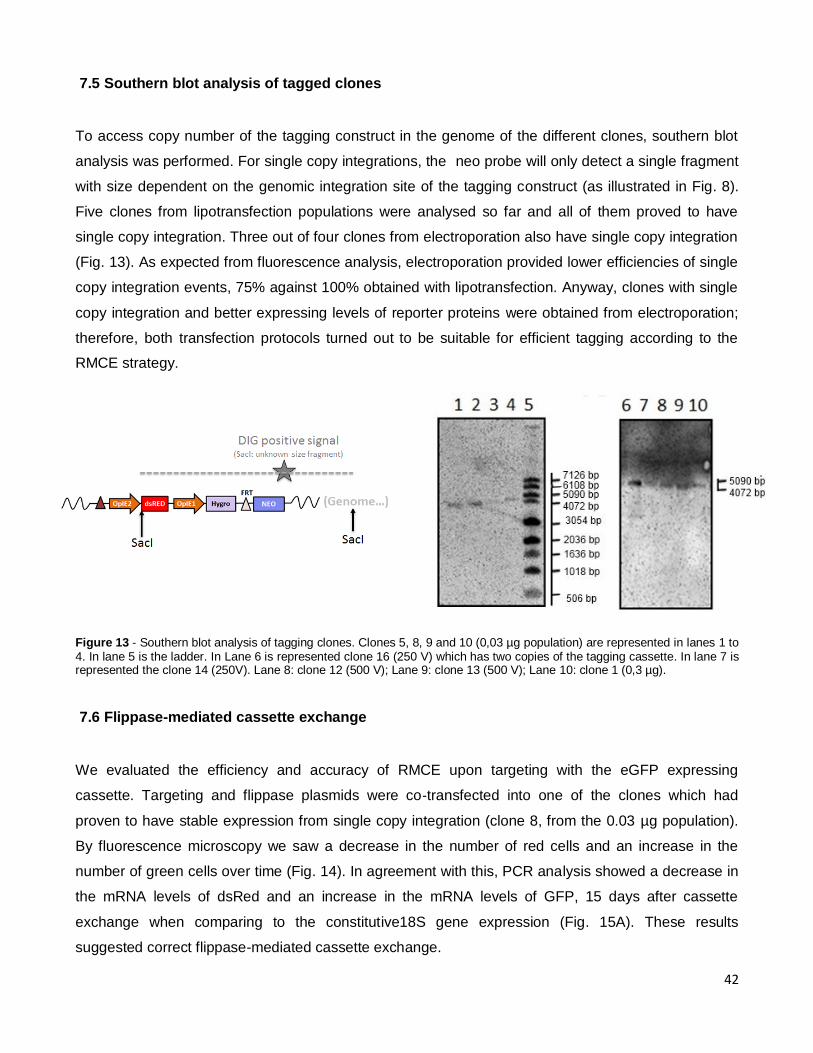
42
7.5 Southern blot analysis of tagged clones
To access copy number of the tagging construct in the genome of the different clones, southern blot
analysis was performed. For single copy integrations, the neo probe will only detect a single fragment
with size dependent on the genomic integration site of the tagging construct (as illustrated in Fig. 8).
Five clones from lipotransfection populations were analysed so far and all of them proved to have
single copy integration. Three out of four clones from electroporation also have single copy integration
(Fig. 13). As expected from fluorescence analysis, electroporation provided lower efficiencies of single
copy integration events, 75% against 100% obtained with lipotransfection. Anyway, clones with single
copy integration and better expressing levels of reporter proteins were obtained from electroporation;
therefore, both transfection protocols turned out to be suitable for efficient tagging according to the
RMCE strategy.
Figure 13 - Southern blot analysis of tagging clones. Clones 5, 8, 9 and 10 (0,03 µg population) are represented in lanes 1 to 4. In lane 5 is the ladder. In Lane 6 is represented clone 16 (250 V) which has two copies of the tagging cassette. In lane 7 is represented the clone 14 (250V). Lane 8: clone 12 (500 V); Lane 9: clone 13 (500 V); Lane 10: clone 1 (0,3 µg).
7.6 Flippase-mediated cassette exchange
We evaluated the efficiency and accuracy of RMCE upon targeting with the eGFP expressing
cassette. Targeting and flippase plasmids were co‐transfected into one of the clones which had
proven to have stable expression from single copy integration (clone 8, from the 0.03 µg population).
By fluorescence microscopy we saw a decrease in the number of red cells and an increase in the
number of green cells over time (Fig. 14). In agreement with this, PCR analysis showed a decrease in
the mRNA levels of dsRed and an increase in the mRNA levels of GFP, 15 days after cassette
exchange when comparing to the constitutive18S gene expression (Fig. 15A). These results
suggested correct flippase-mediated cassette exchange.

43
After 20 days in neomycin selection, there were practically no red cells and some green colonies had
reached a reasonable size, so they could be transferred to 6 well-plates for expansion and further
analysis.
Figure 15 – A) mRNA levels of dsRED and eGFP from clone 8 cells. L – Ladder; 1- red expression of tagging cells; 2- red
expression 15 days after targeting; 3- GFP expression of tagging cells; 4- GFP expression 15 days after targeting and G418
selection; 5- 18S expression of tagging cells (housekeeping gene); 6 – 18S expression 15 days after targeting. B) PCR to
genomic DNA from clone 8 cells. L – Ladder; 1- Tagging cells; 2- Targeted cells after 15 days in G418 selection. C)
Schemetic representation of the result of PCR to genomic DNA, in order to confirm cassette exchange. The same primers
were used ( pOpIE1 and neo marker).
Figure 14 – Fluorescence microscopy images of clone 8 (A) before targeting, (B - D) 72 h after targeting and (E – G) 2 weeks after targeting, using either GFP, dsRED or merging both channels.

44
The cassette exchange was also confirmed by PCR of genomic DNA. The same primers were used in
the tagging clone and in the cells after cassette exchange: the primers used were in OpIE1 promoter
and in neo marker. A difference of 1000 bp in size confirmed the exchange, since the target cassette
lacks the hygromycin gene between OpIE1 promoter and the neo resistance marker. This is the first
proof of flippase-mediated cassette exchange in Sf9 cells (Figs. 15B-C). Subclones of the targeting of
clone 8 can be seen in figure 16. These targeted cell clones still need to be screened for absence of
additional random integration events of the targeting cassette by real-time PCR or Southern Blot
analysis. The remaining clones which have proven to have single copy integration of the tagging
cassette were already targeted and placed in neomycin selection. From fluorescence microscopy
analysis, these clones show an increase in GFP expression and a decrease in dsRed expression
which is a good indication that cassette exchange has occurred well in these tagging clones too.
Figure 16 – Fluorescence microscopy images of the colonies obtained after targeting clone 8 cells when they were picked for
growth as sub-clones.
7.7 Tetracycline Inducible Circuits
In order to control and tune recombinant gene expression in Sf9 cells, a tetracycline-based
transcriptional regulatory circuit, developed originally for mammalian cells, was modified to work in
insect cells. This system is composed by two elements: i) a transactivator being continuously
expressed under the control of OpIE2 promoter, that will bind to ii) a sequence of seven Tet-operators
placed upstream of the Hsp70 minimal promoter and activate the expression of the gene of interest.
The hybrid promoter (Tet-operators and minimal hsp70 promoter) were obtained from a plasmid that
was being used in S2 Drosophila cells. The scheme of the two constructs is illustrated in Fig. 17.

45
Figure 17- Schematic representation of (A) transactivator (ptTA) and (B) TetOmin Hsp70 constructs.
Once the vectors were constructed, Sf9 cells were transfected with the transactivactor construct. The
transfection conditions were the same as in the first study for promoter strength (see materials and
methods). The population of Trans (transactivator) was then selected with zeocin for two weeks and
then analysed for transactivator expression by western blot (Fig. 18A). After confirming that cells were
expressing the transactivator, they were transfected with the second construct, the hybrid promoter
(TetOminHsp70) driving the expression of eGFP. We have also transfected parental Sf9 cells with
TetOminHsp70 to have a control for the basal expression (without transactivator). After two weeks in
hygromycin selection, both populations (with and without transactivator expression) seem to have
similar GFP expression levels (by fluorescence microscopy analysis).
A preliminary study was conducted by adding different doxycycline doses to evaluate how the system
responds. As explained in the Introduction section, when doxycycline is added to cells harboring the
Tet-Off system, it will bind to the transactivator, subsequently preventing the binding of the
transactivator to the promoter and thus reducing expression of the transgene (Fig. 19). Cells were
subjected to different levels of Dox during 7 days and then analysed by flow cytometry. GFP
expression decreased slightly (mean fluorescence intensity decreased from 22.0 to 16.9 as Dox was
increased from 0.01 to 100 ng/ml (Fig. 18C). In Fig. 18B, it is a comparison of GFP expression in three
cell populations: 1) the population with the full Hsp70 promoter, 2) the population of cells not
expressing no legends for the above figure, transactivator but harboring the TetOminhsp70 construct
and 3) the population transfected with both transactivator and TetOminhsp70 constructs. We can see
that there is a huge difference in GFP expression driven by the full and minimal Hsp70 promoter,
which was supposed to be rescued in the transactivator expressing cells. However, only a slight
increase in mean fluorescence intensity was observed suggesting that somehow the transactivator is
not reaching the Tet-operators sequence in the hybrid promoter as expected.

46
Figure 18 – A) Western blot analys of the transactivator (37 kDa). L - Ladder, 1- Non transfected cells, 2- Transfected cells with 0,05µg of transactivator plasmid and 3- Transfected cells with 0,3µg of transactivator plasmid. B) comparison of GFP expression in three cell populations: i) the population with the full Hsp70 promoter, ii) the population of cells not expressing transactivator but harboring the tetOminhsp70 construct and iii) the population transfected with both transactivator and tetOminhsp70 constructs. C) Cells harboring both constructs were subjected to different levels of Dox during 7 days and then analysed by flow cytometry.
Figure 19 – Scheme of the way how the system should work inside the cells. A) First, cells were transfected with the transactivator B) integrated and selected in order to have a population expressing transactivator C) This population was then transfected with tetominhsp70 and selected. D) If no DOX is on the system there is a maximum of expression of GFP. When dox is added, expression decreases in a dose-dependent manner.

47
8. Discussion
The first steps in the development of a reusable insect cell factory were undertaken in this thesis. This
cell line is expected to be advantageous over the baculovirus-insect cell system, the current gold
standard to produce multimeric proteins. Three major tasks were performed during this thesis: 1)
different transcriptional promoters were first compared in terms of reporter protein expression and
genetic stability in Sf9 cells; then 2) a Flippase RMCE system was implemented in Sf9 cells using the
best performing promoter and finally 3) some preliminary work concerning the implementation of an
inducible system for proper stoichiometric expression of multicomponent complex proteins was
performed.
8.1 Comparison of different promoters strength
For expression of recombinant proteins in biological systems, an initial task is the selection of suitable
promoters. Strong promoters with little or almost no tissue specificity must be considered. In what
concerns Sf9 cells, constitutive or inducible promoters have not yet been extensively studied, much
because these cells have been used essentially in the context of the baculovirus expression system
and also because the genome of these cells is not sequenced yet. Thus, constitutive and inducible
promoters from closer species, such as Drosophila, and immediate early promoters from baculovirus
these cells were evaluated. When these promoters were cloned into the same vector systems, their
expression activities could be compared. Two inducible promoters were tested: Drosophila hsp70 and
metallothionein promoters (Steller and Pirrotta, 1986; Hegedus et al., 1998). The Hsp70 for a cellular
promoter is rather strong; it gives rise to high background activity without heat shock treatment, acting
just like a constitutive promoter (Clem and Miller, 1994; Prikhod’ko and Miller, 1996). The
metallothionein promoter revealed to be the worst alternative for stable protein expression in Sf9 cells
due to the high concentration of metals required to have measurable protein expression, leading to
high cytotoxicity. Hegedus et al. (1998) have shown that the Drosophila metallothionein promoter
allows higher level expression in dipteran cells than in lepidopteran cells such as Sf9 cells used in this
work which corresponds to our results. The baculovirus early promoters are stronger, allowing higher
protein levels than the Drosophila-derived promoters. Their capability of inducing high translational
rates in most lepidopteran cell lines have been described previously (Pfeifer et al, 1997). From our
results, OpIE2 promoter allowed a four-fold increase in gene expression comparing to hsp70

48
promoter, but there are some studies reporting up to ten fold increase (Mclachlin et al, 1997).
Moreover, the comparison between OpIE1 and OpIE2 promoters was performed previously in our lab,
with the OpIE2 promoter allowing about five-fold more reporter protein, in agreement with Pfeifer et al,
(1997).
8.2 Flippase-Mediated Cassette Exchange
Our approach to establish a reusable high producer recombinant insect cell line relies on a flippase
site-specific recombination system. This strategy was already successfully implemented in several
mammalian cell lines for the production of various proteins, including secreted monoclonal antibodies
and even to produce highly complex biological entities like retroviral vectors for gene therapy purposes
(Schucht et al. 2006; Coroadinha et al. 2006; Wirth et al. 2007). The success of the establishment of
this producer cell line depends on some critical aspects, such as an efficient tagging of a hotspot
supporting higher expression levels, single copy integration of the tagging cassette, genetic stability
and the ability of targeting the tagged site.
The tagging cassette was randomly integrated in Sf9 cells by two transfection methods, cellfectin-
based lipotransfection and electroporation, as we did not know which one would allow preferentially
single copy integration. Although cellfectin-based transfection allowed higher transfection efficiencies,
so far all seven cellfectin clones analysed by Southern blot revealed to have single copy integration.
From electroporation, only one from the four analyzed clones has two copies of the tagging cassette.
Therefore both transfection methods seem to be adequate for single copy integration purposes.
A remarkable advantage of this tagging/targeting strategy in relation to traditional cell line
development is that the cells are marked by fluorescence and so high producers can be pre-selected
for dsRED expression to ensure only active transcriptional sites to be tested for their single copy
integration and exchangeability (Qiao et al., 2009). The broad expression pattern of the selected
clones indicates variable integration sites.
Correct cassette exchange was confirmed in clone 8, one of the clones of lipotransfection with
0,03 µg of tagging cassette. Our system uses for selection an ATG-deficient neomycin gene
positioned downstream to the FRT site. This system assures correct targeting of recombinants by
ATG complementation of neomycin resistance gene, allowing for neomycin selection after exchange.
If only excision would occur without introduction of the targeting cassette, cells would not be G418
resistant (Seibler, 1999). In the absence of this positive selection with NEO complementation, random
integration would be favored over RMCE since this reaction can only take place at a single site
whereas random integration can be widespread in the whole genome (Oumard et al, 2006; Qiao et al,

49
2009). Nevertheless, high expressing clones can overpass the ATG deficiency and resist to the G418
without exchanging cassettes favoring in this case random integration (Coroadinha et al 2006). To
exclude this scenario, we performed a PCR analysis directly to genomic DNA from clone 8 before and
after co-transfection with flippase and target cassette. The difference in size between the two bands
confirmed undoubtedly cassette exchange. Therefore, we have shown for the first time that with the
right modifications flippase-RMCE systems can be applied also to insect cells. This approach opens
the way to a highly flexible method for protein expression in Sf9 cells.
Once we generate a master cell line, it will be an ideal platform for the integration of various genes of
interest. In contrast to random approaches, screening efforts are minor. Therefore, compared to the
traditional integration and selection that takes up to 12 months (Wurm et al, 2004), the tag-and-
targeting RMCE strategy only takes about 4 – 6 months to have a novel expression clone with
predictable properties (Schucht et al. 2006).
8.3 Transcriptional regulatory circuits
Tightly controlled expression of transgenes is a challenge in the establishment of sophisticated
cellular systems. Two different patterns of control are normally attempted: (1) the restriction of gene
expression to a certain time period while shutting off transgene expression when not needed and (2)
to express adjustable levels of a protein in order to individually modulate the concentration of the
transgene (Schucht et al, 2010). Our aim fits into the second pattern as we intend to control
expression of one protein in relation to other protein, both composing the same particle, to obtain
stoichiometric balanced expression and improve in this way the percentage of correctly assembled
particles. A tetracycline transcriptional control system was implemented in Sf9 cells; the expression of
the mammalian transactivator originally conceived by Gossen and Bujard (1992), was driven by the
strong Opie2 promoter, and the reporter plasmid was modified from one applied to Drosophila (Bello
et al. 1998) in which the seven tandem repeated TetO sequences were placed upstream to a minimal
Hsp70 promoter, and this hybrid promoter drives the expression of GFP.
The first result we obtained was that, comparing to other systems like Drosophila and mammalian
cells (Gossen and Bujard, 2002), this Tet-Off system could not induce GFP expression with the same
strength. Sf9 cells transfected with just the reporter TetOminHsp70 plasmid exhibit only a slight
difference in terms of GFP fluorescence intensity when comparing to cells harboring both parts of the
Tet-system: the transactivator and the reporter TetOminHsp70 construct. Moreover, the presence of
Dox just decreased only slightly GFP expression, even using high doses of Dox (100 ng/ml). This

50
means that there is a problem with one of the parts concerning this system, most likely with the
transactivator as it has been recently reported in other insect cell line (Karasaki et al, 2009) .
One major concern is that the transactivator has to function in the nucleus, thus a nuclear import of
this protein should occur. The recognition is in general well conserved for nuclear protein import
mechanisms but in some pathways is different among species (Riolobos et al, 2010). Indeed some
studies have shown that this type of import can be mediated by recognition of the pattern of
phosphorylation in the proteins and it was observed that recombinant proteins from mammalian virus
when produced in insect cell are not correctly imported (Karasaki et al, 2009, Riolobos et al, 2010).
The transactivator is composed by two proteins fused: the tetracycline repressor protein from E. coli
and VP16 which is an abundant phosphoprotein present in the tegument of the herpes simplex virus.
VP16 enhances gene expression in the tet transactivator (Schmelter et al., 1996). An incorrect
phosphorylation of this mammalian virus protein could lead to an incorrect nuclear importation or
incorrect action as transactivator (Riolobos et al, 2010).In Bombyx mori cells Karasaki et al. (2009)
observed a 100 fold increase in luciferase expression in a Tet-On system when a nuclear locazation
signal (NLS) sequence was added to the transactivator.
This might suggest that nuclear protein import mechanisms are different between the two species.
Despite the benefits of NLS in the closely related Bombyx mori cells, we cannot exclude the possibility
of the transactivator being expressed in limiting amounts in Sf9 cells.

51
9. Conclusion and Future Work Perspectives
In this work it was approached for the first time site‐directed engineering of defined chromosomal site
in Sf9 cells for the development of a flexible insect cell factory for the production of multiple complex
biopharmaceuticals, bypassing the need for baculovirus infection. Although the work is far from being
finished we have proved that the use of RMCE can be implemented into these cells with extremely
success. The RMCE technology, which allows the introduction of heterologous genes at a predefined
chromosomal locus, makes production more predictive and, importantly, allows to reduce the time
necessary to define the best producer clone for a particular product each time new complex
biopharmaceuticals are intended to be expressed in this cell line.
In parallel, the firsts steps, for the implementation of a tetracycline regulatory network in sf9 cells were
conducted. From the results, it is clear that additional changes to the original system must be
performed to obtain a Tet-Off system working properly, as it seems that too few if any transactivator is
reaching the Tet-operators and so the transactivator is not exerting any control in GFP expression.
Therefore, as future work, we intend to optimize the Tetracycline inducible system. We will focus on
the transactivator, in which we can test the effect of adding a nuclear localization signal or even
substitute the VP16 mammalian protein to a transcription factor form of virus infecting Sf9 cells, such
as baculovirus. Also, instead of minimal Hsp70 promoter we can test minimal OpIE2 promoter. We
can anticipate a higher basal expression using this stronger promoter in a Tet-Off system, but this is
not really a disadvantage for the application in question. Ideally, the expression without inducer should
be as higher as possible and then we should be able to decrease it with some precision by adding a
certain amount of inducer.
In addition, we will construct a second cassette system, which will be based on the cre-recombinase
system. The best flippase-tagging clone will be transfected with the second cassette and we will
screen again for clones with high and stable expression of this cassette and obviously with single copy
integration. Then, to be able to control expression of both proteins in relation to each other, the
transcriptional circuit will be implemented in one of the cassettes. Finally, we will exchange cassettes
with the genes of interest, for instance VP2 and VP6 which are structural proteins from rotavirus that
self-assemble to produce a rotavirus-like particle vaccine, and evaluate how this master cell line
performs in terms of yields and quality comparing to the gold standard baculovirus expression system.

52
Bibliography
Alexander H., Booy E., Xiao W, Ezzati P., Baust H, Los M. (2007). Selected Technologies To Control Genes And Their
Products For Experimental And Clinical Purposes. Archivum immunologiae et therapiae experimentalis. 55(3), 139-49.
Baneyx F. (1999). Recombinant protein expression in Escherichia coli. Current Opinion Biotechnoly 10, 411–421
Beljelarskaya S. (2002). A Baculovirus Expression System for Insect Cells. Molecular Biology. 36(3), 281-292.
Bello B., Resendez-Perez D., Gehring W. (1998). Spatial and temporal targeting of gene expression in Drosophila by means
of a tetracycline-dependent transactivator system. Development .125(12): 2193--2202.
Boshart M., Weber F., Jahn G., Dorsch-Hasler K., Fleckenstein B., Schaffner W. (1985). A Very Strong Enhancer Is Located
Upstream of an Immediate Early Gene Of Human Cytomegalovirus. Cell. 41, 521-30.
Branda C. and Dymecki S. (2004). Talking About A Revolution: The Impact Of Site-Specific Recombinases On Genetic
Analyses In Mice. Development cell. 6(1), 7-28.
Broach J. and Hicks J. (1980) Replication and Recombination Functions Associated with the Yeast Plasmid, 2mu
Circle. Cell 17, 501-508.
Buchholz F., Angrand P., Stewart A. (1996). A Simple Assay To Determine The Functionality Of Cre Or FLP Recombination
Targets In Genomic Manipulation Constructs. Nucleic Acids Research. 24(15), 3118-3119.
Butler M. (2005). Animal Cell Cultures: Recent Achievements And Perspectives In The Production Of Biopharmaceuticals.
Applied microbiology and biotechnology. 68(3), 283-91.
Chtarto A., Bender H., Hanemann C., Kemp T., Lehtonen E., Levivier M., (2003). Tetracycline-Inducible Transgene
Expression Mediated By A Single AAV Vector. Gene therapy. 10(1), 84-9.
Clem R. and Miller L. (1994) Control of programmed cell death by the baculovirus genes p35 and iap. Molecular and Cellular
Biology, 14, 5212-5222.
Cobellis G., Nicolaus G., Iovino M., Romito A., Marra E., Barbarisi M. (2005). Tagging genes with cassette-exchange sites.
Nucleic Acids Research, 33(4), 44.
Coroadinha A., Schucht R., Gama-Norton L., Wirth D., Hauser H., Carrondo M., (2006). The Use of Recombinase Mediated
Cassette Exchange In Retroviral Vector Producer Cell Lines: Predictability And Efficiency By Transgene Exchange.
Journal of Biotechnology. 124(2), 457-68.
Frandsen T., Naested H., Rasmussen S., Hauptig P., Wiberg F., Rasmussen L., Jensen A., Persson P., Wikén M., Engström
A., Jiang Y., Thorpe S., Förberg C., Tolstrup A. (2011).Consistent manufacturing and quality control of a highly complex
recombinant polyclonal antibody product for human therapeutic use. Biotechnology Bioengineering.108(9):2171-81.
Fussenegger M., Morris R., Fux C., Rimann M., von Stockar B., Thompson C., Bailey J. (2000) Streptogramin-Based Gene
Regulation Systems For Mammalian Cells. Nature Biotechnology. 18, 1203-8.

53
Fussenegger M. (2001). The impact of mammalian gene regulation concepts on functional genomic research, metabolic
engineering, and advanced gene therapies. Biotechnology Progress. 17, 1–51
French T. and Roy P. (1990). Synthesis of bluetongue virus (BTV) corelike particles by a recombinant baculovirus expressing
the two major structural core proteins of BTV, Journal of Virology. 64, 1530–1536.
Geisse S., Gram H., Kleuser B., Kocher H. (1996). Eukaryotic Expression Systems: A Comparison. Protein expression and
purification, 8(3), 271-82.
Goodrich J., Hoey T., Thut C., Admon A., Tjian R. (1993). Drosophila TAFII40 interacts with both a VP16 activation domain
and the basal transcription factor TFIIB. Cell 75, 519-30
Gossen M., and Bujard H. (2002). Studying Gene Function In Eukaryotes By Conditional Gene Inactivation. Annual Review
of Genetics. 36, 153-73.
Granados R., Li G., Blissard, G. (2007). Insect Cell Culture and Biotechnology. 22(2), 83-93.
Groth A., Olivares E., Thyagarajan B., Calos. M. (2000). A Phage Integrase Directs Efficient Site-Specific Integration In
Human Cells. Proceedings of the National Academy of Sciences 97(11), 5995-6000.
Hegedus D., Pfeifer T., Hendry J., Theilmann D., Grigliatti T.,(1998). A series of broad host range shuttle vectors for
constitutive and inducible expression of heterologous proteins in insect cell lines. Gene 207, 241–24
Huang Y., Li Y., Wang Y., Gu X., Wang Y., Shen B. (2007). An Efficient And Targeted Gene Integration System For High-
Level Antibody Expression. Journal Immunology Methods. 322 (1-2), 28-39.
Hunt I. (2005). From gene to protein: a review of new and enabling technologies for multi-parallel protein expression, Protein
Expression Purifification. 40, 1–22.
Ikonomou, L., Schneider Y., Agathos, S. (2003). Insect Cell Culture For Industrial Production Of Recombinant Proteins.
Applied microbiology and biotechnology. 62(1), 1-20.
Ingles C., Shales M., Cress W., Triezenberg S., Greenblatt J. (1991). Reduced Binding Of TFIID To Transcriptionally
Compromised Mutants Of VP16. Nature. 351, 588-90.
Jarvis D., Fleming J., Kovacs G., Summers M., and Guarino L. (1990). Use Of Early Baculovirus Promoters For Continuous
Expression And Efficient Processing Of Foreign Gene Products In Stably Transformed Lepidopteran Cells. Nature
BioTechnology. 8, 950–955.
Jarvis D., and Summers M. (1989) Glycosylation And Secretion Of Human Tissue Plasminogen Activator In Recombinant
Baculovirus-Infected Insect Cells. Molecular and Cellular Biology. 9, 214–223.
Jarvis,D.. (1997) Baculovirus expression vectors. In Miller,L.K. (ed.), The Baculoviruses. Plenum Press, New York, 389–431
Kaern M., Blake W., Collins J., (2003). The Engineering of Gene Regulatory Networks. Annual Review of Biomedical
Engineering. 5, 179-206.

54
Karasaki N., Mon H., Takahashi M., Lee J., Koga K., Kawaguchi Y., Kusakabe T. (2009). Establishment of tetracycline-
inducible gene expression systems in the silkworm, Bombyx mori. Biotechnology Letters, 31(4), 495-500.
Kito M., Itami S., Fukano Y., Yamana K., and Shibui T. (2002). Construction Of Engineered CHO Strains For High-Level
Production Of Recombinant Proteins. Applied Microbiology and Biotechnology. 60(4), 442-448.
Kramer B., Weber W., Fussenegger M. (2001). Artificial regulatory networks and cascades for research, metabolic
engineering, and advanced gene therapies. Biotechnolology Progress 17, 1-51
Kromenaker S. and Srienc F. (1994). Stability Of Producer Hybridoma Cell Lines After Cell Sorting: A Case Study.
Biotechnology Progress. 10(3), 299-307.
Li J., Menzel C., Meier D., Zhang C., Dubel S., Jostock T. (2007). A Comparative Study Of Different Vector Designs For The
Mammalian Expression Of Recombinant Igg Antibodies. Journal of Immunological Methods 318(1-2), 113-124.
Lin Y., Ha I., Maldonado E., Reinberg D., Green M. (1991). Binding Of General Transcription Factor TFIIB To An Acidic
Activating Region. Nature 353, 569-71.
May T., Hauser H., Wirth D. (2006). Current Status of Transcriptional Regulation Systems. Cytotechnology. 50, 109-119.
McCarroll L. and King L. (1997). Stable Insect Cell Cultures for Recombinant Protein Production. Current Opinion in
Biotechnology. 8(5), 590-4.
Mclachlin J. and Miller L. (1997). Stable Transformation Of Insect Cells To Coexpress A Rapidly Selectable: Opiap. Growth.
In Vitro Cellular & Developmental Biology – Animal. 33, 575-579.
Nakayama G., Kawaguchi Y., Koga K., Kusakabe T. (2006). Site-specific gene integration in cultured silkworm cells mediated
by phiC31 integrase. Molecular genetics and genomics, 275(1), 1-8.
Narlikar G., Fan H., Kingston R. (2002). Cooperation between Complexes that Regulate Chromatin Structure and
Transcription. Cell .108(4): 475-487
Orphanides G. and Reinberg D. (2002). A Unified Theory of Gene Expression. Cell 108(4), 439-451.
Oumard, A., Qiao J., Jostock T., Li J., Bode J. (2006). Recommended Method for Chromosome Exploitation: RMCE-based
Cassette-exchange Systems in Animal Cell Biotechnology. Cytotechnology 50, 93-108
Park J., Kim H., Hwang H., Lee S., Kim H., Hur B., Ryu Y., An C., Kim J. (2004). Large-scale production of rotavirus VLP as
vaccine candidate using baculovirus expression vector system, Journal of Microbiology and Biotechnology. 14(1) 35–40
Pelham H., (1982). A regulatory upstream promoter element in the selectable G418 resistance to Drosophila larvae.
European Molecular Biology Organization Journal. 4,167–171.
Pfeifer T., Hegedus D., Grigliatti T., Theilmann, D. (1997). Baculovirus Immediate-Early Promoter-Mediated Expression Of
The Zeocin Resistance Gene For Use As A Dominant Selectable Marker In Dipteran And Lepidopteran Insect Cell Lines.
Gene. 188(2), 183-90.

55
Pfleger B., Pitera D., Smolke C., Keasling J. (2006). Combinatorial Engineering Of Intergenic Regions In Operons Tunes
Expression Of Multiple Genes. Nature biotechnology. 24(8), 1027-32.
Prikhod'ko E. and Miller L. (1996). Induction of apoptosis by baculovirus transactivator IE1. Journal of Virology. 70,7116-7124
Qiao J., Oumard A., Wegloehner W., Bode J. (2009). Novel Tag-And-Exchange (RMCE) Strategies Generate Master Cell
Clones With Predictable And Stable Transgene Expression Properties. Journal of Molecular Biology. 390(4), 579-94.
Ramachandra C., Shahbazi M., Kwang T., Choudhury Y., Bak X., Yang J. (2011). Efficient Recombinase-Mediated Cassette
Exchange At The AAVS1 Locus In Human Embryonic Stem Cells Using Baculoviral Vectors. Nucleic Acids Research. 1-
13.
Riolobos L., Valle N., Hernando E., Maroto B., Kann M., Almendral J. (2010). Viral oncolysis that targets Raf-1 signaling
control of nuclear transport. Journal of Virology, 84,2090-9
Roebroek A, Wu X, Bram R. (2003). Knockin approaches. Methods Molecular Biology, 209,187–200
Roldão A., Vieira H., Alves P., Oliveira R., Carrondo M. (2006). Intracellular dynamics in rotavirus-like particles production:
Evaluation of multigene and monocistronic infection strategies. Process Biochemistry, 41(10), 2188-2199.
Schaft J., Ashery-Padan R., van der Hoeven F., Gruss P., Stewart A. (2001). Efficient FLP Recombination In Mouse ES
Cells And Oocytes. Genesis, 31(1), 6-10.
Schlake T. and Bode J. (1994). Use Of Mutated FLP Recognition Target (FRT) Sites For The Exchange Of Expression
Cassettes At Defined Chromosomal Loci. Biochemistry, 33(43), 12746-12751.
Schmelter J., Knez J., Smiley J., Capone J. (1996). Identification and characterization of a small modular domain in the
herpes simplex virus host shutoff protein sufficient for interaction with VP16. Journal of Virology. 70:2124–2131.
Schmidt F. (2004). Recombinant Expression Systems In The Pharmaceutical Industry. Applied microbiology and
biotechnology. 65(4), 363-72.
Schucht R., Coroadinha A., Zanta-Boussif, M., Verhoeyen E., Carrondo M., Hauser H., Wirth D. (2006). A new generation of
retroviral producer cells: predictable and stable virus production by Flp-mediated sitespecific integration of retroviral
vectors. Molecular Therapy, 14, 285-92.
Schucht R., Wirth D., May T. (2010) Precise regulation of transgene expression level and control of cell physiology. Cellular
Biology Toxicology. 26:29–42
Seibler J., Schübeler D., Fiering S., Groudine M., Bode J., (1998) DNA cassette exchange in ES cells mediated by Flp
recombinase: an efficient strategy for repeated modification of tagged loci by marker-free constructs. Biochemistry, 37:
6229–6234
Steller H. and Pirrotta V. (1985). A transposable P vector that confers selectable G418 resistance to Drosophila larvae.
European Molecular Biology Organization Journal, 4, 167-171.
Sternberg N., Sauer B., Hoess R., Abremski K. (1986). Bacteriophage P1 Cre Gene And Its Regulatory Region. Evidence
For Multiple Promoters And For Regulation By DNA Methylation. Journal of Molecular Biology. 187(2), 197-212.

56
Thorpe H. and Smith M. (1998). In Vitro Site-Specific Integration Of Bacteriophage DNA Catalyzed By A Recombinase Of
The Resolvase/Invertase Family. Proceedings of the National Academy of Sciences. 95(10), 5505-5510.
Thyagarajan B., Olivares E., Hollis R., Ginsburg D., Calos M. (2001). Site-Specific Genomic Integration In Mammalian Cells
Mediated By Phage Phic31 Integrase. Molecular and Cellular Biology. 21(12), 3926-3934.
Tigges M., Marquez-Lago T., Stelling J., Fussenegger M. (2009) A tunable synthetic mammalian oscillator Nature, 457: 309-
312.
Trill J., Shatzman A., Ganguly S. (1995). Production Of Monoclonal Antibodies In COS And CHO Cells. Current Opinion in
Biotechnology. 6(5), 553-560.
Turan S., Galla M., Ernst E., Qiao J., Voelkel C., Schiedlmeier B., Zehe C., Bode J. (2011). Recombinase-Mediated Cassette
Exchange (RMCE): Traditional Concepts And Current Challenges. Journal of molecular Biology. 407(2), 193-221.
Vaughn J., Goodwin R., Tompkins G., Mccawley A., Al V. (1977). The History And Characteristics Of Two Cell Lines
Developed From Primary Explants Of Key Words : Characterization . In Vitro. 13(4), 0-4.
Vieira H., Estêvão C., Roldão A., Peixoto C., Sousa M., Cruz P., Carrondo M., Alves P. (2005). Triple layered rotavirus VLP
production: kinetics of vector replication, mRNA stability and recombinant protein production. Journal of Biotechnology,
120: 72-82.
Walsh G. (2005). Biopharmaceuticals: Recent Approvals and Likely Directions. Trends in biotechnology. 23(11), 553-8.
Weber W., Fux C., Daoud-El Baba M., Keller B., Weber C., Kramer B., Heinzen C., Aubel D., Bailey J., Fussenegger M.
(2002). Macrolide-Based Transgene Control In Mammalian Cells And Mice. Nature Biotechnology. 20, 901-7.
Weber W. and Fussenegger M. (2009). Insect Cell-Based Recombinant Protein Production. Cell and Tissue Reaction
Engineering. 263-277.
Weber W. and Fussenegger M. (2004) Approaches for trigger-inducible viral transgene regulation in geno-based tissue
engineering, Current Opinion in Biotechnology. 15, 383–391
Wiberg C., Rasmussen K., Frandsen P., Rasmussen K., Tengbjerg K., Coljee V., Sharon J., Yang C., Bregenholt, Nielsen L.,
Haurum J., Tolstrup A. (2006). Production Of Target-Specific Recombinant Human Polyclonal Antibodies In Mammalian
Cells. Biotechnology Bioengineering. 94(2), 396-405.
Wirth D., Gama-Norton L., Riemer P., Sandhu U., Schucht R., Hansjörg H. (2007) Road to precision: recombinase-based
targeting technologies for genome engineering. Current Opinion Biotechnology. 18: 411–419.
Wurm F. (2004). Production Of Recombinant Protein Therapeutics In Cultivated Mammalian Cells. Nature Biotechnology. 22,
1393–1398.
Yamaji H., Manabe T., Watakabe K., Muraoka M., Fujii I., Fukuda H. (2008). Production Of Functional Antibody Fab
Fragment By Recombinant Insect Cells. Biochemical Engineering Journal. 41(3), 203–209.
Zhu X. and Sadowski P. (1995). Cleavage-Dependent Ligation By The FLP Recombinase. Characterization Of A Mutant FLP
Protein With An Alteration In A Catalytic Amino Acid. The Journal of Biological Chemistry. 270(39), 23044-54.

57
Appendix
Figure 20 - Killing Curves for the three antibiotics used for selection of the cells. A) G418, B) Hygro and C) Zeo.
Table 2 – Primers used in the construction of vectors need in this work.