development of immunopathogenesis strategies to treat behçet's
TRANSCRIPT

Hindawi Publishing CorporationPathology Research InternationalVolume 2012, Article ID 261989, 7 pagesdoi:10.1155/2012/261989
Review Article
Development of Immunopathogenesis Strategies to Treat Behcet’sDisease
Osman Kose
Department of Dermatology, School of Medicine, Gulhane Military Medical Academy, Tevfik Saglam Street No. 1,06018 Ankara, Turkey
Correspondence should be addressed to Osman Kose, [email protected]
Received 8 August 2011; Accepted 25 January 2012
Academic Editor: Umit Tursen
Copyright © 2012 Osman Kose. This is an open access article distributed under the Creative Commons Attribution License, whichpermits unrestricted use, distribution, and reproduction in any medium, provided the original work is properly cited.
Behcet disease is a chronic relapsing vasculitis with unclear etiology and immunopathogenesis. Antigenic stimuli, antigenpresenting cells, T cells, monocyte, and neutrophil and endothelial cells are major parts of the pathology of the disease.Understanding of the new pathogenic mechanisms based on molecular structure of the disease helps us in improving the noveltherapeutic modalities. These drugs target specific and nonspecific inhibition of the immun system. These therapies includebiologic agents, new topical and systemic immunosuppressants, tolerizing agents, and immunoablation. Novel treatment will bepromising to treat the especially recalcitrant cases to conventional therapy. In this paper, new aspect of the immunopathogenesisof Behcet’s diseases and novel treatment modalities will be discussed.
1. Introduction
Behcet disease (BD) is a vasculitis that, characterized byrecurrent aphthous stomatitis, genital ulcers, skin lesions,relapsing uveitis, articular, neurologic, urogenital, vascular,intestinal, and pulmonary manifestations [1–3]. BD has beenreported worldwide but has a distinct geographic distrubi-tion, with highest prevalences in countries such as Turkey,Iran, and Japan which are place on silk road. Although muchhas been learned during recent years on the pathogenesisand treatment of the disease, the etiology and pathogenesisof BD have not been fully clarified [4, 5]. Symptoms of thedisease are considered to be based on the correlation betweenthe genetic intrinsic factors and the triggering extrinsicfactors, because more than 60% of BD patients are associatedwith HLA-B 51. Immune-mediated mechanisms play a majorrole in the pathogenesis of the disease, and inflammatorymediators are also involved [4–6]. Nowadays, recent investi-gations have made clear explanations about the pathogenesisof the disease. The hypersensitivity of T lymphocytes todifferent types of antigens plays a crucial role in thepathogenesis [3–6].
The present paper overviews an update on the immun-opathogenesis of BD and also novel treatment based onpathogenesis.
2. Immunopathogenesis of Behcet’s Disease
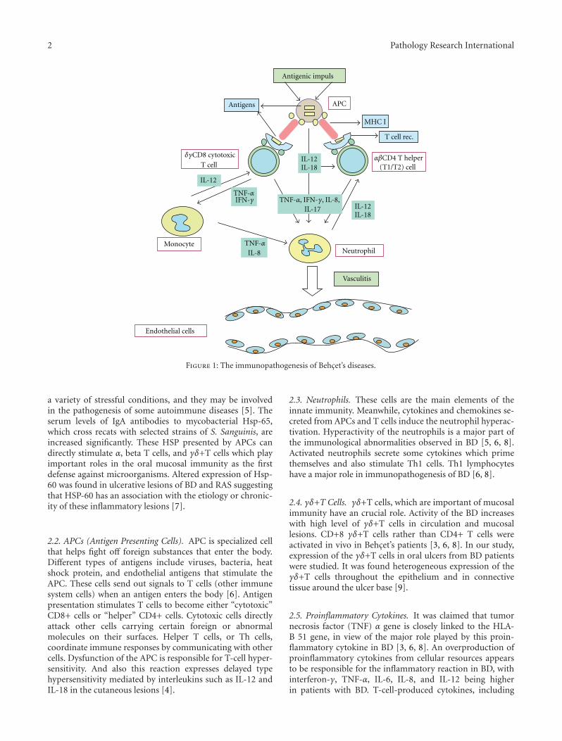
BD is an inflammatory disorder characterized mainly bymucocutaneous findings and uveitis. However, it can bepresent with other cutaneous symptoms such as pseudofolli-culitis, erythema nodosum, and pyoderma gangrenosum [2–5]. It can be present with articular, neurological, pulmonary,intestinal manifestations other than classical triad. The closerelationship between the genetic and triggering external fac-tors is thought to be present in the pathogenesis of BD [3,4]. The immunopathogenesis of BD is shown in Figure 1.Critical region for BD in the human major histocompatibilitycomplex (MHC) gene could be pinpointed to a 46 kb seg-ment between the MHC class I gene and HLA-B51 gene [5].
2.1. Heat Shock Proteins (HSP). HSP, which essentiallyscavenge denatured intracellular proteins, are supposed to beinduced by microorganisms and mammalian tissues under
2 Pathology Research International
Antigenic impuls
Antigens APC
MHC I
T cell rec.
αβCD4 T helper(T1/T2) cell
IL-12
IL-12IL-18
IL-8
IL-18
δγCD8 cytotoxic
T cellIL-12
TNF-α
TNF-α
IFN-γ
MonocyteNeutrophil
Vasculitis
Endothelial cells
TNF-α, IFN-γ, IL-8,IL-17
Figure 1: The immunopathogenesis of Behcet’s diseases.
a variety of stressful conditions, and they may be involvedin the pathogenesis of some autoimmune diseases [5]. Theserum levels of IgA antibodies to mycobacterial Hsp-65,which cross recats with selected strains of S. Sanguinis, areincreased significantly. These HSP presented by APCs candirectly stimulate α, beta T cells, and γδ+T cells which playimportant roles in the oral mucosal immunity as the firstdefense against microorganisms. Altered expression of Hsp-60 was found in ulcerative lesions of BD and RAS suggestingthat HSP-60 has an association with the etiology or chronic-ity of these inflammatory lesions [7].
2.2. APCs (Antigen Presenting Cells). APC is specialized cellthat helps fight off foreign substances that enter the body.Different types of antigens include viruses, bacteria, heatshock protein, and endothelial antigens that stimulate theAPC. These cells send out signals to T cells (other immunesystem cells) when an antigen enters the body [6]. Antigenpresentation stimulates T cells to become either “cytotoxic”CD8+ cells or “helper” CD4+ cells. Cytotoxic cells directlyattack other cells carrying certain foreign or abnormalmolecules on their surfaces. Helper T cells, or Th cells,coordinate immune responses by communicating with othercells. Dysfunction of the APC is responsible for T-cell hyper-sensitivity. And also this reaction expresses delayed typehypersensitivity mediated by interleukins such as IL-12 andIL-18 in the cutaneous lesions [4].
2.3. Neutrophils. These cells are the main elements of theinnate immunity. Meanwhile, cytokines and chemokines se-creted from APCs and T cells induce the neutrophil hyperac-tivation. Hyperactivity of the neutrophils is a major part ofthe immunological abnormalities observed in BD [5, 6, 8].Activated neutrophils secrete some cytokines which primethemselves and also stimulate Th1 cells. Th1 lymphocyteshave a major role in immunopathogenesis of BD [6, 8].
2.4. γδ+T Cells. γδ+T cells, which are important of mucosalimmunity have an crucial role. Activity of the BD increaseswith high level of γδ+T cells in circulation and mucosallesions. CD+8 γδ+T cells rather than CD4+ T cells wereactivated in vivo in Behcet’s patients [3, 6, 8]. In our study,expression of the γδ+T cells in oral ulcers from BD patientswere studied. It was found heterogeneous expression of theγδ+T cells throughout the epithelium and in connectivetissue around the ulcer base [9].
2.5. Proinflammatory Cytokines. It was claimed that tumornecrosis factor (TNF) α gene is closely linked to the HLA-B 51 gene, in view of the major role played by this proin-flammatory cytokine in BD [3, 6, 8]. An overproduction ofproinflammatory cytokines from cellular resources appearsto be responsible for the inflammatory reaction in BD, withinterferon-γ, TNF-α, IL-6, IL-8, and IL-12 being higherin patients with BD. T-cell-produced cytokines, including
Pathology Research International 3
interleukin (IL)-2, tumor necrosis factor (TNF)-α, interferon(IFN)-γ, IL-12 and IL-18, are elevated and probably con-tribute to neutrophil and endothelial cell activation. Andalso IL-12, and IL-18, which are mainly produced by APCs,regulate the neutrophil function and may play important rolein the skewing of immune response [4, 8].
2.6. Th17 Cells. It was reported a marked increase in Th17cell numbers and a decreased frequency of CD4 (+) forkheadbox protein 3 positive Treg cells in the peripheral blood ofpatients with active BD [8–12]. Th17/Th1 ratio was elevatedin BD patients with uveitis or folliculitis compared tothose without it [11]. Th17 cells regulate inflammation viaproduction of distinct cytokines such as IL-17. There is grow-ing evidence that Th17 cells are pathological in many humanautoimmune and inflammatory diseases [8–12]. Th17 cellsrepresent a new subset of Th cells, which mainly produceIL-17A-F, IL-22, and TNF-α. IL-6 and TGF-ß induce thedifferentiation of Th17 cells form naive T cells. Hamzoui etal. found high level of TBX 21 (Th1), RORC (Th17) andFoxp3 (Treg) in neuro-BD. They postulated Th1 and Th17mRNA expressions involving a possible impairment of Tregcells [13].
2.7. IL-21. Geri et al. demonstrated the presence of theIL-21 and IL 17-A producing T cells within the cerebro-spinal fluid, brain parenchyma inflammatory infiltrates, andintracerebral blood vessels form patients with active BDand central nervous system involvement. The stimulation ofCD(+) T cells with IL-21 increased Th17 and Th1 differen-tiation and decreased the frequency of Treg cells. IL-21 re-presents a promising target for novel therapy in BD [14].Jiang et al. found strong association of a single-nucleotidepolymorphism of IL-23R with BD. The results suggested thatIL-23R is predisposing genotypes for BD [15].
2.8. VEGF. In other study, VEGF (vascular endothelialgrowth factor) was measured in the cerebrospinal fluid inneuro-BD and was found significantly increased. They spec-ulated that VEGF may be associated with the increasedpercentages of CD4 cell subpopulation [16].
2.9. Vitamin D. Serum vitamin D concentrations and BDactivity were investigated. Active BD was associated withlower serum vitamin D levels. These results showed that lowlevels of vitamin D were associated with a decrease in Tregcells and a skewing of the Th1/Th2 balance towards Th1 [17].
2.10. Histopathology. Histopathogenesis of this disease, char-acterized by systemic perivasculitis, with presence of earlyneutrophil infiltration, endothelial cell swelling, and fibri-noid necrosis [1–3]. Prominent neutrophil infiltration isseen in all early mucocutaneous lesions. Recurrent aphthousulceration, skin pathergy reaction, nodular cutaneous lesion,and also ocular lesion show this type of histopathologicpattern [3, 4].
3. Novel Treatment of Behcet’s DiseasesBased on Immunopathogenesis
In general, BD patients have been treated for suppressing thesymptoms. Conventional therapeutic approaches suppressthe activity of the leucocytes (antiinflammatory) and lym-phocytes (immunosuppressive) in T-cell-mediated diseases,for the suppression of the immune system. Generally, BDpatients have been treated with the antisymptomatic drugsas follows; immunosuppressants such as nonsteroid anti-inflammatory agents, steroids, colchicine, cyclosporine-A.[1, 3, 4]. Meanwhile, the treatment of BD therapy re- mainsstill empirical, but nowadays new insights into BD im-munopathogenesis have led to novel therapeutic approaches[18–20]. On the other hand, HSP seems to play an importantrole in the pathogenesis of BD. The probability of a newtherapy for BD should be as the immune tolerance utilizingthe peptides of HSP.
3.1. Biologic Agents. Clinical and laboratory observationssuggested an important role of TNF-mediated process inthe pathogenesis of BD [21–25]. During the last ten-yearperiod, 3 licenced TNF antagonists drugs such as infliximab(chimeric anti-TNF-αmonoclonal antibody), adalimumab(humanized anti-TNF-αmonoclonal antibody) and etaner-cept (fusion protein human p75 TNF-α receptor IgG1) areincreasingly used off-label for patients with BD.
Off-label use of antitumor necrosis factor (TNF) agentsfor BD is increasing. It was found 88,12 and 13 primary arti-cles on infliximab, etanercept, and adalimumab, reporting on325, 37, and 28 patients, respectively [21]. Increased levels ofTNF, soluble TNF receptors, and TNF-producing cells werefound in the peripheral blood of patients with active disease.Among inflammatory cytokine-related genes, TNF blockadereduced expression of IL-1 receptor type 2, interferon γ re-ceptors, IL-6, IL-6 receptors, and IL-17 receptors [22]. It wasfound that infliximab is capable of interfering with γδ+T cellfunction in BD characterized by dysregulated cell-mediatedimmunity [23]. Overall, the majority of patients treatedwith either infliximab, etanercept, or adalimumab showedimprovement of their mucocutaneous manifestations [21].
3.2. Anti-TNF Agents
3.2.1. Infliximab. Infliximab most frequently has been usedin BD [24–31]. The dosing regimen for Infliximab was5 mg/kg IV at weeks 0, 2, 6, and every 8 weeks thereafter[24] and most of these patients were treated with infliximab;remission of oral ulcers, genital ulcers, erythema nodosum,and other skin lesions were noticed in 91%, 96%, 81%, and77% of them, respectively. A rapid and dramatic improve-ment of visual acuity and decrease of ocular inflammationstarting 24 hours after infliximab was reported [24, 25]. Andalso long-term effects of repetitive infliximab infusions hadpositive results regarding the prevention of ocular relapsesand tapering of immunosuppressive therapy [25]. Infliximabwas used extensively in entero-Behcet, neuro Behcet, andmucocutaneous BD resistant to conventional therapy [26,

4 Pathology Research International
27]. Infliximab showed satisfactory results in patients withprogressive neuro-BD in different clinical study [25–27].Experience with infliximab for vascular involvement is lim-ited to case reports. But response to this drug was impressive,with resolution of symptoms within days and improvementof laboratory and imaging findings [29]. But in some casesof BD, TNF blockers are not enough for suppressing thesympoms of the BD. In one series, the combination of inflix-imab and methotraxate brings about long-term alleviation ofentero-BD and excellent tolerability. [31]
3.2.2. Etanercept. Etanercept was administered subcuta-neously (SC) in a dose of 25 mg twice a week or 50 mgonce a week. Etanercept was found successful in sustainingremission for mucocutaneous findings in significantly morepatients than placebo [32–34]. Using the etanercept for ocu-lar involvement was found in small case series. Etanerceptwas found effective in more than half of patients treated withetanercept [32, 33]. Isolated patients with central nervoussystem involvement were treated with etanercept with favor-able results [32, 34].
3.2.3. Adalimumab. Adalimumab was administered SC as40 mg every 15 days [21]. Using the etanercept and adal-imumab for ocular involvement was found in small caseseries. Complete remission was achieved in all patientstreated with adalimumab. 3 patients with gastrointestinalinvolvement have been treated successfully with adalimumab[34, 35]. On the other hand, few patients with centralnervous system involvement were treated with adalimumabwith good results [36]. In large clinical study, a total of 69patients with BD have been treated with infliximab. Butseventeen of these (25%) have been switched to adalimumabfor lack or loss of efficacy or infusion reactions. It can bepostulated that patients with BD showing a scarce responseor adverse events to infliximab may successfully be treatedwith adalimumab [37].
There is enough published experience to suggest thatTNF blockade represents an important therapeutic advance-ment for patients with severe and resistant, or intolerant, tostandard immunosuppresive regimens BD.
3.2.4. Rituximab. Rituximab is a chimeric monoclonal anti-body that acts against the specific B cell antigen, CD 20.Rituximab was found effective in retinal vasculitis and ocularmanifestations in BD [38, 39]. Twenty patients of withintractable ocular lesions of BD were randomized to a ritux-imab or cytotoxic drugs such as methotraxate, prednisone,and cyclophosphamide [38]. Rituximab was found effectivein ocular lesions of the diseases [38, 39].
3.2.5. Campath 1-H. Campath 1-h is a humanized anti-CD52 antibody. The CD52 antigen is present on lymphocytesand macrophages, but the predominant effect of anti-CD52antibody therapy (CAMPATH 1-H) is T-cell depletion. Lock-wood et al. explored the therapeutic response to lymphocytedepletion with a humanized anti-CD 52 antibody in active
BD. This drug will be a potential alternative treatment forrefractory BD [40].
3.2.6. Toclizumab. Toclizumab is a humanized anti-inter-leukin 6 receptor antibody. Toclizumab binds both to sol-uble and to membrane-bound IL-6 receptor [41, 42]. Evi-dences showed that IL-6 has a crucial role in the neuro-immunology of neuro-Behcet diseases. Therefore inhibitionof IL-6 signaling could be a new therapeutic regimen forNeuro-Behcet diseases [41]. Tocolizumab was used in 47 yearold female with recraftory BD. Excellent results were ob-tained for 1 year. This experience indicates that tocol- izumabmay constitute a therapeutic option for refractory BD [42].
3.2.7. Gevokizumab. Gevokizumab (XOMA-052) is an Ig G2humanized monoclonal antibody against human IL-1β, forthe potential treatment of BD. In future, this drug will becandidate for the treatment of uveitis in patients with thevasculitic diseases such as BD [43].
3.2.8. Rilonacept and Canakinumab. Two new orphan medi-cines, Rilonacept (Regeneron) and Canakinumab (Ilaris), area human anti-IL-1β monoclonal antibody. Their mode ofaction are based on the neutralization of IL-1β signaling,resulting in supression of inflammation in patients withdisorders of autoinflammation. [44, 45]. IL-1β is one ofthe major cytokines implicated in the pathogenesis of manyinflammatory-associated diseases. IL-1β is, therefore, be-coming a focus for the development of new anti-inflam-matory drug products [44]. Reports from clinical trials sug-gest that two drugs was were well tolerated in most patientsand no serious adverse efeects were observed [45].
3.3. Tolerization Therapy. Heat shock proteins (HSPs) aresynthesized when cells are exposed to nonspecific stimulisuch as trauma, heat, and infection HSP has played majorrole in pathogenesis of BD. Tolerance induction has beenused for the treatment of autoimmune uveitis [18, 20].Within HSP-60, the 336–351 sequence has been shown toinduce uveitis when administered subcutaneously. Oraladministration of the 336–351 peptide linked to recombinantcholera B-toxin B subunit (CTB) was found effective in in-hibiting the development of uveitis. There were not ob-served adverse effects during the therapy. Tolerization couldbecome an appealing therapeutic option because of its lack ofside effects and the possibility of the use of other treatmentmodalities [19, 20].
3.4. Immunoablation. Immunoablation with autologoushematopoietic cell transplantation has shown some effective-ness in the treatment of autoimmune diseases. Myeloablativechemotherapy with immunosuppressive drugs followed byautologous transplantation of T-cell-depleted hematopoieticstem cells was found to be safe and effective in BD [18]. Espe-cially in some cases resistant to immunosuppressive drugs,immunoablations could be alternative treatment modalitiesto control to BD [20].

Pathology Research International 5
3.5. Other Drugs
3.5.1. Rebamipide. Rebamipide can be used for oral aphtousulcers in BD. This drug inhibits free radicals derived fromactivated neutrophils and decreases the inhibiting inflamma-tory cytokine [45, 46]. Matsuda et al. used the rebamipidein a multicenter, double-blind, placebo-controlled study 35patients with BD were randomized 300 mg/day or placebofor 12–24 weeks. In this study, rebamipide was well toleratedand significantly improved the recurrent aphtous stomatitis[46]. In a recent study, Bang et al. used rebamipide pluscolchicine versus colchicine in the treatment to BD-likemice. They found that rebamipide helped the function ofcolchicine to improve the Herpes simplex virus-induced BDsymptoms by inhibiting the expression of NADPH oxidase ina vivo mouse model [47].
3.5.2. Immunomodulators. Tacrolimus and pimecrolimus aremacrolide antiinflammatory drugs with potent immunosup-pressive activity [48–51]. Tacrolimus is used for its capacityto inhibit T-cell cytokines, such as IL-2, IL-4, and TNF-α.Oral and topical tacrolimus were used to treat the intestinaland ocular BD [47, 48]. On the other hand, in a clinical trial,topical pimecrolimus cream plus colchicine tablets versuscolchicine tablets were used in the treatment of genital ulcersin BD. Pimecrolimus cream shortens the pain duration ingenital ulcers [50]. In another clinical study, pimecrolimusversus placebo was used in genital ulcer of BD and alsopimecrolimus was found safe and efficient in the treatmentof genital ulcers, by accelerating the healing [51].
3.5.3. Mycophenolate Sodium. Mycophenolic acid (MPA)is a potent, selective, and reversible inhibitor of ino-sine monophospate dehydrogenase and adenosine deam-inase. Enteric-coated mycophenolate sodium (EC-MPS),monosodium salt of MPA, allows delayed release of MPAinto the small intestine, and it is associated with less adverseeffects [52]. Ten patients received enteric-coated formulationof mycophenolate sodium in a standard dose of 720 mgtwice daily for six months. Treatment with EC-MPS leads tosignificant decrease in BD activity. Side effects were mild anddid not lead to discontinuation of therapy [53].
3.5.4. Mycophenolate Mofetil. It was presented that 4 caseswith parenchymal neuro-BD, where used immunosuppresivedrugs could not be continued to intolerance or inefficacy.These patients benefited well from mycophenolate mofetil.The benefit was sustained during 3–7 years of follow-up[54].
References
[1] S. Hirohata and H. Kikuchi, “Behcet’s disease,” ArthritisResearch and Therapy, vol. 5, no. 3, pp. 139–146, 2003.
[2] O. Kose, “Diagnosis and differential diagnosis in Behcet’s dis-ease,” Turkderm Deri Hastaliklari ve Frengi Arsivi, vol. 43,supplement 2, pp. 87–91, 2009.
[3] I. Krause and A. Weinberger, “Behcet’s disease,” Current Opin-ion in Rheumatology, vol. 20, no. 1, pp. 82–87, 2008.
[4] F. Kaneko, A. Togashi, S. Saito et al., “Behcet’s disease,” Clinicaland Developmental Immunology, vol. 2011, Article ID 681956,2011.
[5] C. Mendoza-Pinto, M. Garcıa-Carrasco, M. Jimenez-Hernandez et al., “Etiopathogenesis of Behcet’s disease,” Auto-immunity Reviews, vol. 9, no. 4, pp. 241–245, 2010.
[6] V. D. Kapsimali, M. A. Kanakis, G. A. Vaiopoulos, and P. G.Kaklamanis, “Etiopathogenesis of Behcet’s disease with em-phasison the role of immunological aberrations,” ClinicalRheumatology, vol. 29, no. 11, pp. 1211–1216, 2010.
[7] E. Deniz, U. Guc, N. Buyukbabani, and A. Gul, “HSP 60expression in recurrent oral ulcerations of Behcet’s disease,”Oral Surgery, Oral Medicine, Oral Pathology, Oral Radiologyand Endodontology, vol. 110, no. 2, pp. 196–200, 2010.
[8] S. Pay, I. Simsek, H. Erdem, and A. Dinc, “Immunopathogen-esis of Behcet’s disease with special emphasize on the possiblerole of antigen presenting cells,” Rheumatology International,vol. 27, no. 5, pp. 417–424, 2007.
[9] O. Kose, J. Stewart, A. Waseem, A. Lalli, and F. Fortune,“Expression of cytokeratins, adhesion and activation mol-ecules in oral ulcers of Behcet’s disease,” Clinical and Experi-mental Dermatology, vol. 33, no. 1, pp. 62–69, 2008.
[10] H. Direskeneli, H. Fujita, and C. A. Akdis, “Regulation ofTH17 and regulatory T cells in patients with Behcet disease,”Journal of Allergy and Clinical Immunology, vol. 128, no. 3, pp.665–666, 2011.
[11] J. Kim, J. A. Park, E. Y. Lee, Y. J. Lee, Y. W. Song, and E. B. Lee,“Imbalance of Th17 to Th1 cells in Behcet’s disease,” Clinicaland Experimental Rheumatology, vol. 28, no. 4, supplement 60,pp. S16–S19, 2010.
[12] K. Hamzaoui, “Thl7 cells In Behcet’s disease: a newimmunoregulatory axis,” Clinical and Experimental Rheuma-tology, vol. 29, no. 4, supplement 67, pp. S71–S76, 2011.
[13] K. Hamzaoui, A. Borhani Haghighi, I. B. Ghorbel, and H.Houman, “RORC and Foxp3 axis in cerebrospinal fluid ofpatients with Neuro-Behcet’s Disease,” Journal of Neuroim-munology, vol. 233, no. 1-2, pp. 249–253, 2011.
[14] G. Geri, B. Terrier, M. Rosenzwajg et al., “Critical role of IL-21in modulating T H17 and regulatory T cells in Behcet disease,”Journal of Allergy and Clinical Immunology, vol. 128, no. 3, pp.655–664, 2011.
[15] Z. Jiang, P. Yang, S. Hou et al., “IL-23R gene confers suscepti-bility to Behcet’s disease in a Chinese Han population,” Annalsof the Rheumatic Diseases, vol. 69, no. 7, pp. 1325–1328, 2010.
[16] K. Hamzaoui, K. Ayed, M. Hamza, and A. Hamzaoui, “VEGFand mRNA VEGF expression in CSF from Behcet’s diseasewith neurological involvement,” Journal of Neuroimmunology,vol. 213, no. 1-2, pp. 148–153, 2009.
[17] K. Hamzaoui, I. B. Dhifallah, E. Karray, F. H. Sassi, andA. Hamzaoui, “Vitamin D modulates peripheral immunityin patients with Behcet’s disease,” Clinical and ExperimentalRheumatology, vol. 28, no. 4, supplement 60, pp. S50–S57,2010.
[18] I. Alexoudi, V. Kapsimali, A. Vaiopoulos, M. Kanakis, and G.Vaiopoulos, “Evaluation of current therapeutic strategies inBehcet’s disease,” Clinical Rheumatology, vol. 30, no. 2, pp.157–163, 2011.
[19] N. Pipitone, I. Olivieri, F. Cantini, G. Triolo, and C. Salvarani,“New approaches in the treatment of Adamantiades-Behcet’sdisease,” Current Opinion in Rheumatology, vol. 18, no. 1, pp.3–9, 2006.
[20] M. H. Houman and K. Hamzaoui, “Promising new therapiesfor Behcet’s disease,” European Journal of Internal Medicine,vol. 17, no. 3, pp. 163–169, 2006.

6 Pathology Research International
[21] A. Arida, K. Fragiadaki, E. Giavri, and P. Sfikakis, “Anti-TNFagents for Behcet’s disease: analysis of published data on 369patients,” Seminars in Arthritis and Rheumatism, vol. 41, no. 1,pp. 61–70, 2011.
[22] H. Keino, T. Watanabe, W. Taki, and A. A. Okada, “Effect ofinfliximab on gene expression profiling in Behcet’s disease,”Investigative Ophthalmology & Visual Science, vol. 52, no. 10,pp. 7681–7686, 2011.
[23] A. Accardo-Palumbo, A. R. Giardina, F. Ciccia et al., “Phe-notype and functional changes of Vγ9/Vδ2 T lymphocytes inBehcet’s disease and the effect of infliximab on Vγ9/Vδ2 T cellexpansion, activation and cytotoxicity,” Arthritis Research andTherapy, vol. 12, no. 3, article R109, 2010.
[24] N. R. Benitah, L. Sobrin, and G. N. Papaliodis, “The use ofbiologic agents in the treatment of ocular manifestations ofBehcet’s disease,” Seminars in Ophthalmology, vol. 26, no. 4-5,pp. 295–303, 2011.
[25] M. Accorinti, M. P. Pirraglia, M. P. Paroli, R. Priori, F. Conti,and P. Pivetti-Pezzi, “Infliximab treatment for ocular and ex-traocular manifestations of Behcet’s disease,” Japanese Journalof Ophthalmology, vol. 51, no. 3, pp. 191–196, 2007.
[26] N. E. Aikawa, C. Goncalves, C. A.A. Silva, C. Goncalves, E.Bonfa, and J. F. De Carvalho, “Late response to anti-TNF-αtherapy in refractory mucocutaneous lesions of Behcet’sdisease,” Rheumatology International, vol. 31, no. 8, pp. 1097–1099, 2011.
[27] N. Markomichelakis, E. Delicha, S. Masselos, K. Fragiadaki, P.Kaklamanis, and P. P. Sfikakis, “A single infliximab infusion vscorticosteroids for acute panuveitis attacks in Behcet’s disease:a comparative 4-week study,” Rheumatology, vol. 50, no. 3,Article ID keq366, pp. 593–597, 2011.
[28] A. Borhani Haghighi, A. Safari, M. A. Nazarinia, Z. Habiba-gahi, and S. Shenavandeh, “Infliximab for patients with neuro-Behcet’s disease: case series and literature review,” ClinicalRheumatology, vol. 30, no. 7, pp. 1007–1012, 2011.
[29] S. Iwata, K. Saito, K. Yamaoka et al., “Effects of anti-TNF-α antibody infliximab in refractory entero-Behcet’s disease,”Rheumatology, vol. 48, no. 8, pp. 1012–1013, 2009.
[30] G. Almoznino and E. Ben-Chetrit, “Infliximab for the treat-ment of resistant oral ulcers in Behcet’s disease: a case reportand review of the literature,” Clinical and Experimental Rheu-matology, vol. 25, no. 4, supplement 45, pp. S–99, 2007.
[31] S. Iwata, K. Saito, K. Yamaoka et al., “Efficacy of combinationtherapy of anti-TNF-α antibody infliximab and methotrexatein refractory entero-Behcet’s disease,” Modern Rheumatology,vol. 21, no. 2, pp. 184–191, 2011.
[32] L. Cantarini, I. Tinazzi, P. Caramaschi, F. Bellisai, A. Brogna,and M. Galeazzi, “Safety and efficacy of etanercept in childrenwith juvenile-onset Behcet’s disease,” International Journal ofImmunopathology and Pharmacology, vol. 22, no. 2, pp. 551–555, 2009.
[33] V. Curigliano, M. Giovinale, C. Fonnesu et al., “Efficacy ofetanercept in the treatment of a patient with Behcet’s disease,”Clinical Rheumatology, vol. 27, no. 7, pp. 933–936, 2008.
[34] M. F. Zhang, C. Zhao, X. Wen, H. Du, and Y. Zhao, “Theshort-term efficacy and safety treatment study of recurrentuveitis in Behcet disease with etanercept,” Chinese Journal ofOphthalmology, vol. 46, no. 2, pp. 145–150, 2010.
[35] A. Bawazeer, L. H. Raffa, and Nizamuddin, “Clinical expe-rience with adalimumab in the treatment of ocular Behcetdisease,” Ocular Immunology and Inflammation, vol. 18, no. 3,pp. 226–232, 2010.
[36] C. De Cassan, B. De Vroey, C. Dussault, E. Hachulla, S. Buche,and J. F. Colombel, “Successful treatment with adalimumab in
a familial case of gastrointestinal Behcet ’s disease,” Journal ofCrohn’s and Colitis, vol. 5, no. 4, pp. 364–368, 2011.
[37] I. Olivieri, P. Leccese, S. D’Angelo et al., “Efficacy of adali-mumab in patients with Behcet’s disease unsuccessfully treatedwith infliximab,” Clinical and Experimental Rheumatology, vol.29, no. 4, supplement 67, pp. S54–S57, 2011.
[38] F. Davatchi, H. Shams, M. Rezaipoor et al., “Rituximab inintractable ocular lesions of Behcet’s disease; randomizedsingle-blind control study (pilot study),” International Journalof Rheumatic Diseases, vol. 13, no. 3, pp. 246–252, 2010.
[39] S. Sadreddini, H. Noshad, M. Molaeefard, and R. Noshad,“Treatment of retinal vasculitis in Behct’s disease with rit-uximab,” Modern Rheumatology, vol. 18, no. 3, pp. 306–308,2008.
[40] C. M. Lockwood, G. Hale, H. Waldman, and D. R. W. Jayne,“Remission induction in Behcet’s disease following lympho-cyte depletion by the anti-CD52 antivbody CAMPATH 1-H,”Rheumatology, vol. 42, no. 12, pp. 1539–1544, 2003.
[41] A. Borhani Haghighi and A. Safari, “Tocilizumab may be apotential addition to our weapons against neuro-Behcet’s dis-ease,” Medical Hypotheses, vol. 71, no. 1, pp. 156–157, 2008.
[42] T. Hirano, N. Ohguro, S. Hohki et al., “A case of Behcet’s dis-ease treated with a humanized anti-interleukin-6 receptor an-tibody, tocilizumab,” Modern Rheumatology. In press.
[43] J. Geiler and M. F. McDermott, “Gevokizumab, an anti-IL-1βmAb for the potential treatment of type 1 and 2 diabetes,rheumatoid arthritis and cardiovascular disease,” CurrentOpinion in Molecular Therapeutics, vol. 12, no. 6, pp. 755–769,2010.
[44] E. A. Dubois, R. Rissmann, and A. F. Cohen, “Rilonacept andcanakinumab,” British Journal of Clinical Pharmacology, vol.71, no. 5, pp. 639–641, 2011.
[45] E. Dhimolea, “Canakinumab,” MAbs, vol. 2, no. 1, pp. 3–13,2010.
[46] T. Matsuda, S. Ohno, S. Hirohata et al., “Efficacy of rebamipideas adjunctive therapy in the treatment of recurrent oral aph-thous ulcers in patients with Behcet’s disease: a randomised,double-blind, placebo-controlled study,” Drugs in R and D,vol. 4, no. 1, pp. 19–28, 2003.
[47] D. Bang, B. Choi, H. J. Kwon, E. S. Lee, S. Lee, and S. Sohn,“Rebamipide affects the efficiency of colchicine for the herpessimplex virus-induced inflammation in a Behcet’s diseasemouse model,” European Journal of Pharmacology, vol. 598,no. 1–3, pp. 112–117, 2008.
[48] K. Matsumura, H. Nakase, and T. Chiba, “Efficacy of oral tac-rolimus on intestinal Behcet’s disease,” Inflammatory BowelDiseases, vol. 16, no. 2, pp. 188–189, 2010.
[49] F. N. Yalcindag, F. Batioglu, and O. Ozdemir, “Penetration oftopically applied tacrolimus into the aqueous humor inBehcet’s disease,” Annals of Ophthalmology, vol. 39, no. 1, pp.15–17, 2007.
[50] O. Kose, A. Dinc, and I. Simsek, “Randomized trial of pime-crolimus cream plus colchicine tablets versus colchicine tabletsin the treatment of genital ulcers in Behcet’s disease,” Derma-tology, vol. 218, no. 2, pp. 140–145, 2009.
[51] C. Chams-Davatchi, B. Barikbin, F. Shahram et al., “Pime-crolimus versus placebo in genital aphthous ulcers of Behcet’sdisease: a randomized double-blind controlled trial,” Interna-tional Journal of Rheumatic Diseases, vol. 13, no. 3, pp. 253–258, 2010.
[52] J. H. Kappen, P. B. F. Mensink, W. Lesterhuis et al., “Mycophe-nolate sodium: effective treatment for therapy-refractoryintestinal Behcet’s disease, evaluated with enteroscopy,” Amer-ican Journal of Gastroenterology, vol. 103, no. 12, pp. 3213–3214, 2008.

Pathology Research International 7
[53] O. Kose, I. Simsek, and S. Pay, “Mycophenolate sodium in thetreatment of mucocutaneous Behcet’s diseases,” InternationalJournal of Dermatology, vol. 50, no. 7, pp. 895–896, 2011.
[54] E. Shugaiv, E. Tuzun, M. Mutlu, A. Kiyat-Atamer, M.Kurtuncu, and G. Akman-Demir, “Mycophenolate mofetil as anovel immunosuppressant in the treatment of neuro-Behcet’sdisease with parenchymal involvement: presentation of fourcases,” Clinical and Experimental Rheumatology, vol. 29, no. 4,supplement 67, pp. S64–S67, 2011.

Submit your manuscripts athttp://www.hindawi.com
Stem CellsInternational
Hindawi Publishing Corporationhttp://www.hindawi.com Volume 2014
Hindawi Publishing Corporationhttp://www.hindawi.com Volume 2014
MEDIATORSINFLAMMATION
of
Hindawi Publishing Corporationhttp://www.hindawi.com Volume 2014
Behavioural Neurology
EndocrinologyInternational Journal of
Hindawi Publishing Corporationhttp://www.hindawi.com Volume 2014
Hindawi Publishing Corporationhttp://www.hindawi.com Volume 2014
Disease Markers
Hindawi Publishing Corporationhttp://www.hindawi.com Volume 2014
BioMed Research International
OncologyJournal of
Hindawi Publishing Corporationhttp://www.hindawi.com Volume 2014
Hindawi Publishing Corporationhttp://www.hindawi.com Volume 2014
Oxidative Medicine and Cellular Longevity
Hindawi Publishing Corporationhttp://www.hindawi.com Volume 2014
PPAR Research
The Scientific World JournalHindawi Publishing Corporation http://www.hindawi.com Volume 2014
Immunology ResearchHindawi Publishing Corporationhttp://www.hindawi.com Volume 2014
Journal of
ObesityJournal of
Hindawi Publishing Corporationhttp://www.hindawi.com Volume 2014
Hindawi Publishing Corporationhttp://www.hindawi.com Volume 2014
Computational and Mathematical Methods in Medicine
OphthalmologyJournal of
Hindawi Publishing Corporationhttp://www.hindawi.com Volume 2014
Diabetes ResearchJournal of
Hindawi Publishing Corporationhttp://www.hindawi.com Volume 2014
Hindawi Publishing Corporationhttp://www.hindawi.com Volume 2014
Research and TreatmentAIDS
Hindawi Publishing Corporationhttp://www.hindawi.com Volume 2014
Gastroenterology Research and Practice
Hindawi Publishing Corporationhttp://www.hindawi.com Volume 2014
Parkinson’s Disease
Evidence-Based Complementary and Alternative Medicine
Volume 2014Hindawi Publishing Corporationhttp://www.hindawi.com