development of itraconazole-loaded polymeric nanoparticle
TRANSCRIPT

Research ArticleDevelopment of Itraconazole-Loaded Polymeric NanoparticleDermal Gel for Enhanced Antifungal Efficacy
Hoang Nhan Ho ,1 Thien Giap Le ,2,3 Thi Thanh Tuyen Dao,3 Thi Ha Le ,3
Thi ThanhHai Dinh,4DangHoa Nguyen,5 Trinh Cong Tran ,6 and Chien Ngoc Nguyen 2,3
1Faculty of Pharmacy, University of Medicine and Pharmacy, Hue University, 6 Ngo Quyen, Hue, Thua Thien Hue, Vietnam2National Institute of Pharmaceutical Technology, Hanoi University of Pharmacy, 15 Le Thanh Tong, Hanoi, Vietnam3Department of Pharmaceutical Industry, Hanoi University of Pharmacy, 15 Le Thanh Tong Hanoi, Vietnam4Department of Organic Chemistry, Hanoi University of Pharmacy, 15 Le Thanh Tong, Hanoi, Vietnam5Department of Pharmaceutics, Hanoi University of Pharmacy, 15 Le Thanh Tong, Hanoi, Vietnam6Department of Biology and Microbiology, Hanoi University of Pharmacy, 15 Le Thanh Tong, Hanoi, Vietnam
Correspondence should be addressed to Trinh Cong Tran; [email protected] Chien Ngoc Nguyen; [email protected]
Received 9 September 2020; Revised 6 November 2020; Accepted 14 December 2020; Published 31 December 2020
Academic Editor: Hassan Azzazy
Copyright © 2020 Hoang NhanHo et al. This is an open access article distributed under the Creative Commons Attribution License,which permits unrestricted use, distribution, and reproduction in any medium, provided the original work is properly cited.
Fungal infection of the skin is one of the most common dermatological diseases in the world. Gel formulations are among the mostsuitable dosage forms for topical use to treat cutaneous infection. Nanotechnology is a promising approach to penetrate the deeperskin layers and enhance permeability of itraconazole (ITZ) through the stratum corneum. ITZ-loaded nanoparticles (ITZ NPs) werefabricated using the evaporation emulsion method, followed by incorporation of NPs into gel using Carbopol 934 as the gel-forming excipient. The physical properties, in vitro release, ex vivo permeation studies, and antifungal activity of ITZ NP gelwere characterized. ITZ NPs were almost spherical in shape with colloidal sizes in the range of 200 nm. The drug encapsulationefficiency was 98:79 ± 1:24%. ITZ NP gel demonstrated a sustained ex vivo permeation of ITZ over 24 h through excised rat skinand a higher drug penetrating capacity than that of a gel containing ITZ-saturated suspension. The in vitro antifungal activity ofthe ITZ-loaded NP incorporated gel was better than that of ITZ dispersion. Incorporation of the ITZ-loaded nanosystem intogel has the potential to enhance antifungal activity through transdermal drug delivery.
1. Introduction
Itraconazole (ITZ), an antifungal agent of the triazole group,has a broad activity spectrum. ITZ is popularly used for thetreatment of fungal infections due to its good tolerance forpatients. ITZ is a drug of choice for immunocompromisedand nonimmunocompromised patients along with patientswho cannot tolerate amphotericin B therapy [1]. However,its hydrophobic properties and a large variation in oralbioavailability between individuals have limited its use.Moreover, its adverse effects have been reported such asconstipation, abdominal pain, headache, and in rareinstances, cardiac failure. Its other disadvantage is it notbeing recommended for patients with renal and/or hepaticimpairment [2, 3].
Topical drug delivery is the conventional administrationroute for the treatment of cutaneous disorders such as acne,aging, or some cutaneous diseases such as skin inflammation.Gel, a semisolid form, is generally prepared by swelling across-linked polymer network in a liquid medium. The inter-action between the solid-state polymer and the liquid compo-nent decides its properties. In comparison to creams andointments, gel formulations for topical use are suitable fordelivering drugs owing to less greasiness and easy removalfrom the skin.
The drug delivery onto the skin is found effective for localtherapy in the treatment of many dermatological diseasessuch as fungal infection. However, the main challenge facingthe efficiency of topical antifungal drugs is the ability topenetrate the required layers of the skin through the stratum
HindawiJournal of NanomaterialsVolume 2020, Article ID 8894541, 11 pageshttps://doi.org/10.1155/2020/8894541

corneum, the first layer of the skin, for a highly effective andsuccessful treatment [4, 5]. Additionally, adequate drug con-centrations in skin layers play an important role in ensuringthe effective therapy via topical administration. Severalattempts have been made to overcome these shortcomingsand to discover and develop appropriate dosage forms whichcan enhance effectiveness and patient compliance whilemaintaining safety, efficacy, and affordability of drug delivery[5]. Among them, nanostructured drug carriers have beenthe alternative approaches in formulations in the past decadefor topical administration [6]. Furthermore, nanocarrierscould enhance drug penetration through the skin, and thus,help eradicate deep fungal infections. Other attractive fea-tures of nanoscale particles include sustained drug release,enhanced drug stability, targeting of infected tissue, reduc-tion of off-target side effects, and improved drug efficacy [7].
Nanosuspensions, one of the nanotechnological prod-ucts, have been growing in popularity with much applicationin various drug administration routes. However, the low vis-cosity of nanosuspensions poses the problem of applicabilityto skin and decreases patient compliance. Nevertheless, com-pared with conventional solution, gel, or cream formulations,nanosuspension-incorporated gels significantly enhance theabsorption of drugs. Consequently, the combination ofnanosuspension with bioadhesive gel may be a promisingapproach to treat topical infection. The nanosuspension-based gel may overcome the gastric side effects of ITZ andbe retained on skin with improved therapeutic outcome foran extended period of time [3].
Polymers derived from cellulose derivatives are oftenused to formulate the drug delivery systems with specific pur-poses such as enteric coating, colon targeting, controlledrelease, and taste masking. Ethylcellulose (EC), one of thewater-insoluble cellulose derivatives, is often used for thepreparation of the abovementioned drug delivery systems[8]. Quercetin-loaded EC nanoparticles (NPs) were reportedto control the drug release and increase drug skin retention[9]. Vitamin C-loaded EC NP-incorporated hydroxypropylmethylcellulose gel used for topical delivery has beenreported to extend drug release, significantly improve thera-peutic effect, and considerably reduce adverse reactions[10]. Dexamethasone-loaded EC NPs controlled drug releaseon the skin surface; hence, this formulation was promising inthe treatment of some skin diseases [11].
Therefore, the present study is aimed at fabricating ITZ-loaded NP-incorporated gels (ITZ NP gel) for topicaladministration and to evaluate their in vitro antifungalactivities. The physicochemical characterization of theseformulations was conducted, such as the particle size, poly-dispersity index (PDI), morphology, and physical state. Thesubsequent in vitro or ex vivo studies of these formulationswere performed to investigate their skin permeation andantifungal efficacy.
2. Materials and Methods
2.1. Materials. All chemicals were used without further puri-fication and obtained from the following commercialsources: ITZ (Yashica Pharmaceuticals Private Limited,
Maharashtra, India), EC (Shanghai Colorcon Coating Tech-nology Ltd., Shanghai, China), dichloromethane (XilongScientific Co., Ltd., Guangdong, China), polyvinyl alcohol(Kuraray Asia Pacific Pte. Ltd., Singapore), polysorbate 80(Tween 80; Duksan Chemical Co., Ansan, South Korea), pro-pylene glycol (PG, Zhejiang Kehong Chemical Co., Zhejiang,China), and Carbopol 934 (CBP 934; Lubrizol Corp., Wick-liffe, Ohio, USA). Methanol was of high-performance liquidchromatography (HPLC) grade. Other chemicals were ofanalytical grade.
Candida albicans ATCC 10231 and Aspergillus nigerATCC 16404 (National Institute of Drug Quality Control,Vietnam) used for the antifungal activity test were a gift fromthe Department of Industrial Pharmacy, Hanoi University ofPharmacy. The rat skin was a gift from the National Instituteof Drug Quality Control (Vietnam).
2.2. Preparation of ITZ NPs. ITZ NPs were prepared usingthe solvent evaporation method [12]. In brief, ITZ and ECwere dissolved in 5mL dichloromethane. The oil phase wasadded slowly to 10mL of the aqueous phase containing 2%polyvinyl alcohol (w/v) with a flow rate of 2mL per minand was homogenized by probe sonication at an input powerof 117W (Ultrasonic processor Vibra–Cell, Sonics & Mate-rials, USA) and magnetic stirring at 1000 rpm (Ika RCT basicIKAMAG, Germany) for 5min in a cold ice water bath (5–10°C). Then, the resultant emulsion was magnetically stirredat 1000 rpm for 3 h to evaporate dichloromethane to obtainthe ITZ suspension. The emulsifier and free drug werewashed three times by a Millipore UFC801008 Amicon®filter (Amicon Ultra, Millipore, USA) with a molecularweight cut-off of 10 kDa.
Lyophilization of the ITZ dispersion was performedusing a freeze dryer (Christ Alpha 1-LD, Germany). Thedispersion was prefrozen (-70°C) for 12 h and subsequentlylyophilized at −48 ± 2°C and 0:120 ± 0:020mbar for 24 h.
2.3. Preparation of Gel Formulations. ITZ NPs were intro-duced into hydrogel using CBP 934 as the gel-forming excip-ient and PG as the permeability enhancer. Batches ofhydrogel formulations (10 g) were fabricated by adding ITZnanosuspensions to an aqueous dispersion containing CBP934 and 5% of PG (w/v, of the total volume). Hydrogels weregently stirred overnight and then stored at 4°C until use [13].Gel containing ITZ-saturated suspension was prepared bymagnetic stirring of ITZ (0.1 g) in 10mL of water overnightfor 72h, followed by mixing with the aqueous dispersioncontaining CBP 934 and PG, and then, stirring overnight toform ITZ-saturated hydrogel as described in the preparationprocess of the ITZ NP gel.
2.4. Characterization of ITZ NPs
2.4.1. Determination of Particle Size (Z) and PDI. The ITZNPs were characterized by measuring Z and PDI using adynamic light-scattering system, Zetasizer Nano 90 (MalvernInstruments, UK). The samples were diluted 20 times withdistilled water prior to Z and PDI analyses (triplicate mea-surements within each sample).
2 Journal of Nanomaterials

2.4.2. Morphological Characterization. The morphology ofITZ NPs and ITZ NP gel was evaluated using a high-resolution scanning electron microscope (SEM; Hitachi S-4800, Tokyo, Japan). For SEM, the samples were droppedon the aluminum foil and dried at room temperature(25 ± 2°C). The aluminum foil was mounted on aluminum/-copper stubs with carbon tapes and without magnetron andsputter-coated by a 3nm gold layer before SEM imaging asdescribed in our previous paper [14].
2.4.3. X-Ray Powder Diffraction (XRPD) Analysis. TheXRPD analysis was performed on ITZ, EC, physical mixture,and lyophilized NPs using a D8 ADVANCE diffractometer(Bruker, Germany) with Cu Kα radiation (λ = 1:5406)between 5° and 50° (2θ) at room temperature (25 ± 2°C) witha step size of 0.02°. The total measuring time was 498 s/step.The operating current and voltage were 40mA and 40 kV,respectively [14].
2.4.4. Fourier Transform Infrared Spectroscopy (FT-IR)Analysis. The surface chemistry and physical interactionbetween the polymers and drug were determined from FT-IR spectra. The samples for FT-IR spectrometry were mixedwith KBr and compressed into transparent tablets prior tothe analysis over the 4000-400 cm-1 range using a Jasco6700 FT-IR spectrometer (Japan).
2.4.5. Encapsulation Efficiency and Drug Loading. The valuesof encapsulation efficiency and drug loading capacity of ITZNPs were determined using HPLC. The ITZ nanosuspension(2mL) was loaded into a Millipore UFC801008 Amicon®filter (Amicon Ultra, Millipore, USA) with a molecularweight cut-off of 10 kDa and centrifuged at 3000× g for30min (Hermle, Germany). Unbound ITZ, which movedacross the filter membrane to the bottom compartment,was assayed, and the drug encapsulation efficiency (EE) andthe drug loading capacity (LC) were then calculated usingthe following equation [13, 15]:
EE %ð Þ = W initial drug −Wunbound drug� �
W initial drug× 100,
LC %ð Þ = W initial drug −Wunbound drug� �
Wnanoparticles× 100,
ð1Þ
where W represents the weight in mg.
2.4.6. HPLC Assay. ITZ was assayed chromatographicallyusing an Agilent HPLC system and a C18 analytical column(Zorbax Eclipse XBD C18; 250 × 4:6mm, 5μm; Agilent,USA) at ambient temperature. The mobile phase comprisedmethanol and purified water (80 : 20, v/v). The flow ratewas 1.5mL/min, the injection volume was 20μL, and theeffluent was monitored at 265nm [16].
2.5. Characterization of ITZ NP Gel
2.5.1. Visual Appearance and pH. Visual appearance andclarity of the prepared ITZ NP gel were observed for the pres-ence of any particulate matter and uniformity. The pH of
each gel batch was determined using a pH meter (MettlerToledo, Switzerland). pH was measured by dispersing 1 g ofeach formulated gel in 10mL of distilled water [4].
2.5.2. In Vitro Release Study. In vitro drug release was deter-mined over a period of 24h using a cellulose acetate syntheticmembrane with a molecular weight cut-off of 12 kDa (Sigma-Aldrich, Missouri, USA). The studies were conducted usingstatic Franz diffusion cells (Hanson Research, CA, USA) witha diffusion area of 1.767 cm2 and a receptor compartmentcontaining 7mL phosphate-buffered saline at pH7.4 [17,18]. This receptor phase was stirred at 400 rpm and main-tained at 37 ± 0:5°C via a thermostatic water pump. Eachsample was applied with an equivalent amount of 2mg drug,and at predetermined points of time, aliquots of 1mL wereremoved and replaced with the fresh medium. All ITZ con-centrations were assayed using the HPLC method describedin Section 2.4.6.
2.5.3. Rheological Analysis. The rheological analysis of theCBP 934 gel with the introduced ITZ NP (ITZ NP gel) wasperformed using a controlled stress rheometer (DiscoveryHybrid Rheometer HR-1; TA Instruments, USA), equippedwith a stainless steel plate (diameter of 40mm and cone angleof 4°). Briefly, ITZ NP gel (1.2 g) was allowed to stand on theplate for 60 s to eliminate the external force caused by puttingthe sample on the plate, and the sample was maintained at ahomogeneous temperature. Next, the viscosity and the corre-sponding shear stress of the ITZ NP gel were determined iso-thermally at 37°Cwith a shear rateworking range of 0–900 s−1.The elastic (G′) and loss (G″) moduli were recorded [19].
2.6. Ex Vivo Permeation Study. Ex vivo permeation studieswere also performed in static Franz diffusion cells (asdescribed in Section 2.5.2) using the male rat epidermis as askin model; this was clamped between the donor and recep-tor compartments with the stratum corneum side facing up[20]. The rat skin was treated as described previously withpermission obtained from the Ethics Commitment Jury[21]. The fur and lipids were removed from the skin. Intactskin specimens were rinsed in 0.9% saline and covered withaluminum foil before storage at -20°C. Before the experi-ments, the skin was warmed to room temperature (25 ± 2°C). The lower face of the skin was then immersed in a0.9% saline solution or release media for 30min.
The retained drug in the skin was assessed by measuringthe drug amount in the skin after permeability studies. Thedrug was extracted into methanol by means of sonicationand centrifugation. Specifically, the skin was rinsed thricewith saline buffer (pH7.4) and then broken into small piecesand placed in a 10mL volumetric flask. After adding 5mLmethanol, the samples were sonicated thrice for 30min each.The collected solutions were centrifuged at 8000 rpm for10min. The supernatants were then filtrated through a0.45μm membrane before quantification via HPLC [13].
2.7. Antifungal Activity. The procedure for evaluating anti-fungal activity was the same as the potato-dextrose agarmethod with some modifications [22]. Sabouraud agar wasdissolved in distilled water to a final concentration of 70 g/L
3Journal of Nanomaterials

(peptone: 10 g, glucose: 40 g, agar: 20 g) and then sterilized at121°C for 15min. The sterilized Sabouraud agar solution wasplaced in a water bath, and the temperature was cooled andmaintained at 55 to 60°C. The antifungal agent stock solu-tions (500-8000ppm, dissolved in dimethyl sulfoxide) orITZ NP gel was mixed with the Sabouraud agar solutionwas used to produce a series of different final concentrations.Drug-free agar containing only 0.5% (v/v) dimethyl sulfox-ide and blank NP gel were used as controls. The mixturesof antifungal agents and Sabouraud agar solutions werepoured directly into the wells (1.0mL/well in 12-well plates).After the plates were cooled to room temperature (25 ± 2°C),10μL of freshly prepared fungal suspension (2 × 10³ cfu/mL)was inoculated onto the agar of each well. The plates wereincubated aerobically at 35°C for 36–48 h until the fungi ofthe control cultures (containing no drug) reached a growthsufficient for identification. A four-grade scoring systemwas used (i.e., the control cultures were scored as 4; nodetectable fungal growth was scored as 0; and 3, 2, and 1represented 75, 50, and 25% the fungal growth of the controlcultures, respectively).
2.8. Data Analysis. The release experiments were performedin triplicate; the results are expressed as the mean ± SD.Differences between groups were analyzed using ANOVAtest. p < 0:05 was considered statistically significant.
3. Results and Discussion
3.1. Preparation of ITZ NPs. Using the emulsion solventevaporation technique, two critical parameters, the drug/po-lymer ratio and drug content, were assessed to achieve opti-mal conditions for the preparation of a nanoscale drugdelivery system. Polymers can form a thin layer coveringdrug particles, which contributes to increasing the stabilityof the nanosuspension.
Figure 1 shows a narrow size distribution range of below300 nm for the polymer-based formulations. When EC con-centration was increased, the average particle size of ECNPs was in the range of 150 to 200nm with PDI value lessthan 0.15 for the formulations containing drug : polymerratios of 1 : 5 to 1 : 1, respectively. When the polymer concen-tration is low, it could be insufficient to cover the surface ofall particles, and uncoated particles tend to accumulate intobigger particles. However, when the polymer content wasfurther increased, the particle conversely exhibited a largersize. This could be attributed to the high viscosity of the oilphase, and a poorer dispersibility of the oil phase in the aque-ous phase. Moreover, the high viscosity of the solutionenhances the aggregation in a more concentrated solution,thereby forming bigger emulsion droplets [10, 23]. Conse-quently, the effects of different ITZ/polymer ratios (1 : 3 to1 : 5) on the size and size distribution were further evaluatedto find the optimal ratios.
As shown in Figure 2,when the ITZ concentration increasedin relation to polymer mass, the particle size and PDI increased.This can be explained by the fact that a greater amount of drugresults in a more viscous dispersed phase, making the mutualdispersion of the phases difficult and originating largerparticles [23]. The ratio of drug : polymer of 1 : 3 and drugconcentration of 1.0% were selected for further study owingto its small size, narrow size distribution range (220:7 ± 3:6nm and 0:095 ± 0:034, respectively, Figure 3(a), p > 0:05),encapsulation efficiency approximately 100% (98:79 ± 1:24%), and high loading capacity (11:11 ± 0:48%) compared toother ratios (9:80 ± 0:33% and 8:58 ± 0:58% for the ratio ofdrug : polymer of 1 : 4 and 1 : 5, respectively, p < 0:05).
3.2. Characterization of ITZ NPs
3.2.1. Morphology. Figure 3(b) shows the representative SEMimage of ITZ-loaded NPs. The NPs are spherical in shapeand a size range of within 200nm.
0.00
0.05
0.10
0.15
0.20
0.25
0.30
0
50
100
150
200
250
300
1:1 1:2 1:3 1:4 1:5 1:6 1:7 1:8 1:9
PDI
Part
icle
size
(nm
)
Ratio of drug/polymer
Particle sizePDI
Figure 1: The size and PDI of ITZ NPs at various ratios of drug/polymer.
4 Journal of Nanomaterials

3.2.2. XRPD Analysis. The XRPD patterns of pure materials(ITZ, EC), lyophilized ITZ-loaded NPs (NPs), and therespective physical mixture (PM) are shown in Figure 4.The diffraction pattern of ITZ showed numerous distinctreflections, thus indicating the crystalline nature of the drug.The reflections of ITZ were observed in the range of 5°–30° atdiffraction angles of 2θ at 14.6°, 17.6°, 18.1°, 19.5°, 20.5°, 23.6°,and 25.6° (Figure 4, marked with “∗”) [24]. The XRPD of ECpolymer showed a broad peak at 20.5°, indicating its amor-phous state [25]. As shown in Figure 4, the distinctive sharpreflections of ITZ disappeared in the XRPD patterns of NPs,although these were observed in the XRPD patterns of acorresponding PM, indicating that ITZ was in an amorphousphase in NPs.
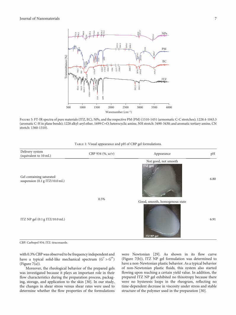
3.2.3. FT-IR Analysis. FT-IR studies were performed to detectthe possible interactions between the ITZ and other compo-nents in the ITZ NPs. Figure 5 shows the IR spectra of ITZ,EC, and the NP formulation. Pure ITZ displays a band witha strong peak at 1699 cm-1 indicative of the C=O stretch ofthe ketone group, a sharp peak at 1510 cm-1 attributed to an
aromatic C-C stretch band, and a distinctive peak at1228 cm-1 owing to aromatic C-H in-plane bending. Thepeaks around 3000 cm-1 were formed owing to stretchingvibrations of amino groups [26]. The EC IR spectrum showeda band at 3476 cm-1, which could be attributed to the OHstretching vibration [27]. The PM spectrum was the sum ofthe spectrum of ITZ, polyvinyl alcohol, and EC. A shift inthe C=O band of ITZ in the NP spectrum from 1699 to1719 cm-1 may be attributed to the change in the solid-stateform of ITZ from crystalline to amorphous during the prep-aration of NPs. The relative similarity of the other areas ofthe spectra of NPs and PM suggests that there was no chem-ical change between the drug and the other components.
3.3. Preparation and Characterization of ITZ NP Gel. Toovercome the disadvantages of polymeric NPs such as lowviscosity and low bioadhesive ability, the gel formulationwas employed for topical administration. Therefore, theinfluence of the gel-forming polymer CBP 934 (triethylaminewas used to adjust to pH6–7, 5% (w/v) of PG) on gel proper-ties was investigated.
0.00
0.05
0.10
0.15
0.20
0.25
0.30
0
50
100
150
200
250
300
350
0.25 0.5 1.0 1.5
PDI
Part
icle
size
(nm
)
Drug concentration (%)(at ratio of drug/polymer = 1:3)
Particle sizePDI
0.25 0.5 1.0 1.5Drug concentration (%)
(at ratio ofo drug/po// lymer = 1:3)
Particle size
(a)
0.00
0.05
0.10
0.15
0.20
0.25
0.30
0
50
100
150
200
250
300
350
0.25 0.5 1.0 1.5
PDI
Part
icle
size
(nm
)
Drug concentration (%)(at ratio of drug/polymer = 1:4)
Particle sizePDI
(b)
0.00
0.05
0.10
0.15
0.20
0.25
0.30
0
50
100
150
200
250
300
350
0.25 0.5 1.0 1.5PD
I
Part
icle
size
(nm
)
Drug concentration (%)(at ratio of drug/polymer = 1:5)
Particle sizePDI
0.25 0.5 1.0 1.5Drug concentration (%)
(at ratio ofo drug/p/ olyl mer = 1:5
Particle size
(c)
Figure 2: The size and PDI of ITZ NPs at different drug concentrations and drug/polymer ratios: (a) at ratio of drug/polymer = 1 : 3, (b) atratio of drug/polymer = 1 : 4, and (c) at ratio of drug/polymer = 1 : 5.
5Journal of Nanomaterials

3.3.1. Visual Appearance and pH. Table 1 shows the physico-chemical properties of several CBP gel formulations such asvisual appearance, pH of gel containing saturated suspen-sion, and ITZ NP gel.
3.3.2. In Vitro Drug Release Study. Figure 6(a) shows therelease profiles of ITZ from gel containing saturated suspen-sion and ITZ NP gel. The NP gel exhibited an initial burstrelease followed by a sustained release for 24h. The burstrelease could be explained by the presence of the drugadsorbed onto or near the nanoparticle surface [10]. Com-pared to gel containing ITZ-saturated suspension, nanosys-tem application led to the formation of a gel with betterperformance and with a sustained release pattern. Theamount of the drug released at 24 h was 0:12 ± 0:02% and
16:92 ± 0:50% for gel containing ITZ-saturated suspensionand ITZ NP gel, respectively. In this study, EC, a release-controlling excipient with hydrophobic but water permeablecharacter influencing the controlled drug release from themodified drug delivery system, was used as the encapsulatingpolymer [10]. This result was consistent with that reported inprevious studies on EC NP incorporated into hydroxypropylmethylcellulose gel [10] or on lipid NP incorporated intoCBP gel [28].
The concentration of gel-forming excipient could adjustthe viscosity and bioadhesive properties of gel. Hence, in thisstudy, CBP 934 concentration from 0.3% to 0.5% (w/v) wasinvestigated.
As shown in Figure 6(b), the concentration of gel-forming excipient exhibited an influence on drug release,e.g., at the concentration of 0.3% CBP 934, maximum drugcontent was released (29:85 ± 0:70% after 24h). With theincrease of CBP 934 concentration from 0.3% to 0.5%, drugrelease was reduced (from 29:85 ± 0:70% to 16:92 ± 0:50%after 24 h, p < 0:05). CBP 934 may influence the viscosity ofa gel in a concentration-dependent manner, which has beenreported to have an effect on the dissolution rate. The higherthe polymer concentration, the greater is gel viscosity, whichresults in a reduction in drug release [13]. Consequently,0.3% CBP 934 was finally chosen owing to its high dissolu-tion rate and good bioadhesive properties.
3.3.3. Rheological Analysis. In oscillatory rheometry, theeffects of oscillatory stress on the viscoelastic properties aremeasured as the storage modulus (G′), a measure of the elas-ticity, and the loss modulus (G′′), representing viscous com-ponents at a given frequency of oscillation. An ideal gelformulation should exhibit a solid-like mechanical spectrum,where G′ >G″ with little frequency dependence [29]. Basedon the results of rheological studies, the ITZ gel prepared
20
15
10
5
00.1 1 10
Size (d.nm)
Size distribution by intensity
Average (d.nm): 219.4PdI: 0.109Intercept: 0.945
Size (d.nm):
Peak 1: 243.4 100.0 82.710.0000.000
0.00.0
0.0000.000
Peak 2:Peak 3:
% Intensity Width (d.nm):
Result quality: Good
Record 1740: EC 1
Inte
nsity
(%)
100 1000 10000
(a)
200 nm
(b)
Figure 3: (a) DLS result and (b) SEM image of ITZ NPs (scale bar = 200 nm).
5 10 15 20 25 30 35 40 45 50
Nor
mal
ised
inte
nsity
(cou
nts)
2𝜃 (°) (Cu K𝛼)
NPs
PM
EC
ITZ
⁎
⁎⁎ ⁎
⁎⁎
⁎ ⁎
⁎⁎⁎⁎
⁎⁎
Figure 4: X-ray powder diffraction patterns (XRPD) of purematerials (ITZ, EC), NPs, and the respective PM (PM).Characteristic reflections of ITZ is marked with (∗). All spectra arenormalized.
6 Journal of Nanomaterials
with 0.3%CBPwas observed to be frequency independent andhave a typical solid-like mechanical spectrum (G′ >G″)(Figure 7(a)).
Moreover, the rheological behavior of the prepared gelswas investigated because it plays an important role in theirflow characteristics during the preparation process, packag-ing, storage, and application to the skin [30]. In our study,the changes in shear stress versus shear rates were used todetermine whether the flow properties of the formulations
were Newtonian [29]. As shown in its flow curve(Figure 7(b)), ITZ NP gel formulation was determined tohave a non-Newtonian plastic behavior. As a typical behaviorof non-Newtonian plastic fluids, this system also startedflowing upon reaching a certain yield value. In addition, theprepared ITZ NP gel exhibited no thixotropy because therewere no hysteresis loops in the rheogram, reflecting notime-dependent decrease in viscosity under stress and stablestructure of the polymer used in the preparation [30].
500 1000 1500 2000 2500 3000 3500 4000
824.
4
944.
910
43.3
1228
.4
1380
.814
51.2
1510
1699
2823
.3
2963
.1
796.
5
918
1110
.8 1376
.9
1638
.217
35.6
1846
.5
1994
2137
.722
40.9
2608
.3
2874
.429
76.6
3476
.1
671.
1
824.
4
1052
1106 13
79.8
1450
.215
10 1699
2155
.122
61.1
2478
.1
2608
.3
2876
.3
2975
.6
3126
3484
.7
733.
883
0.2
998.
911
04 1228
.4
1376
.9
1510
1638
.217
19.2
2939
2973
.7
3433
.6
NPs
PM
EC
ITZ Tran
smitt
ance
(%)
Wavenumber (cm–1)
Figure 5: FT-IR spectra of pure materials (ITZ, EC), NPs, and the respective PM (PM) (1510-1451 (armomatic C-C stretches); 1228.4-1043.3(aromatic C-H in plane bends); 1228 alkyl-aryl ether, 1699 C=O; heterocyclic amine, NH stretch: 3490-3430; and aromatic tertiary amine, CNstretch: 1360-1310).
Table 1: Visual appearance and pH of CBP gel formulations.
Delivery system(equivalent to 10mL)
CBP 934 (%, w/v) Appearance pH
Gel containing saturatedsuspension (0.1 g ITZ/10.0mL)
0.5%
Not good, not smooth
6.80
ITZ NP gel (0.1 g ITZ/10.0mL)
Good, smooth, homogenous state
6.91
CBP: Carbopol 934; ITZ: itraconazole.
7Journal of Nanomaterials
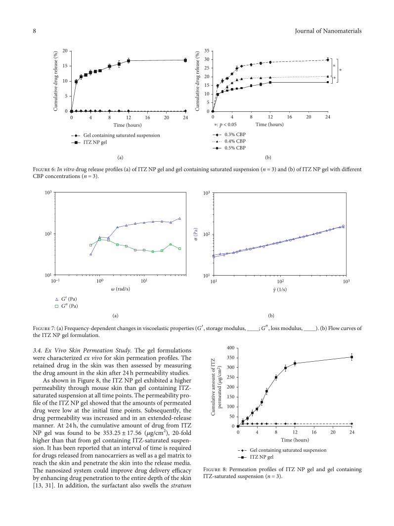
3.4. Ex Vivo Skin Permeation Study. The gel formulationswere characterized ex vivo for skin permeation profiles. Theretained drug in the skin was then assessed by measuringthe drug amount in the skin after 24 h permeability studies.
As shown in Figure 8, the ITZ NP gel exhibited a higherpermeability through mouse skin than gel containing ITZ-saturated suspension at all time points. The permeability pro-file of the ITZ NP gel showed that the amounts of permeateddrug were low at the initial time points. Subsequently, thedrug permeability was increased and in an extended-releasemanner. At 24h, the cumulative amount of drug from ITZNP gel was found to be 353:25 ± 17:56 (μg/cm2), 20-foldhigher than that from gel containing ITZ-saturated suspen-sion. It has been reported that an interval of time is requiredfor drugs released from nanocarriers as well as a gel matrix toreach the skin and penetrate the skin into the release media.The nanosized system could improve drug delivery efficacyby enhancing drug penetration to the entire depth of the skin[13, 31]. In addition, the surfactant also swells the stratum
0
5
10
15
20
0 4 8 12 16 20 24
Cum
ulat
ive d
rug
rele
ase (
%)
Time (hours)
Gel containing saturated suspensionITZ NP gel
(a)
0
5101520253035
0 4 8 12 16 20 24
Cum
ulat
ive d
rug
rele
ase (
%)
Time (hours)
0.3% CBP0.4% CBP0.5% CBP
⁎: p < 0.05
⁎
⁎⁎
(b)
Figure 6: In vitro drug release profiles (a) of ITZ NP gel and gel containing saturated suspension (n = 3) and (b) of ITZ NP gel with differentCBP concentrations (n = 3).
103
102
101
10–1 100
G′ (Pa)G″ (Pa)
101
𝜔 (rad/s)
(a)
103
102
101
101 102 103
𝛾 (1/s)
σ (P
a)
.
(b)
Figure 7: (a) Frequency-dependent changes in viscoelastic properties (G′, storage modulus, ____;G″, loss modulus, ____). (b) Flow curves ofthe ITZ NP gel formulation.
0
50
100
150
200
250
300
350
400
0 4 8 12 16 20 24
Cum
ulat
ive a
mou
nt o
f ITZ
perm
eate
d (𝜇
g/cm
2 )
Time (hours)
Gel containing saturated suspensionITZ NP gel
Figure 8: Permeation profiles of ITZ NP gel and gel containingITZ-saturated suspension (n = 3).
8 Journal of Nanomaterials

corneum and the intact vesicle can penetrate through theintact skin cells [32].
Moreover, the amount of retained drug of the ITZ NPgel in the skin after 24 h was also higher than that of gelcontaining ITZ-saturated suspension (573:88 ± 43:35μg/cm2 and 79:74 ± 23:45μg/cm2, respectively, p < 0:05).This could be explained by the effect of covering the skinsurface with NPs, NPs inside the cell space, and the betterbioadhesive ability of gel. Hence, the ITZ NP gel showedbetter permeation and drug retention than gel containingITZ-saturated suspension.
3.5. Antifungal Activity. Candida albicans forms creamy-greycolonies filamenting along their outer border. The filamen-tous border becomes visible initially, following 24 h incuba-tion, but is prominent following 48 h [33]. As shown inFigure 9(a), after 48 h incubation, the filamentous borderwas easily seen at all wells. Meanwhile, Aspergillus species,which also form filaments or hyphae, are the second mostcommon cause of opportunistic fungal infection in humansafter Candida albicans. Primary cutaneous infection, espe-cially in immunocompetent patients, is extremely rare, butan increase in prevalence has been noted in the last 20 years[34]. As shown in Figure 9(a), at the concentration of ITZ of40 ppm, the ITZ NP gel exhibited better antifungal activityagainst C. albicans than ITZ dispersion (with a total of 5points and 10 points, respectively). Figure 9(b) shows thatthe ITZ NP gel exhibited better antifungal activity againstA. niger than ITZ dispersion at all concentrations of ITZ(with a total of 7 points and 10 points, respectively). Further-more, the blank samples did not show any inhibition on thetwo fungal species. Nanosized liposomal formulations donot negatively affect the ITZ activity and ITZ preserves itsactivity even when it is being loaded in liposomes [31].According to Alhowyan et al. [26], the better inhibition ofITZ NPs (at the concentration of 5μg/mL) could be attrib-uted to the capability of NPs to infiltrate across thesurrounding media and fungal spores. In addition, the sus-tained release of ITZ explains the good inhibition over timewith the ITZ NP gel as compared to that of ITZ aqueous sus-pension. Another reason might be that the smaller size of
NPs and their uniform dispersibility increased the contactduration of the NPs with the spores and cells, hence, leadingto rapid drug internalization [26]. The study by Ling et al.[15] also showed that the nanoparticle formulation of mono-methoxypoly(ethyleneglycol)-b-poly(lactic acid)- (mPEG-b-PLA-) based ITZ-loaded NPs preserved the strong inhibitionof fungal growth as compared to the ITZ solid dispersion sys-tem on C. albicans (strain ATCC 10231) at low ITZ levels (aslow as at 0.0153μg/mL). Similarly, ITZ-loaded nanostruc-tured lipid carriers protected approximately 100% of C. albi-cans fungal infection at 20–40mg/kg; in contrast, ITZdispersion (with dimethyl sulfoxide) at either dose did notpreserve the health status and life of Galleria mellonella lar-vae [35]. For the inhibition against Aspergillus species, Patelet al. [36] showed that ITZ-loaded poly(lactic-co-glycolic)acid NPs exhibited enhanced antifungal activity against A.flavus compared to ITZ emulsion and ITZ in water at0.03mg/mL ITZ. Consequently, the above findings suggestedthat the ITZ NP gel could be promising for the developmentof an effective therapy for cutaneous infection treatment.
4. Conclusion
ITZ NPs were successfully fabricated with nanoscale parti-cle size by selecting the appropriate ratios of drug/polymerand drug concentration. ITZ NP gel with a high loadingcapacity and small particle size ensured a sustained releaseprofile, a good permeation through mouse skin, and a highretained drug content in skin. In addition, compared to theconventional gel, ITZ NP gel exhibited the better in vitroantifungal activity. Furthermore, the optimized gel formula-tion has the potential to deliver ITZ via topical administra-tion through skin.
Abbreviations
ITZ: ItraconazoleEC: EthylcelluloseNPs: NanoparticlesPDI: Polydispersity indexPG: Propylene glycol
ITZ NP gel(40 ppm)
ITZ NP gel(40 ppm)
ITZ NP gel(40 ppm)
ITZ NP gel(40 ppm)
ITZ NP gel(40 ppm)
DMSO(0.5%,v/v)Blank NP gel
ITZ/DMSO(40 ppm)
ITZ/DMSO(40 ppm)
ITZ/DMSO(40 ppm)
ITZ/DMSO(40 ppm)
ITZ/DMSO(40 ppm)
(a)
ITZ NP gel(2.5 ppm)
ITZ NP gel(40 ppm)
Aspergillu
ITZ NP gel(5 ppm)
ITZ NP gel(10 ppm)
ITZ NP gel(20 ppm)
ITZ NP gel(2.5 ppm)
ITZ NP gel(40 ppm)
ITZ NP gel(5 ppm)
ITZ NP gel(10 ppm)
ITZ NP gel(20 ppm)
DMSO(0.5%,v/v)Blank NP gel
(b)
Figure 9: Antifungal activity of ITZ NP gel and ITZ dispersion on (a) Candida albicans and (b) Aspergillus niger.
9Journal of Nanomaterials
CBP: CarbopolSEM: Scanning electron microscopeHPLC: High-performance liquid chromatographyXRPD: X-ray powder diffractionFT-IR: Fourier transform infrared spectroscopyPM: Physical mixture.
Data Availability
No data were used to support this study.
Ethical Approval
All institutional and national guidelines for the care and useof laboratory animals were followed.
Conflicts of Interest
The authors report no conflicts of interest.
Authors’ Contributions
Hoang Nhan Ho and Thien Giap Le contributed equally.
References
[1] S. Alam, Z. Iqbal, A. Ali et al., “Microemulsion as a potentialtransdermal carrier for poorly water soluble antifungal drugitraconazole,” Journal of Dispersion Science and Technology,vol. 31, no. 1, pp. 84–94, 2009.
[2] A. Chudasama, V. Patel, M. Nivsarkar, K. Vasu, andC. Shishoo, “Investigation of microemulsion system for trans-dermal delivery of itraconazole,” Journal of Advanced Pharma-ceutical Technology & Research, vol. 2, no. 1, pp. 30–38, 2011.
[3] K. K. Mali, S. C. Dhawale, and R. J. Dias, “Microemulsionbased bioadhesive gel of itraconazole using tamarind gum:in-vitro and ex-vivo evaluation,” Marmara PharmaceuticalJournal, vol. 21, no. 3, pp. 688–688, 2017.
[4] S. El-Housiny, M. A. Shams Eldeen, Y. A. El-Attar et al., “Flu-conazole-loaded solid lipid nanoparticles topical gel for treat-ment of pityriasis versicolor: formulation and clinical study,”Drug Delivery, vol. 25, no. 1, pp. 78–90, 2018.
[5] M. Mathur and V. K. Devi, “Potential of novel drug deliverysystems in the management of topical candidiasis,” Journal ofDrug Targeting, vol. 25, no. 8, pp. 685–703, 2017.
[6] Z. Zhang, P. C. Tsai, T. Ramezanli, and B. B. Michniak-Kohn,“Polymeric nanoparticles-based topical delivery systems forthe treatment of dermatological diseases,”Wiley Interdisciplin-ary Reviews. Nanomedicine and Nanobiotechnology, vol. 5,no. 3, pp. 205–218, 2013.
[7] G. M. Soliman, “Nanoparticles as safe and effective deliverysystems of antifungal agents: achievements and challenges,”International Journal of Pharmaceutics, vol. 523, no. 1,pp. 15–32, 2017.
[8] G. S. Rekhi and S. S. Jambhekar, “Ethylcellulose - a polymerreview,” Drug Development and Industrial Pharmacy, vol. 21,no. 1, pp. 61–77, 2008.
[9] S. Sahu, S. Saraf, C. D. Kaur, and S. Saraf, “Biocompatiblenanoparticles for sustained topical delivery of anticancer phy-toconstituent quercetin,” Pakistan Journal of Biological Sci-ences, vol. 16, no. 13, pp. 601–609, 2013.
[10] S. Duarah, R. D. Durai, and V. B. Narayanan, “Nanoparticle-in-gel system for delivery of vitamin C for topical application,”Drug Delivery and Translational Research, vol. 7, no. 5,pp. 750–760, 2017.
[11] B. Balzus, F. F. Sahle, S. Honzke et al., “Formulation and_ex vivo_ evaluation of polymeric nanoparticles for controlleddelivery of corticosteroids to the skin and the corneal epithe-lium,” European Journal of Pharmaceutics and Biopharmaceu-tics, vol. 115, pp. 122–130, 2017.
[12] H. T. Nguyen, T. H. Tran, J. O. Kim, C. S. Yong, and C. N.Nguyen, “Enhancing the in vitro anticancer efficacy of artesu-nate by loading into poly-D,L-lactide-co-glycolide (PLGA)nanoparticles,” Archives of Pharmacal Research, vol. 38,no. 5, pp. 716–724, 2015.
[13] C. N. Nguyen, T. T. T. Nguyen, H. T. Nguyen, and T. H. Tran,“Nanostructured lipid carriers to enhance transdermal deliv-ery and efficacy of diclofenac,” Drug Delivery and Transla-tional Research, vol. 7, no. 5, pp. 664–673, 2017.
[14] H. N. Ho, I. Laidmae, K. Kogermann et al., “Development ofelectrosprayed artesunate-loaded core-shell nanoparticles,”Drug Development and Industrial Pharmacy, vol. 43, no. 7,pp. 1134–1142, 2017.
[15] X. Ling, Z. Huang, J. Wang et al., “Development of an itraco-nazole encapsulated polymeric nanoparticle platform for effec-tive antifungal therapy,” Journal of Materials Chemistry B,vol. 4, no. 10, pp. 1787–1796, 2016.
[16] M. Yao and N. R. Srinivas, “Bioanalytical methods for thedetermination of itraconazole and hydroxyitraconazole: over-view from clinical pharmacology, pharmacokinetic, pharma-codynamic and metabolism perspectives,” BiomedicalChromatography, vol. 23, no. 7, pp. 677–691, 2009.
[17] B. Balzus, M. Colombo, F. F. Sahle, G. Zoubari, S. Staufenbiel,and R. Bodmeier, “Comparison of different _in vitro_ releasemethods used to investigate nanocarriers intended for dermalapplication,” International Journal of Pharmaceutics, vol. 513,no. 1-2, pp. 247–254, 2016.
[18] C. Salerno, A. M. Carlucci, and C. Bregni, “Study of in vitrodrug release and percutaneous absorption of fluconazole fromtopical dosage forms,” AAPS PharmSciTech, vol. 11, no. 2,pp. 986–993, 2010.
[19] C. V. Pham, M. C. Van, H. P. Thi et al., “Development ofibuprofen-loaded solid lipid nanoparticle-based hydrogels forenhanced _in vitro_ dermal permeation and _in vivo_ topicalanti-inflammatory activity,” Journal of Drug Delivery Scienceand Technology, vol. 57, p. 101758, 2020.
[20] C. Vitorino, J. Almeida, L. M. Gonçalves, A. J. Almeida, J. J.Sousa, and A. A. Pais, “Co-encapsulating nanostructured lipidcarriers for transdermal application: from experimental designto the molecular detail,” Journal of Controlled Release, vol. 167,no. 3, pp. 301–314, 2013.
[21] C. López-Castillo, C. Rodríguez-Fernández, M. Córdoba, andJ. J. Torrado, “Permeability characteristics of a new antifungaltopical amphotericin B formulation with γ-Cyclodextrins,”Molecules, vol. 23, no. 12, p. 3349, 2018.
[22] Y. Liu, G. Tortora, M. E. Ryan, H. M. Lee, and L. M. Golub,“Potato dextrose agar antifungal susceptibility testing foryeasts and molds: evaluation of phosphate effect on antifungalactivity of CMT-3,” Antimicrobial Agents and Chemotherapy,vol. 46, no. 5, pp. 1455–1461, 2002.
[23] R. M. Mainardes and R. C. Evangelista, “PLGA nanoparticlescontaining praziquantel: effect of formulation variables on size
10 Journal of Nanomaterials

distribution,” International Journal of Pharmaceutics, vol. 290,no. 1-2, pp. 137–144, 2005.
[24] L. De Smet, L. Saerens, T. De Beer et al., “Formulation of itra-conazole nanococrystals and evaluation of their bioavailabilityin dogs,” European Journal of Pharmaceutics and Biopharma-ceutics, vol. 87, no. 1, pp. 107–113, 2014.
[25] T. Mahnaj, S. U. Ahmed, and F. M. Plakogiannis, “Character-ization of ethyl cellulose polymer,” Pharmaceutical Develop-ment and Technology, vol. 18, no. 5, pp. 982–989, 2013.
[26] A. A. Alhowyan, M. A. Altamimi, M. A. Kalam et al., “Antifun-gal efficacy of itraconazole loaded PLGA-nanoparticles stabi-lized by vitamin-E TPGS: _in vitro_ and _ex vivo_ studies,”Journal of Microbiological Methods, vol. 161, pp. 87–95, 2019.
[27] S. E. El-Habashy, A. N. Allam, and A. H. El-Kamel, “Ethyl cel-lulose nanoparticles as a platform to decrease ulcerogenicpotential of piroxicam: formulation and in vitro/in vivo evalu-ation,” International Journal of Nanomedicine, vol. 11,pp. 2369–2380, 2016.
[28] K. Bhaskar, J. Anbu, V. Ravichandiran, V. Venkateswarlu, andY. M. Rao, “Lipid nanoparticles for transdermal delivery offlurbiprofen: formulation, in vitro, ex vivo and in vivo studies,”Lipids in Health and Disease, vol. 8, no. 1, p. 6, 2009.
[29] S. Y. Karavana, E. Gokce, S. Rençber et al., “A new approach tothe treatment of recurrent aphthous stomatitis with bioadhe-sive gels containing cyclosporine A solid lipid nanoparticles:in vivo/in vitro examinations,” International Journal of Nano-medicine, vol. 7, pp. 5693–5704, 2012.
[30] I. M. Abdulbaqi, Y. Darwis, R. Abou Assi, and N. Abdul KarimKhan, “Transethosomal gels as carriers for the transdermaldelivery of colchicine: statistical optimization, characteriza-tion, and ex vivo evaluation,” Drug Design, Development andTherapy, vol. Volume 12, pp. 795–813, 2018.
[31] A. H. Alomrani, G. A. Shazly, A. A. Amara, and M. M. Badran,“Itraconazole-hydroxypropyl-β-cyclodextrin loaded deform-able liposomes: _In vitro_ skin penetration studies and anti-fungal efficacy using _Candida albicans_ as model,” Colloidsand Surfaces. B, Biointerfaces, vol. 121, pp. 74–81, 2014.
[32] M. S. Jangdey, C. D. Kaur, and S. Saraf, “Efficacy ofConcanavalin-A conjugated nanotransfersomal gel of api-genin for enhanced targeted delivery of UV induced skinmalignant melanoma,” Artificial Cells, Nanomedicine, andBiotechnology, vol. 47, no. 1, pp. 904–916, 2019.
[33] C. C. Sheth, E. Johnson, M. E. Baker, K. Haynes, and F. A.Muhlschlegel, “Phenotypic identification of Candida albicansby growth on chocolate agar,” Medical Mycology, vol. 43,no. 8, pp. 735–738, 2005.
[34] C. Tahir, H. A. Nggada, A. M. Abubakar, M. Garbati, and E. H.Terna Yawe, “Primary cutaneous aspergillosis in an immuno-competent patient,” Journal of Surgical Technique and CaseReport, vol. 3, no. 2, pp. 94–96, 2011.
[35] J. S. Passos, L. C. de Martino, V. F. C. Dartora, G. L. B. deAraujo, K. Ishida, and L. B. Lopes, “Development, skin tar-geting and antifungal efficacy of topical lipid nanoparticlescontaining itraconazole,” European Journal of Pharmaceuti-cal Sciences, vol. 149, article 105296, 2020.
[36] N. R. Patel, K. Damann, C. Leonardi, and C. M. Sabliov, “Itra-conazole-loaded poly(lactic-co-glycolic) acid nanoparticles forimproved antifungal activity,” Nanomedicine (London,England), vol. 5, no. 7, pp. 1037–1050, 2010.
11Journal of Nanomaterials