development of laser desorption imaging mass spectrometry ... · development of laser desorption...
TRANSCRIPT
B American Society for Mass Spectrometry, 2015DOI: 10.1007/s13361-015-1123-0
J. Am. Soc. Mass Spectrom. (2015) 26:878Y886
FOCUS: IMAGING MASS SPECTROMETRY: RESEARCH ARTICLE
Development of Laser Desorption Imaging MassSpectrometry Methods to Investigate the MolecularComposition of Latent Fingermarks
Nidia Lauzon,1 Martin Dufresne,1 Vinita Chauhan,2 Pierre Chaurand1
1Department of Chemistry, Université de Montréal, C.P. 6128, Succursale Centre-ville, Montréal, Québec H3C 3J7, Canada2Health Canada, Ottawa, ON, Canada
Abstract. For a century, fingermark analysis has been one of the most important andcommon methods in forensic investigations. Modern chemical analysis technologieshave added the potential to determine the molecular composition of fingermarks andpossibly identify chemicals a suspect may have come into contact with. Improve-ments in analytical detection of the molecular composition of fingermarks is thereforeof great importance. In this regard, matrix-assisted laser desorption ionization(MALDI) and laser desorption ionization (LDI) imaging mass spectrometry (IMS)have proven to be useful technologies for fingermark analysis. In these analyses,the choice of ionizing agent and its mode of deposition are critical steps for theidentification of molecular markers. Here we propose two novel and complementary
IMS approaches for endogenous and exogenous substance detection in fingermarks: sublimation of 2-mercaptobenzothiazol (2-MBT) matrix and silver sputtering.Keywords: Mass spectrometry, Imaging, MALDI, LDI, Fingermarks
Received: 15 December 2014/Revised: 3 March 2015/Accepted: 3 March 2015/Published Online: 7 April 2015
Introduction
Fingerprint evidence is one of the most commonmethods ofbiometric identification. There are a variety of techniques
for the visualization of latent fingerprints on porous (paper,cotton, wood), semi-porous (plastic, waxy surfaces), and non-porous (glass, metal, paint) surfaces, including powdermethods [1], chemical methods (ninhydrin, DFO) [2], spectro-scopic imaging approaches (IR, Raman) [3, 4], andnanotechnology-based technologies [5]. Interestingly, the mo-lecular composition of a latent fingermark can lead to addition-al forensic evidence and may assist law enforcers in the case offingerprint matching process failure. [6–8]. The detection oflatent fingermarks is a considerably challenging analyticalproblem. Fingermarks are complex, composed of a mixtureof contaminants derived from the environment, secretion ofmetabolites from external uptake, and natural secretions fromthe body, including proteins, peptides, amino acids, vitamins,
salts, fatty acids, wax esters, diglycerides (DAGs), triglycerides(TAGs), cholesterol, squalene, and more [9, 10]. Further, latentfingermark deposits behave differently on different substratetypes. In this regard, mass spectrometry (MS) has proven to bea useful technology to monitor the molecular composition offingermarks. Much of the prior research for the detection andidentification of endogenous substances in fingermarks hasbeen carried out using gas chromatography-mass spectrometry(GC-MS). Despite the fact that this technique is destructive andlimited to a small class of compounds (<500 Da), it allows thedetection of endogenous substances, such as amino acids, fattyacids, wax esters, squalene, and cholesterol, as well as thedetection of some exogenous substances coming from hairproduct, parfum residue, cosmetics, and skin lotion [11–16].Many nondestructiveMS techniques have been developped forthe detection of endogenous and exogenous substances fromfingermarks. As an exemple, desorption electrospray ionization(DESI) MS has been successfully applied to several exogenoussubstances and external uptake detection [17]. Surface assisted-laser desorption/ionization-time-of-flight (SALDI-TOF) MShas been carried out for the detection of some fatty acids,dehydrated cholesterol, squalene, as well as the identificationof explosives, drugs, and nicotine residues [8, 18, 19]. Morerecently, matrix-assisted-laser desorption/ionization (MALDI)
Electronic supplementary material The online version of this article(doi:10.1007/s13361-015-1123-0) contains supplementary material, which isavailable to authorized users.
Correspondence to: Pierre Chaurand; e-mail: [email protected]
TOF MS has been reported for the detection of endogenoussubstances, such as amino acids, fatty acids, peptides,dehydrated cholesterol, squalene, DAGs, and TAGs, as wellas surfactants, polymers, drugs, pharmaceuticals, and explo-sives [20–22].
Imaging mass spectrometry (IMS) [23–25] is particularlywell suited for investigating fingermarks because it combinessuspect identification by visualizing the ridge pattern definedby the molecular images, and the chemical composition forfurther forensic evidence. Despite proven capabilities infingermark analysis, for the detection and mapping of endog-enous and exogenous markers, MALDI IMS critically relies onthe nature of the matrix and its mode of deposition that mayneed tailoring to the chemistry of the interested species. In thisregard, extensive research has been performed to improve theanalytical detection for numerous samples by minimizing theanalyte delocalization, to improve the sensitivity and the imag-ing spatial resolution. The first application of IMS has beenreported by Ifa et al. for the detection of drugs of abuse andexplosives on latent fingermark by DESIMS [7]. MALDI IMSintroduced by Francese et al. has also proven to be a powerfultool for fingermark analysis. Initially, fingermark IMS wasperformed by depositing the MALDI matrix, α-cyano-4-hydroxycinnamic acid (CHCA), by spray-coating [26]. Thisprocedure allows both visual and chemical information to beobtained in one analysis. Subsequently, the search for moreefficient protocols for matrix deposition has resulted in thedevelopment of a novel two-step procedure, named Bdry-wet^that consist of dusting the MALDI matrix CHCA onto thefingermark followed by solvent spray deposition [27]. Thismethod has proven to be more efficient on fingermarks byimproving the imaging spatial resolution and increasing theion abundance and intensity. Moreover, they demonstrated theversatility and the robustness of fingermark IMS methods fordifferent forensic situations, contributing to additional evi-dences [28], including the determination of the presence ofblood for criminal investigation [29].
In this paper, we demonstrate the potential of two novelMALDI- and LDI-based imaging methods for the detection ofendogenous and exogenous substances from fingermarks. Re-cently, matrix sublimation was demonstrated to be a powerfulapproach for very homogeneous matrix deposition that allowsfor the detection and IMS of small molecules and proteins withhigh sensitivity and high spatial resolution [30–32]. In the firstapproach proposed herein, we provide an alternative to theBdry-wet^ method using sublimation for homogeneous matrixdeposition followed by automated solvent spray deposition. Tothis end, we screened several matrix candidates and found that2-mercaptobenzothiazole (2-MBT) offered similar and com-plementary results to this method. In the second approach, wetested the potential of silver assisted laser desorption ionization(AgLDI) for IMS of fingermarks. Previous research has dem-onstrated the potential of metal sputtering, such as gold for thedetection of deprotonated fatty acids and hazardous substrates[33] and the use of silver nanoparticles for the detection ofexogenous drug compounds [34] from fingermarks. AgLDI
IMS was recently proposed by our laboratory and allows theimaging of cholesterol, fatty acids, and other olefin-containingmolecules from thinly cut tissue sections with high specificityand sensitivity [35]. Surprisingly, AgLDI has demonstrated tobe a powerful tool for fingermark analysis. The deposition ofmetallic silver by sputtering on fingermark allows high-resolution IMS and higher sensitivity of several class of bio-molecules and some exogenous substances. Ultimately, wefound that both approaches provide complementary results thatcould significantly contribute to forensic investigations.
ExperimentalChemicals and Materials
1,5-Diaminonaphtalene (DAN), 2,5-dihydroxybenzoic acid(DHB), α-cyano-4-hydroxycinnamic acid (CHCA), 2′,4′,6′-trihydroxyacetophenone (THAP), 2-mercaptobenzothiazole( 2 - M B T ) , L - a m i n o a c i d s k i t , s q u a l e n e ,dimethyldioctadecylammonium bromide, oleic acid, stearicacid, 1,2-dioleoyl-sn-glycerol, trifluoroacetic acid (TFA), andliquid chromatography grade solvents were purchased fromSigma-Aldrich (St. Louis, MO, USA). 1,2-Dipalmitoyl-sn-glycero-3-phosphate was purchased from Avanti Polar Lipids(Alabaster, AL, USA). 1-Palmitoyl-2-oleoyl-3-linoleoyl-rac-glycerol was purchased from Cayman Chemical (Orlando,FL, USA). 9-Aminoacridine (9-AA) was purchased fromAcros Organics (Morris Plains, NJ, USA). Ethanol (EtOH)was purchased from Greenfield Ethanol Inc. (Boucherville,QC, Canada). Double-sided carbon conductive tape was pur-chased from Ted Pella, Inc. (Redding, CA, USA).
Fingermark Preparation
The slides were washed in acetone to ensure complete clean-ness prior to fingermark deposition. Latent groomed andungroomed fingermarks were prepared on ITO coated slides(Delta Technologies, Loveland, CO, USA) according to pub-lished preparation methods [26]. The same procedure wasfollowed for fingermark deposition on sheets of paper.Fingermarks were obtained in a de-identified manner. Allprocedures were approved by the Health Canada ResearchEthics Board and comply with the Ethical Principles set outin Tri-Council Policy Statement: Ethical Conduct for ResearchInvolving Humans guidelines.
Matrix Deposition
Solvent-free matrix deposition on fingermarks was carried outin a sublimation apparatus (Chemglass Life Science, Vineland,NJ, USA) as previously described [30]. The temperature andthe time of sublimation were optimized for each matrix toensure the best coverage and MALDI efficiency (number ofsignals detected and signal to noise). To enhance MS signals,recrystallization of the matrix was performed by spraying thesurface with a solution of 70:30 EtOH:water at 30°C using aTM-Sprayer (HTX LC-Transform). Ten passes of solvent over
N. Lauzon et al.: Imaging Mass Spectrometry in Forensic Science 879
the surface of the fingermarks were deposited on the sublimat-ed 2-MBT matrix with a lateral travel velocity and debit of1.2 m/min and 13.5 mL/min, respectively. Each solvent passwas separated by 5 mm and for every even numbered pass anoffset of 2.5 mm was applied leading to a homogenous solventlayer deposition. Recrystallization of the sublimated matrixCHCA and DHB have also been performed by spraying thesurface with a solution of 70:30 ACN:water and 70:30
MeOH:water at 30°C, respectively, using the same instrumentparameters.
Matrix was also deposited on fingermarks using theImagePrep spray system (Bruker Daltonics, Billerica, MA,USA) starting from the default CHCA method and optimizedfor each matrix studied. 2-MBT was prepared at a concentra-tion of 20mg/mL in 70:30 EtOH:water, CHCAwas prepared at7 mg/mL in 50:50 acetonitrile:water, DAN was prepared at
Table 1. Potential and Experiment Settings of Investigated Matrices for Imaging the Molecular Composition of Latent Fingermarks After Sublimation Depositionα
Matrix Positive polarity Negative polarity Sublimation temperature(°C)
Sublimation time(min)
Deposited amount(μg/cm2)
CHCA *** * 180 5,0 200DAN * * 140 4,0 220DHB ** * 180 4,0 2202-MBT **** * 180 3,5 200THAP * * 150 2,0 1809-AA * * 180 7,0 180
α The number of star indicates the observed performance of the matrix according to the polarity with *, **, ***, **** representing low, medium, high, and very highperformances, respectively.
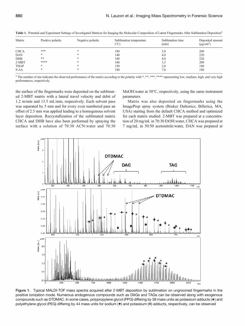
Figure 1. Typical MALDI-TOF mass spectra acquired after 2-MBT deposition by sublimation on ungroomed fingermarks in thepositive ionization mode. Numerous endogenous compounds such as DAGs and TAGs can be observed along with exogenouscompounds such as DTDMAC. In some cases, polypropylene glycol (PPG) differing by 58mass units as potassium adducts (★) andpolyethylene glycol (PEG) differing by 44 mass units for sodium (✦) and potassium (✱) adducts, respectively, can be observed
880 N. Lauzon et al.: Imaging Mass Spectrometry in Forensic Science
5 mg/mL in 70:30 EtOH:water, and DHB was prepared at25 mg/mL and THAP at 10 mg/mL, both in 50:50MeOH:water. Finally, 9-AA was prepared at 10 mg/mL in50:50 EtOH:water. For fingermarks analyzed in positive ioni-zation mode, 0.1% of TFA was added to the matrix solution.
Silver Sputter Coating
Metallic silver was sputtered on top of fingermarks using aCressington 308R sputter coater (Ted Pella, Inc., Redding, CA,USA) as previously described [35]. For optimal results, thedeposited silver layer thickness was optimized at 14±2 nmcorresponding to 30 s of deposition.
IMS of Fingermarks on Paper
Double-sided tape was first applied to a glass slide (Supple-mentary Figure 8). The piece of paper containing thefingermark was then mounted on the tape using an aluminumsheet to press down on the piece of paper to minimize alterationof the fingermark. Subsequent silver deposition was performedas described above.
Mass Spectrometry Instrumentation
Profiling and IMS of fingermarks were performed on either aMALDI TOF/TOF Ultraflextreme mass spectrometer or aMALDI FT/ICR SolariX XR 7 T mass spectrometer bothequipped with SmartBeam II Nd:YAG 355 nm lasers operating
at 1000 Hz or at 250 Hz for porous surface analyses (BrukerDaltonics). For IMS data acquisition, 200 shots were summedper array position with a lateral resolution of 75 μm using theBmedium^ focus setting or with a lateral resolution of 10 μmusing the Bminimum^ focus setting. Using these parameters,we achieved imaging speeds of ~70 pixels/min. For a wholefingermark at 75 μm of spatial resolution, IMS acquisitionrequired approximately 5 h. Profiling and IMS data from theUltraflextreme were performed in reflectron geometry underoptimized delayed extraction conditions with a source ac-celerating voltage of +25 kV, in a mass range of 100–1100 Da. For MALDI IMS, external calibration was per-formed using a homemade mix of standards (lysine, stearicacid, 1,2-dioleoyl-sn-glycerol, and 1-palmitoyl-2-oleoyl-3-linoleoyl-rac-glycerol). Subsequently, internal calibrationwas performed with known species including the [M + H]+
and [2 M – H]+ matrix signal (168.00 and m/z 332.96,respectively) and the [M]+ DTDMAC ion at m/z 550.63(see below). For AgLDI IMS, internal calibration was car-ried out in quadratic mode using the silver clusters (Ag1 toAg9, see Fig. 4) present in all the spectra. IMS data werereconstructed and visualized without normalization usingthe FlexImaging 3.0 software (Bruker Daltonics). Lipididentification was performed by MALDI/AgLDI tandemMS on the Ultraflextreme mass spectrometer in combinationwith the LIPID MAPS prediction tool by comparing accu-rate mass measurements (www.lipidmaps.org/tools/index.html).
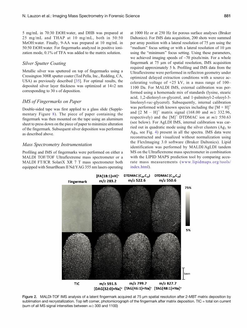
Figure 2. MALDI-TOF IMS analysis of a latent fingermark acquired at 75 μm spatial resolution after 2-MBT matrix deposition bysublimation and recrystallization. Top left corner, photomicrograph of the fingermark after matrix deposition. TIC = total ion current(sum of all MS signal intensities between m/z 300 and 1100)
N. Lauzon et al.: Imaging Mass Spectrometry in Forensic Science 881
Results and DiscussionEvaluation of Matrix Candidates for Sublimationand IMS
Initial sample preparation tests were performed with severalMALDI matrices using two different modes of deposition;direct matrix spray coating (Image Prep system) or sublimationfollowed by solvent spray deposition. The solvent systememployed in matrix spray coating enables a better co-crystallization of some analytes, enhancing their signal. How-ever, depending on the matrix and solvent used, we found thatthis mode of deposition produces some analyte delocalization,which in many cases prevented the proper visualization of thefingerprint motif (Supplementary Figure 3). Similar compari-sons between the dry-wet method and spray coating have alsobeen performed, leading to the conclusion that the initial drydeposition on fingermarks allowed higher ion signal intensityand enhanced clarity of the ridge details [36]. Sublimation is aninteresting approach for matrix deposition on fingermarks be-cause of its high reproducibility, the absence of analyte delo-calization, and capability to detect numerous small moleculesby MALDI IMS. Six MALDI matrices that were previouslyinvestigated by sublimation for lipid IMS analyses from tissuesections were tested on latent fingermarks [37]. IMS analyseswere performed in positive and negative ionization modes. Asshown in Table 1, for each matrix tested, the sublimation time
and temperature were optimized to obtain the best MS signals.Their performance was evaluated using the following analyti-cal criteria: (1) MS signal quality: intensity and number ofcompounds detected based on existing literature for fingermarkanalysis; (2) uniformity and crystal size for optimal IMS spatialresolution (Supplementary Figure 4); and (3) number of back-ground signals. Overall, 2-MBT showed the best performanceand reproducibility for fingermark IMS analysis in positiveionization mode, when considering the sum of MS signalsobserved from fingermarks and the intensity of backgroundsignals (Supplementary Figures 5 and 6). More importantly, itssublimation on latent fingermarks allows the detection of nu-merous exogenous and endogenous substances, possibly mak-ing 2-MBT a valuable matrix for forensic screening (Fig. 1).For example, the ions detected at m/z 494.6, 522.6, and 550.6h a v e p r e v i o u s l y b e e n i d e n t i f i e d a s aditallowdimethylammonium ions (DTDMAC) and originatefrom personal care and domestic products [38]. In a previouspublication, polymer analysis by MALDI MS has been per-formed on fingermarks using dithranol, instead of CHCA,allowing a better detection efficiency [39]. Polymeric sub-stances such as polyethylene glycol (PEG) and polypropyleneglycol (PPG) can also be easily observed on ungroomedfingermarks using the matrix 2-MBT and often originate fromhand lotions and other beauty products (Fig. 1). For endoge-nous substance detection, previous studies have demonstrated
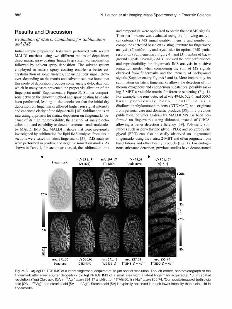
Figure 3. (a) AgLDI-TOF IMS of a latent fingermark acquired at 75 μm spatial resolution. Top left corner, photomicrograph of thefingermark after silver sputter deposition. (b) AgLDI-TOF IMS of a small area from a latent fingermark acquired at 10 μm spatialresolution. (Top) Oleic acid [OA + 109Ag]+ atm/z 391.17 and (Bottom) [TAG(50:1) + Na]+ atm/z 855.74. *Composite image of both oleicacid [OA + 109Ag]+ and stearic acid [SA + 107Ag]+. Stearic acid (SA) is typically observed in much lower intensity than oleic acid infingermarks
882 N. Lauzon et al.: Imaging Mass Spectrometry in Forensic Science
the efficiency of 2-MBT as a MALDI matrix for tissue analysisof lipids after application by spray deposition [40]. Its deposi-tion by sublimation on fingermarks allows the detection ofamino acids, sodiated wax esters, diglycerides (DAGs), triglyc-erides (TAGs), and some fatty acids, such as oleic and stearicacids (Supplementary Table 1). Figure 2 shows IMS results ofthe most abundant exogenous and endogenous substances de-tected in fingermarks. With data acquisition at a spatial resolu-tion of 75 μm, the fingermark ridge pattern is clearly visiblewhen comparing the relative intensities of the displayed ionsand is more clearly observed when looking at the total ionchromatogram (TIC) image (sum of signal intensities betweenm/z 300 and 1100).
Fingermark IMS after Silver Sputtering
Silver sputter deposition is a novel method developed for IMSthat allows the privileged detection of olefin containing mole-cules observed as [M + Ag]+ ions directly from tissue sections[35]. Silver adducts are easily determined by the presence ofthe 107Ag and 109Ag isotopic pattern, with relative abundancesof 52% and 48%, respectively. After silver layer deposition onfingermarks and analysis by LDI-TOF MS, numerous [M +Ag]+ ions have been detected such as cholesterol, squalene,
wax esters, fatty acids, and TAGs (Supplementary Table 2). Inaddition, the presence of Na+ and K+ in fingermark residuesallow us to detect sodium and potassium adducts of DAGs andTAGs. Some exogenous substances were also observed, in-cluding DTDMAC and polymeric substances, such as PEG(Supplementary Figure 7). Figure 3a shows some endogenousand exogenous ion images acquired from a latent fingermarkby AgLDI-TOF MS. The whole fingermark was imaged againwith a lateral resolution of 75 μm, allowing a good definition ofthe fingermark pattern. As expected, most of the compoundswere detected from the ridge of the fingerprint. Silver sputterdeposition, however, also allows IMS acquisition at high spa-tial resolution [35]. As demonstrated in Fig. 3b, a small area ofa fingermark was imaged at 10 μm of lateral resolution, reveal-ing information such as the minutiae points, pores, and ridgeshape. The optical image after silver deposition is enough toprovide this forensic information, but it is interesting to look atthe molecular distribution of compounds, such as oleic acid(OA) at m/z 391.2 and the [TAG(50:1) + Na]+ at m/z 855.7,with the latter displaying some correlation with the fingerprintpores. Fingermark IMS after silver sputter deposition was alsoperformed using a MALDI FT/ICR mass spectrometer, pro-viding higher mass resolving power and increased sensitivitycompared with the TOF MS instrument. The high mass
200 300 400 500 600 700 800 900 1000 m/z0
2
4
6
x107
Inte
ns. (
a.u.
)
400 500 600 700 800 900 m/z
0.25
0.50
0.75
1.00
1.25
x107
Inte
ns. (
a.u.
)
Ag
Ag2
Ag3
Ag5
Ag7 Ag9
FA
DAG & WE TAG
Squalene
Cholesterol
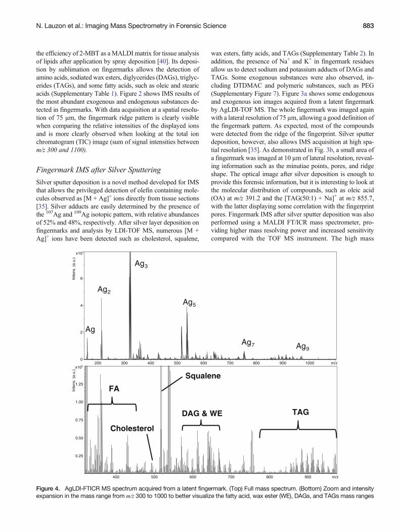
Figure 4. AgLDI-FTICR MS spectrum acquired from a latent fingermark. (Top) Full mass spectrum. (Bottom) Zoom and intensityexpansion in the mass range from m/z 300 to 1000 to better visualize the fatty acid, wax ester (WE), DAGs, and TAGs mass ranges
N. Lauzon et al.: Imaging Mass Spectrometry in Forensic Science 883
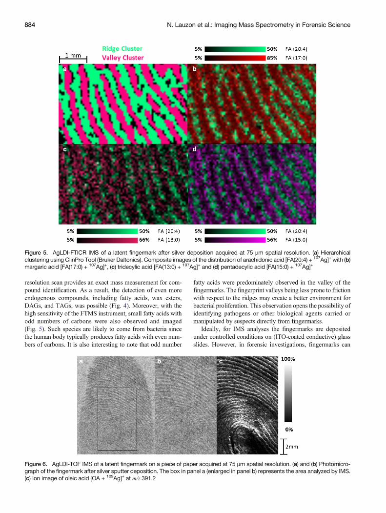
resolution scan provides an exact mass measurement for com-pound identification. As a result, the detection of even moreendogenous compounds, including fatty acids, wax esters,DAGs, and TAGs, was possible (Fig. 4). Moreover, with thehigh sensitivity of the FTMS instrument, small fatty acids withodd numbers of carbons were also observed and imaged(Fig. 5). Such species are likely to come from bacteria sincethe human body typically produces fatty acids with even num-bers of carbons. It is also interesting to note that odd number
fatty acids were predominately observed in the valley of thefingermarks. The fingerprint valleys being less prone to frictionwith respect to the ridges may create a better environment forbacterial proliferation. This observation opens the possibility ofidentifying pathogens or other biological agents carried ormanipulated by suspects directly from fingermarks.
Ideally, for IMS analyses the fingermarks are depositedunder controlled conditions on (ITO-coated conductive) glassslides. However, in forensic investigations, fingermarks can
Figure 5. AgLDI-FTICR IMS of a latent fingermark after silver deposition acquired at 75 μm spatial resolution. (a) Hierarchicalclustering using ClinPro Tool (Bruker Daltonics). Composite images of the distribution of arachidonic acid [FA(20:4) + 107Ag]+ with (b)margaric acid [FA(17:0) + 107Ag]+, (c) tridecylic acid [FA(13:0) + 107Ag]+ and (d) pentadecylic acid [FA(15:0) + 107Ag]+
Figure 6. AgLDI-TOF IMS of a latent fingermark on a piece of paper acquired at 75 μm spatial resolution. (a) and (b) Photomicro-graph of the fingermark after silver sputter deposition. The box in panel a (enlarged in panel b) represents the area analyzed by IMS.(c) Ion image of oleic acid [OA + 109Ag]+ at m/z 391.2
884 N. Lauzon et al.: Imaging Mass Spectrometry in Forensic Science
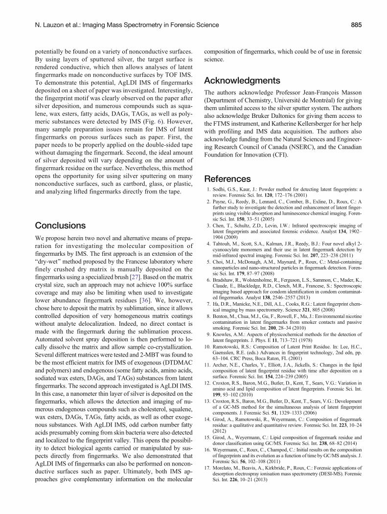
potentially be found on a variety of nonconductive surfaces.By using layers of sputtered silver, the target surface isrendered conductive, which then allows analyses of latentfingermarks made on nonconductive surfaces by TOF IMS.To demonstrate this potential, AgLDI IMS of fingermarksdeposited on a sheet of paper was investigated. Interestingly,the fingerprint motif was clearly observed on the paper aftersilver deposition, and numerous compounds such as squa-lene, wax esters, fatty acids, DAGs, TAGs, as well as poly-meric substances were detected by IMS (Fig. 6). However,many sample preparation issues remain for IMS of latentfingermarks on porous surfaces such as paper. First, thepaper needs to be properly applied on the double-sided tapewithout damaging the fingermark. Second, the ideal amountof silver deposited will vary depending on the amount offingermark residue on the surface. Nevertheless, this methodopens the opportunity for using silver sputtering on manynonconductive surfaces, such as cartbord, glass, or plastic,and analyzing lifted fingermarks directly from the tape.
ConclusionsWe propose herein two novel and alternative means of prepa-ration for investigating the molecular composition offingermarks by IMS. The first approach is an extension of theBdry-wet^ method proposed by the Francese laboratory wherefinely crushed dry matrix is manually deposited on thefingermarks using a specialized brush [27]. Based on the matrixcrystal size, such an approach may not achieve 100% surfacecoverage and may also be limiting when used to investigatelower abundance fingermark residues [36]. We, however,chose here to deposit the matrix by sublimation, since it allowscontrolled deposition of very homogeneous matrix coatingswithout analyte delocalization. Indeed, no direct contact ismade with the fingermark during the sublimation process.Automated solvent spray deposition is then performed to lo-cally dissolve the matrix and allow sample co-crystallization.Several different matrices were tested and 2-MBTwas found tobe the most efficient matrix for IMS of exogenous (DTDMACand polymers) and endogenous (some fatty acids, amino acids,sodiated wax esters, DAGs, and TAGs) substances from latentfingermarks. The second approach investigated is AgLDI IMS.In this case, a nanometer thin layer of silver is deposited on thefingermarks, which allows the detection and imaging of nu-merous endogenous compounds such as cholesterol, squalene,wax esters, DAGs, TAGs, fatty acids, as well as other exoge-nous substances. With AgLDI IMS, odd carbon number fattyacids presumably coming from skin bacteria were also detectedand localized to the fingerprint valley. This opens the possibil-ity to detect biological agents carried or manipulated by sus-pects directly from fingermarks. We also demonstrated thatAgLDI IMS of fingermarks can also be performed on noncon-ductive surfaces such as paper. Ultimately, both IMS ap-proaches give complementary information on the molecular
composition of fingermarks, which could be of use in forensicscience.
AcknowledgmentsThe authors acknowledge Professor Jean-François Masson(Department of Chemistry, Université de Montréal) for givingthem unlimited access to the silver sputter system. The authorsalso acknowledge Bruker Daltonics for giving them access tothe FTMS instrument, and Katherine Kellersberger for her helpwith profiling and IMS data acquisition. The authors alsoacknowledge funding from the Natural Sciences and Engineer-ing Research Council of Canada (NSERC), and the CanadianFoundation for Innovation (CFI).
References1. Sodhi, G.S., Kaur, J.: Powder method for detecting latent fingerprints: a
review. Forensic Sci. Int. 120, 172–176 (2001)2. Payne, G., Reedy, B., Lennard, C., Comber, B., Exline, D., Roux, C.: A
further study to investigate the detection and enhancement of latent finger-prints using visible absorption and luminescence chemical imaging. Foren-sic Sci. Int. 150, 33–51 (2005)
3. Chen, T., Schultz, Z.D., Levin, I.W.: Infrared spectroscopic imaging oflatent fingerprints and associated forensic evidence. Analyst 134, 1902–1904 (2009)
4. Tahtouh, M., Scott, S.A., Kalman, J.R., Reedy, B.J.: Four novel alkyl 2-cyanoacylate monomers and their use in latent fingermark detection bymid-infrared spectral imaging. Forensic Sci. Int. 207, 223–238 (2011)
5. Choi, M.J., McDonagh, A.M., Maynard, P., Roux, C.: Metal-containingnanoparticles and nano-structured particles in fingermark detection. Foren-sic Sci. Int. 179, 87–97 (2008)
6. Bradshaw, R., Wolstenholme, R., Ferguson, L.S., Sammon, C., Mader, K.,Claude, E., Blackledge, R.D., Clench, M.R., Francese, S.: Spectroscopicimaging based approach for condom identification in condom contaminat-ed fingermarks. Analyst 138, 2546–2557 (2013)
7. Ifa, D.R., Manicke, N.E., Dill, A.L., Cooks, R.G.: Latent fingerprint chem-ical imaging by mass spectrometry. Science 321, 805 (2008)
8. Benton, M., Chua, M.J., Gu, F., Rowell, F., Ma, J.: Environmental nicotinecontamination in latent fingermarks from smoker contacts and passivesmoking. Forensic Sci. Int. 200, 28–34 (2010)
9. Knowles, A.M.: Aspects of physicochemical methods for the detection oflatent fingerprints. J. Phys. E 11, 713–721 (1978)
10. Ramotowski, R.S.: Composition of Latent Print Residue. In: Lee, H.C.,Gaensslen, R.E. (eds.) Advances in fingerprint technology, 2nd edn, pp.63–104. CRC Press, Boca Raton, FL (2001)
11. Archer, N.E., Charles, Y., Elliott, J.A., Jickells, S.: Changes in the lipidcomposition of latent fingerprint residue with time after deposition on asurface. Forensic Sci. Int. 154, 224–239 (2005)
12. Croxton, R.S., Baron, M.G., Butler, D., Kent, T., Sears, V.G.: Variation inamino acid and lipid composition of latent fingerprints. Forensic Sci. Int.199, 93–102 (2010)
13. Croxton, R.S., Baron,M.G., Butler, D., Kent, T., Sears, V.G.: Developmentof a GC-MS method for the simultaneous analysis of latent fingerprintcomponents. J. Forensic Sci. 51, 1329–1333 (2006)
14. Girod, A., Ramotowski, R., Weyermann, C.: Composition of fingermarkresidue: a qualitative and quantitative review. Forensic Sci. Int. 223, 10–24(2012)
15. Girod, A., Weyermann, C.: Lipid composition of fingermark residue anddonor classification using GC/MS. Forensic Sci. Int. 238, 68–82 (2014)
16. Weyermann, C., Roux, C., Champod, C.: Initial results on the compositionof fingerprints and its evolution as a function of time by GC/MS analysis. J.Forensic Sci. 56, 102–108 (2011)
17. Morelato, M., Beavis, A., Kirkbride, P., Roux, C.: Forensic applications ofdesorption electrospray ionisation mass spectrometry (DESI-MS). ForensicSci. Int. 226, 10–21 (2013)
N. Lauzon et al.: Imaging Mass Spectrometry in Forensic Science 885
18. Rowell, F., Hudson, K., Seviour, J.: Detection of drugs and their metabo-lites in dusted latent fingermarks by mass spectrometry. Analyst 134, 701–707 (2009)
19. Rowell, F., Seviour, J., Lim, A.Y., Elumbaring-Salazar, C.G., Loke, J., Ma,J.: Detection of nitro-organic and peroxide explosives in latent fingermarksbyDART- and SALDI-TOF-mass spectrometry. Forensic Sci. Int. 221, 84–91 (2012)
20. Kaplan-Sandquist, K., LeBeau, M.A., Miller, M.L.: Chemical analysis ofpharmaceuticals and explosives in fingermarks using matrix-assisted laserdesorption ionization/time-of-flight mass spectrometry. Forensic Sci. Int.235, 68–77 (2014)
21. Sundar, L., Rowell, F.: Detection of drugs in lifted cyanoacrylate-developed latent fingermarks using two laser desorption/ionisation massspectrometric methods. Analyst 139, 633–642 (2014)
22. Francese, S., Bradshaw, R., Ferguson, L.S., Wolstenholme, R., Clench,M.R., Bleay, S.: Beyond the ridge pattern: multi-informative analysis oflatent fingermarks byMALDImass spectrometry. Analyst 138, 4215–4228(2013)
23. Chughtai, K., Heeren, R.M.: Mass spectrometric imaging for biomedicaltissue analysis. Chem. Rev. 110, 3237–3277 (2010)
24. Chaurand, P.: Imaging mass spectrometry of thin tissue sections: a decadeof collective efforts. J. Proteome. 75, 4883–4892 (2012)
25. Norris, J.L., Caprioli, R.M.: Analysis of tissue specimens by matrix-assisted laser desorption/ionization imaging mass spectrometry in biologi-cal and clinical research. Chem. Rev. 113, 2309–2342 (2013)
26. Wolstenholme, R., Bradshaw, R., Clench, M.R., Francese, S.: Study oflatent fingermarks by matrix-assisted laser desorption/ionisationmass spec-trometry imaging of endogenous lipids. Rapid Commun. Mass Spectrom.23, 3031–3039 (2009)
27. Ferguson, L., Bradshaw, R., Wolstenholme, R., Clench, M., Francese, S.:Two-step matrix application for the enhancement and imaging of latentfingermarks. Anal. Chem. 83, 5585–5591 (2011)
28. Bradshaw, R., Bleay, S., Wolstenholme, R., Clench, M.R., Francese, S.:Towards the integration of matrix assisted laser desorption ionisation massspectrometry imaging into the current fingermark examination workflow.Forensic Sci. Int. 232, 111–124 (2013)
29. Bradshaw, R., Bleay, S., Clench, M.R., Francese, S.: Direct detection ofblood in fingermarks byMALDIMS profiling and imaging. Sci. Justice 54,110–117 (2014)
30. Chaurand, P., Cornett, D.S., Angel, P.M., Caprioli, R.M.: From whole-body sections down to cellular level, multiscale imaging of phospholipids
by MALDI mass spectrometry. Mol. Cell. Proteomics 10, O110.004259(2011)
31. Hankin, J.A., Barkley, R.M., Murphy, R.C.: Sublimation as a method ofmatrix application for mass spectrometric imaging. J. Am. Soc. MassSpectrom. 18, 1646–1652 (2007)
32. Yang, J., Caprioli, R.M.: Matrix sublimation/recrystallization for imagingproteins by mass spectrometry at high spatial resolution. Anal. Chem. 83,5728–5734 (2011)
33. Tang, H.W., Lu,W., Che, C.M., Ng, K.M.: Gold nanoparticles and imagingmass spectrometry: double imaging of latent fingerprints. Anal. Chem. 82,1589–1593 (2010)
34. Niziol, J., Ruman, T.: Surface-transfer mass spectrometry imaging on amonoisotopic silver nanoparticle enhanced target. Anal. Chem. 85, 12070–12076 (2013)
35. Dufresne, M., Thomas, A., Breault-Turcot, J., Masson, J.F., Chaurand, P.:Silver-assisted laser desorption ionization for high spatial resolution imag-ing mass spectrometry of olefins from thin tissue sections. Anal. Chem. 85,3318–3324 (2013)
36. Ferguson, L.S., Creasey, S., Wolstenholme, R., Clench, M.R., Francese, S.:Efficiency of the dry-wet method for the MALDI-MSI analysis of latentfingermarks. J. Mass Spectrom. 48, 677–684 (2013)
37. Thomas, A., Charbonneau, J.L., Fournaise, E., Chaurand, P.: Sublimationof new matrix candidates for high spatial resolution imaging mass spec-trometry of lipids: enhanced information in both positive and negativepolarities after 1,5-diaminonapthalene deposition. Anal. Chem. 84, 2048–2054 (2012)
38. Manier,M.L., Cornett, D.S., Hachey, D.L., Caprioli, R.M.: Identification ofdimethyldioctadecylammonium ion (m/z 550.6) and related species (m/z522.6, 494.6) as a source of contamination in mass spectrometry. J. Am.Soc. Mass Spectrom. 19, 666–670 (2008)
39. Bradshaw, R., Wolstenholme, R., Blackledge, R.D., Clench, M.R.,Ferguson, L.S., Francese, S.: A novel matrix-assisted laser desorption/ionisation mass spectrometry imaging based methodology for the identifi-cation of sexual assault suspects. Rapid Commun. Mass Spectrom. 25,415–422 (2011)
40. Astigarraga, E., Barreda-Gomez, G., Lombardero, L., Fresnedo, O.,Castano, F., Giralt, M.T., Ochoa, B., Rodriguez-Puertas, R., Fernandez,J.A.: Profiling and imaging of lipids on brain and liver tissue by matrix-assisted laser desorption/ ionization mass spectrometry using 2-mercaptobenzothiazole as a matrix. Anal. Chem. 80, 9105–9114 (2008)
886 N. Lauzon et al.: Imaging Mass Spectrometry in Forensic Science