development of myxobolus dispar (myxosporea: myxobolidae) in
TRANSCRIPT

15
Development of Myxobolus dispar (Myxosporea: Myxobolidae) in anoligochaete alternate host, Tubifex tubifex
Kálmán Molnár1, Amina El-Mansy2,3, Csaba Székely1 and Ferenc Baska1
1Veterinary Medical Research Institute, Hungarian Academy of Sciences, H–1581 Budapest, P.O. Box 18, Hungary;2National Institute of Oceanography and Fisheries, Cairo, Egypt;3Department of Zootaxonomy, Synbiology and Hydrobiology, University of Eötvös Loránd, Budapest, Hungary
Key words: Myxobolus dispar, Myxozoa, raabeia stages, development, Tubifex tubifex
Abstract. The development of Myxobolus dispar Thélohan, 1895, a myxosporean parasite of the gills of common carp (Cyprinuscarpio L.) was studied in experimentally infected oligochaetes Tubifex tubifex Müller. After infection of uninfected tubificidswith mature spores of M. dispar, development of actinosporean stages was first observed light microscopically 21 days afterinitial exposure. In histological sections, early pansporocysts were located in the gut epithelium of experimental oligochaetes,while advanced stages occupied mostly the outer layers of the gut and the coelozoic space. Mature pansporocysts, eachcontaining 8 raabeia spores, appeared 199 days after initial exposure. Following damage of the intestinal wall and rupture of thepansporocysts, free actinosporean stages were found in the gut lumen of the oligochaetes. Actinospores of M. dispar emergedfrom the worms after 217 days of intra-oligochaete development. They were floating in the water and showed a unique raabeiaform. Each raabeia spore had three pyriform polar capsules and a cylindrical-shaped sporoplasm with approximately 32secondary cells. The spore body joined the three caudal projections without a style. Caudal projections were bifurcated at the endand the two main branches had further small bifurcations. The total length of the raabeia spore was approximately 158 µm. Theprevalence of infection in 240 experimentally infected Tubifex specimens was 99.2%. No infection was found in the controloligochaetes.
Following the revelation by Wolf and Markiw (1984)that the extrapiscine development of Myxobolus cere-bralis Hofer took place in an oligochaete alternate host(Tubifex tubifex Müller), several teams made efforts toreproduce their results or perform similar experimentswith other myxosporeans. The life cycle of the follow-ing species belonging to the genus Myxobolus has beenstudied: M. cotti El-Matbouli et Hoffmann, a parasite ofthe bullhead Cottus gobio, M. pavlovskii (Akhmerov), aparasite of the silver carp Hypophthalmichthys molitrix,M. carassii Clokacheva, a parasite of the orfe Leuciscusidus, M. arcticus Pugachev et Khokhlov, a parasite ofthe sockeye salmon Oncorhynchus nerca, and M. cultusYokoyama, Ogawa et Wakabayashi, a parasite of thegoldfish Carassius auratus (El-Matbouli and Hoffmann1989, Ruidisch et al. 1991, El-Matbouli and Hoffmann1993, Kent et al. 1993, Yokoyama et al. 1995). Fromother myxosporean genera successful life-cycle studieshave been completed for Hoferellus, Ceratomyxa,Zschokkella, Myxidium, Thelohanellus spp. and for thecausative agent of proliferative gill disease of channelcatfish (Styer et al. 1991, El-Matbouli et al. 1992,Grossheider and Körting 1992, Benajiba and Marques1993, Yokoyama et al. 1993, Uspenskaya 1995, Trouil-lier et al. 1996, Bartholomew et al. 1997, Yokoyama1997). In each case, various species of oligochaete wereshown to be alternate hosts.
Myxobolus dispar was first described by Thélohan(1895). This parasite is one of the most commonly oc-curring myxosporeans of the gill of common carp(Cyprinus carpio L.), causing economic losses in fishfarms (Molnár and Szakolczai 1980).
The work presented in this paper is a part of con-tinuing experimental life cycle studies conducted on themost common myxosporeans of Hungarian fish species(El-Mansy and Molnár 1997a,b). In the experiments re-ported here, the oligochaete Tubifex tubifex was ex-perimentally infected with Myxobolus dispar spores,and actinosporean stages belonging to the raabeia typedeveloped in it.
MATERIALS AND METHODS
Spores of Myxobolus dispar were collected from maturecysts from the gills of 4- to 5-year-old common carp (Cypri-nus carpio) collected from the Kis-Balaton Water Reservoir,Hungary.
Oligochaetes Tubifex tubifex and Limnodrilus hoffmeisteri(Claparéde), identified according to Brinkurst (1963), werecollected from a muddy pool in a forest near the top of a hillnorth of Budapest, Hungary, where no fishes live. They weretransferred to sterilised mud, and propagated in the laboratoryin aerated aquaria. The worms were fed on some drops ofgranulated fish food, and pieces of chicken faeces were addedto increase the organic matter content of the mud. In addition,
FOLIA PARASITOLOGICA 46: 15-21, 1999
Address for correspondence: K. Molnár, Veterinary Medical Research Institute, Hungarian Academy of Sciences, H-1581 Budapest, P.O. Box 18,Hungary. Phone: ++36 1 252 2455; Fax: ++36 1 252 1069; E-mail: [email protected]
16
oligochaete specimens of Branchiura sowerbyi (Beddard)collected from a fish pond were also used in the experiments.Normal tap-water was used throughout the experiments. Thetemperature of the room varied between 18°C and 22°C.
Between 300 and 700 oligochaetes were placed into a 5-litre aquarium, and 50 to 100 worms were placed into a smallplastic cup of 500 ml volume. All dishes were permanentlyaerated and regularly supplied with fresh water to preventevaporation and to refresh the water for the oligochaetes. Theoligochaetes were infected by adding about 0.5 million M.dispar spores to the content of both dishes.
Oligochaete specimens from the same stock were main-tained in a 5-litre aquarium as a control.
From the infected stocks, 240 T. tubifex and 240 L. hoff-meisteri specimens were examined for the presence of devel-opmental stages. The same number of oligochaetes waschecked from the control stock. In addition, 20 exposed and20 control specimens of B. sowerbyi were checked regularlyafter infection with M. dispar spores.
The development of actinosporean stages of M. dispar waschecked regularly by the following methods: (1) Twice aweek 2 to 5 oligochaetes were selected from both dishes forexamination. First the gross appearance (colour and move-ment) of the worms was examined. Afterwards they werecarefully placed under a coverslip and examined alive at 200-fold magnification of the microscope for the presence of de-velopmental stages. One or two of the worms showing altera-tions in gross appearance were squashed. (2) Three weeks af-ter initial exposure, 72 oligochaetes were placed into 2-mlcell-well plates three times a week (Yokoyama et al. 1991),and after one day of incubation they were examined for therelease of actinospores under a compound microscope. (3)Every second day, water from the aquaria and the small disheswas filtered through a fine mesh of 10 µm pore size. The fil-trates were taken up in a drop of water and examined for thepresence of actinospores. (4) As far as possible, 5 oligochaeteswere sacrificed for histological purposes every week. A totalof 38 infected T. tubifex specimens were fixed in Bouin’s so-lution, embedded in Paraplast® wax, cut into 4 to 8 µm thicksections, and stained with haematoxylin and eosin. For elec-tron microscopy, 5 infected T. tubifex specimens were fixed in2% osmium tetroxide, washed several times with cacodylatebuffer, dehydrated and embedded in Durcupan ACM resin.Semithin sections (0.5-1 µm) were made and stained with0.1% methylene blue solution. Ultrathin sections were cutwith glass knives, contrasted with uranyl acetate and lead cit-rate, and examined with a JEOL-100 transmission electronmicroscope.
Raabeia spores released by the oligochaetes were exam-ined under a coverslip. They were recorded with the help of avideo image program on videotapes as described by Székely(1997). Photographs were taken, drawings made and meas-urements of 50 actinospores recorded. In the description, allmeasurements are given in µm. The actinosporean stage of M.dispar was described using the terminology of Janiszewska(1957) as modified by Lom et al. (1997).
RESULTS
Light microscopyThe development of Myxobolus dispar was followed
in Tubifex tubifex. Over a period of 217 days, 238 of theexamined 240 T. tubifex specimens (99.2%) were infec-ted by actinosporeans (including developmental stagesand released spores). Both T. tubifex specimens kept inthe aquarium and those in the plastic cups were in-fected. Heavily infected specimens could be selected bytheir pale colour and sluggish movement. No infectionwas found in Limnodrilus hoffmeisteri and in the con-trol Tubifex specimens. B. sowerbyi specimens, both theinfected ones and the controls, released some aurantiac-tinomyxon stages. In live T. tubifex the first sign of in-fection was recorded 21 days after initial exposure withM. dispar spores. In that phase developmental stageswere seen in the gut epithelium and in the coelom ofsome of the segments of the worms. Later on thesestages grew in number and size. From crushed T. tubifexspecimens pansporocysts were obtained, each of whichcontained 8 developing raabeia spores.
From live Tubifex tubifex, actinospores were first re-leased into the water 217 days post infection, and theactinospore-release continued for about 1.5 months aftertheir first appearance. The released actinosporean stagesproved to be raabeia types but, due to their bifurcatedtails, they represented an unusual form of raabeiastages.Histology
Developing actinosporean stages were found only inT. tubifex. L. hoffmeisteri and B. sowerbyi specimenswere free of infection. The first developing stages in thegut epithelium were young pansporocysts 21 days afterinitial exposure. Forty-eight days after initial exposurewith M. dispar myxospores, larger pansporocysts of ad-vanced stage were located also in the muscular layer ofthe intestine. These stages were round to oval in shapewith a dark cytoplasm (Fig. 1). As the developmentprogressed, in addition to pansporocysts still being lo-cated in the epithelium, more and more pansporocystswere found in the muscular layer of the gut and amongchloragogen cells surrounding the intestine (Fig. 2). Inaddition, specimens were located in the ovary amongthe oocytes.
Mature pansporocysts were formed 199 days afterinitial exposure. They were mostly located in the coe-lom between the chloragogen cells of the gut and theworm’s cuticle (Fig. 3), with fewer pansporocysts re-maining in the epithelium and in the muscular layer ofthe gut. At that time each pansporocyst contained 8 raa-beia spores in which the three polar capsules, the sporo-plasm containing approximately 32 secondary cells andthe projection of caudal processes were easily detected(Figs. 3, 4). At higher magnifications the contours of

Molnár et al.: Development of Myxobolus dispar
17
Fig. 1. Histological section of the intestinal wall of a Tubifex tubifex 48 days after initial exposure. More developed pansporo-cysts (arrowheads) of Myxobolus dispar are located in the epithelium (e) and muscular layer of the intestinal wall. H&E, ×400.Fig. 2. Cross-section of a T. tubifex 199 days after initial exposure. Pansporocysts containing 8 actinospores are located in themuscular layer (m) of the intestinal wall and inside chloragogen cells (c). l – lumen of the gut. Semithin section, ×200. Fig. 3.Cross-section of a T. tubifex 199 days after initial exposure. The space between chloragogen cells (c) and the cuticle (cu) is filledwith pansporocysts containing 8 raabeia spores each. Semithin section, ×300. Fig. 4. Cross-section of a T. tubifex 199 days afterinitial exposure. Note a pansporocyst with transversally sectioned spores between the cuticle (cu) and the chloragogen cells (c),and a pansporocyst with longitudinally sectioned spores among the chloragogen cells. Semithin section, ×1000.
18
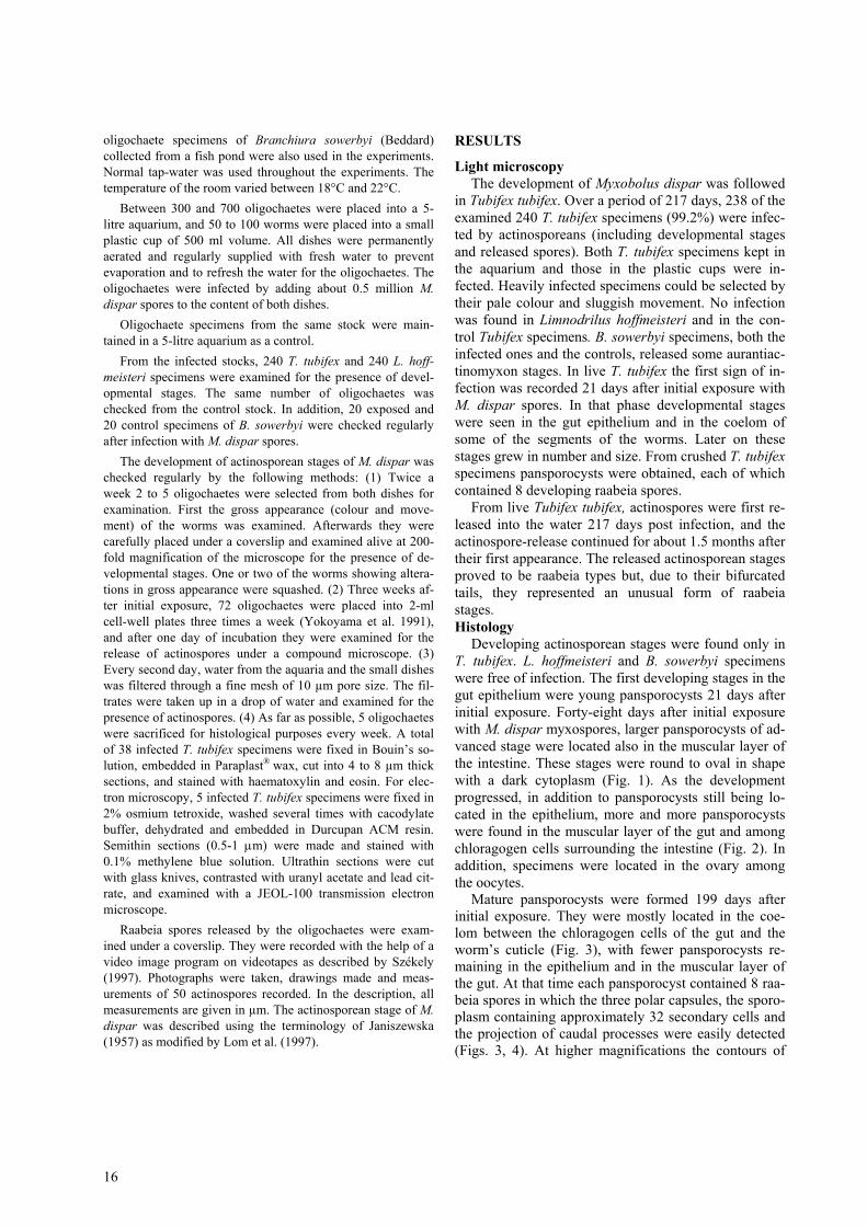
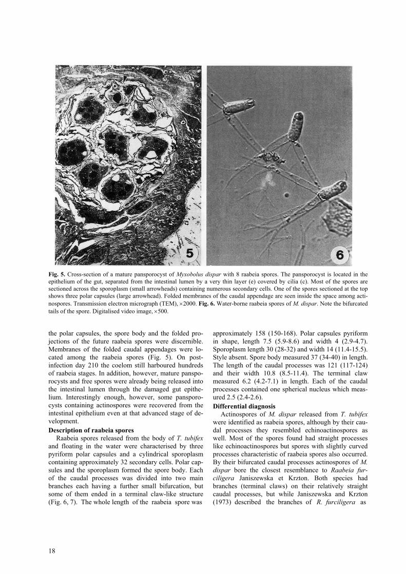
Fig. 5. Cross-section of a mature pansporocyst of Myxobolus dispar with 8 raabeia spores. The pansporocyst is located in theepithelium of the gut, separated from the intestinal lumen by a very thin layer (e) covered by cilia (c). Most of the spores aresectioned across the sporoplasm (small arrowheads) containing numerous secondary cells. One of the spores sectioned at the topshows three polar capsules (large arrowhead). Folded membranes of the caudal appendage are seen inside the space among acti-nospores. Transmission electron micrograph (TEM), ×2000. Fig. 6. Water-borne raabeia spores of M. dispar. Note the bifurcatedtails of the spore. Digitalised video image, ×500.
the polar capsules, the spore body and the folded pro-jections of the future raabeia spores were discernible.Membranes of the folded caudal appendages were lo-cated among the raabeia spores (Fig. 5). On post-infection day 210 the coelom still harboured hundredsof raabeia stages. In addition, however, mature panspo-rocysts and free spores were already being released intothe intestinal lumen through the damaged gut epithe-lium. Interestingly enough, however, some pansporo-cysts containing actinospores were recovered from theintestinal epithelium even at that advanced stage of de-velopment.Description of raabeia spores
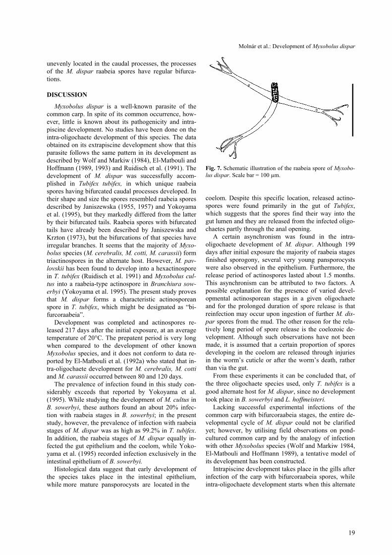
Raabeia spores released from the body of T. tubifexand floating in the water were characterised by threepyriform polar capsules and a cylindrical sporoplasmcontaining approximately 32 secondary cells. Polar cap-sules and the sporoplasm formed the spore body. Eachof the caudal processes was divided into two mainbranches each having a further small bifurcation, butsome of them ended in a terminal claw-like structure(Fig. 6, 7). The whole length of the raabeia spore was
approximately 158 (150-168). Polar capsules pyriformin shape, length 7.5 (5.9-8.6) and width 4 (2.9-4.7).Sporoplasm length 30 (28-32) and width 14 (11.4-15.5).Style absent. Spore body measured 37 (34-40) in length.The length of the caudal processes was 121 (117-124)and their width 10.8 (8.5-11.4). The terminal clawmeasured 6.2 (4.2-7.1) in length. Each of the caudalprocesses contained one spherical nucleus which meas-ured 2.5 (2.4-2.6).Differential diagnosis
Actinospores of M. dispar released from T. tubifexwere identified as raabeia spores, although by their cau-dal processes they resembled echinoactinospores aswell. Most of the spores found had straight processeslike echinoactinospores but spores with slightly curvedprocesses characteristic of raabeia spores also occurred.By their bifurcated caudal processes actinospores of M.dispar bore the closest resemblance to Raabeia fur-ciligera Janiszewska et Krzton. Both species hadbranches (terminal claws) on their relatively straightcaudal processes, but while Janiszewska and Krzton(1973) described the branches of R. furciligera as
Molnár et al.: Development of Myxobolus dispar
19
unevenly located in the caudal processes, the processesof the M. dispar raabeia spores have regular bifurca-tions.
DISCUSSION
Myxobolus dispar is a well-known parasite of thecommon carp. In spite of its common occurrence, how-ever, little is known about its pathogenicity and intra-piscine development. No studies have been done on theintra-oligochaete development of this species. The dataobtained on its extrapiscine development show that thisparasite follows the same pattern in its development asdescribed by Wolf and Markiw (1984), El-Matbouli andHoffmann (1989, 1993) and Ruidisch et al. (1991). Thedevelopment of M. dispar was successfully accom-plished in Tubifex tubifex, in which unique raabeiaspores having bifurcated caudal processes developed. Intheir shape and size the spores resembled raabeia sporesdescribed by Janiszewska (1955, 1957) and Yokoyamaet al. (1995), but they markedly differed from the latterby their bifurcated tails. Raabeia spores with bifurcatedtails have already been described by Janiszewska andKrzton (1973), but the bifurcations of that species haveirregular branches. It seems that the majority of Myxo-bolus species (M. cerebralis, M. cotti, M. carassii) formtriactinospores in the alternate host. However, M. pav-lovskii has been found to develop into a hexactinosporein T. tubifex (Ruidisch et al. 1991) and Myxobolus cul-tus into a raabeia-type actinospore in Branchiura sow-erbyi (Yokoyama et al. 1995). The present study provesthat M. dispar forms a characteristic actinosporeanspore in T. tubifex, which might be designated as “bi-furcoraabeia”.
Development was completed and actinospores re-leased 217 days after the initial exposure, at an averagetemperature of 20°C. The prepatent period is very longwhen compared to the development of other knownMyxobolus species, and it does not conform to data re-ported by El-Matbouli et al. (1992a) who stated that in-tra-oligochaete development for M. cerebralis, M. cottiand M. carassii occurred between 80 and 120 days.
The prevalence of infection found in this study con-siderably exceeds that reported by Yokoyama et al.(1995). While studying the development of M. cultus inB. sowerbyi, these authors found an about 20% infec-tion with raabeia stages in B. sowerbyi; in the presentstudy, however, the prevalence of infection with raabeiastages of M. dispar was as high as 99.2% in T. tubifex.In addition, the raabeia stages of M. dispar equally in-fected the gut epithelium and the coelom, while Yoko-yama et al. (1995) recorded infection exclusively in theintestinal epithelium of B. sowerbyi.
Histological data suggest that early development ofthe species takes place in the intestinal epithelium,while more mature pansporocysts are located in the
Fig. 7. Schematic illustration of the raabeia spore of Myxobo-lus dispar. Scale bar = 100 µm.
coelom. Despite this specific location, released actino-spores were found primarily in the gut of Tubifex,which suggests that the spores find their way into thegut lumen and they are released from the infected oligo-chaetes partly through the anal opening.
A certain asynchronism was found in the intra-oligochaete development of M. dispar. Although 199days after initial exposure the majority of raabeia stagesfinished sporogony, several very young pansporocystswere also observed in the epithelium. Furthermore, therelease period of actinospores lasted about 1.5 months.This asynchronism can be attributed to two factors. Apossible explanation for the presence of varied devel-opmental actinosporean stages in a given oligochaeteand for the prolonged duration of spore release is thatreinfection may occur upon ingestion of further M. dis-par spores from the mud. The other reason for the rela-tively long period of spore release is the coelozoic de-velopment. Although such observations have not beenmade, it is assumed that a certain proportion of sporesdeveloping in the coelom are released through injuriesin the worm’s cuticle or after the worm’s death, ratherthan via the gut.
From these experiments it can be concluded that, ofthe three oligochaete species used, only T. tubifex is agood alternate host for M. dispar, since no developmenttook place in B. sowerbyi and L. hoffmeisteri.
Lacking successful experimental infections of thecommon carp with bifurcoraabeia stages, the entire de-velopmental cycle of M. dispar could not be clarifiedyet; however, by utilising field observations on pond-cultured common carp and by the analogy of infectionwith other Myxobolus species (Wolf and Markiw 1984,El-Matbouli and Hoffmann 1989), a tentative model ofits development has been constructed.
Intrapiscine development takes place in the gills afterinfection of the carp with bifurcoraabeia spores, whileintra-oligochaete development starts when this alternate

20
host becomes infected with the myxosporean spores ofM. dispar.Acknowledgements. The study was supported by the Hun-garian Scientific Research Fund (OTKA, project no. T02
0044) and by a joint grant given by the Balaton Secretariat of thePrime Minister’s Office. The authors thank Ms. Emese Papp forher help in collecting and culturing tubificids. Grateful thanksare due to the Balaton Fisheries Co. for providing fish.
REFERENCES
BARTHOLOMEW J. L., WHIPPLE M. J., STEVENS D. G.,FRYER J. L. (1997): The life cycle of Ceratomyxa shasta,a myxosporean parasite of salmonids requires a freshwaterpolychaete as an alternate host. J. Parasitol: 83: 859-868.
BENAJIBA M.H., MARQUES A. 1993: The alternation ofactinomyxidian and myxosporidian sporal forms in thedevelopment of Myxidium giardi (parasite of Anguilla an-guilla) through oligochaetes. Bull. Eur. Ass. Fish Pathol.13: 100-103.
BRINKHURST R.O. 1963: A guide for the identification ofBritish aquatic Oligochaeta. Freshwater Biological Asso-ciation, Scientific Publication No. 22, 52 pp.
EL-MANSY A., MOLNÁR K. 1997a: Extrapiscine develop-ment of Myxobolus drjagini Achmerov, 1954 (Myxospo-rea: Myxobolidae) in oligochaete alternative hosts. ActaVet. Hung. 45: 427-438.
EL-MANSY A., MOLNÁR K. 1997b: Development of Myxo-bolus hungaricus Jaczó, 1940 (Myxosporea: Myxoboli-dae) in oligochaete alternate hosts. Dis. Aquat. Org. 31:227-232.
EL-MATBOULI M., FISCHER-SCHERL T., HOFFMANNR.W. 1992a: Present knowledge on the life cycle, taxon-omy, pathology, and therapy of some Myxosporea spp.important for freshwater fish. Annu. Rev. Fish Dis. 3:367-402.
EL-MATBOULI M., FISCHER-SCHERL T., HOFFMANNR.W. 1992b: Transmission of Hoferellus carassii Ach-merov, 1960 to goldfish Carassius auratus via an aquaticoligochaete. Bull. Eur. Ass. Fish Pathol. 12: 54-56.
EL-MATBOULI M., HOFFMANN R.W. 1989: Experimentaltransmission of two Myxobolus spp. developing by spo-rogony via tubificid worms. Parasitol. Res. 75: 461-464.
EL-MATBOULI M., HOFFMANN R.W. 1993: Myxoboluscarassii Klokacheva, 1914 also requires an aquatic oligo-chaete, Tubifex tubifex as intermediate host in its life cy-cle. Bull. Eur. Ass. Fish Pathol. 13: 189-192.
GROSSHEIDER G., KÖRTING W. 1992: First evidence thatHoferellus cyprini (Doflein, 1898) is transmitted by Naissp. Bull. Eur. Ass. Fish Pathol. 12: 17-20.
JANISZEWSKA J. 1955: Actinomyxidia. Morphology, ecol-ogy, history of investigations, systematics, devel-opment.Acta Parasitol. Pol. 2: 405-443.
JANISZEWSKA J. 1957: Actinomyxidia II. New systematics,sexual cycles, description of new genera and species.Zool. Pol. 8: 3-34.
JANISZEWSKA J., KRZTON M. 1973: Raabeia furciligerasp. n. (Cnidosporidia: Actinomyxidia) from the body cav-ity of Limnodrilus hoffmeisteri Claparéde, 1862. ActaProtozool. 12: 165-167.
KENT M.L., WHITAKER D.J., MARGOLIS L. 1993:Transmission of Myxobolus arcticus Pugachev and Khok-
hlov, 1979, a myxosporean parasite of Pacific salmon, viaa triactinomyxon from the aquatic oligochaete Stylodrilusheringianus (Lumbriculidae). Can. J. Zool. 71: 1207-1211.
LOM J., McGEORGE J., FEIST S.W., MORRIS D., ADAMSA. 1997: Guidelines for the uniform characterisation ofthe actinosporean stages of parasites of the phylum Myxo-zoa. Dis. Aquat. Org. 30: 1-9.
MOLNÁR K., SZAKOLCZAI J. 1980: Halbetegségek. Pub-lishing House Mezőgazdasági Kiadó, 254 pp. (In Hun-garian)
RUIDISCH S., EL-MATBOULI M., HOFFMANN R.W.1991: The role of tubificid worms as an intermediate hostin the life cycle of Myxobolus pavlovskii (Akhmerov,1954). Parasitol. Res. 77: 663-667.
STYER E.L., HARRISON L.R., BURTLE G.J. 1991: Ex-perimental production of proliferative gill disease in chan-nel catfish exposed to a myxozoan-infected oligochaete,Dero digitata. J. Aquat. Anim. Hlth. 3: 288-291.
SZÉKELY C. 1997: Possible applications of video technologyand digital image processing in fish parasitology: mor-phological examination of the groups Apicomplexa andMyxosporea-Actinosporea by video-technology. Bull.Eur. Ass. Fish Pathol. 17: 81-82.
THÉLOHAN P. 1895: Recherches sur les Myxosporidies.Bull. Sci. Fr. Belg. 26: 100–394.
TROUILLIER A., EL-MATBOULI M., HOFFMANN R.W.1996: A new look at the life-cycle of Hoferellus carassiiin the goldfish (Carassius auratus auratus) and its relationto “kidney enlargement disease” (KED). Folia Parasitol.43: 173-187.
USPENSKAYA A.V. 1995: Alternation of actinosporean andmyxosporean phases in the life cycle of Zschokkella nova(Myxozoa). J. Euk. Microbiol. 42: 665-668.
WOLF K., MARKIW M.E. 1984: Biology contravenes taxon-omy in the myxozoa: new discoveries show alternation ofinvertebrate and vertebrate hosts. Science 225: 1449-1452.
YOKOYAMA H. 1997: Transmission of Thelohanellus ho-vorkai Achmerov, 1960 (Myxosporea: Myxozoa) to com-mon carp Cyprinus carpio through the alternate oligo-chaete host. Syst. Parasitol. 36: 79-84.
YOKOYAMA H., OGAWA K., WAKABAYASHI H. 1991:A new collection method of actinosporeans. - A probableinfective stage of myxosporeans to fishes from tubificidsand experimental infection of goldfish with the actino-sporean, Raabeia sp. Fish Pathol. 26: 133-138.
YOKOYAMA H., OGAWA K., WAKABAYASHI H. 1993:Involvement of Branchiura sowerbyi (Oligochaeta: An-nelida) in the transmission of Hoferellus carassii (Myxo-sporea: Myxozoa), the causative agent of kidney enlarge-ment disease (KED) of goldfish Carassius auratus. FishPathol. 28: 135-139.
Molnár et al.: Development of Myxobolus dispar
21
FIFTH INTERNATIONAL SYMPOSIUM ONFISH PARASITES
9 - 13 AUGUST 1999ČESKÉ BUDĚJOVICE, CZECH REPUBLIC
Organized by the Institute of Parasitology, Academy of Sciencesof the Czech Republic
Contact:Dr. František Moravec or Dr. Jiří Lom
Institute of ParasitologyAcademy of Sciences of the Czech RepublicBranišovská 31, 370 05 České Budějovice
CZECH REPUBLIC
Phone: ++420 38 7775432; Fax: ++420 38 47743E-mail: [email protected], or [email protected]
http://www.paru.cas.cz/Sympos.htm
YOKOYAMA H., OGAWA K., WAKABAYASHI H. 1995:Myxobolus cultus n. sp. (Myxosporea: Myxobolidae) inthe goldfish Carassius auratus transformed from the acti-
nosporean stage in the oligochaete Branchiura sowerbyi.J. Parasitol. 81: 446-451.
Received 26 January 1998 Accepted 15 April 1998