development of phenylthiourea derivatives as allosteric
TRANSCRIPT
University of Groningen
Development of phenylthiourea derivatives as allosteric inhibitors of pyoverdine maturationenzyme PvdP tyrosinaseWibowo, Joko P; Xiao, Zhangping; Voet, Julian M; Dekker, Frank J; Quax, Wim J
Published in:Bioorganic & Medicinal Chemistry Letters
DOI:10.1016/j.bmcl.2020.127409
IMPORTANT NOTE: You are advised to consult the publisher's version (publisher's PDF) if you wish to cite fromit. Please check the document version below.
Document VersionPublisher's PDF, also known as Version of record
Publication date:2020
Link to publication in University of Groningen/UMCG research database
Citation for published version (APA):Wibowo, J. P., Xiao, Z., Voet, J. M., Dekker, F. J., & Quax, W. J. (2020). Development of phenylthioureaderivatives as allosteric inhibitors of pyoverdine maturation enzyme PvdP tyrosinase. Bioorganic &Medicinal Chemistry Letters, 30(17), [127409]. https://doi.org/10.1016/j.bmcl.2020.127409
CopyrightOther than for strictly personal use, it is not permitted to download or to forward/distribute the text or part of it without the consent of theauthor(s) and/or copyright holder(s), unless the work is under an open content license (like Creative Commons).
The publication may also be distributed here under the terms of Article 25fa of the Dutch Copyright Act, indicated by the “Taverne” license.More information can be found on the University of Groningen website: https://www.rug.nl/library/open-access/self-archiving-pure/taverne-amendment.
Take-down policyIf you believe that this document breaches copyright please contact us providing details, and we will remove access to the work immediatelyand investigate your claim.
Downloaded from the University of Groningen/UMCG research database (Pure): http://www.rug.nl/research/portal. For technical reasons thenumber of authors shown on this cover page is limited to 10 maximum.
Contents lists available at ScienceDirect
Bioorganic & Medicinal Chemistry Letters
journal homepage: www.elsevier.com/locate/bmcl
Development of phenylthiourea derivatives as allosteric inhibitors ofpyoverdine maturation enzyme PvdP tyrosinaseJoko P. Wibowoa,b,c, Zhangping Xiaoa,c, Julian M. Voeta, Frank J. Dekkera, Wim J. Quaxa,⁎
a Department of Chemical and Pharmaceutical Biology, Groningen Research Institute of Pharmacy, University of Groningen, the Netherlandsb Faculty of Pharmacy, University of Muhammadiyah Banjarmasin, Banjarmasin, Indonesia
A R T I C L E I N F O
Keywords:Pseudomonas aeruginosaIronPyoverdinePvdP
A B S T R A C T
Infections caused by Pseudomonas aeruginosa become increasingly difficult to treat because these bacteria haveacquired various mechanisms for antibiotic resistance, which creates the need for mechanistically novel anti-biotics. Such antibiotics might be developed by targeting enzymes involved in the iron uptake mechanism be-cause iron is essential for bacterial survival. For P. aeruginosa, pyoverdine has been described as an importantvirulence factor that plays a key role in iron uptake. Therefore, inhibition of enzymes involved in the pyoverdinesynthesis, such as PvdP tyrosinase, can open a new window for the treatment of P. aeruginosa infections.Previously, we reported phenylthiourea as the first allosteric inhibitor of PvdP tyrosinase with high micromolarpotency. In this report, we explored structure-activity relationships (SAR) for PvdP tyrosinase inhibition byphenylthiourea derivatives. This enables identification of a phenylthiourea derivative (3c) with a potency in thesubmicromolar range (IC50 = 0.57 + 0.05 µM). Binding could be rationalized by molecular docking simulationand 3c was proved to inhibit the bacterial pyoverdine production and bacterial growth in P. aeruginosa PA01cultures.
Pseudomonas aeruginosa is a Gram-negative pathogen that causesinfection in many organs. Although these bacteria barely infect healthyindividuals, cystic fibrosis patients and immunocompromised in-dividuals suffer from high morbidity and mortality in P. aeruginosa in-fections.1 However, the development of antibiotic resistance increas-ingly limits the treatment options for P. aeruginosa infections.2 P.aeruginosa employs intrinsic, acquired and adaptive mechanisms tocounter most antibiotics including aminoglycosides, quinolones, and β-lactams. Expulsion of antibiotic molecules by efflux pumps is one of theexamples of the intrinsic and adaptive mechanisms. The acquired re-sistance mechanisms can be obtained through either horizontal transferof resistance genes or mutational changes. Biofilm formation is one ofthe examples of adaptive antibiotic resistance.3 In 2017, the WorldHealth Organization (WHO) has classified carbapenem-resistant P.aeruginosa into category 1, which is a category of the most urgent needfor developing new antibiotics to treat the infection.4
Bacterial transport mechanisms to acquire iron provide attractivetargets to develop new antibiotics since iron is an essential element thatacts as a cofactor in many biological reactions in bacteria including P.
aeruginosa.5 Moreover, the low availability of iron in the human body isone of the growth-limiting factors for P. aeruginosa. Since iron III (Fe3+)is hardly soluble and cannot passively diffuse into the bacterial cells,these bacteria secrete siderophore pyoverdine, an iron III (Fe3+) che-lating molecule, to transport iron into the cell via the FpvA transporter.6
Pyoverdine biosynthetic enzymes have been proposed as novel anti-biotic targets because small molecule inhibitors would downregulatethe production of pyoverdine resulting in limited iron acquisition. Insupport of this concept, several studies have reported that P. aeruginosapyoverdine-deficient mutants have reduced virulence in animal andplant infection models.7,8 Therefore, small molecule inhibitors that in-terfere with the pyoverdine biosynthetic enzymes have potency for thedevelopment of new treatments for P. aeruginosa infections.
In a low iron condition, the biosynthesis of pyoverdine is induced.Initially, in the absence of complex Fe-ferri uptake regulator (FUR)which otherwise binds to the PvdS promoter, to block the production ofthe extracytoplasmic function (ECF)-sigma factor PvdS. After transla-tion, PvdS binds to the IS boxes of PVDI genes, starting the transcriptionand translation of PVDI proteins. The non-ribosomal peptide
https://doi.org/10.1016/j.bmcl.2020.127409Received 1 April 2020; Received in revised form 9 July 2020; Accepted 10 July 2020
Abbreviations: IPTG, isopropyl 1-thio-β-D-galactopyranoside; PTU, phenylthiourea⁎ Corresponding author at: Department of Chemical and Pharmaceutical Biology, Groningen Research Institute of Pharmacy, University of Groningen, Antonius
Deusinglaan 1, Groningen 9713 AV, the Netherlands.E-mail address: [email protected] (W.J. Quax).
c These authors contributed equally.
Bioorganic & Medicinal Chemistry Letters 30 (2020) 127409
Available online 15 July 20200960-894X/ © 2020 The Authors. Published by Elsevier Ltd. This is an open access article under the CC BY license (http://creativecommons.org/licenses/BY/4.0/).
T
synthetases (PvdL, PvdI, PvdJ, and PvdD) together with MbtH, PvdG,PvdH, PvdA and PvdF assemble the precursor siderophore PvdIq in thecytoplasm. Subsequently, PvdE transports PvdIq to the periplasm thenit is cleaved by PvdQ to form ferribactin and myristoleic acid. Theconversion of ferribactin to pyoverdine involves at least four differentenzymes: PvdM, PvdN, PvdO, and PvdP at the final step of the bio-synthesis.9,10 PvdP is a tyrosinase known from its role for the matura-tion of pyoverdine chromophore.11 Even though, tyrosinases are foundin almost every organism: human, animal, plant, mushroom, and bac-teria, the PvdP enzyme is different compared to other tyrosinases. Theclosest homolog is a tyrosinase of Bacillus megaterium (PDB ID = 3NQ5)sharing sequence identity 30,7% (Clustal Omega, http://www.ebi.ac.uk/Tools). Moreover, PvdP also has a unique homodimer structurewhere each monomer consists of 2 different domains i.e.: beta-barreldomain (BBD) and tyrosinase domain (TYD) are not found in the othertyrosinase structure based on the recent reports. As in other tyrosinases,its active site is formed by a di-copper center of six histidine residues,12
but in the case of PvdP, this is uniquely covered by a C-terminal lid inthe apo state.13
In line with the facts above, we have reported that most of thetyrosinase inhibitors, for example, mushroom tyrosinase inhibitors donot inhibit PvdP tyrosinase activity. Surprisingly, phenylthiourea (PTU)was found to inhibit PvdP tyrosinase activity at micromolar con-centration. Moreover, a crystal structure demonstrated the binding ofPTU to an allosteric binding site instead of the tyrosinase active site.13
This provides opportunities to develop inhibitors that target the allos-teric binding site, which seems to be confined to fluorescent pseudo-monads. Therefore, identifying PvdP tyrosinase inhibitors that selec-tively interfere with the allosteric binding site, might result in a specificantibiotic with limited side effects as it does not bind to other tyr-osinases.
To obtain sub-micromolar inhibitors, we designed and synthesized aseries of PTU derivatives. Some of the generated derivatives show in-hibition at the low micromolar range. Kinetic studies were performed todetermine the type of inhibition and, with molecular docking revealingthe binding of the compound at the allosteric binding site, the highaffinity was being explained.
Design and synthesis of PTU derivatives
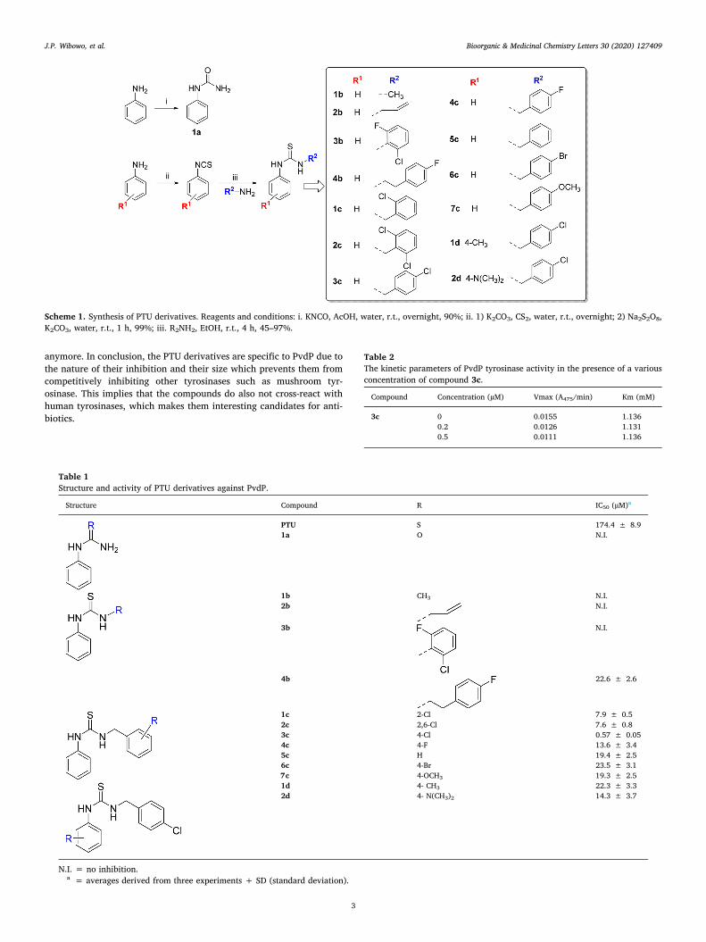
In our previous study, PTU was discovered as an allosteric inhibitorthat binds at the interface of the beta-barrel domain (BBD) and C-terminal domain (TYD) of PvdP and thereby inhibits the tyrosinaseenzymatic activity.13 A focused compound collection of PTU derivativeswas synthesized to improve the PvdP tyrosinase inhibitory potency andto explore structure–activity relationships. The desired PTU derivativeswere generated by the synthesis of the required phenylthiocyanates thatwere reacted with the corresponding amines to provide the desiredphenylisothiocyanates as final products (Scheme 1). The phenyli-sothiocyanates were prepared through a one-pot procedure, in whichthe phenylamines and carbon disulfide were reacted to provide thedithiocarbamate that was desulfurized with sodium persulfate to givethe products in good yields after filtration and wash with water.14 Thephenylisothiocyanates were applied without prior purification forcoupling with the corresponding amines to generate the disubstitutedthioureas (Table 1). The compounds were subjected to filtration toobtain the pure products as white precipitates, except for 1b and 3b forwhich flash column purification was needed. The final product wasobtained in variable but sufficient yields (38–99%) over two steps.Phenylurea (1a) was synthesized as an equivalent of phenylthiourea bycharging aniline with potassium cyanate in an aqueous 10% AcOHsolution. The product 1a was formed and isolated by extraction andrecrystallization to provide 1a in a yield of 98%.15
Structure-activity relationship of PTU derivatives
The structure-activity relationships for PvdP tyrosinase inhibitionby the 14 membered compound collection were investigated. First,modification of the thiourea functionality of phenylthiourea providedphenylurea 1a, which provided less than 50% inhibition of PvdP tyr-osinase activity at a concentration up to 100 μM. The crystal structureof PvdP bound to PTU (PDB ID = 6RRP) showed the thioketone groupinteracting with Trp320 and Ser329. The substitution of this group witha ketone group breaks the interaction between the compound and thoseresidues resulting in the loss of its inhibitory activity on PvdP. Thismeans the thio ketone group is important for the inhibitory activity ofPTU on the tyrosinase activity of PvdP.
Next, the substitution of the thiourea functionality of PTU was ex-plored (Table 1) and it was found that methyl (1b), vinyl (2b) or phenyl(3b) substitution decreased the potency. In contrast, ethyl phenyl orbenzyl substitution of phenylthiourea improved the inhibitory potencyby a factor 10 or more. Structural variation of the benzyl substitutionprovided compound 3c with a more than 100-fold improved potencycompared to non-substituted PTU with an IC50 of 0.57 μM. Other sub-stitution patterns, such as 2-Cl, 2,6-Cl, 4-F, 4-H, 4-Br or 4-OCH3, pro-vided IC50 between 7.6 and 23.5 μM. Further substitutions of the phenylring of PTU were investigated based on 3c as a starting point. However,1d and 2d with a 4-methyl or 4-dimethylamino provided IC50 valuesthat indicated reduced potency compared to 3c.
Kinetic study and inhibition activity on PvdP-trunc
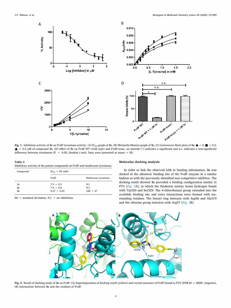
To establish the mechanism of PvdP tyrosinase inhibition,Michaelis–Menten enzyme kinetics analysis in the presence of inhibitor3c was performed. The Lineweaver–Burk double reciprocal plot showsthat inhibitor 3c causes a decrease in the Vmax values, whereas the Km
values remain constant (Table 2), indicating a non-competitive inhibi-tion. These results are in line with the data obtained previously for PTUfor which we also reported non-competitive inhibition. Obviously, thesubstitution on the thiourea functionality of PTU with 4-chlorobenzylimproved the inhibition activity but did not change the mechanism ofinhibition. Previously, we described that PTU mediated inhibition ofPvdP tyrosinase activity involves a C-terminal lid rearrangement andthe absence of this lid precludes PTU mediated inhibition of PvdP tyr-osinase activity. Inhibitor 3c was tested for inhibition of wild-type PvdPin comparison to a PvdP variant that is truncated for the C-terminal lid.Similar to the parent compound, this PTU derivative lost its potentinhibition activity on the truncated enzyme, which indicates that in-hibitor 3c has the same mechanism of inhibition as described for PTUthat stabilizes the C-terminal lid covering the active site (Fig. 1D).
Mushroom tyrosinase inhibition
In a follow-up experiment, three most potent of the PTU derivativeswere tested on mushroom tyrosinase to investigate their effect on othertyrosinases. The IC50 values of the compounds increased dramatically,the IC50 of 1c and 2c were greater than 500 µM and the IC50 of the mostpotent compound (3c) increased up to 328 ± 27 µM, an increase ofmore than 500 times compared to PvdP (Table 3). It means that thederivatives of PTU are not only non-competitive inhibitors, but alsovery specific inhibitors for PvdP.
As reported, PTU is a competitive inhibitor of mushroom tyr-osinase16 and it binds at the active site of tyrosinase.17 Knowing thatthe inhibition of the new derivatives on mushroom tyrosinase is lost,this suggests that its ability to act as a competitive inhibitor is gone.This is most likely attributed to the change in the size and shape of themolecule. The addition of a chlorinated phenyl ring has increased thesize of the molecule in such a way that it does not fit into the active site
J.P. Wibowo, et al. Bioorganic & Medicinal Chemistry Letters 30 (2020) 127409
2
anymore. In conclusion, the PTU derivatives are specific to PvdP due tothe nature of their inhibition and their size which prevents them fromcompetitively inhibiting other tyrosinases such as mushroom tyr-osinase. This implies that the compounds do also not cross-react withhuman tyrosinases, which makes them interesting candidates for anti-biotics.
Scheme 1. Synthesis of PTU derivatives. Reagents and conditions: i. KNCO, AcOH, water, r.t., overnight, 90%; ii. 1) K2CO3, CS2, water, r.t., overnight; 2) Na2S2O8,K2CO3, water, r.t., 1 h, 99%; iii. R2NH2, EtOH, r.t., 4 h, 45–97%.
Table 1Structure and activity of PTU derivatives against PvdP.
Structure Compound R IC50 (µM)a
PTU S 174.4 ± 8.91a O N.I.
1b CH3 N.I.2b N.I.
3b N.I.
4b 22.6 ± 2.6
1c 2-Cl 7.9 ± 0.52c 2,6-Cl 7.6 ± 0.83c 4-Cl 0.57 ± 0.054c 4-F 13.6 ± 3.45c H 19.4 ± 2.56c 4-Br 23.5 ± 3.17c 4-OCH3 19.3 ± 2.51d 4- CH3 22.3 ± 3.32d 4- N(CH3)2 14.3 ± 3.7
N.I. = no inhibition.a = averages derived from three experiments + SD (standard deviation).
Table 2The kinetic parameters of PvdP tyrosinase activity in the presence of a variousconcentration of compound 3c.
Compound Concentration (µM) Vmax (A475/min) Km (mM)
3c 0 0.0155 1.1360.2 0.0126 1.1310.5 0.0111 1.136
J.P. Wibowo, et al. Bioorganic & Medicinal Chemistry Letters 30 (2020) 127409
3
Molecular docking analysis
In order to link the observed SAR to binding information, 3c wasdocked at the allosteric binding site of the PvdP enzyme in a similarfashion as with the previously identified non-competitive inhibitor. Thedocking result showed 3c provided a binding configuration similar toPTU (Fig. 2A), in which the thioketon moiety forms hydrogen bondswith Trp320 and Ser329. The 4-chlorobenzyl group extended into theavailable binding site and extra interactions were formed with sur-rounding residues. The benzyl ring interacts with Asp56 and Ala319and the chlorine group interacts with Arg57 (Fig. 2B).
Fig. 1. Inhibition activity of 3c on PvdP tyrosinase activity. (A) IC50 graph of 3c, (B) Michaelis-Menten graph of 3c, (C) Lineweaver-Burk plots of 3c; ● = 0, ■ = 0.2,▲ = 0.5 µM of compound 3c, (D) effect of 3c on PvdP WT (wild type) and PvdP-trunc, an asterisk (*) indicates a significant and n.s. indicates a non-significantdifference between treatments (P < 0.05, Student t test). Data were presented as mean + SD.
Table 3Inhibitory activity of the potent compounds on PvdP and mushroom tyrosinase.
Compound IC50 + SD (µM)
PvdP Mushroom tyrosinase
1c 7.9 + 0.5 N.I.2c 7.6 + 0.8 N.I.3c 0.57 + 0.05 328 + 27
SD = standard deviation, N.I. = no inhibition.
Fig. 2. Result of docking study of 3c on PvdP. (A) Superimposition of docking result (yellow) and crystal structure of PvdP bound to PTU (PDB ID = 6RRP, magenta),(B) interactions between 3c and the residues of PvdP.
J.P. Wibowo, et al. Bioorganic & Medicinal Chemistry Letters 30 (2020) 127409
4
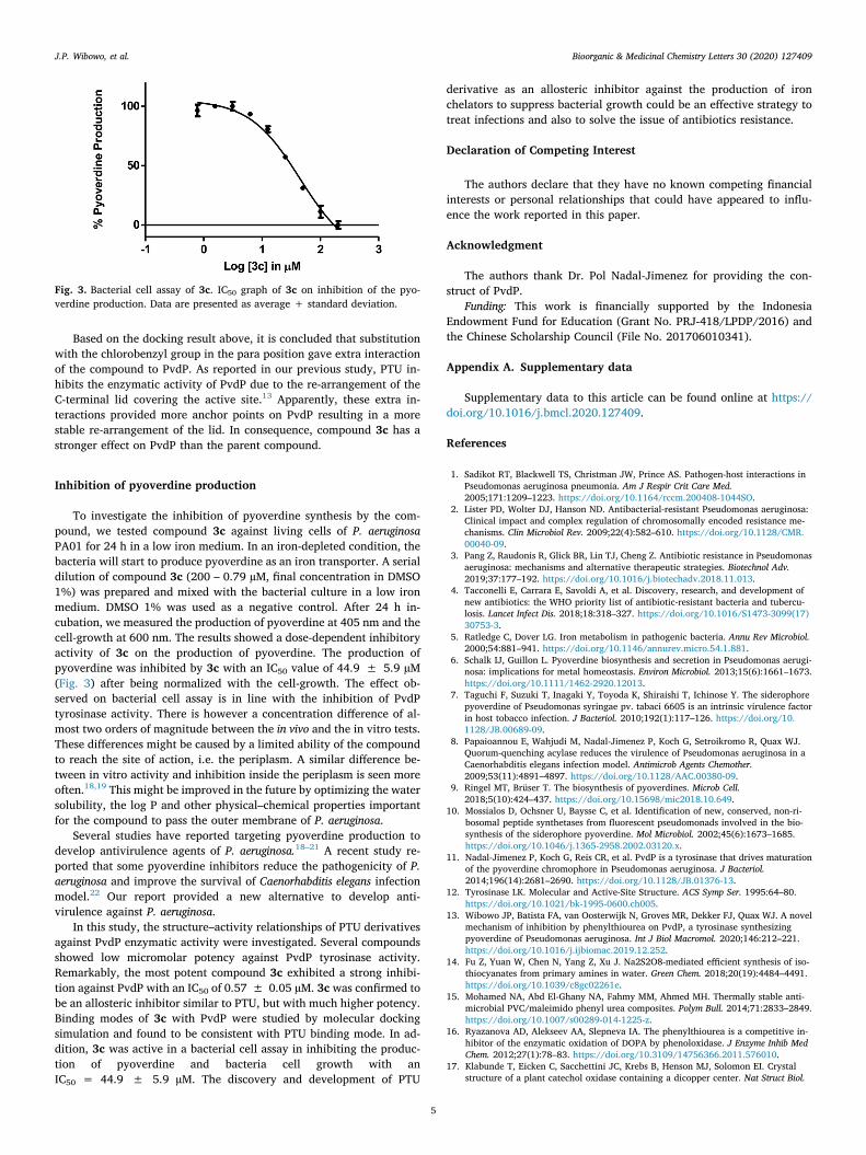
Based on the docking result above, it is concluded that substitutionwith the chlorobenzyl group in the para position gave extra interactionof the compound to PvdP. As reported in our previous study, PTU in-hibits the enzymatic activity of PvdP due to the re-arrangement of theC-terminal lid covering the active site.13 Apparently, these extra in-teractions provided more anchor points on PvdP resulting in a morestable re-arrangement of the lid. In consequence, compound 3c has astronger effect on PvdP than the parent compound.
Inhibition of pyoverdine production
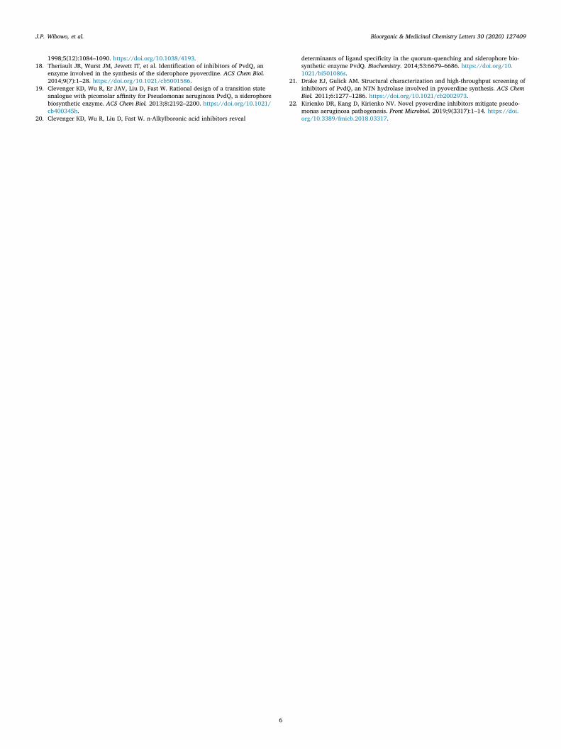
To investigate the inhibition of pyoverdine synthesis by the com-pound, we tested compound 3c against living cells of P. aeruginosaPA01 for 24 h in a low iron medium. In an iron-depleted condition, thebacteria will start to produce pyoverdine as an iron transporter. A serialdilution of compound 3c (200 – 0.79 µM, final concentration in DMSO1%) was prepared and mixed with the bacterial culture in a low ironmedium. DMSO 1% was used as a negative control. After 24 h in-cubation, we measured the production of pyoverdine at 405 nm and thecell-growth at 600 nm. The results showed a dose-dependent inhibitoryactivity of 3c on the production of pyoverdine. The production ofpyoverdine was inhibited by 3c with an IC50 value of 44.9 ± 5.9 µM(Fig. 3) after being normalized with the cell-growth. The effect ob-served on bacterial cell assay is in line with the inhibition of PvdPtyrosinase activity. There is however a concentration difference of al-most two orders of magnitude between the in vivo and the in vitro tests.These differences might be caused by a limited ability of the compoundto reach the site of action, i.e. the periplasm. A similar difference be-tween in vitro activity and inhibition inside the periplasm is seen moreoften.18,19 This might be improved in the future by optimizing the watersolubility, the log P and other physical–chemical properties importantfor the compound to pass the outer membrane of P. aeruginosa.
Several studies have reported targeting pyoverdine production todevelop antivirulence agents of P. aeruginosa.18–21 A recent study re-ported that some pyoverdine inhibitors reduce the pathogenicity of P.aeruginosa and improve the survival of Caenorhabditis elegans infectionmodel.22 Our report provided a new alternative to develop anti-virulence against P. aeruginosa.
In this study, the structure–activity relationships of PTU derivativesagainst PvdP enzymatic activity were investigated. Several compoundsshowed low micromolar potency against PvdP tyrosinase activity.Remarkably, the most potent compound 3c exhibited a strong inhibi-tion against PvdP with an IC50 of 0.57 ± 0.05 µM. 3c was confirmed tobe an allosteric inhibitor similar to PTU, but with much higher potency.Binding modes of 3c with PvdP were studied by molecular dockingsimulation and found to be consistent with PTU binding mode. In ad-dition, 3c was active in a bacterial cell assay in inhibiting the produc-tion of pyoverdine and bacteria cell growth with anIC50 = 44.9 ± 5.9 µM. The discovery and development of PTU
derivative as an allosteric inhibitor against the production of ironchelators to suppress bacterial growth could be an effective strategy totreat infections and also to solve the issue of antibiotics resistance.
Declaration of Competing Interest
The authors declare that they have no known competing financialinterests or personal relationships that could have appeared to influ-ence the work reported in this paper.
Acknowledgment
The authors thank Dr. Pol Nadal-Jimenez for providing the con-struct of PvdP.
Funding: This work is financially supported by the IndonesiaEndowment Fund for Education (Grant No. PRJ-418/LPDP/2016) andthe Chinese Scholarship Council (File No. 201706010341).
Appendix A. Supplementary data
Supplementary data to this article can be found online at https://doi.org/10.1016/j.bmcl.2020.127409.
References
1. Sadikot RT, Blackwell TS, Christman JW, Prince AS. Pathogen-host interactions inPseudomonas aeruginosa pneumonia. Am J Respir Crit Care Med.2005;171:1209–1223. https://doi.org/10.1164/rccm.200408-1044SO.
2. Lister PD, Wolter DJ, Hanson ND. Antibacterial-resistant Pseudomonas aeruginosa:Clinical impact and complex regulation of chromosomally encoded resistance me-chanisms. Clin Microbiol Rev. 2009;22(4):582–610. https://doi.org/10.1128/CMR.00040-09.
3. Pang Z, Raudonis R, Glick BR, Lin TJ, Cheng Z. Antibiotic resistance in Pseudomonasaeruginosa: mechanisms and alternative therapeutic strategies. Biotechnol Adv.2019;37:177–192. https://doi.org/10.1016/j.biotechadv.2018.11.013.
4. Tacconelli E, Carrara E, Savoldi A, et al. Discovery, research, and development ofnew antibiotics: the WHO priority list of antibiotic-resistant bacteria and tubercu-losis. Lancet Infect Dis. 2018;18:318–327. https://doi.org/10.1016/S1473-3099(17)30753-3.
5. Ratledge C, Dover LG. Iron metabolism in pathogenic bacteria. Annu Rev Microbiol.2000;54:881–941. https://doi.org/10.1146/annurev.micro.54.1.881.
6. Schalk IJ, Guillon L. Pyoverdine biosynthesis and secretion in Pseudomonas aerugi-nosa: implications for metal homeostasis. Environ Microbiol. 2013;15(6):1661–1673.https://doi.org/10.1111/1462-2920.12013.
7. Taguchi F, Suzuki T, Inagaki Y, Toyoda K, Shiraishi T, Ichinose Y. The siderophorepyoverdine of Pseudomonas syringae pv. tabaci 6605 is an intrinsic virulence factorin host tobacco infection. J Bacteriol. 2010;192(1):117–126. https://doi.org/10.1128/JB.00689-09.
8. Papaioannou E, Wahjudi M, Nadal-Jimenez P, Koch G, Setroikromo R, Quax WJ.Quorum-quenching acylase reduces the virulence of Pseudomonas aeruginosa in aCaenorhabditis elegans infection model. Antimicrob Agents Chemother.2009;53(11):4891–4897. https://doi.org/10.1128/AAC.00380-09.
9. Ringel MT, Brüser T. The biosynthesis of pyoverdines. Microb Cell.2018;5(10):424–437. https://doi.org/10.15698/mic2018.10.649.
10. Mossialos D, Ochsner U, Baysse C, et al. Identification of new, conserved, non-ri-bosomal peptide synthetases from fluorescent pseudomonads involved in the bio-synthesis of the siderophore pyoverdine. Mol Microbiol. 2002;45(6):1673–1685.https://doi.org/10.1046/j.1365-2958.2002.03120.x.
11. Nadal-Jimenez P, Koch G, Reis CR, et al. PvdP is a tyrosinase that drives maturationof the pyoverdine chromophore in Pseudomonas aeruginosa. J Bacteriol.2014;196(14):2681–2690. https://doi.org/10.1128/JB.01376-13.
12. Tyrosinase LK. Molecular and Active-Site Structure. ACS Symp Ser. 1995:64–80.https://doi.org/10.1021/bk-1995-0600.ch005.
13. Wibowo JP, Batista FA, van Oosterwijk N, Groves MR, Dekker FJ, Quax WJ. A novelmechanism of inhibition by phenylthiourea on PvdP, a tyrosinase synthesizingpyoverdine of Pseudomonas aeruginosa. Int J Biol Macromol. 2020;146:212–221.https://doi.org/10.1016/j.ijbiomac.2019.12.252.
14. Fu Z, Yuan W, Chen N, Yang Z, Xu J. Na2S2O8-mediated efficient synthesis of iso-thiocyanates from primary amines in water. Green Chem. 2018;20(19):4484–4491.https://doi.org/10.1039/c8gc02261e.
15. Mohamed NA, Abd El-Ghany NA, Fahmy MM, Ahmed MH. Thermally stable anti-microbial PVC/maleimido phenyl urea composites. Polym Bull. 2014;71:2833–2849.https://doi.org/10.1007/s00289-014-1225-z.
16. Ryazanova AD, Alekseev AA, Slepneva IA. The phenylthiourea is a competitive in-hibitor of the enzymatic oxidation of DOPA by phenoloxidase. J Enzyme Inhib MedChem. 2012;27(1):78–83. https://doi.org/10.3109/14756366.2011.576010.
17. Klabunde T, Eicken C, Sacchettini JC, Krebs B, Henson MJ, Solomon EI. Crystalstructure of a plant catechol oxidase containing a dicopper center. Nat Struct Biol.
Fig. 3. Bacterial cell assay of 3c. IC50 graph of 3c on inhibition of the pyo-verdine production. Data are presented as average + standard deviation.
J.P. Wibowo, et al. Bioorganic & Medicinal Chemistry Letters 30 (2020) 127409
5
1998;5(12):1084–1090. https://doi.org/10.1038/4193.18. Theriault JR, Wurst JM, Jewett IT, et al. Identification of inhibitors of PvdQ, an
enzyme involved in the synthesis of the siderophore pyoverdine. ACS Chem Biol.2014;9(7):1–28. https://doi.org/10.1021/cb5001586.
19. Clevenger KD, Wu R, Er JAV, Liu D, Fast W. Rational design of a transition stateanalogue with picomolar affinity for Pseudomonas aeruginosa PvdQ, a siderophorebiosynthetic enzyme. ACS Chem Biol. 2013;8:2192–2200. https://doi.org/10.1021/cb400345h.
20. Clevenger KD, Wu R, Liu D, Fast W. n-Alkylboronic acid inhibitors reveal
determinants of ligand specificity in the quorum-quenching and siderophore bio-synthetic enzyme PvdQ. Biochemistry. 2014;53:6679–6686. https://doi.org/10.1021/bi501086s.
21. Drake EJ, Gulick AM. Structural characterization and high-throughput screening ofinhibitors of PvdQ, an NTN hydrolase involved in pyoverdine synthesis. ACS ChemBiol. 2011;6:1277–1286. https://doi.org/10.1021/cb2002973.
22. Kirienko DR, Kang D, Kirienko NV. Novel pyoverdine inhibitors mitigate pseudo-monas aeruginosa pathogenesis. Front Microbiol. 2019;9(3317):1–14. https://doi.org/10.3389/fmicb.2018.03317.
J.P. Wibowo, et al. Bioorganic & Medicinal Chemistry Letters 30 (2020) 127409
6