development of the dopaminergic system: from stem cells to ... · session 1 . development of the...
TRANSCRIPT
®
SUPPORTED BY:
Development of the Dopaminergic System:from Stem Cells to CircuitsFodele Beach Resort
Crete
13-15 May 2019

Development of the Dopaminergic System - from Stem Cells to Circuits13-15 May 2019 - Fodele Beach Resort, Crete
®
2
Table of Contents
3. Introduction
4. Programme
5. Venue Information
6. Partners
7. Session 1
Development of the Dopaminergic System - from Stem Cells to Circuits13-15 May 2019 - Fodele Beach Resort, Crete
®
3
Introduction
The mammalian brain is anatomically and functionally complex, and susceptible to diverse forms of neuropathology. A fundamental goal of developmental neuroscience is to understand the molecular, cellular and activity-based mechanisms that control the formation and maintenance of neural circuits. This knowledge is fundamental to better understand how these mechanisms become compromised in neurodevelopmental and neurodegenerative/psychiatric disorders.
In recent years, the development and function of dopamine neurons has come under intense focus, driven by the ambition of generating dopaminergic neurons for cell replacement strategies in Parkinson’s disease (PD).To deepen our understanding of dopamine biology in the healthy brain and to develop strategies to ameliorate disease states, it is essential to bring together neurodevelopmental research, approaches to dissect complex neuronal networks, and advanced pluripotent stem cell technologies.
The 2019 conference “Development of the dopaminergic system-from stem cells to circuits” will feature an exciting and diverse scientific programme focused on recent advances and future directions in fundamental and applied developmental neuroscience centred on the midbrain dopaminergic system. We look forward to you joining us in this interesting and informative meeting and help us form a growing network of interactions and collaborations aiming at pushing the boundaries of research in the field of dopaminergic development.

Development of the Dopaminergic System - from Stem Cells to Circuits13-15 May 2019 - Fodele Beach Resort, Crete
®
4
Scientific Organising Committee
Prof. Rajeshwar Awatramani ASSOCIATE PROFESSOR OF NEUROLOGY, NORTHWESTERN UNIVERSITY, USA The focus of Prof. Rajeshwar Awatramani’s research has been the development and diversity of dopamine neurons. His lab has described the floor plate origin of DA neurons, and the key role of Wnt signaling in DA neuron production. They are continuing to explore mechanisms of DA neuron development. Recently, the
Awatramani Lab has developed a logical strategy to classify DA neurons. This involved a multilayered approach, initially involving single cell profiling which provided genetic entry points to dissect the DA system. The lab is now developing sophisticated genetic approaches to manipulate DA subtypes, to determine their transcriptome, projection and functions.
Prof. Sandra Blaess HEISENBERG-PROFESSORSHIP IN NEURODEVELOPMENT, UNIVERSITY OF BONN, GERMANY Prof. Sandra Blaess holds a diploma degree in Molecular Biology from the University of Basel, Switzerland. She finished her PhD under the supervision of Prof. Denis Monard and Dr. Ulrich Müller at the Friedrich Miescher Institute and
the University of Basel in 2002. Subsequently, she joined the research group of Prof. Alexandra Joyner at the Skirball Institute, New York University and the Memorial Sloan Kettering Cancer Center, New York as a Postdoctoral Fellow. Since 2008 she is head of the Neurodevelopmental Genetics Group at the Institute of Reconstructive Neurobiology at the University. In 2017, she was awarded a Heisenberg-Professorship in Neurodevelopment. Her present research is focused on elucidating the mechanisms that underlie the generation of neuronal and functional diversity in the dopaminergic system.
Dr Emmanouil Metzakopian TEAM LEADER, UK DEMENTIA RESEARCH INSTITUTE (UK DRI), UNIVERSITY OF CAMBRIDGE Dr Emmanouil Metzakopian holds a BSc in Biochemistry and Biotechnology from the University of Thessaly, Greece. He received his PhD in midbrain development from University College London under the supervision of Dr. Siew-Lan Ang at the National Institute for Medical Research. In the last 5 years
Emmanouil has been working on genome scale genetic screens using the CRISPR-Cas9 gene editing tool in the Lab of Dr. Allan Bradley at the Wellcome Trust Sanger Institute. Emmanouil now leads a team at the UK Dementia Research Institute (UK DRI) in Cambridge. The aim of his projects is to identify genes which confer resistance to stress (oxidative and ER) and synaptic maintenance in dopamine neurons.
Prof. Martin Lévesque ASSOCIATE PROFESSOR AT LAVAL UNIVERSITY, CANADA & HEAD OF NEURODEVELOPMENTAL GROUP AT CERVO BRAIN RESEARCH CENTRE Prof. Martin Lévesque holds a bachelor degree in Biology from Laval University (Canada). He then completed a PhD in Neurobiology under the supervision of Prof. André Parent at Laval University in 2006. Afterward, he joined the laboratory of Dr Frederic Charron at the Montreal Clinical Research Institute
(Canada) as postdoctoral fellow working on developmental neurobiology. He then completed a second postdoctoral training on the development of dopamine neurons in the group of Dr Siew-Lan Ang at the National Institute for Medical Research (London, UK). Since 2012, he is head of Neurodevelopmental group at the CERVO Brain Research Centre and associate professor at Laval University (Canada). His current research focuses on understanding the mechanisms regulating dopaminergic circuit development and the mechanisms leading to neurodegeneration in Parkinson’s disease.
Development of the Dopaminergic System - from Stem Cells to Circuits13-15 May 2019 - Fodele Beach Resort, Crete
®
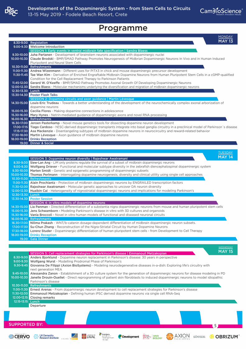
RegistationWelcome IntroductionSESSION 1: Early events in ventral midbrain fate specification | Sandra BlaessJuha Partanen - Development of brainstem neurons associated with dopaminergic nucleiClaude Brodski - BMP/SMAD Pathway Promotes Neurogenesis of Midbrian Dopaminergic Neurons In Vivo and in Human Induced Pluripotent and Neural Stem CellsRefreshmentsAndrea Wizenmann - Different uses for PITX3 in chick and mouse dopaminergic precursor developmentTae Wan Kim - Derivation of Enriched Engraftable Midbrain Dopamine Neurons from Human Pluripotent Stem Cells in a cGMP-qualified Condition for the Cell Replacement Therapy to Parkinson PatientsGerard W. O’Keeffe - BMP/SMAD Pathway Promotes Axonal Growth Of Developing Dopaminergic NeuronsSandra Blaess - Molecular mechanisms underlying the diversification and migration of midbrain dopaminergic neuronsLunchPoster Flash TalksSESSION 2: Dopamine circuits and axon guidance | Martin LévesqueLouis-Eric Trudeau - Towards a better understanding of the development of the neurochemically complex exonal arborization of dopamine neuronsCecilia Flores - Making dopamine connections in adolescenceMary Hynes - Netrin-mediated guidance of dopaminergic axons and novel RNA processingRefreshments Jeroen Pasterkamp - Novel mouse genetics tools for dissecting dopamine neuron developmentTiago Cardoso - hESC-derived dopaminergic transplants integrate into basal ganglia circuitry in a preclinical model of Parkinson´s diseaseAsa Mackenzie - Disentangeling subtypes of midbrain dopamine neurons in neurocircuitry and reward-related behaviorMartin Lévesque - Axon guidance of midbrain dopamine neuronsDrinks ReceptionDinner & Social
8.30-9.009.00-9.30
9.30-10.0010.00-10.30
10.30-11.0011.00-11.3011.30-11.45
11.45-12.0012.00-12.3012.30-13.3013.30-14.30
14.30-15.00
15.00-15.3015.30-16.0016.00-16.3016.30-17.0017.00-17.1517.15-17.30
17.30-18.0018.00-19.00
19.00
SESSION 3: Dopamine neuron diversity | Rajeshwar Awatramani Siew-Lan Ang - LIM only proteins regulate the survival of a subset of midbrain dopaminergic neuronsWolfgang Driever - Functional and molecular subtype diversity in the zebrafish diencephalospinal dopaminergic systemMarten Smidt - Genetic and epigenetic programming of dopaminergic subsetsThomas Perlmann - Interrogating dopamine neurogenesis, diversity and clinical utility using single cell approachesRefreshmentsAlain Prochiantz - Protection of midbrain dopaminergic neurons by ENGRAILED transcription factorsRajeshwar Awatramani - Molecular genetic approaches to uncover DA neuron diversityHuaibin Cai - Heterogeneity of nigrostriatal dopaminergic neurons and implications for modeling Parkinson’s LunchPoster SessionSESSION 4: In vitro models of dopamine neuronsLia Panman - Directed differentiation of a substantia nigra dopaminergic neurons from mouse and human pluripotent stem cellsJens Schwamborn - Modeling Parkinson’s disease in vitro with 3D cultures and organoidsVania Broccoli - Novel in vitro human models of functional and diseased neuronal circuitsRefreshmentsNilima Prakash - WNT/b-catenin dosage-dependent differentiation of midbrain dopaminergic neuron subsets.Su-Chun Zhang - Reconstruction of the Nigra-Striatal Circuit by Human Dopamine Neurons Lorenz Studer - Dopaminergic differentiation of human pluripotent stem cells - from Development to Cell TherapyDrinks ReceptionGala Dinner
8.30-9.009.00-9.30
9.30-10.0010.00-10.3010.30-11.0011.00-11.3011.30-12.0012.00-12.3012.30-13.3013.30-14.30
14.30-15.0015.00-15.3015.30-16.0016.00-16.3016.30-17.0017.00-17.3017.30-18.0018.00-19.00
19.00
SESSION 5: Cell replacement strategies for Parkinson’s disease | Emmanouil MetzakopianAnders Bjorklund - Dopamine neuron replacement in Parkinson’s disease: 30 years in perspectiveWolfgang Wurst - Modelling Prodromal Phase of Parkinson’sGiovanna De Filippi (Axion BioSystems) - Modeling neurodegenerative diseases in-a-dish: Exploring life’s circuitry with next generation MEAAlessandra Zanon - Establishment of a 3D culture system for the generation of dopaminergic neurons for disease modeling in PDJanelle Drouin-Ouellet - Direct reprogramming of patient skin fibroblasts to induced dopaminergic neurons to model idiopathic Parkinson's diseaseRefreshmentsErnest Arenas - From dopaminergic neuron development to cell replacement strategies for Parkinson’s diseaseEmmanouil Metzakopian - Defining human iPSC derived dopamine neurons via single cell RNA-SeqClosing remarksLunchDeparture
8.30-9.009.00-9.309.30-9.45
9.45-10.0010.00-10.30
10.30-11.0011.00-11.3011.30-12.0012.00-12.1512.15-13.15
MONDAY
MAY 13
TUESDAY
MAY 14
SUPPORTED BY:
WEDNESDAY
MAY 15
Programme
5

Development of the Dopaminergic System - from Stem Cells to Circuits13-15 May 2019 - Fodele Beach Resort, Crete
®
6
Venue Information
The Fodele Beach is situated in wonderful bay of Fodele. The hotel's terraced layoutmeans that stunning views are to be had out over the sea and the surrounding beautiful landscape. The hotel offers a fun-filled waterpark, stylish accommodation and a range of dining venues. Airy rooms and suites offer flat-screens, plus features such as balconies, direct pool access, and/or terraces with garden access. Some suites add living or dining rooms, bars, private pools and/or kitchens.
MAP
SEA VIEWS
POOL VIEWS

Development of the Dopaminergic System - from Stem Cells to Circuits13-15 May 2019 - Fodele Beach Resort, Crete
®
7
Partners
Research Topic: Frontiers Submit your abstracts now on the website: https://www.frontiersin.org/research-topics/9844/development-of-the-dopaminergic-system---from-stem-cells-to-circuits
Gold Partner:
Silver Partner:
Bronze Partner:
Session Sponsor:
Academic supporters:
Development of the Dopaminergic System - from Stem Cells to Circuits13-15 May 2019 - Fodele Beach Resort, Crete
®
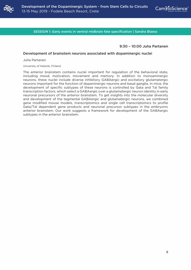
SESSION 1: Early events in ventral midbrain fate specification | Sandra Blaess
8
9:30 – 10:00 Juha Partanen
Development of brainstem neurons associated with dopaminergic nuclei
Juha Partanen
University of Helsinki, Finland
The anterior brainstem contains nuclei important for regulation of the behavioral state, including mood, motivation, movement and memory. In addition to monoaminergic neurons, these nuclei include diverse inhibitory GABAergic and excitatory glutamatergic neurons important for the function of dopaminergic neurons and basal ganglia. In mice, the development of specific subtypes of these neurons is controlled by Gata and Tal family transcription factors, which select a GABAergic over a glutamatergic neuron identity in early neuronal precursors of the anterior brainstem. To get insights into the molecular diversity and development of the tegmental GABAergic and glutamatergic neurons, we combined gene modified mouse models, transcriptomics and single cell transcriptomics to profile Gata/Tal dependent gene products and neuronal precursor subtypes in the embryonic anterior brainstem. Our work suggests a framework for development of the GABAergic subtypes in the anterior brainstem.

Development of the Dopaminergic System - from Stem Cells to Circuits13-15 May 2019 - Fodele Beach Resort, Crete
®
9
10:00 – 10:30 Claude Brodski
BMP/SMAD Pathway Promotes Neurogenesis of Midbrain Dopaminergic Neurons In Vivo and in Human Induced Pluripotent and Neural Stem Cells
Claude Brodski
Ben Gurion University, Israel
Studying the molecular pathways controlling the development of midbrain dopaminergic (mDA) neurons is essential to better understand the maintenance, function and vulnerability of these cells in adulthood. Moreover, investigating the embryonic formation of mDA neurons in vivo provides critical guidelines for the in vitro differentiation of mDA neurons from stem cells, currently being developed for Parkinson’s disease cell replacement therapy. BMP/SMAD inhibition is routinely used during early steps of stem cell differentiation protocols, including for the generation of mDA neurons. However, the function of the BMP/SMAD pathway for in vivo specification of mammalian mDA neurons is virtually unknown. We found that BMP5/7 deficient mice (Bmp5-/-; Bmp7-/-) lack mDA neurons, caused by reduced neurogenesis in the mDA progenitor domain, but not in the adjacent basal plate. As molecular mechanisms accounting for these alterations in Bmp5-/-; Bmp7-/- mutants, we identified expression changes of the BMP/SMAD target genes MSX1/2 and SHH. Conditionally inactivating SMAD1 in neural stem cells of mice in vivo (Smad1Nes) hampered the differentiation of progenitor cells into mDA neurons by preventing cell cycle exit, especially of TH+SOX6+ and TH+GIRK2+ substantia nigra neurons. In contrast, red nucleus neurons that develop in the basal plate formed normally. Notably, BMP5/7 robustly increased the in vitro differentiation of human induced pluripotent stem cells and induced neural stem cells to mDA neurons by up to 3-fold. In conclusion, we have identified BMP/SMAD signaling as a novel critical pathway orchestrating essential steps of mammalian mDA neurogenesis in vivo that balances progenitor proliferation and differentiation. Moreover, we demonstrate the potential of BMPs to improve the generation of stem cell-derived mDA neurons in vitro, highlighting the importance of sequential BMP/SMAD inhibition and activation in this process.
Development of the Dopaminergic System - from Stem Cells to Circuits13-15 May 2019 - Fodele Beach Resort, Crete
®
10
11:00 – 11:30 Andrea Wizenmann
Dopaminergic precursor marker PITX3 differs in spatiotemporal expression and
epistatic gene regulation during chick and mouse development
Ruth Klafkea, A. Alwin Prem Anandb, Wolfgang Wursta,c,d,e§, Nilima Prakasha,c,1§ and Andrea
Wizenmannb§
a Institute of Developmental Genetics, Helmholtz Zentrum München, Deutsches Forschungszentrum für Gesundheit und Umwelt (GmbH), Ingolstädter Landstr. 1, 85764 Neuherberg, Germany b Institute of Clinical Anatomy and Cell Analysis, University of Tuebingen, Oesterbergstrasse 3, 72074 Tuebingen, Germany c Technische Universität München-Weihenstephan, Lehrstuhl für Entwicklungsgenetik c/o Helmholtz Zentrum München, Ingolstädter Landstr. 1, 85764 Neuherberg, Germany d Deutsches Zentrum für Neurodegenerative Erkrankungen (DZNE) Standort München, Schillerstr. 44, 80336 München, Germany e Munich Cluster for Systems Neurology (SyNergy), Adolf-Butenandt-Institut, Ludwig-Maximilians-Universität München, Schillerstrasse 44, 80336 München, Germany
The mesodiencephalic dopaminergic (mdDA) neurons are located in the ventral
mesencephalon and caudal diencephalon of all tetrapod species studied so far, and these
are the most prominent DA neuronal population. These neurons are implicated in the control
and modulation of motor, cognitive and rewarding/affective behaviors, and their
degeneration or dysfunction is intimately linked to several neurological and
neuropsychiatric human diseases. To gain further inhsights into the generation of mdDa
neurons, we studied the spatiotemporal expression patterns and epistatic interactions in
developing chick embryos of selected marker genes and signaling pathways associated with
mdDA neuron development in the mouse. We detected striking differences in the
spatiotemporal expression patterns of the chick orthologs of mouse mdDA marker genes
Pitx3 and Aldh1a1 suggesting important differences in the generation of these cells between
these species. We also discovered that the Sonic hedgehog signaling pathway is both
necessary and sufficient for the induction of ectopic Pitx3 expression in chick
mesencephalon downstream of chick WNT9A-induced Lmx1a transcription. These aspects
of early chicken development resemble more the ontogeny of zebrafish diencephalic DA
neuronal populations, and suggest that they have diverged between birds and mammals
during evolution.
Development of the Dopaminergic System - from Stem Cells to Circuits13-15 May 2019 - Fodele Beach Resort, Crete
®
11
11:30 – 11:45 Selected Abstract: Tae Wan Kim
Derivation of Enriched Engraftable Midbrain Dopamine Neurons from Human Pluripotent Stem Cells in a cGMP-qualified Condition for the Cell Replacement Therapy to Parkinson Patients
Tae Wan Kim1,4, So Yeon Koo1, Jinghua Piao2, Eveline M Gutzwiller1, Se Joon Choi3, Eugene V Mosharov3, Mark J Tomishima1, Viviane Tabar2, Lorenz Studer1
1Center for Stem Cell Biology, Memorial Sloan Kettering Cancer Center, New York 2Department of neurosurgery, Memorial Sloan Kettering Cancer Center, New York 3Department of Neurology, Columbia University Medical Center, New York, New York, USA 4New York Stem Cell Foundation – Druckenmiller Fellow
hPSC-derived midbrain dopamine (mDA) neurons are considered a promising avenue for cell replacement therapy in Parkinson’s disease (PD). Despite rapid developments for deriving mDA neuron from hPSC towards human translation, several questions remain as to define the most optimal cell product for treating PD patients. Here we developed our protocol for deriving clinical relevant mDA neurons by optimizing activation and timing of WNT signaling. The conditions yield mDA neurons giving rise to authentic functional TH positive neurons in-vitro and in-vivo, and rescuing amphetamine-induced rotation in rat Parkinson model. However, when applying those optimized mDA neuron induction conditions to EN1-Knockout hPSC, we observe expression of contaminating PAX6 and STN markers, suggesting a pivotal role for EN1 in mediating induction of mDA neuron and suppression of alternative markers. Furthermore, we established a NURR1-H2B-GFP reporter line that enables us to derive nearly pure population of engraftable mDA neurons, which was used for candidate surface markers. These studies take advantage of our ability to generate pure population of mDA neurons in a clinically relevant culture system. The work should be geared towards developing a “2.0” version of our GMP mDA neuron product to offer the best possible cell therapy to PD patients in the near future.

Development of the Dopaminergic System - from Stem Cells to Circuits13-15 May 2019 - Fodele Beach Resort, Crete
®
12
11:45 – 12:00 Selected Abstract: Gerard W. O’Keeffe
BMP/SMAD Pathway Promotes Axonal Growth Of Developing Dopaminergic Neurons Gerard W. O’Keeffe
Department of Anatomy and Neuroscience, Cork Neuroscience Centre, University College Cork, Cork, Ireland.
The establishment of midbrain dopaminergic connectivity requires neuronal specification, differentiation, axon growth, and target innervation. While the specification and differentiation of midbrain dopaminergic neurons are being extensively studied, the molecular mechanisms that regulate axon growth and target innervation of midbrain dopaminergic neurons are less clear. One group of developmental signals that are expressed in the embryonic midbrain are the bone-morphogenetic proteins (BMP) which are a group of neurotrophic factors that are the largest subgroup of the transforming growth factor beta superfamily. These ligands and their receptors show a developmental expression profile that coincides with the period of development during which midbrain dopaminergic axons are growing towards their targets in vivo. These ligands signal through BMP receptors (BMPRs) to activate intracellular transcriptions factors called Smads. Exposure of developing midbrain dopaminergic neurons to specific BMPs promotes axon growth and branching. Moreover pharmacological and siRNA-based manipulation of the BMPR-Smad pathway shows that the axon growth promoting effects of BMPs require BMPR1B-Smad1/5 signalling. In agreement with this, overexpression of constitutively active BMPR1B in isolated midbrain dopaminergic neurons is sufficient to promote axon growth. Therefore, these findings show that as well as recently its described role in promoting neurogenesis of midbrain dopaminergic neurons, BMP-Smad signalling may also be important for promoting midbrain dopaminergic axon growth during development, and for the protection of midbrain dopaminergic axons in Parkinson’s disease.

Development of the Dopaminergic System - from Stem Cells to Circuits13-15 May 2019 - Fodele Beach Resort, Crete
®
13
12:00 – 12:30 Sandra Blaess
Molecular mechanisms underlying the diversification and migration of midbrain dopaminergic neurons Ankita Ravi Vaswani1, Beatrice Weykopf2, Cathleen Hagemann1, Hans-Ulrich Fried2, Oliver Brüstle2 and Sandra Blaess1
1Neurodevelopmental Genetics, Institute of Reconstructive Neurobiology, University of Bonn School of Medicine & University Hospital Bonn, Bonn, Germany. 2Institute of Reconstructive Neurobiology, University of Bonn School of Medicine & University Hospital Bonn, Bonn, Germany. 3Light Microscope Facility, German Center for Neurodegenerative Diseases, Bonn, Germany.
Midbrain dopaminergic (mDA) neurons migrate from their progenitor domain in the ventral midbrain floor plate to form the laterally-located substantia nigra pars compacta (SN) and medially-located ventral tegmental area (VTA). SN and VTA-mDA neurons migrate radially away from the floor plate, followed by a tangential migration step of SN-mDA neurons that allows them to take up lateral positions. Still, little is known about the underlying cellular and molecular processes of these migratory processes. Using two-photon excitation time-lapse imaging to monitor SN-mDA tangential migration in organotypic slice cultures, we demonstrate that slow migration is the default mode in SN-mDA neurons, while fast, laterally-directed migration occurs infrequently and is strongly associated with bipolar cell morphology. We show that Reelin signaling directly regulates lateral, tangential migration of mDA neurons by promoting the lateral directionality of small, slow movements, by increasing the frequency of laterally-directed fast migration events that cover larger distances and by stabilizing the morphology of migrating SN-mDA neurons. We thus provide new mechanistic insight into how Reelin signaling regulates the formation of the SN and how Reelin signaling controls tangential neuronal migration.
Development of the Dopaminergic System - from Stem Cells to Circuits13-15 May 2019 - Fodele Beach Resort, Crete
®
14
14:30 – 15:00 Louis-Eric Trudeau
Towards a better understanding of the development of the neurochemically complex axonal arborization of dopamine neurons
Louis-Eric Trudeau
Professor, Department of Physiology and Physiology, Faculty of Medicine, CNS Research Group (GRSNC), Université de Montréal
Dopamine neurons in the brain play key roles in a number of key physiological functions including motivation, learning and movement selection. A gradual loss of these neurons and of their axonal connections is tightly linked with the appearance of the cardinal motor symptoms of Parkinson’s disease. A striking characteristic of these neurons is their highly branched nature, with the axonal arbor of single neurons covering a substantial portion of their target fields such as the striatum. We are presently exploring the development of the axonal arbor of mouse dopamine neurons, focussing on the links between axonal arborization size and bioenergetics, in an effort to explain the selective vulnerability of subsets of dopamine neurons in Parkinson’s disease. We are also characterizing the development of the neurochemical identity and structure of release sites established by these neurons, highlighting the surprizing dichotomy between the minority of release sites that have a synaptic structure and that appear to be specialized for release of glutamate and GABA and the majority of release sites that have a non-synaptic structure and that appear to mediate dopamine volume transmission.
SESSION 2: Dopamine circuits and axon guidance | Martin Lévesque
Development of the Dopaminergic System - from Stem Cells to Circuits13-15 May 2019 - Fodele Beach Resort, Crete
®
15
15:00 – 15:30 Cecilia Flores
Making dopamine connections in adolescence
Cecilia Flores
Department of Psychiatry, McGill University, Canada
Adolescence is a period of increased vulnerability to mental health disorders. Yet, there is a significant gap in our knowledge about basic mechanisms of adolescent brain development and about how they are influenced by experience, including drugs of abuse and stressors. This talk focuses on the emerging role of guidance cues in the adolescent development of dopamine systems and on its implications for susceptibility and resilience to psychiatric disorders. I discuss our recent findings from rodent and human studies on the role of the Netrin-1 receptor, DCC, on the development of the dopamine projections to the prefrontal cortex in adolescence and how this process is impacted by exposure to drugs of abuse and to social stressors. I show that DCC receptors on dopamine neurons in adolescence control the development of the prefrontal cortex itself, impacting cognitive abilities in adulthood. Variations in DCC expression are linked to psychiatric conditions of prefrontal cortex dysfunction and may be a key determinant of adolescent vulnerability or resilience. This new line of research may have significant implications for the development of data-driven prevention and intervention strategies for the youth.
Development of the Dopaminergic System - from Stem Cells to Circuits13-15 May 2019 - Fodele Beach Resort, Crete
®
16
15:30 – 16:00 Mary Hynes
Netrin-1-dependent guidance of dopaminergic axons; RNA expression in early neurogenesis
Ze Yang1, Shaoyi Yi1, Leonardi Gozali1, Arif Kocabas2, Jie Li, Ana Marija Sola2, Caitlin Gilbert1, Eliza Adams1, Marc Tessier-Lavigne1, and Mary Hynes1
1Stanford University, Biology, Stanford, CA 2Rockefeller University, Biology, New York , NY
Axons from the two main groups of midbrain dopaminergic (DA) neurons, the ventral tegmental area (VTA), and the more laterally located substantia nigra (SN) neurons show spatially segregated innervation of the striatum. VTA axons project ventro–laterally (VL) and SN axons doro–medially (DM). Unlike axons that project to layered structures, DA axons in the striatum do not innervate discrete regions and instead arborize widely within either the VL or DM zones- however they do not stray from one zone to the other. Here we show that Netrin-1 acts in a novel fashion to topographically pattern midbrain DA axons into these two striatal zones by a gradient of Netrin-1 in the striatum and differential attraction of axons to overlapping but distinct concentrations of Netrin-1. In mice lacking Netrin-1, DA axons that reach the striatum fail to segregate into their two terminal zones. Netrin-1 signaling via the Netrin-1 receptor DCC has also been implicated in patterning of DA innervation of the mPFC. To assess how changes in Netrin-1 signaling impact DA circuit function, we performed unbiased whole-brain c-Fos mapping in DCC heterozygous mice. We show, using iDISCO tissue clearing and automated ClearMap voxel mapping, that DCC heterozygous mice show a blunted functional response to stimulants, and region-specific reductions in neural activity as compared to WT and saline controls. We have also been examining the role of the widespread expression of isolated 3’UTR sequences, first noticed in RNAseq samples from embryonic DA neurons. Early studies examining the role of such isolated 3’UTRs in early cell fate decisions and neurogenesis will be presented.
Development of the Dopaminergic System - from Stem Cells to Circuits13-15 May 2019 - Fodele Beach Resort, Crete
®
17
16:30 – 17:00 Jeroen Pasterkamp
Using mouse genetics approaches for dissecting dopamine neuron subset-specific axon guidance and cell migration events
Brignani S1, Raj D1, Schmidt ERE1, Adolfs Y1, Moreno-Bravo JA2, Van Battum EY1, Chedotal A2, Pasterkamp RJ1
1Department of Translational Neuroscience, Brain Center Rudolf Magnus UMC Utrecht, Utrecht University, The Netherlands 2Sorbonne Universités, UPMC Université Paris 06, INSERM, CNRS, Institut de la Vision, Paris, France
Most regions of our central nervous system (CNS) have classically been considered as homogeneous structures on basis of biochemical markers or neuronal morphology. However, techniques such as single cell RNA sequencing and advanced neuronal tracing challenge this view and support the idea that most CNS regions are composed of many different neuron types with their own molecular make-up, connectivity patterns and functional roles. We exploit the midbrain dopamine system to understand how such neuronal subsets develop and function. The midbrain dopamine (mDA) system is involved in the control of various cognitive and motor behaviours. mDA neurons are grossly divided into two anatomically and functionally distinct subpopulations: substantia nigra pars compacta (SNc) and ventral tegmental area (VTA) neurons. SNc neurons make precise connections with dorsal striatum (nigrostriatal projections), while VTA neurons target ventral striatum and cortex (mesocorticolimbic projections). Both pathways collectively run in the medial forebrain bundle (MFB) towards the forebrain. Recent data support the idea that the SNc and VTA can be subdivided into smaller and functionally more relevant neuronal subsets on basis of gene expression and connectivity patterns. However, tools to study such subsets, especially during embryonic and postnatal development, are lacking. Therefore, we recently developed novel mouse genetics tools to distinguish between different subsets of dopaminergic projections in vivo (called Pitx3-ITC mice). The subtractive genetic strategy we have developed relies on the expression of different fluorescent proteins in different subsets of mDA neurons in a single mouse. Pitx3-ITC mice display labelling of SNc neurons and selective visualization of nigrostriatal projections in the MFB and striatum, from early embryonic development onwards. Combination of Pitx3-ITC mice with 3D-imaging of solvent cleared organs (3DISCO) technology and light sheet microscopy allows for 3D analysis of neuronal subset-specific migration and axonal/dendritic development of mDA neurons. Using this approach, we have identified a previously unexplored role for Netrin-1 in the positioning of SNc mDA neurons. Intriguingly, we find that the lateral-ventral positioning of SNc mDA neurons and the ventrally directed outgrowth of their dendrites in the midbrain relies of the Netrin-1-dependent migration of GABAergic interneurons. This work identifies a novel mechanism in which the positioning of one brain nucleus relies on the migration of molecularly distinct neurons that will occupy an adjacent brain structure.
Development of the Dopaminergic System - from Stem Cells to Circuits13-15 May 2019 - Fodele Beach Resort, Crete
®
18
17:00 – 17:15 Selected Abstract: Tiago Cardoso
hESC-derived dopaminergic transplants integrate into basal ganglia circuitry in a
preclinical model of Parkinson´s disease
Tiago Cardoso1,2, Andrew F. Adler1,2, Sara Nolbrant1,2, Bengt Mattsson1, Deirdre B. Hoban1,2, Ulla Jarl1, Jenny Nelander Wahlestedt1,2, Shane Grealish1, Anders Björklund1, Malin Parmar1,2*
1Developmental and Regenerative Neurobiology, Department of Experimental Medical Science, Wallenberg Neuroscience Center, Lund University, 22184 Lund, Sweden 2Lund Stem Cell Center, Lund University, 22184 Lund, Sweden
Dopamine (DA) neurons derived from human embryonic stem cells (hESCs) are a promising cell source for cell replacement therapy in Parkinson´s disease (PD). When transplanted into preclinical models of PD, these neurons survive long-term, release dopamine, extensively innervate target host structures and provide functional recovery. Using rabies-based monosynaptic tracing, we have recently shown that grafted hESC-derived neurons appropriately integrate into the host circuitry. In this study, ventral midbrain (VM) and forebrain (FB) patterned hESCs-derived neural progenitors were transplanted into the striatum or into the substantia nigra of 6-OHDA-lesioned rats, to elucidate factors controlling target-appropriate innervation and synaptic integration 6 months post-transplantation. We show that cell intrinsic factors determine the pattern of graft-derived axonal innervation, while synaptic inputs from the host primarily reflect the location of the graft. Furthermore, we provide evidence that hESC-derived DAergic grafts transplanted to the striatum (reflecting clinical graft placement) receive synaptic input from the same subtypes of host cortical, striatal, and pallidal neurons which are known to regulate the function of endogenous nigral DA neurons. This refined understanding of factors controlling graft-derived axonal outgrowth and circuitry integration will be important for the optimization of the design of clinical cell replacement therapies for PD.
Development of the Dopaminergic System - from Stem Cells to Circuits13-15 May 2019 - Fodele Beach Resort, Crete
®
19
17:00 – 17:15 Selected Abstract: Åsa Mackenzie
Disentangeling subtypes of midbrain dopamine in neurocircuitry and reward-related behavior
Åsa Wallén-Mackenzie, Zisis Bimpisidis, Niclas König, Stefanos Stagkourakis, Vivien Zell, Bianca Vlcek, Bruno Giros, Christian Broberger, Thomas S. Hnasko, Sylvie Dumas
Reward-related behavior is complex and its dysfunction correlated with neuropsychiatric illness and non-motor symptoms of Parkinson´s disease. Some of this complexity is likely correlated with the striking heterogeneity of some of the brain areas that regulate the behavior. Dopamine neurons of the ventral tegmental area (VTA) have long been associated with different aspects of reward function, but it remains to be disentangled how distinct VTA dopamine neurons contribute to the full range of behaviors ascribed to the VTA. In our studies, we implement conditional mouse genetics and optogenetics to achieve selective manipulation of molecularly defined subtypes of VTA dopamine neurons for the study of their role in neurocircuitry and behavior. The findings that will be presented show that upon comparison, molecularly defined subtypes of VTA dopamine neurons show different patterns of contribution to the regulation of reward behavior. By disentangling the role of subtypes that together form the heterogeneous habitat of the VTA, the current understanding of VTA neurocircuitry in health and in disorders of the midbrain dopamine system can be improved.
Development of the Dopaminergic System - from Stem Cells to Circuits13-15 May 2019 - Fodele Beach Resort, Crete
®
20
17:30 – 18:00 Martin Lévesque
Axon Guidance of midbrain dopamine neurons
The central pathology of Parkinson’ disease (PD) is characterized by the selective loss ofdopaminergic (DA) neurons in the substantia nigra pars compacta (SNpc). There is a large unmetneed for a treatment that addresses the cause of the disease and modifies its clinical course. Graft of 19dopaminergic neurons newly generated from stem cells represents a promising therapeutic avenue.However, a major factor limiting success in transplantation studies is the inappropriate re-innervation of the grafted neurons. It is thus primordial to identify factors regulating axon projection and connectivity of mDA neurons. The midbrain dopaminergic neurons are organized in two major populations: the neurons of the SNpc, which project their axons to the dorsal striatum and are highly vulnerable in PD; and the neurons of the ventral tegmental area (VTA), which innervate parts of the ventral striatum, the nucleus accumbens and the prefrontal cortex and are relatively spared in PD. Here, I will present our recent data revealing the role of Semaphorin7A-PlexinC1 pathway controlling axon innervation of DA neurons. PlexinC1 is a transmembrane receptor that binds Semaphorin7A (Sema7a). Upon binding to PlexinC1, Sema7a act as a chemo-repulsive cue that repels DA axons. Sema7a is expressed in the dorsal region of the striatum. Our results show that the Sema7a-PlexinC1 interaction mediates the segregation of dopaminergic axon projections of VTA and SNpc. Our in vitro and in vivo experiments also show that Sema7a-PlexinC1 guidance effect are mediated by Src family kinases. At the light of our data, we propose a novel model to explain the segregation of the nigrostriatal and mesolimbic pathways. Data generated here will shed a new light on mechanisms that regulate dopamine neuron connectivity and will certainly help in the effort to understand the molecular factors contributing to the efficiency of cell replacement therapies in PD.
Development of the Dopaminergic System - from Stem Cells to Circuits13-15 May 2019 - Fodele Beach Resort, Crete
®
21
Sponsored by The Royal Society: Open Biology
8:30-9:00 Siew-Lan Ang
LIM-only domain proteins define and regulate the survival of specific subsets of midbrain dopaminergic neurons Mary J Green1, Felicia Mueller-Braun2, Francois Guillemot1, Jochen Roeper2, and Siew-Lan Ang1
1Francis Crick institute, London, UK; 2 Institute of Neurophysiology, Neuroscience Centre, Frankfurt, Goethe University, Germany
Midbrain dopaminergic (mDA) neurons are a heterogeneous group of neurons important for control of voluntary movement, reward processing and a range of cognitive and emotional behaviours. Underpinning these diverse processes are distinct subgroups of cells with diverse axon projections, cellular properties and molecular signatures which are only beginning to be uncovered. In this study, we demonstrate that LMO3, a LIM-domain only transcriptional co-factor, is expressed heterogeneously within mDA neurons throughout development and marks a specific population of mature mDA neurons in the ventral medial Substantia Nigra pars compacta (SNc). Through retrograde labelling we demonstrate that this LMO3+ population preferentially projects to the dorsal medial striatum, but not to the more lateral dorsal striatum, corresponding to specific functional connections that have previously been described. Furthermore, we demonstrate that LMO3 acts in a redundant manner with another LIM-domain only factor, LMO4, to promote survival of specific subgroups of mDA neurons. Using Lmo3-deficient LacZ knock-in mice and mice with conditional deletion of Lmo4, we show that loss of Lmo3 or Lmo4 alone does not affect mDA neuron development but that when both Lmo3 and Lmo4 are removed from developing mDA neurons, approximately 40% of mDA neurons die at late embryonic stages. Double mutant mice show a preferential loss of ventral tier SNc neurons and of the corresponding axon projections to the dorsal striatum. Together these results demonstrate that LIM-only domain factors are important for developmental survival of SNc neurons, in particular ventral tier SNc neurons that are vulnerable in neurodegenerative Parkinson’s disease.
SESSION 3: Dopamine neuron diversity | Rajeshwar Awatramani
Development of the Dopaminergic System - from Stem Cells to Circuits13-15 May 2019 - Fodele Beach Resort, Crete
®
22
9:00-9:30 Wolfgang Driever
Functional and molecular subtype diversity in the zebrafish diencephalospinal dopaminergic system
Wolfgang Driever
Dept. Developmental Biology, University of Freiburg, Hauptstrasse 1, 79104 Freiburg, Germany
Dopaminergic neurons constitute a major neuromodulatory system in vertebrates, affecting a wide range of circuits and behaviors. The diencephalospinal dopaminergic system appears highly conserved from fish to mammals, and provides the sole dopaminergic input into hindbrain and spinal cord. Dopaminergic neurons of this system (called A11 in mammals) have been linked to several diseases, including Restless Legs Syndrome / Willis-Ekbom Disease and chronic pain. In zebrafish, these dopaminergic neurons are located in the anatomical region of the posterior tuberculum in the ventral forebrain. Interestingly, they do not only send descending projections, but also ascending projections into subpallial / striatal regions. In addition, the same dopaminergic somata may also send peripheral projections to the ear, peripheral ganglia, and all lateral line sensory organs. Therefore, the diencephalospinal dopaminergic system has the potential to be a major modulator of both sensory and motor circuits. Given the absence of dopaminergic neurons from the zebrafish ventral midbrain, potential similarities between zebrafish posterior tubercular and mammalian VTA / midbrain A8-A10 dopaminergic neurons have been discussed extensively in the literature.
We will report on two aspects of the diencephalospinal dopaminergic system: Molecular and functional subtype diversity.
Correlation of calcium activity with specific sensory stimuli types or motor behavior revealed that these ventral diencephalic dopaminergic neurons may constitute a ‘‘sensory’’ dopamine system: anatomically distinct dopaminergic subgroups selectively respond to mechanosensory or visual stimulation. Mechanosensory-related dopaminergic activity is tuned to stimulus intensity. The activation of posterior tubercular dopaminergic neurons by sensory stimuli, and their projections onto peripheral mechanosensory systems, suggests a participation of these A11-type neurons in the feedback regulation of sensory systems. Together with the adjacent hypothalamic dopaminergic neurons, they may serve to set basic behavioral states. We have previously shown that diencephalospinal dopaminergic neurons have a dual neurotransmitter phenotype, dopaminergic and glutamatergic, while other dopaminergic groups are also gabaergic. To begin to dissect dopaminergic from second neurotransmitter-based activities of the diencephalospinal system, we have genetically engineered catecholamine-free zebrafish. These zebrafish develop morphologically normal “non-dopaminergic” diencephalospinal A11-type neurons.
Given the complexity of the projection patterns of the posterior tubercular, A11-type dopaminergic neurons, we aim at understanding the molecular basis of their anatomical and functional diversity. Therefore, we performed single cell RNAseq experiments on neurons FACS sorted based on coexpression of the tyrosine hydroxylase catecholaminergic marker and orthopedia, which encodes the transcription factor Otp required for specification of these dopaminergic neurons. Cluster analysis of the single cell data reveals subtype diversity both with respect to neurotransmitter content as well as receptor expression. We will discuss potential mechanisms how this subtype diversity may be generated.
Development of the Dopaminergic System - from Stem Cells to Circuits13-15 May 2019 - Fodele Beach Resort, Crete
®
23
9:30-10:00 Marten Smidt
Genetic and epigenetic programming of dopaminergic subsets
Marten P. Smidt
University of Amsterdam, The Netherlands
Over the last decade several components have been identified to be differentially expressed in subsets of mesodiencephalic dopaminergic (mdDA) neurons. These differences in molecular profile have been implied to be involved in the selective degeneration of the SNc neurons in Parkinson’s disease. The emergence and maintenance of individual subsets is dependent on different transcriptional programs already present during development. In addition to the influence of transcription factors, recent studies have led to the hypothesis that modifications of histones might also influence the developmental program of neurons. Here we focus on the histone methyltransferase EZH2 and its role in the specification of the mid-hindbrain region and its direct influence on the development and maintenance of mdDA neurons. We generated two different conditional knock out (cKO) mice; an En1Cre driven cKO, for deletion of Ezh2 in regional progenitors and a Pitx3Cre driven cKO, to study the effect of post-mitotic deletion of Ezh2 on mdDA neurons maturation and neuronal survival. Loss of Ezh2 changed the molecular coding of the anterior ventral hindbrain leading to a fate switch and the appearance of ectopic dopaminergic neurons. The correct specification of the isthmic region is dependent on the signaling factors produced by the Isthmic organizer (IsO), located at the border of the mid- and hindbrain. We propose that the change of cellular fate is a result of the presence of Otx2 in the hindbrain of Ezh2 conditional knock-outs and a dysfunctional IsO, as represented by the loss of Fgf8 and Wnt1. During dopaminergic development Ezh2 was found to be important for the generation of the proper amount of TH+ neurons. The loss of neurons primarily affected a rostrolateral population, which is also reflected in the analysis of the subset marks, Ahd2 and Cck. In contrast to early genetic ablation, post-mitotic deletion of Ezh2 did not lead to major developmental defects at E14.5. Finally, Pitx3Cre driven deletion of Ezh2 led to a progressive loss of TH+ cells in the VTA and these animals display reduced climbing behavior. Together, our data demonstrates that Ezh2 is important for the early regionalization and decision between dopaminergic or serotonergic programming. Moreover, Ehz2 is essential for the correct generation of mdDA neurons during development and that during adult stages Ezh2 is important for the preservation of proper neuronal subset identity and survival.

Development of the Dopaminergic System - from Stem Cells to Circuits13-15 May 2019 - Fodele Beach Resort, Crete
®
24
10:00-10:30 Thomas Perlmann
Interrogating dopamine neurogenesis, diversity and clinical utility using single cell approaches Thomas Perlmann Department of Cell and Molecular Biology, Karolinska Institutet, Sweden
Stem cell engineering and grafting of midbrain dopamine (mDA) neurons provides a promising strategy for brain repair in Parkinson’s disease. However, essential improvement of differentiation protocols will require deeper interrogation of mDA neuron lineage development. Here we present findings from studies using single-cell RNA sequencing (scRNA-seq) of mDA neurons. We have analyzed early specification steps and revealed a remarkably close relationship between developing mDA and suthalamic nucleus neurons. Combined with additional analysis, these results were essential in efforts to improve human embryonic stem (hESC) engineering protocols for cell replacement therapy. In additional studies we analyzed the maturation of Pitx3-expressing cells during late embryogenesis up into adult stages. The studies revealed several sub-lineages of mDA neurons that are subdivided very early in development, and also identified Pitx3-expressing ventral midbrain non-dopaminergic neuronal lineages. Finally, we have used scRNA-seq to analyze the graft composition of both ventral midbrain human fetal and hESC-derived cells after transplantation to rat striatum. The analysis revealed interesting differences between grafts from either hESCs or human fetal cells. Accordingly, while both cell preparations gave rise to neurons and astrocytes, oligodendrocytes were only detected in grafts of fetal cells. On the other hand, a cell type closely resembling a class of newly identified barrier forming fibroblasts was identified as a unique component of hESC-derived grafts. Thus, these experiments have addressed an important question in the field of cell replacement in neurological disease by revealing graft composition and differences between hESC- and fetal cell-derived grafts.

Development of the Dopaminergic System - from Stem Cells to Circuits13-15 May 2019 - Fodele Beach Resort, Crete
®
25
11:00-11:30 Alain Prochiantz
Protection of midbrain dopaminergic neurons by ENGRAILED transcription factors
Alain Prochiantz
Collège de France, Paris, France
ENGRAILED transcription factor is expressed in different adult central nervous system (CNS) regions, including the Substantia Nigra Pars Compacta (SNpc) and Ventral Tegmental Area (VTA). Studies from different groups have established that ENGRAILED-1 (EN1) and ENGRAILED-2 (EN2) are involved in the survival of mesencephalic dopaminergic (mDA) neurons in the SNpc and, at a lesser degree, in the VTA. Protection is through the regulation of Complex I mitochondrial mRNA translation, gene transcription and heterochromatin maintenance.
As most homeoprotein transcription factors, EN1 and EN2 pass between cells and are thus secreted and internalized. The latter property has allowed us to infuse or inject EN1 and EN2 at the level of the SNpc and to verify that the resulting cytoplasmic and nuclear gain of function has curative effects in rodents and non-human primate models of Parkinson Disease. Curiously, a single injection of the EN1 protein protects the cells for several weeks or months, suggesting an epigenetic mechanism. This was verified leading to the observation that EN1 internalization is followed by changes in chromatin marks disrupted after an acute oxidative stress.
To be more specific, heterochromatin disruption that accompanies ageing or follows oxidative stress is paralleled by an upregulation of mobile elements of the LINE-1 family. As a consequence, the expression of the endonuclease encoded by LINE-1 Open Reading Frame 2 (ORF2) is also increased, leading to the formation of an abnormally high number of DNA-brakes and to cell death. Conversely, the repression of LINE-1 expression and activity, using reverse transcriptase inhibitors, anti-ORF1 siRNAs or the LINE repressor Piwil1 rescues mDA neurons from oxidative stress deleterious effects.
The link between mobile elements and ENGRAILED protective activity is through the ability of the transcription factor to repress LINE-1 expression through chromatin refolding, as already mentioned, but also through direct transcriptional repression following its high affinity binding to LINE-1 promoters.
Key recent references
A. Di Nardo, J. Fuchs, R.L. Joshi, K.L. Moya & A. Prochiantz. The physiology of homeoprotein transduction. Physiological Reviews, 98: 1943-1982, 2018.
D. Alvarez-Fisher (co-first), J. Fuchs (co-first), F. Castagner, O. Stettler, O. Massiani-Beaudoin, K.L. Moya, C. Bouillot, W.H. Oertel, A. Lombès, W. Faigle, R.L. Joshi*, A. Hartmann* & A. Prochiantz* (2011) Engrailed proteins protect mouse midbrain dopaminergic neurons against mitochondrial complex I insults and regulate their physiology. Nature Neurosci., 14, 1260-1266, U. Nordström, G. Beauvais, A. Ghosh, B. Chakrapani, P. Sasidharan, M. Lundblad, J. Fuchs, R.L. Joshi, J.W. Lipton, A. Roholt, T.N. Feinstein, J.A. Steiner, M.L. Escobar, A. Prochiantz & P. Brundin (2015). Progressive nigrostriatal terminal dysfunction and degeneration in engrailed 1 heterozygous model of Parkinson’s disease. Neurobiology of Disease, 73, 70-82.
H. Rekaik (co-first), F.-X. Blaudin de Thé (co-first), J. Fuchs, O. Massiani-Beaudoin, A. Prochiantz* & R. Joshi (2015). Engrailed homeoprotein protects mesencephalic dopaminergic neurons from oxidative stress. Cell Reports, 13, 1-9.
Development of the Dopaminergic System - from Stem Cells to Circuits13-15 May 2019 - Fodele Beach Resort, Crete
®
26
11:30-12:00 Rajeshwar Awatramani
Molecular genetic approaches to uncover DA neuron diversity
Rajeshwar Awatramani
Northwestern University, USA
Midbrain dopamine (DA) neurons have diverse functions that can in part be explained by their heterogeneity. Although molecularly distinct subtypes of DA neurons have been identified by single-cell gene expression profiling, fundamental features such as their projection patterns, and developmental basis of diversification has not been fully elucidated. Progress in this regard has been hindered by the lack of genetic tools to study DA neuron subtypes. Here, we develop intersectional genetic labeling strategies, based on combinatorial gene expression, to map the projections of molecularly defined DA neuron subtypes. We reveal distinct genetically-defined DAergic pathways arising from the substantia nigra pars compacta and from the ventral tegmental area that innervate specific regions of the caudate putamen, nucleus accumbens and amygdala. We also use these approaches to begin to study the diversification of DA neurons. Together our work, in conjunction with other recent studies, paints a richly heterogeneous picture of midbrain DA neurons, and will provide a foundation for functional interrogation of DA neuron subtypes, in normal and diseased states.

Development of the Dopaminergic System - from Stem Cells to Circuits13-15 May 2019 - Fodele Beach Resort, Crete
®
27
12:00-12:30 Huaibin Cai
Distinct connectivity and functionality of nigrostriatal ALDH1A1-positive dopaminergic neurons in motor control
Junbing Wu1, Justin Kung1, Jie Dong1,2, Lisa Chang1, Chengsong Xie1, Nannan Yang1,3, Vivian Chen1, Zhenhua Liu1,3, Rebekah Evans4, Sarah Hawes1, Bo Liang5, Lixin Sun1, Jinhui Ding6, Jia Yu7, Sara Saez-Atienzar1, Ahsan Habib1, Weidong Le2, Beisha Tang3, Zayd Khaliq4, Da-Ting Lin5, and Huaibin Cai1
1Transgenic Section, Laboratory of Neurogenetics, National Institute on Aging, National Institutes of Health, Bethesda, MD 20892, U.S.A. 2Clinical Research Center on Neurological Diseases, the First Affiliated Hospital, Dalian Medical University, Dalian 116011, P. R. China 3National Clinical Research Center for Geriatric Disorders, Xiangya Hospital, Central South University, Changsha, 410008, Hunan, China 4Cellular Neurophysiology Unit, National Institute of Neurological Diseases and Stroke, National Institutes of Health, Bethesda, MD 20892, U.S.A. 5Neural Engineering Unit, National Institute of Drug Abuse, National Institutes of Health, Baltimore, MD 21224, U.S.A. 6Computational Biology Group, Laboratory of Neurogenetics, National Institute on Aging, National Institutes of Health, Bethesda, MD 20892, U.S.A. 7Institute for Geriatrics and Rehabilitation, Beijing Geriatric Hospital, Beijing University of Chinese Medicine, Beijing 100095, P. R. China
We and others demonstrated previously that Parkinson’s disease preferentially affects the nigrostriatal aldehyde dehydrogenase 1A1-positive dopaminergic neuron (NalDAN) subpopulations located in the ventral tier of substantia nigra pars compacta (SNC). The connectivity and functionality of NalDANs, however, remain poorly understood. In this study, we constructed a comprehensive input and output map of NalDANs in the rodent brain and discovered an essential physiological function of NalDANs in motor control. We found that NalDANs receive diverse monosynaptic inputs from a variety of brain regions, but project predominantly to the dorsal portions of dorsal striatum, a striatal subregion particularly important in motor control. Our current work represents an initial attempt to understand specific physiological functions of a molecularly defined midbrain dopaminergic neuron subpopulation in regulating specific behavioral phenotypes.

Development of the Dopaminergic System - from Stem Cells to Circuits13-15 May 2019 - Fodele Beach Resort, Crete
®
28
14:30-15:00 Lia Panman
Directed differentiation of substantia nigra dopaminergic neurons from mouse and human pluripotent stem cells
1,2Tony Oosterveen, 1,2Pedro Garcao, 2Emma Garcia Moles, 1,2Clement Soleilhavoup, 1Marco Travaglio, and 1,2Lia Panman
1MRC Toxicology Unit, University of Cambridge, Lancaster Road, Leicester, United Kingdom 2University of Leicester
Midbrain dopaminergic neurons (mDA) constitute a highly diverse neuronal population controlling important brain functions such as voluntary movements, cognition and reward. These neurons can be broadly subdivided into two major groups, which form the substantia nigra (SN) and ventral tegmental area (VTA). SN DA neurons selectively degenerate in Parkinson’s disease, while the neighbouring VTA neurons remain relatively unaffected despite their commonalities in developmental origin and gene expression profile. How these subpopulations are specified and innervate their target areas during embryonic development and the reason for the difference in vulnerability are not fully understood.
Based on our gained insight into the specification of SN and VTA neurons we have developed an ES/iPS cell-based model system that allows us to investigate the differences in vulnerability between distinct dopaminergic subpopulations. DA cultures containing mainly VTA neurons lack the sensitivity to mitochondrial toxicity and hampers PD disease modelling in vitro. Therefore, we identified culture conditions that can direct the differentiation of ES/iPS cells into enriched dopaminergic cultures displaying SN specific characteristics. Our platform is highly amendable for compound screening and led to the identification of pathways that can protect SN from degeneration.
SESSION 4: In vitro models of dopamine neurons | Lia Panman
Development of the Dopaminergic System - from Stem Cells to Circuits13-15 May 2019 - Fodele Beach Resort, Crete
®
29
15:00-15:30 Jens Schwarmborn
Modeling Parkinson’s disease in vitro with 3D cultures and organoids Jens Schwamborn University of Luxembourg, Luxembourg
Modeling Parkinson’s disease (PD) using advanced experimental in vitro models is a powerful tool to study disease mechanisms and to elucidate unexplored aspects of this neurodegenerative disorder. Here, we demonstrate that 3D differentiation of expandable midbrain floorplate neural progenitor cells (mfNPCs) leads to organoids that resemble key features of the human midbrain. These organoids are composed of midbrain dopaminergic neurons (mDANs), which produce and secrete dopamine. Midbrain-specific organoids derived from PD patients carrying the LRRK2-G2019S mutation recapitulate disease-relevant phenotypes. Automated high-content image analysis shows a decrease in the number and complexity of mDANs in LRRK2-G2019S compared to control organoids. The floor plate marker FOXA2, required for mDAN generation, increases in PD patient-derived midbrain organoids, suggesting a neurodevelopmental defect in mDANs expressing LRRK2-G2019S. Thus, we provide a robust method to reproducibly generate 3D human midbrain organoids containing mDANs to investigate PD-relevant patho-mechanisms.

Development of the Dopaminergic System - from Stem Cells to Circuits13-15 May 2019 - Fodele Beach Resort, Crete
®
30
15:30-16:00 Vania Broccoli
Novel in vitro human models of functional and diseased neuronal circuits Vania Broccoli San Raffaele Scientific Institute/CNR Institute of Neuroscience, Italy Neuronal cultures obtained by in vitro differentiation of pluripotent stem cells are an ideal model to assess physiological and disease-relevant processes occurring in human biology. However, stem cell-derived neurons are obtained in mass cultures that lack spatial organization and without any meaningful connectivity. We implemented a novel system for long-term culture of human neurons with patterned organization of projections and synaptic terminals. Co-culture of human midbrain dopaminergic and striatal medium spiny neurons on a chip established an orchestrated nigro-striatal circuitry with functional dopaminergic synapses. We employed this platform to evaluate the impact of mitochondrial dysfunctions associated with a genetic form of Parkinson’s disease (PD) with OPA1 mutations. Remarkably, we found that axons of OPA1 mutant dopaminergic neurons exhibited a significantly reduction of mitochondria mass, functionality and dynamics.
Development of the Dopaminergic System - from Stem Cells to Circuits13-15 May 2019 - Fodele Beach Resort, Crete
®
31
16:30-17:00 Nilima Prakash
WNT/b-catenin dosage-dependent differentiation of midbrain dopaminergic neuron subsets Nilima Prakash Hamm-Lippstadt University of Applied Sciences, Department Hamm 2, Hamm/Germany
Dopamine (DA)-synthesizing nerve cells located in the human midbrain are involved in the control and modulation of voluntary movements, rewarding/aversive behaviors and other cognitive functions of the brain. The age-dependent and progressive degeneration of these neurons leads to the motor symptoms of Parkinson’s Disease (PD), whereas their dysfunction is associated with neuropsychiatric disorders such as addiction, schizophrenia, attention deficit hyperactivity disorder and depression. The etiology of these diseases, in particular of the neuropsychiatric disorders, is thought to have a neurodevelopmental component. Therefore, a precise knowledge of the developmental pathways directing the generation of the midbrain dopaminergic (mDA) neurons is needed for a better understanding of these still incurable neurodegenerative and psychiatric disorders, and for the design of new therapeutic approaches to these diseases. The mammalian mDA neurons arise from the ventral midline (floor plate) of the mesencephalon (midbrain) and caudal diencephalon (forebrain) between embryonic days 9.5 to 14.5 (E9.5-14.5) of mouse gestation. Several intercellular signaling pathways control the early steps of mDA neuron generation, including the WNT1/b-catenin pathway. Initially (around E10.5), WNT1/b-catenin signaling is implicated in the establishment of the mDA domain in the ventral midbrain and promotes the proliferation of floor plate progenitors, particularly in the medial and caudal midbrain. From E11.5 onwards, this signaling pathway directs the specification of the mDA cell fate in these progenitors and their correct differentiation into the two main mDA neuron subsets in the substantia nigra pars compacta (SNc) and ventral tegmental area (VTA). Moreover, WNT1/b-catenin signaling promotes the survival of mature mDA neurons through a network of downstream transcription and neurotrophic factors. My talk will focus on the identification, quantification and transcriptome profiling of the WNT-responsive cells in the developing ventral midbrain of WNT/b-catenin signaling reporter (BAT-gal) mouse embryos, which revealed several novel and/or additional pathway components and putative WNT/b-catenin target genes in these cells. I will also present data showing that an attenuation of WNT/b-catenin signaling in the mDA progenitors appears to be essential for their correct differentiation into specific mDA neuron subsets, thus providing new insights into the stem cell-based in vitro modeling of mDA neuron-associated neurological and psychiatric diseases.

Development of the Dopaminergic System - from Stem Cells to Circuits13-15 May 2019 - Fodele Beach Resort, Crete
®
32
17:00-17:30 Su-Chun Zhang
Reconstruction of the Nigra-Striatal Circuit by Human Dopamine Neurons
Man XIONG, Yezheng TAO, Yuejun CHEN, Su-Chun ZHANG University of Wisconsin-Madison, Duke-NUS Medical School
Degeneration of the midbrain dopamine (mDA) neurons results in disruption of the nigra-striatal circuit, which underlies Parkinson’s disease. Transplantation of neural cells to repair the damaged circuitry is a potential treatment, but whether and to what extent by which the function of the repaired circuit is restored in the adult brain is not known. By transplanting mDA neurons, derived from human pluripotent stem cells (hPSCs), into the substantia nigra (SN) of adult Parkinson’s disease model mice, we found that mDA neurons projected axons predominantly to the dorsal striatum via the nigra-striatal pathway. The grafted mDA neurons also received area-specific synaptic inputs and these inputs became functional 3-6 months after transplantation. The transplanted animals showed motor functional recovery, which was abrogated or enhanced by regulating the activity of the grafted mDA neurons via DREADDs. These results highlight the capacity of hPSC-derived mDA neurons for functionally reconstructing the nigra-striatal circuit in the adult brain, contributing to therapeutic outcomes.

Development of the Dopaminergic System - from Stem Cells to Circuits13-15 May 2019 - Fodele Beach Resort, Crete
®
33
17:30-18:00 Lorenz Studer
Dopaminergic differentiation of human pluripotent stem cells – from Development to Cell Therapy
Development of the Dopaminergic System - from Stem Cells to Circuits13-15 May 2019 - Fodele Beach Resort, Crete
®
34
8:30-9:00 Anders Björklund
Use of embryonic stem cells for dopamine replacement in Parkinson´s disease
Anders Björklund,
Wallenberg Neuroscience Center, Lund University, BMC A11, S-22184 Lund, Sweden
Cell replacement therapy for Parkinson´s disease is based on the idea that implanted dopamine
neurons can substitute for the lost nigrostriatal neurons, restore dopaminergic neurotransmission
and reverse the Parkinson-like motor impairments induced by damage to the nigrostriatal system.
Open-label clinical trials in patients with PD have shown that dopamine neuroblasts obtained
from fetal human midbrain tissue can survive and function over many years in the brain of PD
patients, restore striatal dopamine release, and provide sustained and long-lasting improvements
in motor behavior. The ethical and practical problems associated with the use of fetal tissue is a
serious obstacle to further developments of this approach. Further progress, therefore, is
critically dependent on the development of transplantable dopamine neurons from stem cells.
The most promising results so far have been obtained using pluripotent stem cells, ESCs or iPSCs,
as starting material. Recently developed and optimized protocols allow efficient generation of
midbrain dopamine neurons from human ES cells that survive well following transplantation to
the striatum, in the absence of any contaminating tumor-forming cells, and differentiate into
genuine midbrain dopamine neurons of both A9 and A10 subtypes. In recent experiments
performed in immunosuppressed and immunodeficient rats we have shown that the hESC-
derived neurons grow to form extensive axonal terminal networks in appropriate striatal, limbic
and and cortical targets and reverse PD-like motor impairments. The results indicate that
transplantable and fully functional midbrain dopamine neurons can be generated from human ES
cells. Clinical trials using these cells are now under way.
SESSION 5: Cell replacement strategies for Parkinson’s disease | Emmanouil Metzakopian
Development of the Dopaminergic System - from Stem Cells to Circuits13-15 May 2019 - Fodele Beach Resort, Crete
®
35
9:00-9:30 Wolfgang Wurst
Prodromal Models of Parkinson Disease
Wolfgang Wurst
Helmholtz Centre Munich, Institute of Developmental Genetics, Munich/Neuherberg, Germany
Parkinson´s Disease (PD) is clinically characterized by the progressive loss of dopaminergic neurons in the ventral midbrain but increasingly recognized also by the loss and dysfunction of other neuronal populations. Based on human genome-wide association studies we have previously generated mouse mutants reproducing mutations in human PD patients: DJ-1, PINK1, and LRRK2. All these mouse models exhibit several symptoms of PD indicative for the prodromal phase of the disease, including gait deficits resembling the human phenotype and represent ideal models to interrogate the biological basis of gait disturbances in PD. We further characterized these mutants and identified alterations in behavior and metabolic, and bioenergetics parameters. Furthermore, we established idiopathic PD patient-derived iPSC, NPCs and DA neurons, which are currently molecularly, and cellular characterized as well. The comparative analysis of human cellular in vitro and mouse in vivo models of PD will be presented and discussed.

Development of the Dopaminergic System - from Stem Cells to Circuits13-15 May 2019 - Fodele Beach Resort, Crete
®
36
9:30-9:45 Partner Talk: Giovanna De Filippi – Axion BioSystems
Modeling neurodegenerative diseases in-a-dish: Exploring life’s circuitry with next generation MEA
Development of the Dopaminergic System - from Stem Cells to Circuits13-15 May 2019 - Fodele Beach Resort, Crete
®
37
9:45-10:00 Selected Abstract: Alessandra Zanon
Establishment of a 3D culture system for the generation of dopaminergic neurons for disease modeling in PD
Zanon A 1, Von Troyer M 1, Riekschnitz DA 1, Lavdas AA 1, Casarosa S 2, 3, Pramstaller PP 1, 4, 5, Hicks AA 1, Pichler I 1
1 Institute for Biomedicine, Eurac Research, Affiliated Institute of the University of Lübeck, 39100 Bolzano, Italy; 2 Center for Integrative Biology (CIBIO), University of Trento, 38123 Trento, Italy; 3 CNR Neuroscience Institute, 56124 Pisa, Italy; 4 Department of Neurology, General Central Hospital, 39100 Bolzano, Italy; 5
Human induced pluripotent stem cells (hiPSCs) represent an unlimited cell source for the generation of 3D in vitro models for neurological diseases. Neurons cultured in 3D aggregates might not only serve to facilitate the development of more precise human brain models for basic mechanistic studies but also represent better predictors of drug responses in vivo. To evaluate the capacity of alginate to support the differentiation of hiPSCs to midbrain-specific dopaminergic neurons for Parkinson’s disease (PD) modeling, we encapsulated iPSCs in alginate microcapsules (1% or 2%) with or without fibronectin (Fn). Cells grown in 1% alginate-Fn and 2% alginate-Fn present increased differentiation capacity towards neural lineages with respect to both alginate (1%-2%) alone and 2D conditions. Gene expression analysis suggests an increase in TH+ neurons and a higher maturity compared to the neurons differentiated in 2D. Immunofluorescence analysis further supports these results, showing expression of synaptic markers as well as specific DA neuronal markers. The 3D neurons can be maintained in culture for more than 200 days. We envision that this differentiation protocol might enable the generation of midbrain-specific dopaminergic neurons in a shorter time-frame as compared to 2D cultures and will allow successful phenotype assessment and advanced therapy development.

Development of the Dopaminergic System - from Stem Cells to Circuits13-15 May 2019 - Fodele Beach Resort, Crete
®
38
10:00-10:30 Janelle Drouin-Ouellet
Direct reprogramming of patient skin fibroblasts to induced dopaminergic neurons to model idiopathic Parkinson's disease
Understanding the pathophysiology of Parkinson’s disease (PD) has been hampered by the lack of models that recapitulate all the critical factors underlying its development. We have developed a novel and highly efficient approach to generate functional induced dopaminergic neurons (iDANs) that are directly reprogrammed from dermal fibroblasts of patients with idiopathic PD. We further investigate whether such cells have deficits in autophagy and show that iDANs derived from PD patients exhibit lower basal chaperone-mediated autophagy as compared to iDANs of healthy donors. Furthermore, stress-induced autophagy resulted in an accumulation of macroautophagic structures in induced neurons derived from PD patients, independently of the specific neuronal subtype but dependent on the age of the donor. Our results show that iDANs provides a patient-specific model to study neuronal features relevant to idiopathic PD.
Development of the Dopaminergic System - from Stem Cells to Circuits13-15 May 2019 - Fodele Beach Resort, Crete
®
39
11:00-11:30 Ernest Arenas
From dopaminergic neuron development to cell replacement strategies for Parkinson’s disease
Ernest Arenas
Laboratory of Molecular Neurobiology, MBB, Karolinska Institute, Biomedicum 6C, Solnavägen 9, Stockholm, Sweden.
Parkinson’s disease (PD) is a progressive neurodegenerative disorder characterized by the loss of midbrain dopaminergic neurons (mDA) neurons in the substantia nigra. Current therapeutic interventions focus on restoring the levels of the neurotransmitter dopamine, by pharmacological means, or on balancing neurotransmission, by stimulating the subthalamic nucleus with deep brain stimulation. Three factors in recent years have contributed to the development of cell replacement therapies for PD: 1) Proof of concept that clinical fetal ventral midbrain tissue grafting can change the course of disease in PD patients. 2) The advent of human pluripotent stem cells (hPSCs) and reprogramming technologies. 3) Progress in understanding mDA neuron development.
However, our knowledge of the developing human midbrain is still limited. In order to address this challenge, we recently examined of the human embryonic ventral midbrain by single-cell RNA-sequencing (scRNA-seq) and identified 25 distinct cell-types as well as their transcriptional signature. This data together with newly available single-cell data covering the entire adult mouse brain, new genome wide association studies of PD as well as bioinformatics analysis and functional studies are currently being used to address critical questions in the field, such as:
1) What are the transcriptional networks controlling mDA neurogenesis?
2) What are the cell types affected in PD that need replacement?
3) What is the quality of current hPSC-derived midbrain cell preparations aimed for PD cell replacement therapy?
4) Can single cell technologies contribute to develop next generation hPSC- or reprogramming-based cell replacement therapies for PD?
Development of the Dopaminergic System - from Stem Cells to Circuits13-15 May 2019 - Fodele Beach Resort, Crete
®
40
11:30-12:00 Emmanouil Metzakopian
Defining human iPSC derived dopamine neurons via single cell RNA-Seq
UK Dementia Research Institute (UK DRI), University of Cambridge
In vitro human induced pluripotent stem cell derived neurons consist a versatile model of studyingdiseases such as Parkinson’s and Alzheimer’s. However, the differentiation course is susceptible toexperimental variability which can skew experimental outcomes from the resulting culture. The cellulardevelopment and heterogeneity of these in vitro models require proper characterization before use fordownstream experiments such as CRSIPR-Cas9 genetic screens. Single cell transcriptomics hasproven to be a powerful tool to achieve in depth understanding of cellular states and tissue culturecomplexities. Here we are capturing the tissue culture states using single-cell transcriptomics bystudying the developmental time course and assessing clonal heterogeneity. Results highlight thedifferent culture outcomes and the intermediate state transitions through the course of differentiation.

Development of the Dopaminergic System - from Stem Cells to Circuits13-15 May 2019 - Fodele Beach Resort, Crete
®
41
Poster Abstracts
Abstract number Name Title
1 Milagros Pereira Luppi
Development and application of a Nestin-anchored intersectional fate mapping strategy to reveal Dopaminergic neuron cell fate
2 Mateja Rybiczka-Te ulov Circular RNAs in developing midbrain dopamine neurons
3 Parivash NouriLEF1-mediated WNT1/b-Catenin signaling in subtype-specific midbrain dopaminergic neuron differentiation
4 Tae Wan Kim
Derivation of Enriched Engraftable Midbrain Dopamine Neurons from Human Pluripotent Stem Cells in a cGMP-qualified Condition for the Cell Replacement Therapy to Parkinson Patients
5 Alessandro PeteseInvestigating Lmo3 (LIM-domain Only Protein 3) as a Marker for a Subset of midbrain Dopaminergic Neurons
6 So Yeon KooMolecular Characterization of Human Stem Cell-Derived Dopaminergic Neuron Subtypes
7 Marco Travaglio
Altered striatal innervation in Nolz1 deficient embryos provides novel insight into dopaminergic circuitry formation
8 Marianna TolveThe Role of the Transcription Factor Bcl11a in Midbrain Dopaminergic Neurons
9 Yojet Sharma Role of ion-channels in development of mouse mDA neurons
10 Tiago Cardoso
hESC-derived dopaminergic transplants integrate into basal ganglia circuitry in a preclinical model of Parkinson´s disease
11 Alessandra ZanonEstablishment of a 3D culture system for the generation of dopaminergic neurons for disease modeling in PD
12 Laura Lahti
Single-cell RNA sequencing reveals midbrain dopamine neuron diversity emerging during mouse brain development
13 Zachary Gaertner Molecular Classification of Dopamine Neurons: Towards a consensus
14 Nikolaos Patikas and Stefanie Foskolou
Single-cell characterization of mid-brain Dopamine & Cortical Neurons from in vitro models
15 Divya RajRole of long-range Netrin-1 signaling in dopaminergic migration and cellular organization in the ventral midbrain
Development of the Dopaminergic System - from Stem Cells to Circuits13-15 May 2019 - Fodele Beach Resort, Crete
®
42
Abstract 1: Milagros Pereira Luppi
Development and application of a Nestin-anchored intersectional fate mapping strategy to reveal Dopaminergic neuron cell fate Milagros Pereira Luppi, Jean-Francois Poulin, Pei-Ken Hsu, Raj Awatramani Department of Neurology, Northwestern University
Lineage analysis has led to a better understanding of how the embryonic neural tube progenitors give rise to the enormous complexity of cell types in the adult CNS. Lineage tracing methods indelibly label a progenitor or subset of progenitors. In particular, site-specific recombinases driven by gene-specific promoters activate a reporter in a permanent manner, allowing tracking of the descendants of the progenitor of interest. The accuracy of these methods, however, is limited by how specific to such progenitors is the promoter driving the recombinase. Intersectional approaches increase the specificity of the analysis, rendering more refined fate maps. Nonetheless, driver genes are often expressed in progenitors and in related or unrelated postmitotic neurons simultaneously. Thus, in such cases, progenitor–progeny relationships cannot be correctly ascertained. Inducible-recombinase approaches restrict labeling to a specific time window avoiding reporter induction in postmitotic cells expressing the driver gene, yet, this method is hampered by mosaicism and toxicity. To circumvent these limitations, we propose a new platform for lineage analysis anchored in the Nestin locus. This method is compatible with most Cre drivers; thus, it will be broadly applicable to study important lineage related questions. Here, we demonstrate the validity and efficiency of this approach and preliminary studies towards understanding DA neuron fates.

Development of the Dopaminergic System - from Stem Cells to Circuits13-15 May 2019 - Fodele Beach Resort, Crete
®
43
Abstract 2: Mateja Rybiczka-Tešulov
Circular RNAs in developing midbrain dopamine neurons Mateja Rybiczka-Tešulov 1 , Roland E. van Dijk 1, Morten T. Venø 2, Lieke L. van de Haar 1, Jørgen Kjems 2, R. Jeroen Pasterkamp 1 1 UMC Utrecht Brain Center, Department of Translational Neuroscience, University Medical Center Utrecht, Utrecht University, The Netherlands 2 Department of Molecular Biology and Genetics, Aarhus University, Denmark
Non-coding (nc) RNAs play a crucial role during neuronal development, but their precise function in midbrain dopamine (mDA) neurons remains incompletely understood. Circular (circ) RNAs are a novel class of ncRNAs that form as the result of covalent linkage of 5’ and 3’ pre-mRNA splice sites. CircRNAs can originate from many different genes, while expression levels are tissue-, cell type- and developmental stage-dependent. The mammalian brain is particularly rich in this novel class of ncRNAs, where circRNAs have been proposed to regulate processes such as synaptogenesis and synaptic activity. Here, we study the spatio-temporal expression and function of circRNAs during the early development of the mDA system. Using novel mouse reporter lines and immunohistochemical approaches, we generated a detailed map of mouse mDA neuron development. On basis of this analysis, we selected a number of developmental timepoints at which mDA neurons were isolated by FACS and subjected to RNAseq and circRNA prediction. We aim to dissect the functional role of specific circRNAs during mDA neuron development by knockdown or overexpression of circRNAs in primary cultures or in vivo by in utero electroporation. Overall, these experiments will provide insight into the role of a novel class of ncRNAs in mDA neuron development.

Development of the Dopaminergic System - from Stem Cells to Circuits13-15 May 2019 - Fodele Beach Resort, Crete
®
44
Abstract 3: Parivash Nouri
LEF1-mediated WNT1/b-Catenin signaling in subtype-specific midbrain dopaminergic neuron differentiation Parivash Nouri1*, Sebastian Götz2*, Benedict Rauser2*, Martin Irmler3, Changgeng Peng2, Dietrich Trümbach2, Yojet Sharma1, Andrea Dlugos1, Wolfgang Wurst2,4,5,6, Johannes Beckers3,7,8 and Nilima Prakash1 1Hamm-Lippstadt University of Applied Sciences, Department Hamm 2, 59063 Hamm, Germany 2Institute of Developmental Genetics, Helmholtz Center Munich, German Research Center for Environmental Health, 85764 Neuherberg, Germany 3Institute of Experimental Genetics, Helmholtz Center Munich, German Research Center for Environmental Health, 85764 Neuherberg, Germany 4Technical University of Munich, Chair of Developmental Genetics/Helmholtz Center Munich, 85764 Neuherberg, Germany 5German Center for Neurodegenerative Diseases, 80336 Munich, Germany 6Munich Cluster for Systems Neurology (SyNergy), 80336 Munich, Germany 7Technical University of Munich, Chair of Experimental Genetics, 85350 Freising-Weihenstephan, Germany 8German Center for Diabetes Research (DZD), 85764 Neuherberg, Germany *These authors contributed equally to this work
The dopamine producing neurons located in substantia nigra pars compacta (SNc) and the ventral tegmental area (VTA) neurons, which are subgroups of Mesodiencephalic dopaminergic (mdDA) neurons, play significant roles in the neural network responsible for movement, cognition and emotion. The development of mdDA neurons has attracted particular attention, due to their critical functions in neuropsychiatric and neurodegenerative disorders, such as Parkinson disease (PD). Previous research on the importance of the WNT1/b-catenin signaling pathway in the differentiation and survival of mdDA neurons suggests that a disrupted activation or inhibition of the WNT1/b-catenin signaling pathway leads to the incorrect specification or differentiation of mdDA neurons and/or their subsets?. This study set out to investigate the identity of the WNT/b-catenin-responsive cells in the murine ventral midbrain (VM) and the precise mechanism of WNT/b-catenin action in the mdDA cells. We found that only a fraction of all mdDA progenitors, precursors, and neurons in the murine VM respond to this pathway. The WNT/b-catenin-responsive cells are located mostly in the Wnt1+, Rspo2+ and Lef1+ lateral floor plate of the medial and caudal midbrain, and give preferentially rise to caudomedial (VTA) mdDA neurons. We also show that the strong activation of the WNT/b-catenin signaling pathway by RSPO2, a WNT/b-catenin agonist, and LEF1, a nuclear effector of this pathway, inhibits the differentiation of WNT/b-catenin-responsive mdDA progenitors into mature mdDA neurons. This is in part due to the direct repression of the murine Pitx3 gene promoter by LEF1-mediated WNT/b-catenin signaling. Our results indicate that the correct differentiation of mdDA progenitors into specific mdDA neuron subtypes, particularly SNc DA neurons, requires the attenuation of WNT/b-catenin signaling in these cells, thus providing new insights to the directed differentiation of these cells means for stem cell-based regenerative therapies of PD and other in vitro models of neuropsychiatric diseases.
Development of the Dopaminergic System - from Stem Cells to Circuits13-15 May 2019 - Fodele Beach Resort, Crete
®
45
Abstract 4: Tae Wan Kim
Derivation of Enriched Engraftable Midbrain Dopamine Neurons from Human Pluripotent Stem Cells in a cGMP-qualified Condition for the Cell Replacement Therapy to Parkinson Patients.
Tae Wan Kim1,4, So Yeon Koo1, Jinghua Piao2, Eveline M Gutzwiller1, Se Joon Choi3, Eugene V Mosharov3, Mark J Tomishima1, Viviane Tabar2, Lorenz Studer1
1Center for Stem Cell Biology, Memorial Sloan Kettering Cancer Center, New York 2Department of neurosurgery, Memorial Sloan Kettering Cancer Center, New York 3Department of Neurology, Columbia University Medical Center, New York, New York, USA 4New York Stem Cell Foundation – Druckenmiller Fellow
hPSC-derived midbrain dopamine(mDA) neurons are considered a promising avenue for cell replacement therapy in Parkinson’s disease (PD). Despite rapid developments for deriving mDA neuron from hPSC towards human translation, several questions remain as to define the most optimal cell product for treating PD patients. Here we developed our protocol for deriving clinical relevant mDA neurons by optimizing activation and timing of WNT signaling. The conditions yield mDA neurons giving rise to authentic functional TH positive neurons in-vitro and in-vivo, and rescuing amphetamine-induced rotation in rat Parkinson model. However, when applying those optimized mDA neuron induction conditions to EN1-Knockout hPSC, we observe expression of contaminating PAX6 and STN markers, suggesting a pivotal role for EN1 in mediating induction of mDA neuron and suppression of alternative markers. Furthermore, we established a NURR1-H2B-GFP reporter line that enables us to derive nearly pure population of engraftable mDA neurons, which was used for candidate surface markers. These studies take advantage of our ability to generate pure population of mDA neurons in a clinically relevant culture system. The work should be geared towards developing a “2.0” version of our GMP mDA neuron product to offer the best possible cell therapy to PD patients in the near future.
Development of the Dopaminergic System - from Stem Cells to Circuits13-15 May 2019 - Fodele Beach Resort, Crete
®
46
Abstract 5: Alessandro Petese
Investigating Lmo3 (LIM-domain Only Protein 3) as a Marker for a Subset of midbrain Dopaminergic Neurons Alessandro Petese1, Cathleen Hagemann1, Pietro Pilo Boyl2, Philipp Hatzfeld3, Joachim Degen4, Walter Witke2, Irmgard Förster3, Sandra Blaess1 1Neurodevelopmental Genetics, Institute of Reconstructive Neurobiology, University of Bonn School of Medicine & University Hospital Bonn, Bonn, Germany 2Institute of Genetics, University of Bonn 3Immunology and Environment, Life & Medical Sciences (LIMES) Institute, University of Bonn 4Animal Facility, Life & Medical Sciences - Genetic Resource Center (LIMES-GRC), University of Bonn
Midbrain dopaminergic (mDA) neurons are located in in three distinct anatomical regions, the substantia nigra pars compacta (SNc), the ventral tegmental area and the retrorubal field. mDA neurons are diverse in their projection targets, physiological properties and impact on behavior. On a molecular level, single-cell RNA sequencing studies of developing and adult mDA neurons have identified mDA subpopulations defined by specific gene expression profiles. To gain insight into how the functional heterogeneity of mDA neurons is linked to these molecular profiles, it is crucial to understand whether genetically-defined subpopulations contribute to specific circuits and functions. Lmo3, a transcriptional co-regulator, is expressed in ventral SNc neurons (E15.5 and E18.5) during embryonic development. This expression pattern is largely maintained in adulthood. Thus, Lmo3 is a suitable candidate to define a subset of mDA neurons. Our goal is to characterize Lmo3-expressing mDA neurons within the context of neurodevelopment, circuits and functions using cell-specific tracing and optogenetic approaches. To this end we are generating a knock-in line in which the Cre recombinase gene is expressed under the control of the Lmo3 promoter. To establish this Lmo3-Cre knock-in line, we are using CRISPR/Cas-mediated targeted integration in zygotes.

Development of the Dopaminergic System - from Stem Cells to Circuits13-15 May 2019 - Fodele Beach Resort, Crete
®
47
Abstract 6: So Yeon Koo
Molecular Characterization of Human Stem Cell-Derived Dopaminergic Neuron Subtypes
So Yeon Koo1, 2, Tae Wan Kim1, Lorenz Studer1,2 1Neuroscience Program, Weill Cornell Medicine, New York NY 2Develpmental Biology Program, Memorial Sloan Kettering Cancer Center, New York NY
Parkinson’s disease (PD) is due by the progressive loss of dopaminergic neurons in the narrow region of the brain called substantia nigra (SN). The Studer lab has recently developed a clinically-relevant protocol to derive functionally authentic midbrain dopaminergic neurons (mDA) from human pluripotent stem cells (hPSCs) under defined cGMP conditions. However, the differentiation of hPSC in vitro does not yield pure population of A9 substantia nigra neurons but likely contains other mDA subtypes. Thus, we set out to define molecular signatures for each mDA neuron subtype (A9 vs. A10) and investigate the developmental origin of such subtype identity in hPSC-derived neurons.
SOX6 and OTX2 have been reported to exhibit exclusive expression in dopamine neurons of the SN and VTA respectively in both the developing and adult murine brain (Panman et al., 2014; Di Salvio et al., 2010; Vernay et al., 2005). However, their role during human mDA neuron development remains unknown. I developed an endogenous SOX6-tdTomato reporter in our well-characterized NURR1-GFP hPSC reporter line. This double reporter may enable us to denote a potential SN-type mDA neuron subtype. Furthermore, I established inducible SOX6 and OTX2 knockout approaches to manipulate expression of either gene at defined temporal windows. These tools will enable us to examine the SN(A9)/VTA(A10) mDA neuron subtype ratio in our culture system, facilitate efforts to further optimize the protocol for maximum SN yield and clarify the role of SOX6 and OTX2 during mDA neuron subtype specification and maintenance in vitro and in vivo.
Development of the Dopaminergic System - from Stem Cells to Circuits13-15 May 2019 - Fodele Beach Resort, Crete
®
48
Abstract 7: Marco Travaglio
Altered striatal innervation in Nolz1 deficient embryos provides novel insight into dopaminergic circuitry formation
Marco Travaglio1,2, Clement Soleilhavoup1,2, Kieran Patrick1,2, Brian Brooks3, Elangovan Boobalan3, Jeroen Pasterkamp4, Fokkoliena Panman1,2
1MRC Toxicology Unit, Lancaster Rd, Leicester LE1 7HB, UK 2The University of Cambridge, School of Biological Sciences, Mill Lane, Cambridge, CB2 1RX, UK 3Ophthalmic Genetics and Visual Function Branch, National Eye Institute, National Institutes of Health, Bethesda, MD 20892, USA 4Department of Translational Neuroscience, Brain Center Rudolf Magnus, University Medical Center Utrecht,
Utrecht, the Netherlands. Dopaminergic (DA) neurons in the midbrain give rise to ascending axonal projections to the striatum. Despite their fundamental role in locomotion, reward and memory, relative little is known about how DA neurons innervate their target areas. Although multiple axon guidance molecules have been suggested to direct DA axon growth toward their final targets, the identity of secreted cues expressed by the striatum to promote DA innervation remains unknown. Here, we found that that the transcriptional regulator Nolz1 expressed in both dopaminergic neurons and the striatum has a key role in establishing dopaminergic circuitry. Using a conditional gene targeting approach, we show that genetic ablation of Nolz1 leads to reduced nigrostriatal axonal projections, indicating its involvement in DA axon development. Transcriptomic analysis of striatal cells from Nolz1 deficient mice further indicates that a repertoire of striatum-enriched genes is dysregulated in mutant embryos. This finding was confirmed by immunohistochemistry and the chemotrophic effect of these genes is currently being investigated through integrated microfluidic platforms. Our results indicate a fundamental role of Nolz1 in dopaminergic axon guidance and shed light on novel mechanisms involved in DA circuitry formation.
Keywords: axon guidance, dopaminergic neurons, striatum, transcriptional regulator

Development of the Dopaminergic System - from Stem Cells to Circuits13-15 May 2019 - Fodele Beach Resort, Crete
®
49
Abstract 8: Marianna Tolve
The Role of the Transcription Factor Bcl11a in Midbrain Dopaminergic Neurons Marianna Tolve1, Ayse Ulusoy2, Alesja Dernst1, Gabriela O. Bodea1, Pentao Liu3, Walid Khaled4, Neal Copeland5, Heinz Beck6, Stephan Baader7, Donato Di Monte2, Sandra Blaess1 1Neurodevelopmental Genetics, Institute of Reconstructive Neurobiology, University of Bonn School of Medicine & University Hospital Bonn, Bonn, Germany 2German Center for Neurodegenerative Diseases (DZNE), Bonn, 3The Wellcome Trust Sanger Institute, Cambridge, UK, 4Department of Pharmacology, Cambridge University, UK 5Institute for Academic Medicine, Houston Methodist, USA, 6Laboratory of Experimental Epileptology and Cognition Research, Department of Epileptology, University of Bonn, 7Institute of Anatomy, University of Bonn
Midbrain dopaminergic (mDA) neurons of the substantia nigra pars compacta (SNc) and the ventral tegmental area (VTA) project to multiple forebrain target regions and modulate various behaviors. Transcription factor codes established during development may define the identity of specific mDA neuronal subsets and could ultimately contribute to the determination of their projection targets, their functional properties and their susceptibility to neurodegeneration. Bcl11a is a Krüppel-like zinc finger transcription factor expressed in a subset of mDA neurons in the lateral VTA and the SNc. Using viral tracing experiments, we show that the olfactory tubercles are a major projection target of Bcl11a-expressing VTA-mDA neurons. To examine Bcl11a function in development and maintenance of mDA neurons, we inactivated Bcl11a specifically in mDA neurons during development (Bcl11a cko mice). We find that differentiation and maintenance of Bcl11a-positive mDA neurons is not obviously affected in Bcl11a cko mice. However, Bcl11a cko mice show behavioral impairments suggesting that the loss of Bcl11a leads to functional deficits in mDA neurons. Moreover, Bcl11a could play a neuroprotective role within SN-mDA neurons, since overexpression of alpha-synuclein in the SNc leads to a significantly greater reduction in the number of SNc-mDA neurons in Bcl11a cko animals as compared to controls.

Development of the Dopaminergic System - from Stem Cells to Circuits13-15 May 2019 - Fodele Beach Resort, Crete
®
50
Abstract 9: Yojet Sharma
Role of ion-channels in development of mouse mDA neurons Yojet Sharma1*, Benedict Rauser2*, Sebastian Götz2*, Martin Irmler3, Parivash Nouri1*, Johannes Beckers3,4,5 Birgit Liss6 and Nilima Prakash1
1Hamm-Lippstadt University of Applied Sciences, Department Hamm 2, 59063 Hamm, Germany 2Institute of Developmental Genetics, Helmholtz Center Munich, German Research Center for Environmental Health, 85764 Neuherberg, Germany 3Institute of Experimental Genetics, Helmholtz Center Munich, German Research Center for Environmental Health, 85764 Neuherberg, Germany 4Technical University of Munich, Chair of Experimental Genetics, 85350 Freising-Weihenstephan, Germany 5German Center for Diabetes Research (DZD), 85764 Neuherberg, Germany 6Institute of Applied Physiology, University of Ulm, 89081 Ulm, Germany *These authors contributed equally to this work
The dopamine (DA)-synthesizing neurons are crucial for facilitation of voluntary movements, regulation of emotional behavior and reward pathways. These neurons are localized in the adult ventral midbrain. To precisely outline the developmental profile of mDA neurons, could prove beneficial in modeling neurological disorders such as Parkinson’s disease (PD). The degeneration of DA neurons in the substantia nigra pars compacta (SNpc) leads to anomalies in the motor functions of the PD patients. One of the most important signaling pathways in the generation of midbrain-DA (mdDA) neurons is the canonical WNT/ -catenin signaling pathway. We have found out that a WNT1 signal directs the specification and differentiation of progenitor cells into mdDA neurons and their subsets. To understand which genes are expressed in the developing WNT-responsive mdDA domain, we carried out a microarray-based comparative transcriptome analyses. To our surprise, around 51% of the overrepresented transcripts in the WNT-responsive mdDA domain encode proteins belonging to the ion channel and neurotransmitter receptor/transporter gene ontology categories. Further, in situ validation of the voltage-gated potassium channel Kcnd3 (Kv4.3), the inwardly-rectifying potassium channel Kcnj6 (Girk2) and the voltage-dependent calcium channel subunit Cacna1d (Cav1.3 1), revealed that they are selectively transcribed in the ventral midbrain of the mouse embryo, exhibiting in some cases a very localized expression pattern in what appears to be mdDA subdomains. Because the neural connectivity of the mdDA neurons has not yet been established at these early developmental stages, our data points out alternative roles of these ion channels during early mdDA neuron development.
Development of the Dopaminergic System - from Stem Cells to Circuits13-15 May 2019 - Fodele Beach Resort, Crete
®
51
Abstract 10: Tiago Cardoso
hESC-derived dopaminergic transplants integrate into basal ganglia circuitry in a
preclinical model of Parkinson´s disease
Tiago Cardoso1,2, Andrew F. Adler1,2, Sara Nolbrant1,2, Bengt Mattsson1, Deirdre B. Hoban1,2, Ulla Jarl1, Jenny Nelander Wahlestedt1,2, Shane Grealish1, Anders Björklund1, Malin Parmar1,2*
1Developmental and Regenerative Neurobiology, Department of Experimental Medical Science, Wallenberg Neuroscience Center, Lund University, 22184 Lund, Sweden 2Lund Stem Cell Center, Lund University, 22184 Lund, Sweden
Dopamine (DA) neurons derived from human embryonic stem cells (hESCs) are a promising cell source for cell replacement therapy in Parkinson´s disease (PD). When transplanted into preclinical models of PD, these neurons survive long-term, release dopamine, extensively innervate target host structures and provide functional recovery. Using rabies-based monosynaptic tracing, we have recently shown that grafted hESC-derived neurons appropriately integrate into the host circuitry.
In this study, ventral midbrain (VM) and forebrain (FB) patterned hESCs-derived neural progenitors were transplanted into the striatum or into the substantia nigra of 6-OHDA-lesioned rats, to elucidate factors controlling target-appropriate innervation and synaptic integration 6 months post-transplantation. We show that cell intrinsic factors determine the pattern of graft-derived axonal innervation, while synaptic inputs from the host primarily reflect the location of the graft. Furthermore, we provide evidence that hESC-derived DAergic grafts transplanted to the striatum (reflecting clinical graft placement) receive synaptic input from the same subtypes of host cortical, striatal, and pallidal neurons which are known to regulate the function of endogenous nigral DA neurons.
This refined understanding of factors controlling graft-derived axonal outgrowth and circuitry integration will be important for the optimization of the design of clinical cell replacement therapies for PD.

Development of the Dopaminergic System - from Stem Cells to Circuits13-15 May 2019 - Fodele Beach Resort, Crete
®
52
Abstract 11: Alessandra Zanon
Establishment of a 3D culture system for the generation of dopaminergic neurons for disease modeling in PD
Zanon A 1, Von Troyer M 1, Riekschnitz DA 1, Lavdas AA 1, Casarosa S 2, 3, Pramstaller PP 1, 4, 5, Hicks AA 1, Pichler I 1
1 Institute for Biomedicine, Eurac Research, Affiliated Institute of the University of Lübeck, 39100 Bolzano, Italy; 2 Center for Integrative Biology (CIBIO), University of Trento, 38123 Trento, Italy; 3 CNR Neuroscience Institute, 56124 Pisa, Italy; 4 Department of Neurology, General Central Hospital, 39100 Bolzano, Italy; 5
Human induced pluripotent stem cells (hiPSCs) represent an unlimited cell source for the generation of 3D in vitro models for neurological diseases. Neurons cultured in 3D aggregates might not only serve to facilitate the development of more precise human brain models for basic mechanistic studies but also represent better predictors of drug responses in vivo. To evaluate the capacity of alginate to support the differentiation of hiPSCs to midbrain-specific dopaminergic neurons for Parkinson’s disease (PD) modeling, we encapsulated iPSCs in alginate microcapsules (1% or 2%) with or without fibronectin (Fn). Cells grown in 1% alginate-Fn and 2% alginate-Fn present increased differentiation capacity towards neural lineages with respect to both alginate (1%-2%) alone and 2D conditions. Gene expression analysis suggests an increase in TH+ neurons and a higher maturity compared to the neurons differentiated in 2D. Immunofluorescence analysis further supports these results, showing expression of synaptic markers as well as specific DA neuronal markers. The 3D neurons can be maintained in culture for more than 200 days. We envision that this differentiation protocol might enable the generation of midbrain-specific dopaminergic neurons in a shorter time-frame as compared to 2D cultures and will allow successful phenotype assessment and advanced therapy development.
Development of the Dopaminergic System - from Stem Cells to Circuits13-15 May 2019 - Fodele Beach Resort, Crete
®
53
Abstract 12: Laura Lahti
Single-cell RNA sequencing reveals midbrain dopamine neuron diversity emerging during mouse brain development
Katarína Tiklová1, Åsa K. Björklund2, Laura Lahti1, Alessandro Fiorenzano3, Sara Nolbrant3, Linda Gillberg1, Nikolaos Volakakis1, Chika Yokota4, Markus M. Hilscher4, Thomas Hauling4, Fredrik Holmström1, Eliza Joodmardi1, Mats Nilsson4, Malin Parmar3 & Thomas Perlmann1,5
1 Ludwig Institute for Cancer Research, Box 240, SE-17177, Stockholm, Sweden 2 Department of Cell and Molecular Biology, National Bioinformatics Infrastructure Sweden, Science for Life Laboratory, Uppsala University, Husargatan 3, SE-75237, Uppsala, Sweden 3 Developmental and Regenerative Neurobiology, Wallenberg Neuroscience Center, and Lund Stem Cell Center, Department of Experimental Medical Science, Lund University, SE-22184, Lund, Sweden 4 Science for Life Laboratory, Department of Biochemistry and Biophysics, Stockholm University, SE-17165, Solna, Sweden 5 Department of Cell and Molecular Biology, Karolinska Institutet, SE-17177, Stockholm, Sweden
Midbrain dopamine (mDA) neurons constitute a heterogenous group of cells that have been intensely studied, not least because their degeneration causes major symptoms in Parkinson’s disease. Understanding the diversity of mDA neurons – previously well characterized anatomically – requires a systematic molecular classification at the genome-wide gene expression level. Here, we use single cell RNA sequencing of isolated mouse neurons expressing the transcription factor Pitx3, a marker for mDA neurons. Analyses include cells isolated during development up until adulthood and the results are validated by histological characterization of newly identified markers. This identifies seven neuron subgroups divided in two major branches of developing Pitx3-expressing neurons. Five of them express dopaminergic markers, while two express glutamatergic and GABAergic markers, respectively. Analysis also indicate evolutionary conservation of diversity in humans. This comprehensive molecular characterization will provide a valuable resource for elucidating mDA neuron subgroup development and function in the mammalian brain.
Development of the Dopaminergic System - from Stem Cells to Circuits13-15 May 2019 - Fodele Beach Resort, Crete
®
54
Abstract 13: Zachary Gaertner
Molecular Classification of Dopamine Neurons: Towards a consensus
Zachary Gaertner1, Jean-Francois Poulin2, Oscar Moreno Ramos1, Rajeshwar Awatramani1.
1Department of Neurology, Feinberg School of Medicine, Northwestern University, Chicago, IL, USA 2Department of Neurology and Neurosurgery, McGill University, Montreal, Quebec, Canada
Dopamine (DA) influences a spectrum of behaviors including locomotion, learning, reward, motivation, and cognition. DA dysfunction is prominently implicated in a wide range of disorders, including Parkinson’s disease (PD), schizophrenia, ADHD, addiction, chronic pain and depression. How a small group of neurons underpins a multitude of unrelated behaviors remains enigmatic. We have hypothesized that the midbrain DAergic system is composed of distinct DA neuron subtypes, each serving a precise purpose. Thus, characterizing DA neuron subtypes could be essential to understanding DA-related diseases. Towards this end, our lab and others have categorized DA neurons based on their molecular signatures obtained by single-cell gene expression profiling. While not in full agreement, these studies have shown overlapping results. Here we aim to synthesize points of congruence as well as highlight key differences between classification schemes to derive a consensus set of DA subtypes and their presumed anatomic localizations. Doing so will provide a theoretical framework for investigating the diversity within the dopaminergic system.

Development of the Dopaminergic System - from Stem Cells to Circuits13-15 May 2019 - Fodele Beach Resort, Crete
®
55
Abstract 14: Nikolaos Patikas and Stefanie Foskolou
Single-cell characterization of mid-brain Dopamine & Cortical Neurons from in vitro models
Nikolaos Patikas, Hugo Fernandes, Muhammad Kaiser Bin Abdul Karim, Stefanie Foskolou,Mark Cotter ,Andrew Bassett , Emmanouil Metzakopian
UK Dementia Research Institute, University of Cambridge, Wellcome Sanger Institute
In vitro differentiations of neurons consist a versatile model of studying diseases such as Parkinson’s and Alzheimer’s. However, the reprogramming course is susceptible to experimental variability that can skew the resulting culture. Here, we focus on the development of cortical neurons using the induced NGN2 protocol and the heterogeneity of midbrain dopaminergic neurons using the Kriks protocol. We are capturing the culture states using single-cell transcriptomics by studying the developmental time course and assessing the clonal heterogeneity. Results highlight the different culture outcomes and the intermediate state transitions through the course of reprogramming and differentiation.

®
SUPPORTED BY: