development of the heart tube
TRANSCRIPT

Development of heart tube and fetal circulation
Dr. Mohamed El fikyProfessor of anatomy and embryology

Mohamed el fiky

Embryo after foldingHead swelling
Cardiac swelling
Umbilical cord
Y.S
GUT
Mohamed el fiky

Embryonic disc
Mohamed el fiky
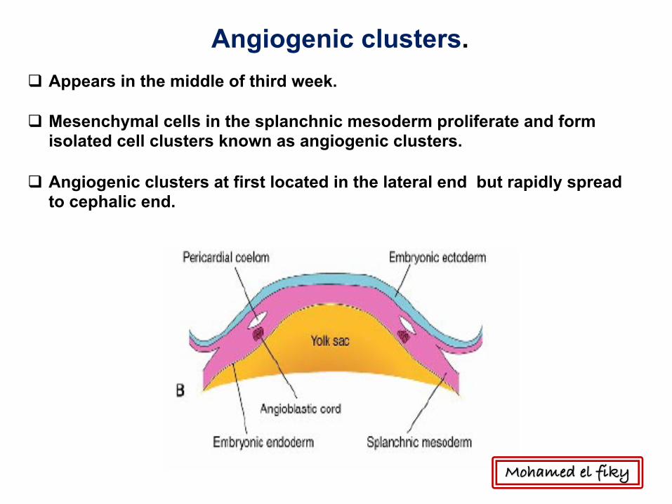
q Appears in the middle of third week.
q Mesenchymal cells in the splanchnic mesoderm proliferate and form isolated cell clusters known as angiogenic clusters.
q Angiogenic clusters at first located in the lateral end but rapidly spread to cephalic end.
Angiogenic clusters.
Mohamed el fiky

Mohamed el fiky

Cardiac swelling
Formation of endocardial heart tubes
endocardial heart tubes
Mohamed el fiky

q Appears in the middle of third week.
q Mesenchymal cells in the splanchnic mesoderm proliferate and form isolated cell clusters known as angiogenic clusters.
q Angiogenic clusters at first located in the lateral end but rapidly spread to cephalic end.
q The angiogenic clusters acquire lumen.
q They unite to form a horseshoe-shaped plexus of small blood vessels.
q The anterior portion of the plexus is called cardiogenic area.
q The intraembryonic coleomic cavity located over the plexus later form pericardial cavity.
Formation of endocardial heart tubes
Mohamed el fiky
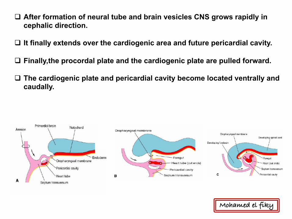
q After formation of neural tube and brain vesicles CNS grows rapidly in cephalic direction.
q It finally extends over the cardiogenic area and future pericardial cavity.
q Finally,the procordal plate and the cardiogenic plate are pulled forward.
q The cardiogenic plate and pericardial cavity become located ventrally and caudally.
Mohamed el fiky

q The embryo folds in cephalocaudal and transversely bringing the two heart tubes closer.
q The two endocardial heart tube fuse in cephalo-caudal direction.
q The tube is attached to the dorsal side of the pericardial cavity by dorsal mesocardium.
Mohamed el fiky

q The mesoderm adjacent to the endocardial tube form epimyocardial mantle.
q The epimyocardial mantle separated from endocardial tube by cardiac jelly.
q Tube consists of endocardium, myocardium and epicardium.
Mohamed el fiky

Development of primitive heart tube
Ø It develops early in the middle of 3rd week , from aggregation of
splanchnic mesodermal cells, in cardiogenic area.
Ø They form 2 angioblastic cords that canalize to form
2 endocardial heart tubes.
Ø The embryo folds in cephalocaudal and
transversely bringing the two heart tubes closer.
Ø The two endocardial heart tube fuse in cephalo-caudal direction.
Ø The tube is attached to the dorsal side of the pericardial cavity by dorsal mesocardium. Mohamed el fiky
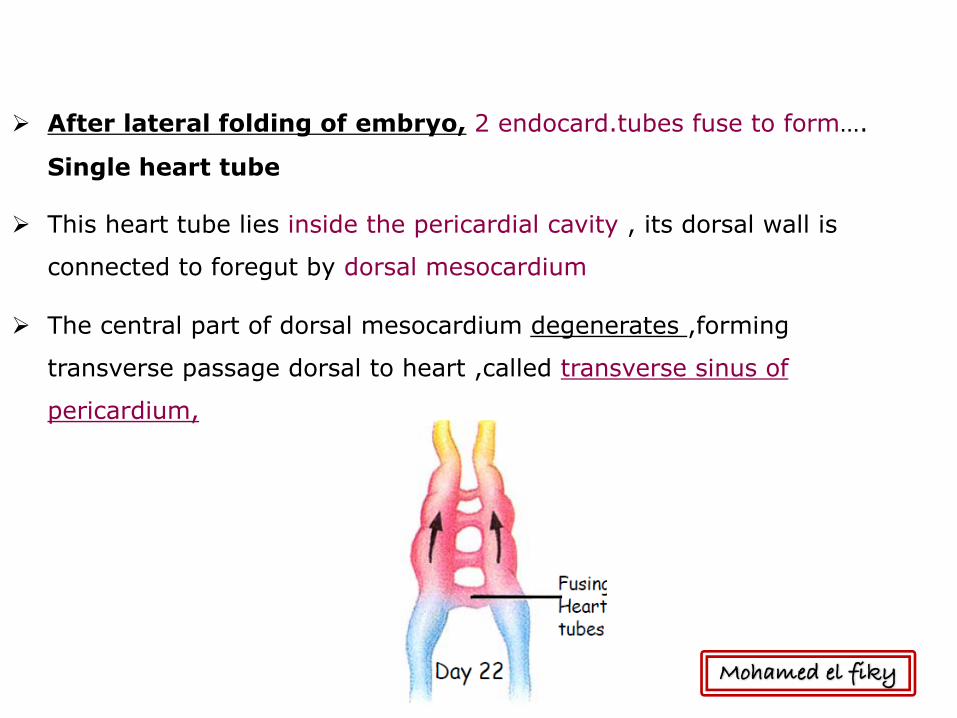
Ø After lateral folding of embryo, 2 endocard.tubes fuse to form….
Single heart tube
Ø This heart tube lies inside the pericardial cavity , its dorsal wall is
connected to foregut by dorsal mesocardium
Ø The central part of dorsal mesocardium degenerates ,forming
transverse passage dorsal to heart ,called transverse sinus of
pericardium,
Mohamed el fiky
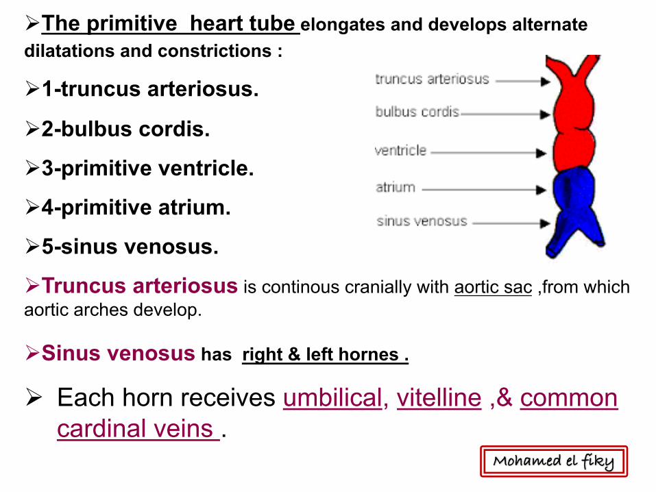
ØThe primitive heart tube elongates and develops alternate dilatations and constrictions :
Ø1-truncus arteriosus.
Ø2-bulbus cordis.
Ø3-primitive ventricle.
Ø4-primitive atrium.
Ø5-sinus venosus.
ØTruncus arteriosus is continous cranially with aortic sac ,from which aortic arches develop.
ØSinus venosus has right & left hornes .
Ø Each horn receives umbilical, vitelline ,& common cardinal veins .
Mohamed el fiky

Main subdivisions of heart tube
Ø Bulbus cordis & ventricle grow faster than other regions, so the heart
bends upon itself,forming U-shaped bulboventricular loop.
Ø The atrium & sinus venosus also come to lie dorsal to truncus arteriosus, bulbus cordis & ventricle .(S-shaped heart tube).
Mohamed el fiky

Formation of cardiac loopq Heart tube elongates and bends.
q The cehpalic portion: bends in ventral and caudal direction to the right.
q The caudal portion: shifts in a dorsocranial direction and to the left.Thebendings creates a cardiac loop.
Mohamed el fiky

Primitive heart tube Twists
Mohamed el fiky

q Local expansion become visible after cardiac loop is formed.
q The atrial portion lie outside the pericardial cavity,
q later incorporated inside the cavity.
q The atrioventricular junction remains narrow and form atrioventricular canal.
q The bulbus cordis is narrow except its proximal third which later forms trabeculated part of right ventricle.
q The distal part of bulbus called the truncus arteriosus.
q The conus cordis forms the outflow tract of both ventricles. Mohamed el fiky

q The proximal portion of the bulbus form the primitive right ventricle.
q The primitive ventricle becomes trabeculated and form the primitive left ventricle.
Mohamed el fiky
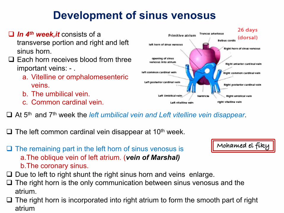
Development of sinus venosusq In 4th week,it consists of a
transverse portion and right and left sinus horn.
q Each horn receives blood from three important veins: - .
a. Vitelline or omphalomesenteric veins.
b. The umbilical vein.c. Common cardinal vein.
q At 5th and 7th week the left umbilical vein and Left vitelline vein disappear.
q The left common cardinal vein disappear at 10th week.
q The remaining part in the left horn of sinus venosus isa.The oblique vein of left atrium. (vein of Marshal)b.The coronary sinus.
q Due to left to right shunt the right sinus horn and veins enlarge.q The right horn is the only communication between sinus venosus and the
atrium.q The right horn is incorporated into right atrium to form the smooth part of right
atrium
Mohamed el fiky

q The entrance the sinoatrial orifice is flanked On each side by right and left venous valves.
q Dorsocranially , the valves fuse , forming a ridge known as septum spurium
q The superior portion of right venous valve disappear.q The inferior part form two parts:
a.The valve of inferior vena cava.b. The valve of coronary sinus.
q The crista terminalis originates from right sinus horn.
Mohamed el fiky

Dividing of A-V canal , primitive atrium & primitive ventricle….. Begins at the middle or end of 4th week. It is completed by the end of 5th week.Endocardial cushions: these are masses of cells and extracellular matrices develop in the atrioventricular and conotruncal regions . • in the atrioventricularregion they are :
– Dorsal & ventral swellings– Fuse, dividing the single AV canal into paired canals– Involved in formation of interatrial & interventricular septa– Derived from neural crest
Partitioning
Mohamed el fiky

•Attheendofthefourthweek,asickle-
shapedcrestgrowsfromtheroofofthe
commonatriumintothelumen.Thiscrest
isthefirstportionoftheseptumprimum.
•Thetwolimbsofthisseptumextend
towardtheendocardialcushionsinthe
atrioventricularcanal.
•Theopeningbetweenthelowerrimof
theseptumprimumandtheendocardial
cushionsistheostiumprimum
Septum formation in the common atrium
Mohamed el fiky
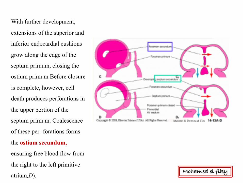
With further development,
extensions of the superior and
inferior endocardial cushions
grow along the edge of the
septum primum, closing the
ostium primum Before closure
is complete, however, cell
death produces perforations in
the upper portion of the
septum primum. Coalescence
of these per- forations forms
the ostium secundum,
ensuring free blood flow from
the right to the left primitive
atrium,D). Mohamed el fiky

When the lumen of the right atrium expands as a result of incorporation of the sinus horn, a
new crescent-shaped fold appears. This new fold, the septum secundum never forms a
complete partition in the atrial cavity . Its anterior limb extends downward to the septum in
the atrioventricu- lar canal. When the left venous valve and the septum spurium fuse with the
right side of the septum secundum, the free concave edge of the septum secundum begins to
overlap the ostium secundum.The opening left by the septum secundum is called the oval
foramen (foramen ovale).
Mohamed el fiky

When the upper part of the septum primum gradually disappears, the remaining
part becomes the valve of the oval foramen. The passage between the two atrial
cavities consists of an obliquely elongated cleft through which blood from the
right atrium flows to the left side.
After birth, when lung circulation begins and pressure in the left atrium
increases, the valve of the oval foramen is pressed against the septum secundum,
obliterating the oval foramen and separating the right and left atria. In about
20% of cases, fusion of the septum primum and septum secundum is incomplete,
and a narrow oblique cleft remains between the two atria.This condition is called
probe patency of the oval foramen; it does not allow intracardiac shunting of
blood.
Mohamed el fiky

Fetus• right side high pressure (high pulmonary resistance, etc.)• well oxygenated blood streams through foramen ovale.• valve of foramen ovale closes with left atrial contraction.
After birth• right side low pressure (low pulmonary resistance).• valve remains closed (physiological closure).• valve eventually fuses (anatomical closure): fossa ovalis.
Mohamed el fiky
•Bytheendofthefourthweek,thetwoprimitiveventriclesbegintoexpand.Thisis
accomplishedbycontinuousgrowthofthemyocardiumontheoutsideandcontinuous
diverticulationandtrabeculaformationontheinside.
•Themedialwallsoftheexpandingventriclesbecomeapposedandgraduallymerge,
formingthemuscularinterventricularseptum.Sometimes,thetwowallsdonotmerge
completely,andamoreorlessdeepapicalcleftbetweenthetwoventriclesappears.
Thespacebetweenthefreerimofthemuscularventricularseptumandthefused
endocardialcushionspermitscommunicationbetweenthetwoventricles.
•Theinterventricularforamen,abovethemuscularportionoftheinterventricular
septum,shrinksoncompletionoftheconusseptum.Duringfurtherdevelopment,out-
growthoftissuefromtheanterior(inferior)endocardialcushionalongthetopofthe
muscularinterventricularseptumclosestheforamen.Thistissuefuseswiththeabut-
tingpartsoftheconusseptum.Completeclosureoftheinterventricularforamen
formsthemembranouspartoftheinterventricularseptum
SeptumFormationintheVentricles
Mohamed el fiky

Mohamed el fiky

• Continuous set of ridges in bulbus cordis(bulbar ridges) and truncus arteriosus (truncal ridges).
• Grow toward each other, spiraling 180º.
Partitioning of Truncus Arteriosus
Mohamed el fiky

• Fuse to form spiraling aorticopulmonaryseptum, dividing aorta & pulmonary trunk
• Bulbar ridges involved in formation of IV septum
• Bulbar & truncal ridges derived from neural crest cells—clinical implications
Partitioning of Truncus Arteriosus
Mohamed el fiky
Fetal circulation

Placenta
Umbilical vein
Portal vein
Inferior vena cava
Right atrium
Left Atrium
Left ventricle
Aorta
Whole body tissue
Fetal circulation(oxygenated)
Mohamed el fiky

Superior & inferior vena cava
Right atrium
Right ventricle
Pulmonary trunk
Arch of Aorta
Descending aorta
Common and internal iliac
2 Umbilical arteries
Placenta
Fetal circulation(Deoxygenated)
Mohamed el fiky

PATHWAY
Placenta
Umbilical Vein
Umbilical Arteries
Liver
Ductus VenosusInferior VenacavaRight AtriumForamen Ovale
Right LungArch of Aoarta
Ductus Arteriosus
Left Atrium
Left Ventricle
Right Ventricle
Portal Vein
Mohamed el fiky
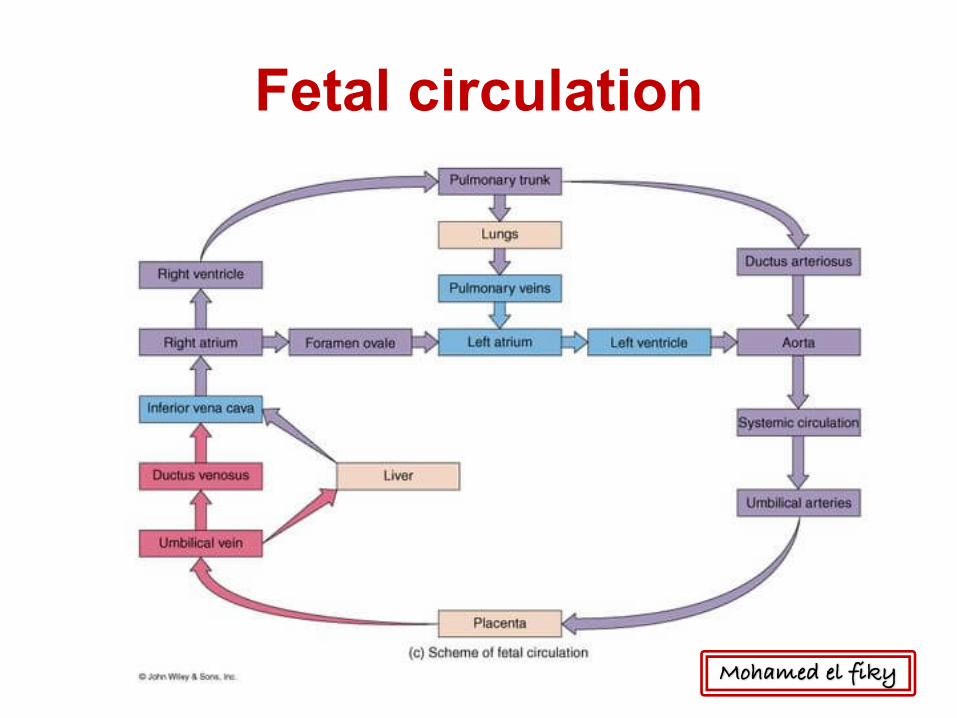
Fetal circulation
Mohamed el fiky
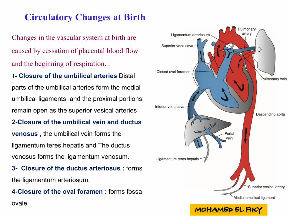
Circulatory Changes at Birth
Changes in the vascular system at birth are
caused by cessation of placental blood flow
and the beginning of respiration. :
1- Closure of the umbilical arteries Distal
parts of the umbilical arteries form the medial
umbilical ligaments, and the proximal portions
remain open as the superior vesical arteries
2-Closure of the umbilical vein and ductus
venosus , the umbilical vein forms the
ligamentum teres hepatis and The ductus
venosus forms the ligamentum venosum.
3- Closure of the ductus arteriosus : forms
the ligamentum arteriosum.
4-Closure of the oval foramen : forms fossa
ovale

Congenital anomalies of the heart1- Defects of the atrial septum:
a)Patent foramen ovale (ASD):
b)Is caused by incomplete anatomic fusion of septum primum and septum secudum.
Present in approximately 25% of people.
c)Foramen secundum defect : is caused by excessive resorption of septum primum
or septum secundum. This results in a large opening between the right and left atria.
d)Common atrium : caused by complete failure of septum primum and septum
secundum to develop.
e)Premature closure of foramen oval: closure of foramen ovale during pre-natal life.
This results in hypertrophy of the right side of the heart and under-development of the
left side of the heart.
Mohamed el fiky

Congenital anomalies of the heart
• 2- Defects of atrio-ventricular canal :
a) Persistent AV canal: caused by failure of AV cushions to fuse, accompanied
by abnormal tricuspid and bicuspid valves.
b) Tricuspid atresia: Obliteration of the right AV canal, characterized by absence
of the tricuspid valve and accompanied by the following :
– 1) Patent foramen ovale.
– 2) I.V. septal defect.
– 3) Over-developed left ventricle.
– 4) Under-developed right ventricle.
Mohamed el fiky
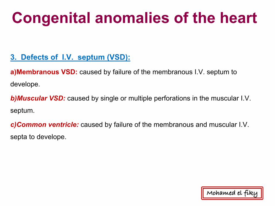
Congenital anomalies of the heart
3. Defects of I.V. septum (VSD):
a)Membranous VSD: caused by failure of the membranous I.V. septum to
develope.
b)Muscular VSD: caused by single or multiple perforations in the muscular I.V.
septum.
c)Common ventricle: caused by failure of the membranous and muscular I.V.
septa to develope.
Mohamed el fiky

Congenital anomalies of the heart4, Defects of the aortico- pulmonary septum:a)Persistent truncus arteriosus: This is due to failure of development of the spiral aortico-pulmonary septum. The truncus overrides the inter-ventricular septum and receives blood from both ventricles. b)Tetralogy of Fallot : characterized by four classic malformations :c)Pulmonary stenosis, overriding aorta, inter-ventricular septal defect and right ventricular hypertrophy. d)Congenital aortic valve stenosis : this is due to fusion of the cusps of the aortic valve leading to a very narrow aortic orifice. The left ventricle is markedly hypertrophied. e)Aortic valve atresia : The aortic orifice is completely closed. The left ventricle is under-developed and the ascending aorta is narrow. The ductus arteriosus is patent to carry blood into the aorta. f)Pulmonary valve stenosis and atresia: The pulmonary trunk is narrow and the right ventricle is under-developed. The ductus arteriosus remains patent and carries blood in an opposite direction from the arch of aorta to the pulmonary arteries. The foramen ovale remains patent. g)Transposition of ascending aorta and pulmonary trunk: This is due to a reversal development of the spiral aortico-pulmonary septum. Mohamed el fiky

Congenital anomalies of the heart
5. Abnormal positions of the heart:
a)Isolated dextro-cardia : the heart is abnormally positioned on the right side of
the thorax. It is usually associated with other severe cardiac anomalies.
b) Dextro-cardia with situs inversus: is dextro-cardia with inversion of the
viscera.
Mohamed el fiky

Thank You
Dr. Fiky Thorax