developmental and age-related processes that lnfluence the … · the plant cell, vol. 5, 553-564,...
TRANSCRIPT

The Plant Cell, Vol. 5, 553-564, May 1993 O 1993 American Society of Plant Physiologists
Developmental and Age-Related Processes That lnfluence the Longevity and Senescence of Photosynthetic Tissues in Arabidopsis
L inda L. Hensel, Vojislava Grbic, David A. Baumgarten, and Anthony 6. Bleecker' Botany Department, Birge Hall, University of Wisconsin, Madison, Wisconsin 53706
Factors that influence the longevity and senescence of photosynthetic tissues of Arabidopsis were investigated. To de- termine the influence of reproductive development on the timing of somatic tissue senescence, the longevity of rosette leaves of the Landsberg efecta strain and of isogenic mutant lines in which flowering is delayed (co-2) or sterile flowers are produced (ms7-7) were compared. No difference in the timing of senescence of individual leaves was observed be- tween these lines, indicating that somatic tissue longevity is not governed by reproductive development in this species. To examine the role of differential gene expression in the process of leaf senescence, cDNA clones representing genes that are differentially expressed in senescing tissues were ísolated. Sequence analysis of one such clone indicated ho- mology to previously cloned cysteine proteinases, which is consistent with a role for the product of this gene in nitrogen salvage. RNA gel blot analysis revealed that increased expression of senescence-associated genes is preceded by declines in photosynthesis and in the expression of photosynthesis-associated genes. A model is presented in which it is postu- lated that leaf senescence is triggered by age-related declines in photosynthetic processes.
INTRODUCTION
The coupling of whole-plant senescence to reproduction is a life history trait that is common to many annual and some perennial plant species and is referred to as the monocarpic habit (Hildebrand, 1881). In nature, monocarpy is most char- acteristic of plant species classified as ruderals (Stebbins, 1950; Grimes, 1979). This ecological classlfication refers to plants, such as Arabidopsis, that are adapted to growth in disturbed environments. According to theory, ruderals have evolved life history traits that are compatible with the high mortality risks from the environment: a propensity for early reproductive de- velopment and high fecundity are two particularly noteworthy traits. The early diversion of resources from vegetative to reproductive development in ruderals is thought to have con- tributed to the evolution of the monocarpic habit characteristic of these species (Molisch, 1928).
The partitioning of resources between vegetative and re- productive development in monocarpic plants involves a complex interplay of generative and degenerative processes that are thought to be under developmental control. The phys- iological basis for these processes has been extensively studied in a few annual crop species, such as soybean, cot- ton, and maize (Eaton, 1955; Wittenbach, 1982; Crafts- Brandner et al., 1984a; Crafts-Brandner and Egli, 1987; Ford and Shibles, 1988). In general, monocarpic plant senescence involves a degeneration of existing vegetative tissues and a
1 To whom correspondence should be addressed.
concomitant cessation of vegetative meristem activity that prevents the development of new photosynthetic tissues (Woolhouse, 1983; Noodén, 1988b). The two processes appear to be coordinately regulated in some cases but independently controlled in other species (see Noodén, 1988a, for a discussion).
The timing of both leaf senescence and apical arrest is thought to involve interacting signals between the affected tis- sues and the developing fruit. These interactions between vegetative and reproductive structures are generally referred to as correlative controls. A number of hypotheses concern- ing the nature of these correlative control signals have been presented (reviewed in Woolhouse, 1983; Kelly and Davies, 1988; Noodén, 1988b), but the biochemical nature of the sig- naling mechanisms has not been unequivocally established for any higher plant.
Although the regulatory mechanisms that govern the tim- ing of leaf senescence remain elusive, the actual processes associated with Senescence of photosynthetic tissue have been extensively characterized in a number of plant species. Com- mon features observed across a range of species have led to the concept of the senescence syndrome: an orderly se- quence of events involving the turnover of macromolecules and lipids and the transport of mobilized nutrients out of the senescing tissue. The most widely used biomarker for the senescence syndrome is the rapid loss of chlorophyll as- sociated with the degeneration of chloroplast interna1 structure

554 The Plant Cell
(Thomson and Plat-Aloia, 1987; see also Woolhouse, 1982).Experimental evidence indicates that the senescence syn-drome is under genetic control by the nucleus (Yoshida, 1961;Ness and Woolhouse, 1980; Thomas et al., 1992). Recentstudies indicate that differential expression of specific genesis associated with the senescence syndrome (Davies andGrierson, 1989; Graham et al., 1992).
The monocarpic habit is exemplified by Arabidopsis, whichmay undergo its entire life cycle in 8 to 10 weeks. Sexualreproduction in Arabidopsis involves the generation of thou-sands of offspring. Associated with this massive reproductiveeffort, the leaves, stems, and fruits of the adult plant undergoprogressive senescence that ultimately results in the deathof the plant. We are interested in determining the mechanismsinvolved in the coordination of these processes. In this report,we make use of single-gene mutations that affect reproduc-tive development to evaluate the role of reproduction in thepatterns of growth, development, and senescence of the Ara-bidopsis rosette leaf. In addition, we examine the differentialexpression of genes in the leaf associated with the senescencesyndrome.
RESULTS
Experimental Set Up
Environmental conditions such as light quality and quantity,nutrient and water availability, temperature, and humidity havea strong influence on the course of development in Arabidop-sis. For this reason, we established culture conditions thatminimized environmental fluctuations. Although these condi-tions of constant light, temperature, humidity, and soil moistureare not necessarily optimum for growth and development,plants were healthy and uniform in appearance, and the Lands-berg erecfa (Ler) strain produced more than 10,000 seeds withinthe 50-day life cycle. Under these same conditions, delayedflowering varieties grew vigorously for several months. In someexperiments, plants were grown in a 16-hr-light/8-hr-dark pho-toperiod. Plants of the Ler strain grown under these conditionswere indistinguishable from plants grown under constant light.
Life History Traits in Arabidopsis
Figures 1A through 1D depict the life history of an Arabidop-sis plant. Under the above constant environmental conditions,the Ler strain of Arabidopsis developed initially as a rosetteof seven to eight leaves (Figure 1A). At ~12 days postgermi-nation, the transition from vegetative to inflorescence meristemoccurred at the apex, resulting in the production of the highlybranched inflorescence (Figures 1B and 1C) (Vaughan, 1955;Bowman et al., 1989; Schultz and Haughn, 1991; Shannon andMeeks-Wagner, 1991). The most active period of flowering and
Figure 1. Stages in the Life Cycle of the Landsberg erecta Strain ofArabidopsis and Age-Related Changes in Rosette Leaf Number Five.
(A) 14 days after planting.(B) 21 days after planting.(C) 37 days after planting.(D) 53 days after planting.(E) 0 days after full leaf expansion.(F) 3 days after full leaf expansion.(G) 5 days after full leaf expansion.(H) 7 days after full leaf expansion.(I) 9 days after full leaf expansion.(J) 11 days after full leaf expansion.

Longevity and Senescence in Arabidopsis 555
fruit development is accompanied by the sequential senes- cence of the original leaves of the rosette, as indicated by an initial stage of chlorophyll loss, followed by the complete dis- integration of the leaf tissue (Figure 1C). Developmental arrest of the rnain inflorescence stem, characterized by a cessation of proliferative activity at the apex and a degeneration of the youngest flower buds, occurred at approximately day 40 (Vaughan, 1955; Shannon and Meeks-Wagner, 1991; Alvarez et al., 1992). During this later phase, the stem, cauline leaves, and seed pods became senescent and the seeds of the next generation reached maturity (Figure 1D).
Based on these general observations, we considered two distinct processes that govern the c o m e of monocarpic senes- cence in Arabidopsis: (1) the senescence of the developed organ systems, such as leaves, stems, and diques, which we define as somatic senescence and (2) the cessation of gener- ative activity at the inflorescence meristem, which we refer to as apical arrest. In this study, we focused on the senescence of somatic tissues using primarily the fifth and sixth leaves of the rosette as our model.
The fifth and sixth leaves of the rosette represent the first consistently adult leaves of the Ler strain (as defined for the Wassilewskija strain by Medford et al., 1992). Under the cul- ture conditions used, individual leaves progressed through full expansion to maturity followed by a period of progressive leaf yellowing and, finally, a degeneration and complete desicca- tion of the tissue. This developmental sequence is shown for leaf 5 in Figures 1E through 1J. To obtain datafrom large popu- lations of plants in a noninvasive way, we performed our initial experiments to asses tissue longevity by simply recording the time from visible emergence (1 to 2 mm) of an individual leaf or stem internode to the time at which more than 50% visible degreening of that tissue had occurred. For leaves, the latter stage was invariably followed by a complete degenera- tion and death of the tissue within 2 days. In the case of stem tissue, senescence refers only to the degreening of the pho- tosynthetic cortical tissues because stems may continue to function in nutrient transport beyond this stage. Preliminary ultrastructural analysis of the leaf and stem indicated that degreening of both mesophyll cells of the leaf and cortical cells of the stem is associated with the loss of thylakoid structure and increases in plastoglobuli characteristic of the senescence syndrome (data not shown) (Thomson and Platt-Aloia, 1987).
Relationship between Reproduction and Somatic Tissue Senescence
The sequential senescence of the leaves of the rosette is tem- porally correlated with inflorescence development (Figure 1). In correlative control models for monocarpic senescence, it is hypothesized that signals or processes associated with in- florescence andlor fruit development are responsible for the induction of the senescence syndrome in leaves (reviewed in Noodén, 1988b). To investigate the relationship between re- productive development and senescence in Arabidopsis,
somatic tissue senescence was first compared between the wild-type Ler strain and ms7-7, a male-sterile mutant line in the Ler background (Koornneef et al., 1983). As shown in Fig- ure 2, the absence of fruit development in the msl-7 line did not appreciably affect the timing of Senescence of either leaf or inflorescence stem tissue. A delay in reproductive develop- ment also failed to have an effect on the longevity of rosette leaves. Leaves of a late-flowering mutant line, co-2(Rédei, 1962; Koornneef et al., 1991), senesce over the same period of time as leaves of wild-type plants, as shown in the mortality curves of Figure 3A. A second late-flowering line, fca (Martinez-Zapatsr and Somerville, 1990; Koornneef et al., 1991), was also exam- ined with similar results (data not shown).
An alternative hypothesis for correlative control is the nutri- ent drain hypothesis in which it is postulated that changes in source-sink relationships between tissues may govern the tim- ing of leaf senescence (Molisch, 1928). One could argue that sink demand resulting from inflorescence development in the male-sterile line or from the development of additional leaves in the delayed flowering lines was governing leaf senescence. To test this possibility, the longevity of rosette leaves was mea- sured in the ff/7-2 (terminal flower) mutant line (Shannon and
n v)
o U )r
> a w E O a v) VI
W
c .I
I
a
i=
20
15
10
5
o 11.2 3.4 5 6 1 I 1
1
I1 211 I - 1-
Leaf number lnternode Figure 2. Longevity of Individual Leaves and Stem Segments of Wild- Type (Ler) and Male-Sterile (ms7-7) Lines.
Plants were grown in l&hr-light/Shr-dark cycles. Mean tissue longevity (+ SE) for rosette leaves 1 through 6 (numbered by order of emer- gente from the meristem) is shown. Data for the first two pairs of leaves are averaged because the time of emergence is often indistinguish- able. Internode 1 is defined as the internode between the first and the second flower produced on the primary inflorescence meristem. Tis- sue longevity was measured as the time from visual emergence (1 to 2 mm) to more than or equal to 50% degreening. For leaf Ler, n = 137 plants; ms7-7, n = 45 plants. For internode Ler, n = 19; ms7-7, n = 11.

556 The Plant Cell
125
1 O0
- o > 75 'E 3 v)
' 25
O
1 O0 - o > > .- f 75
c o 3 50
A
m B
o I+
o
0 0 O 5 10 15 20 25 30
Days from leaf emergence
Figure 3. Survival Curves of Adult Leaves for Wild-Type (Ler), Late- Flowering (co-2), Male-Sterile (ms7-7), and Terminal Flower (ff/l-2) Lines.
Data from leaves 5 and 6 were combined for this analysis. Leaf lon- gevity was measured as given in Figure 2. (A) Ler, n = 322; co-2, n = 54; msl-7, n = 74. (8) Ler, n = 25; ffl7-2, n = 23.
Rosette Leaf Senescence as an Age-Related Process
Results of the above experiments indicated that the timing of leaf and stem senescence is not coupled to reproductive de- velopment in Arabidopsis. Rather, somatic tissue longevity appears to be an intrinsic age-related property of the organ system.
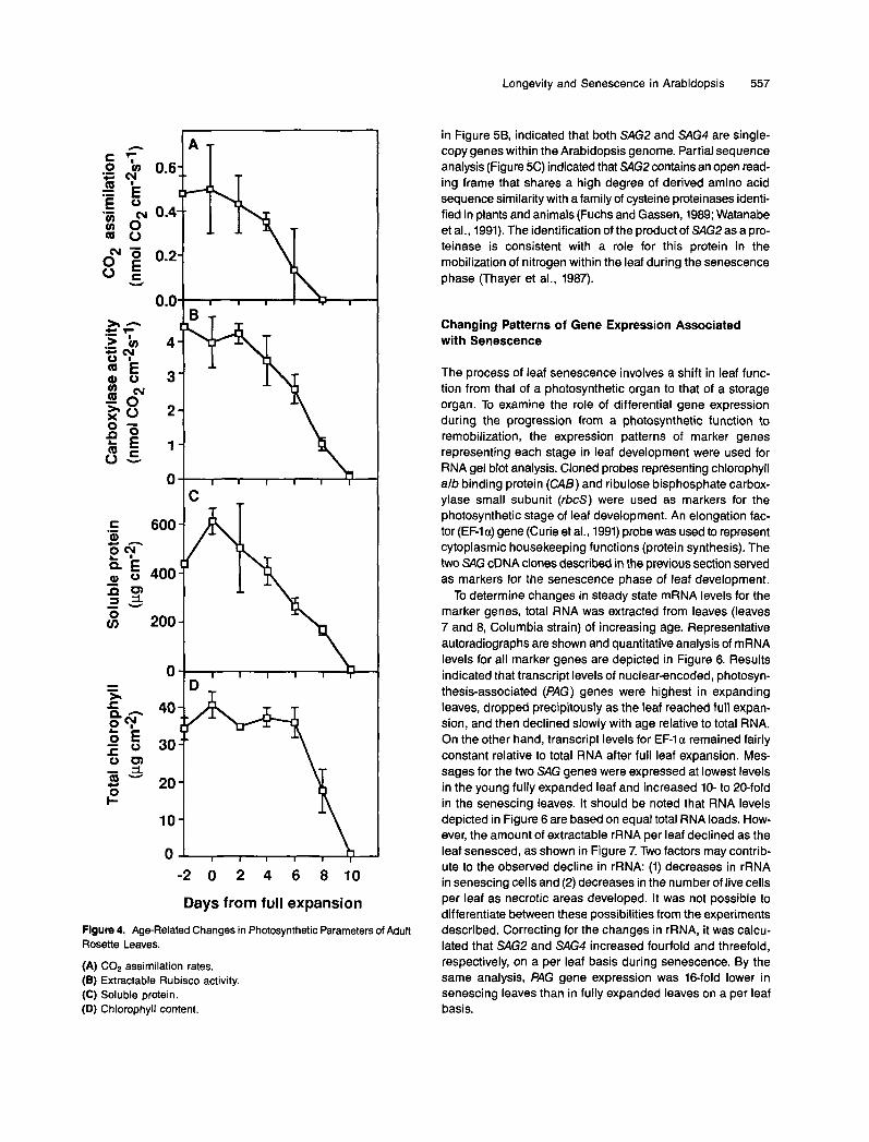
To investigate the role of processes intrinsic to the leaf in the timing of leaf senescence, we examined a number of cel- lular parameters for age-related changes. Because the primary function of the mature leaf is photosynthesis, we focused on biomarkers associated with photosynthetic function. Results shown in Figure 4 indicate that C02 fixation rates, while ini- tially variable, decline with leaf age almost from the time of full leaf expansion (Figure 4A). Extractable ribulose bisphos- phate carboxylase (Rubisco) activity and soluble protein showed similar declines associated with age (Figures 46 and 4C). The slower decline in Rubisco activity relative to COn fix- ation is due in part to the fact that fixation rates were measured on the whole leaf, and Rubisco activity was determined from leaf discs taken from the center of the leaf that senesces last (Figure 1). For leaf discs, the decline in Rubisco preceded the rapid loss in chlorophyll that is characteristic of the senescence syndrome (Figure 4D).
lsolation of cDNA Clones Representing Genes That Are Differentially Expressed during Senescence
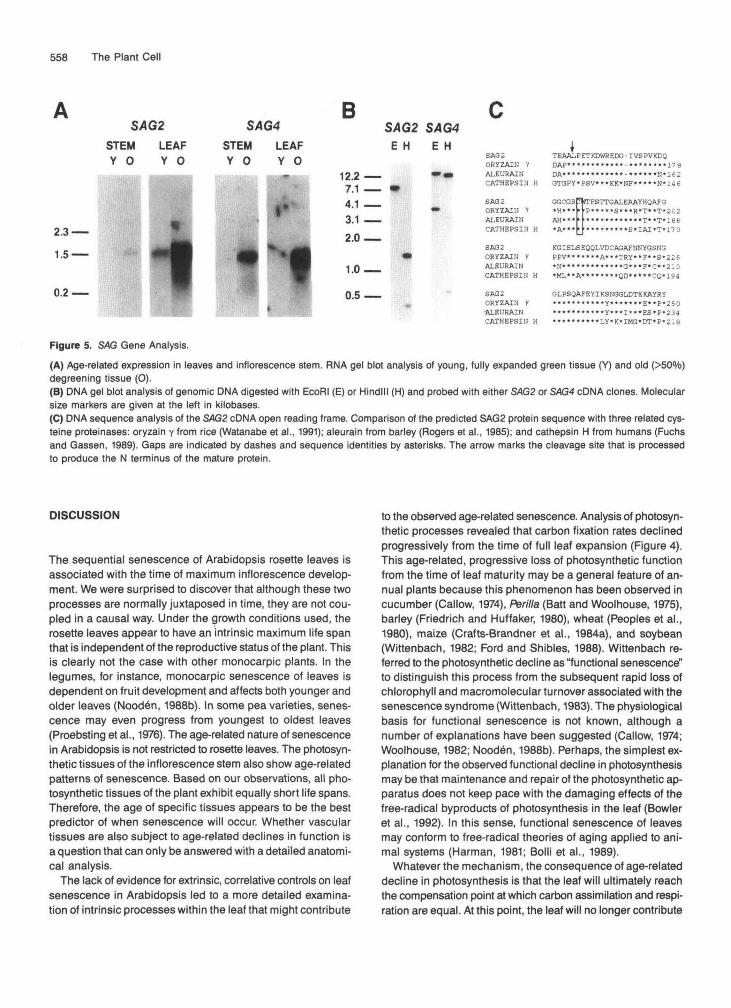
Whereas biological markers for photosynthetic function were known, no such markers were known for the senescence phase of leaf development. As mentioned in the Introduction, severa1 lines of evidence support the view that leaf senes- cence is a developmentally controlled process that requires expression of nuclear genes. To identify senescence-related biomarkers, we screened a cDNA library made from mRNA of senescing leaves using differential hybridization (Sambrook et al., 1989). Nine cDNA families that do not cross-hybridize were identified and further characterized. Data for two of these cDNA clones are shown here. These two senescence- associated (SAG) gene clones were designated SAG2 and SAGA As shown in Figure 5A, RNA gel blot analysis indicated that mRNA levels for both genes are elevated in senescing leaves and inflorescence stems. DNA gel blot analysis, shown
Meeks-Wagner, 1991; Alvarez et al., 1992). The Wl-2 line pro- duces a normal rosette and initiates flower development at the same time as the isogenic Ler wild type. However, the devel- opment of aberrant flower structures at the inflorescence
Flowers Produced per Dry Weight, meristem causes early termination of inflorescence develop-
ment resulting in a 10-fold decrease in biomass accumulation Line Primary lnflorescence Grams k SE relative to wild type, as shown in Table 1. Results shown in
34.5 f 0.8 1.32 f 0.16 Figure 36 indicate that leaf longevity was not appreciably al- Ler tlf 7 -2 <4 0.17 f 0.02 tered by the diminished sink demand in ffll-2.
Table I . Plant Dry Weight Produced by Wild Type (Ler) and Terminal Flower (ffl7-2)
Longevity and Senescence in Arabidopsis 557
a = 0.2 8 : v
0.0
O
40
30 gq -
10
O
' T
I I I I I I .
I I I I I T
) T
- 2 0 2 4 6 8 1 0
Days from full expansion
Figure 4. Age-Related Changes in Photosynthetic Parameters of Adult Rosette Leaves.
(A) COp assimilation rates. ( E ) Extractable Rubisco activity. (C) Soluble protein. (D) Chlorophyll content.
in Figure 5B, indicated that both SAGP and SAG4 are single- copy genes within the Arabidopsis genome. Partia1 sequence analysis (Figure 5C) indicated that SAGP contains an open read- ing frame that shares a high degree of derived amino acid sequence similarity with a family of cysteine proteinases identi- fied in plants and animals (Fuchs and Gassen, 1989; Watanabe et al., 1991). The identification of the product of SAGP as a pro- teinase is consistent with a role for this protein in the mobilization of nitrogen within the leaf during the senescence phase (Thayer et al., 1987).
Changing Patterns of Gene Expression Associated with Senescence
The process of leaf senescence involves a shift in leaf func- tion from that of a photosynthetic organ to that of a storage organ. To examine the role of differential gene expression during the progression from a photosynthetic function to remobilization, the expression patterns of marker genes representing each stage in leaf development were used for RNA gel blot analysis. Cloned probes representing chlorophyll alb binding protein (CAB) and ribulose bisphosphate carbox- ylase small subunit (rbcS) were used as markers for the photosynthetic stage of leaf development. An elongation fac- tor (EFla) gene (Curie et al., 1991) probe was used to represent cytoplasmic housekeeping functions (protein synthesis). The two SAG cDNA clones described in the previous section served as markers for the senescence phase of leaf development.
To determine changes in steady state mRNA levels for the marker genes, total RNA was extracted from leaves (leaves 7 and 8, Columbia strain) of increasing age. Representative autoradiographs are shown and quantitative analysis of mRNA levels for all marker genes are depicted in Figure 6. Results indicated that transcript levels of nuclear-encoded, photosyn- thesis-associated (PAG) genes were highest in expanding leaves, dropped precipitously as the leaf reached full expan- sion, and then declined slowly with age relative to total RNA. On the other hand, transcript levels for EF-1 a remained fairly constant relative to total RNA after full leaf expansion. Mes- sages for the two SAG genes were expressed at lowest levels in the young fully expanded leaf and increased 10- to 20-fold in the senescing leaves. It should be noted that RNA levels depicted in Figure 6 are based on equal total RNA loads. How- ever, the amount of extractable rRNA per leaf declined as the leaf senesced, as shown in Figure 7. Two factors may contrib- ute to the observed decline in rRNA: (1) decreases in rRNA in senescing cells and (2) decreases in the number of tive cells per leaf as necrotic areas developed. It was not possible to differentiate between these possibilities from the experiments described. Correcting for the changes in rRNA, it was calcu- lated that SAGP and SAG4 increased fourfold and threefold, respectively, on a per leaf basis during senescence. By the same analysis, PAG gene expression was 16-fold lower in senescing leaves than in fully expanded leaves on a per leaf basis.
558 The Plant Cell
SAG2STEM LEAFY 0 Y 0
2.3 —
1.5 —
0.2 —
SAG4STEM LEAFY 0 Y O
B
12.2 •7.1 .4.1 .3.1 .2.0.
1.0.
0.5.
SAG2 SAG4E H E H
SAG2 TEAALPETKDWREDG-IVSPVKDQORYZAIU V DAP*'*"«*«'*"**-*"«-"«17eALEURAIN DA****••••• .......... «N'1€2CATHEPSIH H GTSPY*PSV***KK*NF*«***N*146
SAG 2 GGCGSpVTFSTTGALEAAYHQAFaORYZAIH V *H***MP*****S***R*T**T*202ALEURAIN AH**«t|****«****«**T**T*186CATHEPSIH H 'A*'*!-!"-* ****«*S*IAI *T»17 0
SAG2 KGISLSEQQLVDCAGAFNNYGSN3ORYZAIN V PPV*******A***TRY**F**S*226ALEURAIN *N*«*««********G***F*C**210CATHEPSIU H *ML**A********QD*****CQ*194
SAG2 GLPSQAFEYIKSNGGLDTEKAYRYORYZAIN V **•*«*•«.**Y««««*.*E*«p«250-ALEURAIN ...........Y***I***ES*P*234CATHEPSIH H ***«******LY*K*IMG*DT*P*2:8
Figure 5. SAG Gene Analysis.
(A) Age-related expression in leaves and inflorescence stem. RNA gel blot analysis of young, fully expanded green tissue (Y) and old (>50°/o)degreening tissue (O).(B) DMA gel blot analysis of genomic DMA digested with EcoRI (E) or Hindlll (H) and probed with either SAG2 or SAG4 cDNA clones. Molecularsize markers are given at the left in kilobases.(C) DMA sequence analysis of the SAG2 cDNA open reading frame. Comparison of the predicted SAG2 protein sequence with three related cys-teine proteinases: oryzain y from rice (Watanabe et al., 1991); aleurain from barley (Rogers et al., 1985); and cathepsin H from humans (Fuchsand Gassen, 1989). Gaps are indicated by dashes and sequence identities by asterisks. The arrow marks the cleavage site that is processedto produce the N terminus of the mature protein.
DISCUSSION
The sequential senescence of Arabidopsis rosette leaves isassociated with the time of maximum inflorescence develop-ment. We were surprised to discover that although these twoprocesses are normally juxtaposed in time, they are not cou-pled in a causal way. Under the growth conditions used, therosette leaves appear to have an intrinsic maximum life spanthat is independent of the reproductive status of the plant. Thisis clearly not the case with other monocarpic plants. In thelegumes, for instance, monocarpic senescence of leaves isdependent on fruit development and affects both younger andolder leaves (Nooden, 1988b). In some pea varieties, senes-cence may even progress from youngest to oldest leaves(Proebsting et al., 1976). The age-related nature of senescencein Arabidopsis is not restricted to rosette leaves. The photosyn-thetic tissues of the inflorescence stem also show age-relatedpatterns of senescence. Based on our observations, all pho-tosynthetic tissues of the plant exhibit equally short life spans.Therefore, the age of specific tissues appears to be the bestpredictor of when senescence will occur. Whether vasculartissues are also subject to age-related declines in function isa question that can only be answered with a detailed anatomi-cal analysis.
The lack of evidence for extrinsic, correlative controls on leafsenescence in Arabidopsis led to a more detailed examina-tion of intrinsic processes within the leaf that might contribute
to the observed age-related senescence. Analysis of photosyn-thetic processes revealed that carbon fixation rates declinedprogressively from the time of full leaf expansion (Figure 4).This age-related, progressive loss of photosynthetic functionfrom the time of leaf maturity may be a general feature of an-nual plants because this phenomenon has been observed incucumber (Callow, 1974), Perilla (Batt and Woolhouse, 1975),barley (Friedrich and Huffaker, 1980), wheat (Peoples et al.,1980), maize (Crafts-Brandner et al., 1984a), and soybean(Wittenbach, 1982; Ford and Shibles, 1988). Wittenbach re-ferred to the photosynthetic decline as "functional senescence"to distinguish this process from the subsequent rapid loss ofchlorophyll and macromolecular turnover associated with thesenescence syndrome (Wittenbach, 1983). The physiologicalbasis for functional senescence is not known, although anumber of explanations have been suggested (Callow, 1974;Woolhouse, 1982; Nooden, 1988b). Perhaps, the simplest ex-planation for the observed functional decline in photosynthesismay be that maintenance and repair of the photosynthetic ap-paratus does not keep pace with the damaging effects of thefree-radical byproducts of photosynthesis in the leaf (Bowleret al., 1992). In this sense, functional senescence of leavesmay conform to free-radical theories of aging applied to ani-mal systems (Harman, 1981; Bolli et al., 1989).
Whatever the mechanism, the consequence of age-relateddecline in photosynthesis is that the leaf will ultimately reachthe compensation point at which carbon assimilation and respi-ration are equal. At this point, the leaf will no longer contribute

Longevity and Senescence in Arabidopsis 559
- 3 0 3 6 9
- 3 0 3 6 9Days from full expansion
Figure 6. Age-Related Changes in Transcript Levels for SelectedGenes.Equal loads of total RNA were loaded in each lane and blots were hy-bridized with 32P-labeled probes.(A) Representative autoradiographs of (B), (C), (D), (E), and (F).(B) CAB.(C) rbcS.(D) EF-1a.(E) SAG2.(F)SAG4.
as a photosynthetic organ to the assimilatory needs of the restof the plant. However, the functionally senescent leaf containsa significant pool of nutrients in the form of lipids and macro-molecules. The chloroplasts of a mesophyll cell are of particularimportance in this regard because it has been estimated thatthese organelles may contain over 50% of the protein and 75%of the lipid of the cell (Forde and Steer, 1976; Dean and Leech,1982). The mobilization of these nutrients is thought to occurvia salvage pathways that increase in activity at the terminalstages of leaf development (Woolhouse, 1984; Nooden, 1988a).
As outlined below, we postulate that in Arabidopsis the acti-vation of salvage pathways associated with senescence isdirectly coupled to photosynthetic decline. This regulatory fea-ture may be the critical one that distinguishes Arabidopsis fromother monocarpic plants such as soybean, in which the senes-cence syndrome is correlatively controlled by reproductivedevelopment. Interestingly, depodding of soybean plants willdelay the final stages of senescence in the leaf but does notprevent photosynthetic decline (Wittenbach, 1982; Crafts-Brandner et al., 1984b). In fact, the Rubisco enzyme may bedegraded in leaves of depodded plants, but the mobilized Nis apparently refixed in the paraveinal mesophyll as storageprotein (Franceschi et al., 1983; Wittenbach, 1983). Thus, insoybean, depodding appears to result in uncoupling of photo-synthetic decline from activation of the senescence syndrome.In this case, full activation of salvage pathways presumably
100
<DC
o(AO
75-
50-
DC 25-
- 3 0 3 6 9
Days from full expansionFigure 7. Age-Related Changes in Extractable Ribosomal RNA perLeaf.
RNA quantities were evaluated by slot-blot analysis of RNA obtainedon a per leaf basis. Values were corrected for RNA losses duringpurification.

560 The Plant Cell
requires some form of signal from the developing fruit. In con- trast, we have not yet found a similar situation in Arabidopsis leaves. In addition to the analysis of the Arabidopsis mutants affecting reproduction reported in this paper, we have exam- ined existing hormone-deficient and insensitive mutants as well as newly isolated mutants with delayed leaf senescence. In no case have we found a condition in which the senescence syndrome has been uncoupled from photosynthetic decline (L.L. Hensel, V. GrbiC, D.A. Baumgarten, and A.B. Bleecker, unpublished results).
To consider the possible mechanisms by which the activa- tion of salvage pathways may be coupled to photosynthetic decline, we turn to the paradigm of differential gene expres- sion. Based on our analysis of PAG and SAG gene expression, we consider these biomarkers as representatives of two com- peting developmental programs within the leaf. PAG genes code for components of the photosynthetic apparatus and are consequently expressed at highest levels during leaf expan- sion when chloroplast biogenesis is occurring (Figure 6; see also Mullet, 1988). The lower PAG transcript levels observed after full leaf expansion presumably represent messages uti- lized in the maintenance and repair of existing chloroplasts because chloroplast fission and biogenesis are thought to cease at full leaf expansion (Pyke and Leech, 1992). The SAG genes appear to code for salvage-related functions that are antagonistic to photosynthetic maintenance. The observed age- related expression patterns of SAG genes are consistent with this concept: SAG transcripts are expressed at low levels in young leaf tissues and increase in expression as photosynthe- sis declines (Figure 6).
Potential regulatory processes that drive the expression of PAG and SAG gene transcripts are presented diagrammatically in Figure 8. These possible regulatory pathways can form the basis for mechanistic models that simulate the regulation of these two sets of genes. As indicated in the diagram, the regu- lation of the genes under consideration is complex. As one working model from which to generate and test specific hy- potheses, we suggest a two-step process in which (1) age-related declines in photosynthesis trigger the activation of SAG gene expression and (2) the products of the SAG genes act to mobilize nutrients and, as a consequence, reduce the viability of the tissues to the point of a loss of homeostasis and death. According to this model, PAG transcript levels, which influence the rate of damage repair in the mature leaf, may be determinants in the rate of photosynthetic decline and, thus, contribute indirectly to the timing of leaf senescence. An addi- tional key feature of this model is the speculation that SAG gene expression is repressed in younger leaves by metabolites of photosynthesis, and it is the derepression of these genes result- ing from photosynthetic decline that initiates the senescence syndrome. According to this model, the rate of decline in pho- tosynthesis after full leaf expansion will determine the timing of SAG gene expression and, therefore, the longevity of the leaf. The chemical identity of the metabolite(s) responsible is not known, but it is unlikely to be sucrose because this trans- portable form of carbon may occur at high levels in senescent
1 Export Export
Figure 8. Schematic of Regulatory Pathways That May lnfluence Leaf Longevity and Senescence.
The emphasis of this schematic is on the transcriptional control of PAG and SAG genes. The darker lines outline a hypothetical model in which the following sequence of events is proposed: (A) maturation signals suppress PAG gene expression at full leaf expansion; (6) photosyn- thetic function declines as a result of inadequate maintenance; (C) metabolites of photosynthesis, which act to repress SAG gene expres- sion in the young leaf, decline in concentration, resulting in derepression of SAG genes; (D) SAG gene products act to mobilize nutrients and, as a consequence, promote the senescence of the leaf tissues.
tissues (Crafts-Brandner et al., 1984a). The source of carbon for sucrose synthesis in this case is unlikely to be photosynthe- sis, but is rather through the P-oxidation of lipids because glyoxysomal pathways are known to increase during senes- cence (Gut and Matile, 1988; DeBellis et al., 1990). In this regard, the metabolite repression model has previously been suggested for the senescence-associated expression of ma- late synthase in cucumber cotyledons (Graham et al., 1992).
The limited longevity of Arabidopsis somatic tissues is con- sistent with the early reproductive development and high fecundity of this species. In terms of evolutionary theory, the disposable soma hypothesis (Kirkwood and Cremer, 1982; Kirkwood and Rose, 1991) predicts that species adapted to early reproduction will tend to invest less energy in somatic main- tenance because such maintenance requires resources that are then unavailable for reproductive effort. In specific terms, the dynamics of the Arabidopsis leaf representa trade-off be- tween high photosynthetic output, which supports new growth, particularly of reproductive structures, and maintenance and repair, which contribute to longevity of somatic tissues once they develop. The measure of this evolutionary trade-off for photosynthetic tissues may be in the age-related rate of pho- tosynthetic decline characteristic of a particular species. At the opposite end of the continuum from Arabidopsis would be
Longevity and Senescence in Arabidopsis 561
the needles of some conifers, which are purported to main- tain photosynthetic competence for years but are characterized by relatively low photosynthetic output (Oren et al., 1986).
The processes we have detailed also conform to the genetic theory of life span evolution known as the antagonistic pleio- tropy hypothesis first elaborated by Williams (1957) and more recently discussed by Rose (Kirkwood and Rose, 1991; Rose, 1991). According to this hypothesis, individual genes that have a positive effect on reproductive fecundity but a negative ef- fect on postreproductive survival of the parent will be selected for in evolution. The accumulation of these genes over evolu- tionary time is thought to determine the maximum life span for a given species. Although this theory is generally applied to animals, specific genes and associated mechanisms for an- tagonistic pleiotropy have not been well characterized in animal systems (Finch, 1990). On the other hand, the salvage path- ways associated with the senescence syndrome in plants provide a clear example of the operation of processes that may favor reproductive success through remobilization of nutrients but also lead to decreased fitness (i.e., senescence) of the tis- sues in which they are expressed.
The SAG genes we have identified by differential hybridization conform to the theoretical definition of antagonistic pleiotropy if the encoded proteins function in the mobilization of nutrients, favoring reproduction, and also decrease the viability of the tissues in which they operate. The SAGP gene, which encodes a cysteine proteinase, may provide a specific case. The de- rived amino acid sequence of this gene shares a high degree of amino acid identity with oryzain y and aleurain, which are specifically expressed in germinating grain seeds and are thought to function in the mobilization of nitrogen from protein reserves (Rogers et al., 1985; Watanabe et al., 1991). Thus, a role for the SAGP gene in nutrient mobilization seems likely. It is also plausible that the SAGP gene, in concert with other SAG genes, contributes actively to the autolytic process of senescence that leads to the death of the somatic tissues in which these genes are expressed. Considering the evolutionary origin of senescence syndrome-associated salvage pathways, most of the SAG genes we have identified are expressed at detectable levels in the young photosynthetically active leaf (Figure 6; V. GrbiC, unpublished results), which is consistent with a role for some of these genes in the maintenance-related turnover of macromolecules (e.g., damaged proteins). This con- cept is supported by the high degree of amino acid sequence identity between SAGP and cathepsin H from animals, which is thought to function in general protein turnover in animal cell lysosomes Vakio et al., 1983). Sequence analysis of additional SAG gene clones should provide information about how these differentially expressed genes function in cellular maintenance and senescence.
Assuming that SAG genes are responsible for the progres- sion of senescence in somatic tissues of plants, the regulatory pathways that govern their expression patterns may actually determine the longevity of these tissues. We have presented an argument that in Arabidopsis, SAG genes are activated as a consequence of age-related declines in photosynthesis. Not
all monocarpic plants conform to the predictions of this model. As discussed above, the senescence of leaves can be delayed by removal of fruit in a number of species (Noodh, 1988b) even though this treatment does not prevent age-related declines in photosynthesis. In these systems, the activation of SAG gene expression is apparently not coupled to photosyn- thetic decline but rather depends on other or additional developmental signals for activation. This possibility is ac- counted for in our model by the “developmental and age-related signals” box in Figure 8. The developing Arabidopsis plant ap- parently either does not require these additional regulatory systems or cannot afford them. In any case, the timing of leaf senescence in Arabidopsis is adapted to the reproductive re- quirements of the plant; the sequential senescence of the rosette leaves coincides with the period of maximum primary inflorescence development and seed fill (Figure 1).
What impact does the short life span of Arabidopsis somatic tissues have on the longevity of the whole plant? In theory, the iterative nature of plant development could allow for an indefinite life span. In practice, this is true for Arabidopsis only in cases where reproductive development is delayed. In this case, the primary meristem continues to generate new rosette leaves for many months (Napp-Zinn, 1985). The transition to flowering, however, marks the beginning of the end for the adult plant because all aerial meristems are converted over to reproductive development. While the tissues produced by the inflorescence and flower meristems are capable of photosyn- thesis, inflorescence meristems have a limited proliferative capacity; they cease meristematic activity after the produc- tion of afew dozen flowers (Shannon and Meeks-Wagner, 1991; Alvarez et al., 1992). Thus, the combination of limited longev- ity of somatic tissues and the limited proliferative capacity of the inflorescence meristems ultimately limit the life span of the adult Arabidopsis plant.
METHODS
Strains
Seeds for Landsberg erecta (Ler) and the isogenic mutant lines co-2, msl-I, and fca were obtained from A.R. Kranz (Kranz, 1978; Kirchheim and Kranz, 1981). Columbia seeds were obtained from Chris Somer- ville (Michigan State University, East Lansing). M7-2 seeds were provided by D. Smyth (Monash University, Melbourne, Australia) (Alvarez et al., 1992).
Plant Material and Measurements
All plants were grown at a density of approximately one plant per 25 cm2, at 22OC, and 65 to 85% relative humidity under fluorescent illu- mination supplemented with incandescent light (100 to 150 WE m-* sec-l) on a 21 mixture of Jiffy Mix (Jiffy Products of America, Bata- via, IL) to perlite with a continuous wicking system of 10% Hoagland’s solution (Hoagland and Arnon, 1938). All plants were grown under
562 The Plant Cell
continuous illumination unless otherwise stated. Seeds were surface sterilized with a 30% bleach and 0.5% Triton solution and stratified at 4% for 24 to 48 hr prior to planting. Leaf and internode longevity were measured as the time from visual emergence (1 mm) to more than or equal to 50% degreening. Dry weight was determined from fully mature plants harvested 52 days after planting. Plants were dried for 3 days at 65% prior to weighing.
Nucleic Acid Preparation
DNA for gel blot analysis was prepared from 5-weekald plants according to the method of Shure et al. (1983). Total RNA was prepared as de- scribed by Puissant and Houdebine (1990). Total RNA was isolated from a pool of 20 adult leaves (the seventh and eighth leaves to emerge from the primary inflorescence meristem). All nucleic acid prepara- tions were isolated from tissues of the Columbia strain, except the RNA for Figure 5A, which was isolated from tissues of the Landsberg strain.
To correct for losses in total RNA during isolation, 35S-labeled RNA was added as a tracer to leaf homogenates. The %Aabeled tracer was made from the 1.15-kb Hindlll fragment of the 5'end of the TMK7 gene (Chang et al., 1992), which was cloned into the pGEM-7 plasmid (Promega). In vitro transcription from the SP6 promoter (Riboprobe kit; Promega) produced 35S-labeled RNA of -1 kb in length.
Screening the cDNA Library
The cDNA library made from RNA of senescing leaves was provided by M. Michael (CalGene Pacific, Australia). Clones from the amplified cDNA library were screened with 3ZP-labeled cDNA from green (young) and yellow (old) leaves according to the method of Sambrook et al. (1989). The screening of 7000 recombinant cDNA clones resulted in identification of 40 differentially expressed clones belonging to nine families that do not cross-hybridize, designated senescence-asscciated (SAG,)7 through SAGS. :
DNA and RNA Gel Blot Analysis
For DNA gel blots, 3 Wg of DNA was digested with EcoRl and Hindlll (Promega), fractionated on an 0.8% agarose gel (O5 x Tris-boratdEDTA electrophoresis buffer), transferred to a nylon membrane (MSI-Magna NT; Micron Separations Inc., Westborough, MA), and hybridized at 68% with random primer =P-labeled probes (Prime-AGene; Promega) ac- cording to standard protocols (Sambrook et al., 1989). For RNA gel blot analyses, 3 pg of total RNA was denatured in 70% formamide, 20% formaldehyde, 10Yo 10 x running buffer (0.25 3-(N-morpholino) propanesulfonic acid, 50 mM sodium acetate, 10 mM EMA, pH 7.0), fractionated on an 0.8% agarose gel(1 x running buffer), transferred to a nylon membrane (MSI-Magna NT; Micron Separations Inc.) with CE (10 mM sodium citrate, pH 7.0, 1 mM EDTA), and hybridized as was done for DNA gel blot analysis. The chlorophyll alb binding pro- tein (CAS) probe, pAB 140, was obtained from E. Tobin (University of California, Los Angeles) (Leutwiler e1 al., 1986). The ribulose bisphosphate carboxylase small subunit (rbcS) probe, pATS-3, is a genomic clone that spans the promoter and the 5' half of the coding sequence of the ATS3B member of the rbcS gene family (Krebbers et al., 1988). The elongation factor (EF)-Ia probe was made by poly- merase chain reaction amplification with oligonucleotides designed to be unique to EF-laA4 (Curie et al., 1991), and the amplified frag- ment was cloned into Bluescript KS+ (Stratagene). Hybridized filters
were washed in 1 x SSPE (0.15 M NaCI, 10 mM sodium phosphate, 1 mM EDTA, pH 7.2), 0.1% SDS, and then in 0.2 x SSPE, 0.1Oh SDS, each at 68% for 45 min, followed by autoradiography overnight. Ra- dioactivity was quantified with a Betagen Betascope 630.
For quantification of rRNA per leaf, 10-fold seria1 dilutions (103 to 106 of total RNA) were slot blotted on nylon membranes and probed with a pea rRNA gene, pHA2 (Polans et al., 1986). rRNA levels were calculated from the linear range of signals on the slot blot and cor- rected for losses during extraction using recovery of the 35S-labeled tracer RNA.
DNA Sequenclng Analysis
The dideoxynucleotide chain termination method (Sanger et al., 1977) was used for sequencing SAG2. Sequencing was done using the Se- quenase kit (U.S. Biochemicals) according to the manufacturer's protocol. The deduced amino acid sequence of the SAG2 polypeptide was compared to the GenBank sequence data base using FASTA (Pearson and Lipman, 1988).
Biochemical Assays
For protein, chlorophyll, and carboxylase activity, each data point represents the average of three pools of three 05cmz leaf discs taken from the middle of the leaf. The discs were frozen immediately in liq- uid nitrcgen and stored at -80°C until measurements were made. Discs used for total chlorophyll measurements were ground in 2 mL of 80% acetone and quantified photometrically using the method of Arnon (1949). lnitial carboxylase activity was determined by grinding the tis- sue in 2 mL of buffer (100 mM Bicine, pH 7.8, 5 mM MgCl2, 1 mM EDTA, 5 mM DTT, 15% polyvinylpolypyrrolidone) and analyzed pho- tometrically using the method of Sharkey et al. (1991). Soluble protein was quantified using 30 pL of the soluble extract from the carboxy- lase measurements by the method of Bradford (1976) using BSA as the standard.
Gas Exchange Measurements
COz assimilation measurements were performed on the fifth leaf using the method of Loreto and Sharkey (1990). A 2.0-cmz area of leaf was clamped in a 1.59-cm3 aluminum cuvette with glass windows, maintained at 24OC,, and illuminated with 150 pE m-2 sec-l white light. Data points represent the average of measurements from three leaves.
ACKNOWLEDGMENTS
Linda L. Hensel and Vojislava GrbiC contributed equally to this work. We appreciate the invaluable technical contributions of Sara Patterson and the laboratory of Rick Amasino. We thank Michael Michael of Cal- gene Pacific (Victoria, Australia) for the cDNA library from senescent Arabidopsis leaves. We thank Tony Cashmore and Elaine Tobin for the rbcS gene and the CAB gene, respectively. We also thank Tom Sharkey and Rick Amasino for critica1 reviews of this manuscript. The work presented here was funded by grants from the National Science Foundation (NO. DMB-9005164) and National lnstitute on Aging (No. 5F32AG05542-02). V.G. and.D.A.6. are graduate students in the Univer- sity of Wisconsin Genetics Prcgram, which is supported by the National lnstitutes of Health (No. GM07133-181).

Longevity and Senescence in Arabidopsis 563
Received January 26, 1993; accepted March 31, 1993.
REFERENCES
Alvarez, J., Guli, C.L., b, X., and Smyth, D.R. (1992). terminalflowec a gene affecting inflorescence development in Arabidopsis thaliana. Plant J. 2, 103-116.
Arnon, D.I. (1949). Copper enzymes in isolated chloroplasts, poly- phenoloxidase in Beta vulgaris. Plant Physiol. 24, 1-15.
Batt, T., and Woolhouse, H.W. (1975). Changing activities during senescence and sites of synthesis of photosynthetic enzymes in leaves of labiate, krilla frutenscens (L.) Br. J. Exp. Bot. 26,569-579.
Bolli, R., Jeroudi, M.O., Patel, B.S., DuBose, CM., Lai, E.K., Roberts, R., and McCay, P.B. (1989). Direct evidence that oxygen-derived free radicals contribute to postischemic myocardial dysfunction in the intact dog. Proc. Natl. Acad. Sci. USA 86, 4695-4699.
Bowler, C., Van Montagu, M.,.and Inze, D. (1992). Superoxide dis- mutase and stress tolerance: Annu. Rev. Plant Physiol. Plant MOI. Biol. 43, 83-116.
Bowman, J.L., Smyth, D.R., and Meyerowitz, E.M. (1989). Genes directing flower development in Arabidopsis. Plant Cell 1, 37-52.
Bradford, M.M. (1976). A rapid and sensitive method for the quantifi- cation, of microgram quantities of protein utilizing the principle of protein-dye binding. Anal. Biochem. 72, 248-254.
Callow, J.A. (1974). Ribosomal RNA, fraction I protein synthesis, and ribulose diphosphate carboxylase activity in developing and senesc- ing leaves of cucumber. New Phytol. 73, 13-20.
Chang, C., Schaller, G.E., Patterson, S.E., Kwok, S.F., Meyerowitz, E.M., and Bleecker, A.B. (1992). The TMKl gene from Arabidop- sis codes for a protein with structural and biochemical characteristics of a receptor protein kinase. Plant Cell 4, 1263-1271.
Crafts-Brandner, S.J., and Egli, D.B. (1987). Sink removal and leaf senescence in soybean. Plant Physiol. 85, 662-666.
Crafts-Erandner, S.J., Below, F.E., Wittenbach, V.A., Harper, J.E., and Hageman, R.H. (1984a). Differential senescence of maize hybrids following ear removal. II. Selected 1eaf:Plant Physiol. 74,
Crafts-Brandner, S.J., Below, F.E., Wittenbach, V.A., Harper, J.E., and Hageman, R.H. (1984b). Effects of pod removal on metabo- lism and senescence of nodulating, and nonnodulating soybean isolines. II. Enzymes and chlorophyll. Plant Physiol. 75, 318-322.
Curie, C., Liboz, T., Bardet, C., Gander, E., Medale, C., Axelos, M., and Lescure, B. (1991). cis- and trans-acting elements involved in the activation of Arabidopsis thaliana A I gene encoding the trans- lation elongation factor EF-Ia. Nucl. Acids Res. 19, 1305-1310.
Davies, K.M., and Grierson, D. (1989). ldentification of cDNA clones for tomato (Lycopersicon esculentum Mill.) mRNAs that accumulate during fruit ripening and leaf senescence in response to ethylene. Planta 179, 73-80.
Dean, C., and Leech, R.M. (1982). Genome expression during nor- mal leaf development. Plant Physiol. 69, 904-910.
DeBellis, L., Picciarelli, P., Pisteli, L., and Alpi, A. (1990). Localiza- tion of glyoxylatecycle marker enzymes in peroxisomes of senescent leaves and green cotyledons. Planta 180, 435-439.
Eaton, F.M. (1955). Physiology of the cotton plant. Annu. Rev. Plant Physiol. 6, 299-328.
368-373.
Finch, CE. (1990). Longevity, Senescence, and the Genome. (Chicago: University of Chicago Press).
Ford, D.M., and Shibles, R. (1988). Photosynthesis and other traits in relation to chloroplast number during soybean leaf senescence. Plant Physiol. 86, 108-111.
Forde, J., and Steer, M.W. (1976). The use of quantitative electron mi- croscopy in the study of lipid composition of membranes. J. Exp. Bot. 27, 1137-1141.
Franceschi, V.R., Wittenbach, V.A., and Giaquinta, R.T. (1983). Para- veinal mesophyll of soybeán leaves in relation to assimilate transfer and compartmentation. 111. lmmunohistochemical localization of spe-
. cific glycopeptides in the vacuole after depodding. Plant Physiol.
Friedrich, J.W., and Huffaker, R.C. (1980). Photosynthesis, leaf re- sistance, and ribulose-I ,5-bisphosphate carboxylase degradation in senescing leaves. Plant Physiol. 65, 1103-1107.
Fuchs, R., and Gassen, H.G. (1989). Nucleotide sequence of human preprocathepsin H, a lysosomal cysteine proteinase. Nucl. Acids Res. 17, 9471.
Graham, I.A., Leaver, C.J., and Smith, S.M. (1992). lnduction of'ma- late synthase gene expression in senescent and detached organs of cucumber. Plant Cell 4, 349-357.
Grimes, J.P. (1979). Plant Strategies and Vegetation Processes. (New York: John Wiley and Sons):
Gut, H., and Matile, P. (1988). Apparent induction of key enzymes of the glyoxylic acid cycle in senescent barley leaves.,Planta 176,
Harman, D. (1981). The aging process. Proc. Natl. Acad. Sci. USA 78,
Hildebrand, F. (1881). Die Lebensdauer und Vegetationsweise der Pflanzen, ihre Ursache und ihre Entwicklung. Bot. Jahrb. 2,51-135.
Hoagland, D.R., and Arnon, D.I. (1938). The water-culture method for growing plants without soil. Calif. Agr. Expt. Sta. Cir. 347, Berkeley.
Kelly, M.O., and Davies, P.J. (1988). The control of whole plant senes- cence. CRC Crit. Rev. Plant Sci. 7, 139-172.
Kirchheim, B., and Kranz, A.R. (1981). New populations samples of the AIS-seed bank. Arab. Inform. Serv. 18, 173-176.
Kirkwood,"T.B., and Cremer, T. (1982). CytogerontoÍÓgy since 1881: A reappraisal of August Weismann and a review of:modern prog- ress. Hum. Genet. 60, 101-212.
Kirkwood, T.B., and Rose, M.R. (1991). Evolution of senescence: Late survival sacrificed for reproduction. Philos. .Trans. Roy. SOC. Lond.
Koornneef, M., van Eden, J., Hanhart, C.J., Stam, P., Braaksma, F.K., and Feenstra, W.J. (1983). Linkage map of Arabidopsis thaliana.
Koornneef, M., Hanhart, C.J., and van der Veen, J.H. (1991). A genetic and physiological analysis of late flowering mutants in Arabidopsis thaliana. MOI. Gen. Genet. 229, 57-66: '
Kranz, A. R. (1978). Demonstration of new and additional populations samples and mutant lines of the AIS-seed bank. Arab. Inform. Serv.
Krebbers, E., Seurinck, J., Herdies, L., Cashmore, AR., and Timko, M.P. (1988). Four genes in two diverged subfamilies encode the ribulose-l,5-bisphosphate carboxylase small subunit polypeptides of Arabidopsis thaliana. Plant MOI. Biol. 11, 745-759.
Leutwiler, L.S., Meyerowitz, E M , and Tobin, E.M. (1986). Struc- ture and expression of three light-harvesting chlorophyll alb-binding
'72, 586-589.
. . 548-550. .
71 24-71 28.
. .,
332, 15-24.
J. Hered. 74, 265-272. ,. ..
.
15, 118-139.

564 The Plant Cell
protein genes in Arabidopsis thaliana. Nucl. Acids Res. 14,
Loreto, F., and Sharkey, T.D. (1990). A gas-exchange study of pho- tosynthesis and isoprene emission in Quercus rubra L. Planta 182,
Martinez-Zapater, J.M., and Somervllle, C.R. (1990). Effect of light quality and vernalization on late-flowering mutants of Arabidopsis thaliana. Plant Physiol. 92, 770-776.
Medford, J.I., Behringer, F.J., Callos, J.D., and Feldmann, K.A. (1992). Normal and abnormal development in the Arabidopsis vegeta- tive shoot apex. Plant Cell 4, 631-643.
Molisch, H. (1928). Der Lebensdauer der Pflanze. (Translated by F.H. Fulling, 1938) In The Longevity of Plants (New York: H. Fulling).
Mullet, J.E. (1988). Chloroplast development and gene expression. Annu. Rev. Plant Physiol. Plant MOI. .Biol. 39, 475-502.
Napp-Zinn, K. (1985). Arabidopsis thaliana. In Handbook of Flower- ing, Vol.1, H.A. Halevy, ed (Boca Raton, FL: CRC Press), pp. 492-503.
Ness, P.J., and Woolhouse, H.W. (1980). RNA synthesis in Phaseo- lus chloroplasts. II. Ribonucleic acid synthesis in chloroplasts from developing and senescing leaves. J. Exp. Bot. 31, 235-245.
NoodBn, L.D. (1988a). The phenomena of senescence and aging. Senescence and Aging in Plants, L.D. Noodbn and A.C. Leopold, eds (San Diego: Academic Press), pp. 1-50.
NoodBn, L.D. (1988b). Whole plant senescence. In Senescence and Aging in Plants, L.D. Noodbn and A.C. Leopold, eds (San Diego: Academic Press), pp. 391-439.
Oren, R., Schulze, E.D., Matyssek, R., and Zimmermann, R. (1986). Estimating photosynthetic rate and annual carbon gain in conifers from specific leaf weight and leaf biomass. Oecologia 70, 187-193.
Pearson, W., and Lipman, D.J. (1988). lmproved tools for biological sequence comparison. Proc. Natl. Acad. Sci. USA 85,2444-2448.
Peoples, M.B., Beilharz, V.C., Waters, S.P., Simpson, R.J., and Dalling M.J. (1980). Nitrogen redistribution during grain growth in wheat (Fificum aesrivum L.) II. Chloroplast senescence and the degradation of ribuIose-l,8bisphosphate carboxylase. Planta 149,
Polans, N.O., Weeden, N:F., and Thompson, W.F. (1986). Distribu- tion, inheritance and linkage relationships of ribosomal DNA spacer length variants in pea. Theor. Appl. Genet. 72, 289-295.
Proebsting, W.M., Davies, PJ., and Marx, G.A. (1976). Photoperi- odic control of apical senescence in a genetic line of peas. Plant Physiol. 58, 800-802.
Pulssant, C., and Houdebine, L.M. (1990). An improvement of the single step method of RNA isolation by acid guanidinium thiocyanate- phenol-chloroform extraction. Biotechniques 8, 148-149.
Pyke, K.A., and Leech, R.M. (1992). Chloroplast division and expan- sion is radically altered by nuclear mutations in Arabidopsis thaliana. Plant Physiol. 99, 1005-1008.
RBdei, G.P. (1962). Supervital mutants of Arabidopsis. Genetics 47,
Rogers, J.C., Dean, D., and Heck, G.R. (1985). Aleurain: A barley thiol protease closely related to mammalian cathepsin H. Proc. Natl. Acad. Sci. USA 82, 6512-6516.
Rose, M.R. (1991). Evolutionary Biology of Aging. (Oxford: Oxford University Press).
Sambrook, J., Fritsch, E.F., and Maniatis, T. (1989). Molecular Cion- ing: A Laboratory Manual, 2nd ed. (Cold Spring Harbor, NY Cold Spring Harbor Laboratory Press).
4051-4064.
523-531.
241-251.
443-460.
Sanger, F., Nicklen, S., and Coulson, A.R. (1977). DNAsequencing with chain-terminating inhibitors. Proc. Natl. Acad. Sci. USA 74,
Schuitz, E.A., and Haughn, G.W. (1991). LEAFY, a homeotic gene that regulates inflorescence development in Arabidopsis. Plant Cell
Shannon, S., and Meeks-Wagner, D.R. (1991). A mutation in the Arabidopsis TFLl gene affects inflorescence meristem development. Plant Cell 3, 877-892. "
Sharkey, T.D., Savitch, L.V., and Butz, N.D. (1991). Photometric method for the routine determination of kat and carbamylation of Rubisco. Photosyn. Res. 28, 41-48.
Shure, M., Wessler, S., and Fedoroff, N. (1983). Molecular identifi- cation and isolation of the waxy locus in maize. Cell 35, 225-233.
Stebbins, G.L. (1950). Variation and Evolution in Plants. (New York: Columbia University Press).
Takio, K., Towatari, T., Katunuma, N., Teller, D.C.; and Titani, K. (1983). Homology of amino acid sequences of rat liver cathepsins B and H with that of papain. Proc. Natl. Acad. Sci. USA 80,
Thayer, S.S., Choe, H.T., Tang, A., and Huffaker, R.C (1987). Protein turnover during senescence. In Plant Senescence: Its Biochemis- try and Physiology, W.W. Thomson, E.A. Nothnagel, and R.C. Huffaker, eds (Rockville, MD: American Society of Plant Physiolo- gists), pp. 71-80.
Thomas, H., Ougham, H.J., and Davies, T.G.E. (1992). Leaf senes- cence in a non-yellowing mutant of Festuca pratensis. Transcripts and translation products. J. Plant Physiol. 139, 403-412.
Thomson, W.W., and Plat-Aloia, K.A. (1987). Ultrastructure and senescence in plants. In Plant Senescence: Its Biochemistry and Physiology, W.W. Thomson, E.A. Nothnagel, and R.C. Huffaker, eds (Rockville, MD: American Society of Plant Physiologists), pp. 20-30.
Vaughan, J.G. (1955). The morphology and growth of the vegetative and reproductive apices of Arabidopsis thaliana (L.) Heynh., Cap- sella bursa-pastoris (L.) Medic., and Anagallis arvensis L. J. Linn. Soc. Lond. Bot. 55, 279-301.
Watanabe, H., Abe, K., Emori, Y., Hosoyama, H., and Arai, S. (1991). Molecular cloning and gibberellin-induced expression of multiple cysteine proteinases of rice seeds (oryzains). J. Biol. Chem. 266,
Wllliams, G.C. (1957). Pleiotropy, natural selection, and the evolution of senescence. Evolution 11, 398-411.
Wittenbach, V.A. (1982). The effect of pod remwal on leaf senescence in soybeans. Plant Physiol. 70, 1544-1548.
Wittenbach, V.A. (1983). Effect of pod removal on leaf photosynthe- sis and soluble protein composition of field-grown soybeans. Plant Physiol. 73, 121-124.
Woolhouse, H.W. (1982). Leaf senescence. In the Molecular Biology of Plant Development, H. Smith and D. Grierson, eds (Berkeley, CA: University of California Press), pp. 256-281.
Woolhouse, H.W. (1983). Hormonal control of senescence allied to reproduction in plants. in Beltsville Symposia in Agricultura1 Re- search-Strategies of Plant Reproduction. (Totowa, NJ: Allanheld, Osmun, and Co. Publisher, Inc.), pp. 201-236.
Woolhouse, H.W. (1984). The biochemistry and regulation of senes- cence in chloroplasts. Can. J. Bot. 62, 2934-2942.
Yoshida, Y. (1961). Nuclear control of chloroplast activity in Elodea leaf cells. Protoplasma 54, 476-492.
5463-5467.
3, 771-781.
3666-3670.
16897-16902.

DOI 10.1105/tpc.5.5.553 1993;5;553-564Plant Cell
L L Hensel, V Grbic, D A Baumgarten and A B Bleeckerphotosynthetic tissues in arabidopsis.
Developmental and age-related processes that influence the longevity and senescence of
This information is current as of May 14, 2020
Permissions https://www.copyright.com/ccc/openurl.do?sid=pd_hw1532298X&issn=1532298X&WT.mc_id=pd_hw1532298X
eTOCs http://www.plantcell.org/cgi/alerts/ctmain
Sign up for eTOCs at:
CiteTrack Alerts http://www.plantcell.org/cgi/alerts/ctmain
Sign up for CiteTrack Alerts at:
Subscription Information http://www.aspb.org/publications/subscriptions.cfm
is available at:Plant Physiology and The Plant CellSubscription Information for
ADVANCING THE SCIENCE OF PLANT BIOLOGY © American Society of Plant Biologists