developmental- and eye-specific transcriptional control elements in an intronic region of a...
TRANSCRIPT
DEVELOPMENTAL BIOLOGY 177, 536–543 (1996)ARTICLE NO. 0183
Developmental- and Eye-Specific TranscriptionalControl Elements in an Intronic Regionof a Ca2/-Activated K/ Channel Gene
Robert Brenner and Nigel AtkinsonDepartment of Zoology, The University of Texas at Austin, Austin, Texas 78712-1064
The range of electrical properties that a neuron or muscle cell can manifest is determined by which ion channel genes itexpresses and in what amounts. The Drosophila slowpoke Ca2/-activated K/ channel gene has four distinct promoters.Here we assess the role that a downstream intronic region, called the C2/C3 region, plays in modulating Promoter C1 andPromoter C2 activity. Promoter C1 and Promoter C2 appear to be responsible for all neuronal and muscle expression,respectively. Transgenic flies were used to determine the expression pattern from each promoter in the presence andabsence of the C2/C3 region. Deletion of this region silences Promoter C1 in adult but not larval CNS and causes asubstantial reduction in Promoter C2 activity in adult but not larval muscle. The C2/C3 region also activates PromoterC1 in the animal’s eye. By placing the C2/C3 region adjacent to a basal HSP70 promoter we have demonstrated that itcontains elements that can specifically activate a heterologous promoter in the eye and in adult but not larval muscle.These results demonstrate that the C2/C3 region has a important role in regulating slowpoke developmental expressionin the CNS and musculature and in regulating eye expression. q 1996 Academic Press, Inc.
INTRODUCTION cell can adopt, we would like to understand more abouthow channel genes are transcriptionally regulated. Towardthis end, we are studying the transcriptional control of theNerve cells and muscle fibers transmit information in theDrosophila slowpoke (slo) gene. This gene encodes a Ca2/-form of electrical impulses. These impulses are producedactivated K/ (CAK) channel that is molecularly and func-by the activity of and the interplay between different iontionally homologous to the vertebrate BK or Maxi-K CAKchannels in the cell membrane. In many systems, electro-channels (Atkinson et al., 1991; Butler et al., 1993). CAKphysiological and molecular techniques have been used tochannels respond to a change in internal Ca2/ by openingcatalog the channel types expressed in different cells. Differ-a transmembrane K/-specific ion pore. Once activated byent Na/ channel subtypes are expressed in the adult andCa2/, the channel’s activity can be modulated by changingembryonic CNS and in skeletal and cardiac muscle. In turn,membrane potential.a brain can be subdivided into overlapping patterns of K/
The complete expression pattern of the gene has beenchannel gene expression (Perney and Kaczmarek, 1993).documented using anti-Slo antibodies, in situ hybridizationFrom these analyses, it is clear that ion channel gene expres-and reporter gene constructs (Becker et al., 1995). The slosion is both temporally and spatially regulated in speciesgene is widely expressed in the CNS, PNS, musculature,ranging from Drosophila to man (Mandel, 1992; Broadieand tracheal cells and in a limited fashion in the eye andand Bate, 1993; Perney and Kaczmarek, 1993; Hong andmidgut. We would like to understand how the slo transcrip-Ganetzky, 1994; Sheng et al., 1994; Becker et al., 1995).tional control region is organized. Previously, we used 5*Furthermore, the transcription of K/ channels can be alteredRACE, RNase protection, and cDNA clones to map fourby electrical stimulation, growth factors, hormones, andtranscription start sites. These four promoters are distrib-seizures, leading to the supposition that alterations in chan-uted over a 5-kb region of genomic DNA and are callednel expression could function in modulating the synapticPromoters C1, C1b, C1c, and C2. During RNA processingplasticity of the mature nervous system (Perney and Kacz-the unique 5* exon produced by each promoter is spliced tomarek, 1993).a common exon called C3.Since the transcriptional control of channel genes ulti-
mately determines the range of electrical properties that a Deletion mapping of the transcription control region
536
0012-1606/96 $18.00Copyright q 1996 by Academic Press, Inc.
All rights of reproduction in any form reserved.
AID DB 8273 / 6x10$$$$41 06-25-96 10:14:24 dba AP: Dev Bio
537K/ Channel Transcriptional Control
moters C1b, C1c, and C2 but not Promoter C1. Expression fromidentified the position of sequences required for expressionthe slo promoters will cause the expression of b-galactosidase.in the CNS, musculature, trachea, and midgut. None of the
To build P9, the XhoI–ApaI fragment that contains the entiredeletions advanced further 3* than Promoter C2 (the mosttranscriptional control region was subcloned into the XhoI–ApaI3* promoter to be mapped). Downstream of this promotersites of pBluescript(SK/) to produce plasmid pBSXA. pBSXA wasis exon C2, an intron, and exon C3. In this work we demon-digested with BamHI. The enzyme cuts in the vector and at the
strate that this downstream, promoterless region contains BamHI site in exon C1. This releases a 5-kb DNA fragment thatelements required for CNS expression, modulates expres- includes all sequences 5* to the BamHI site of exon C1. This frag-sion in a developmental stage-specific manner, and directs ment was subcloned into the BamHI of pCasperAUGbgal to pro-expression to the animal’s eye. duce plasmid P9. P9 contains the CNS1 box and Promoter C1, but
not Promoters C1b, C1c, or C2.In construct p10a a minimal HSP70 promoter was placed adjacent
to the C2/C3 intronic region. PCR was used to specifically amplifyMETHODS the 057 to /85 region of the HSP70 promoter (Ingolia et al., 1980).
The 5* primer introduced a XhoI and a PstI site into the sequence.The 3* primer was downstream of a PstI and a BamHI site. TheThe construction of slo promoter gene constructs P1, P3, and P6amplification product was digested with XhoI and BamHI. The P1has been previously described (Becker et al., 1995; Brenner et al.,transformation construct (Becker et al., 1995) was also digested1996). All deletion constructs were carried in the vector pCaSperb-with XhoI and BglII. In P1, XhoI is the most 5* restriction site ingal (Thummel et al., 1988) and were transformed into embryos bythe P1 genomic DNA and BglII cuts within exon C2. This releasedco-injecting the transformation constructs (1 mg/ml) and a helperfrom the vector a large fragment that includes Promoters C1, C1b,plasmid pp25.7wc (200 ng/ml) into w1118 embryos (Spradling, 1986).C1c, and C2. This fragment was replaced with the HSP70 promoter-Transformed chromosomes were maintained in either the homozy-bearing XhoI/BamHI fragment to produce P10a. The remaining slogous state or by using the balancer chromosomes CyO and TM6genomic DNA in p10a begins at the BglII site within exon C2 and(Lindsley and Zimm, 1992). b-Galactosidase staining of sectionedends at the ApaI site in exon C3. Therefore, it contains 213 n ofadult tissues and dissected larval tissues was performed as de-exon C2, the 629-n C2/C3 intron, and 204 n of exon C3. This DNAscribed in Becker et al. (1995). The adult pattern was assayed in atdoes not contain any previously identified slo promoters. P10b wasleast three independent transformants except for the P6 construct.built by digesting P10a with PstI and then ligating closed all freeOnly two independent transformants were used to determine theends. P10b was the ligation product which had lost the HSP70P6 adult pattern. In adults, at least 5 different animals were stainedpromoter containing PstI fragment.for each transformation construct. The larval expression pattern
was established by examining a minimum of four independenttransformants. At least 10 different animals were examined for
Anti-HRP Staining of Frozen Adult Sectionseach construct.
Following b-galactosidase activity staining the coverslips werefloated off in PBS. The sections were washed once for 5 min inPBS and then incubated in a PBS, 0.1% Tween 20 solution (PBT)Construction of slowpoke Transformationcontaining a 1/25 dilution of goat fluorescein-conjugated anti-HRPConstructsantibody (Cappell Research) for 1 hr at room temperature. Thesections were then washed three times in PBT for 15 min. Anti-Restriction maps helpful in understanding this section are foundHRP staining was viewed using fluorescence microscopy (Zeissin Fig. 1. The P1 transformation construct was described in Becker450-490/FT510/LP520 filter set).et al. (1995) and the P3 and P6 constructs were described in Brenner
et al. (1996). For convenience they will be briefly described here.To build P1, cosmid S10 (Atkinson et al., 1991) was digested withXhoI and ApaI. An 11-kb XhoI–ApaI genomic fragment containing RESULTSthe slowpoke promoters was isolated and subcloned into a modifiedpCaSperbgal (Thummel et al., 1988). This produced a translational
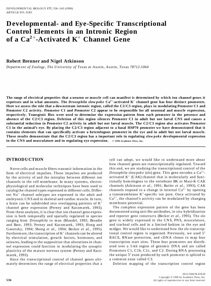
In previous work we mapped the location of four slo pro-fusion between slo exon C3 and the lacZ gene such that transcrip-moters and DNA elements that are required for expressiontion from any of the slo promoters will cause the production of b-in specific tissue types (Brenner et al., 1996). The four slogalactosidase. P3 was built by digesting P1 with BamHI and BglII.promoters are called Promoters C1, C1b, C1c, and C2 andCohesive end ligation joined the BamHI site in exon C1 to the
BglII site in exon C2, resulting in the loss of material between the are named after the unique 5* exon whose synthesis theysites. P3 contains Promoter C1 but not Promoters C1b, C1c, or C2. specify. Deletion analysis had identified two regions, calledP6 was built by digesting P1 with XhoI and PstI, converting the CNS box 1 and CNS box 2, required for expression in theends to blunt ends, and ligating the plasmid shut. This results in CNS, one region required for midgut expression, and a re-the loss of all material 5* of the PstI site and the removal of Promot- gion required for expression in tracheal cells and muscula-ers C1, C1b, and C1c. ture. The position of each of these regions and the four
To build P8, BglII was used to digest P1. This DNA was thenpromoters is summarized in Fig. 1A for convenience. Insubjected to partial BamHI digestion and a 4-kb DNA fragmentthis work we test the function of the C2/C3 intron-bearingwas gel purified. The 5* end of this fragment is the BamHI siteregion in the expression of the slo gene. This intron is lo-within exon C1 and its 3* end is the BglII site within exon C2. Thecated between exon C2 and exon C3.fragment was subcloned into the BamHI site of the transformation
vector pCasperAUGbgal (Thummel et al., 1988). P8 includes Pro- To determine the role played by the intron in gene regula-
Copyright q 1996 by Academic Press, Inc. All rights of reproduction in any form reserved.
AID DB 8273 / 6x10$$$$41 06-25-96 10:14:24 dba AP: Dev Bio
538 Brenner and Atkinson
FIG. 1. (A) Transcriptional control region of slo gene. The horizontal line represents a restriction map of genomic DNA in the vicinityof the slo promoters. Position of all mapped promoters is identified. Above the map is shown the splicing pattern of the first four exonsof the gene. Open boxes represent exons and lines denote their splicing pattern. ATG represents putative translation start sites (Brenneret al., 1996). Labeled gray boxes below the genomic map represent regions that when deleted cause loss of expression in a particular tissue(Brenner et al., 1996). The single open box below the genomic map identifies the position of the C2/C3 intron. (B) slo reporter geneconstructs. This map is aligned with the map in A. A thin line represents DNA not part of a transcription unit, open boxes representexons, and thick lines represent introns. Refer to A for position of promoters and exon names. For each construct a lacZ gene is insertedat the 3* end of the insert (rightmost position in the map above). The terminal restriction sites of all DNA fragments are identified bythe abbreviations Xh, B, Bg, P, or A. The large gray box in P10a is a HSP70 minimal promoter element which can respond to enhancerelements in the C2/C3 intronic region. Abbreviations: CNS, central nervous system; A, ApaI; B, BamHI; Bg, BglII; C, ClaI; E, EcoRI; H,HindIII; K, KpnI; M, MunI; N, NheI; P, PstI; S, SmaI; Sp, SpeI; X, XbaI; Xh, XhoI; Y, XmnI; Z, SphI.
tion we compared Promoter C1 and C2 activity in the pres- developmental-specific expression patterns of the con-structs were determined by staining sectioned adults, dis-ence and absence of the intron. As an additional test for
regulatory elements the intron was placed adjacent to a sected larval brains, and body wall muscles for b-galactosi-dase activity. The P1 construct includes the full-length slominimal HSP70 promoter and tested for its ability to cause
this promoter to be expressed in a tissue-specific manner. transcriptional control region. It has been previously de-scribed and shown to be capable of reproducing the entireTo delete the intron we relied on conveniently located re-
striction sites. Consequently, when the intron was removed slo expression pattern in all developmental stages (Beckeret al., 1995). Figure 1B shows maps of the P1 construct andportions of both exons C2 and C3 were also removed. Simi-
larly, when the intron was tested for enhancer activity (as the various deletions used to determine the function of theC2/C3 region.in P10a) or promoter activity (as in P10b), we were actually
testing the intron and flanking portions of the C2 and C3exons. Therefore, our analysis really maps control elements
The C2/C3 Region Does Not Contain a Promoterto the C2/C3 region. However, the most likely residenceElementfor these elements is in the intron itself. The C2/C3 region
is identified by the open box in Fig. 1A. All expression con- It is important to note that the C2/C3 region does notitself contain a promoter. This is most convincingly demon-structs include a lacZ reporter gene. Each construct was
transformed into Drosophila embryos and transformed strated by the P10b construct. In three independent chromo-some insertions, P10b showed no expression in muscle fi-stocks of animals were produced. Then the tissue- and the
Copyright q 1996 by Academic Press, Inc. All rights of reproduction in any form reserved.
AID DB 8273 / 6x10$$$$41 06-25-96 10:14:24 dba AP: Dev Bio
539K/ Channel Transcriptional Control
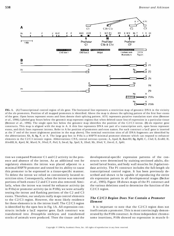
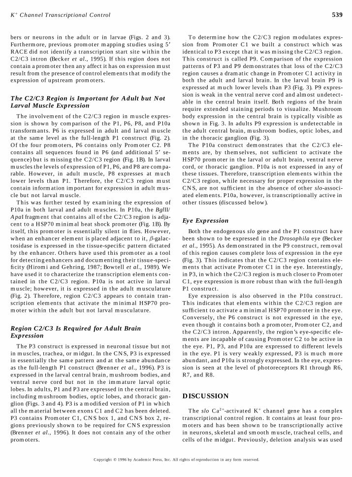
bers or neurons in the adult or in larvae (Figs. 2 and 3). To determine how the C2/C3 region modulates expres-sion from Promoter C1 we built a construct which wasFurthermore, previous promoter mapping studies using 5*
RACE did not identify a transcription start site within the identical to P3 except that it was missing the C2/C3 region.This construct is called P9. Comparison of the expressionC2/C3 intron (Becker et al., 1995). If this region does not
contain a promoter then any affect it has on expression must patterns of P3 and P9 demonstrates that loss of the C2/C3region causes a dramatic change in Promoter C1 activity inresult from the presence of control elements that modify the
expression of upstream promoters. both the adult and larval brain. In the larval brain P9 isexpressed at much lower levels than P3 (Fig. 3). P9 expres-sion is weak in the ventral nerve cord and almost undetect-
The C2/C3 Region is Important for Adult but Not able in the central brain itself. Both regions of the brainLarval Muscle Expression require extended staining periods to visualize. Mushroom
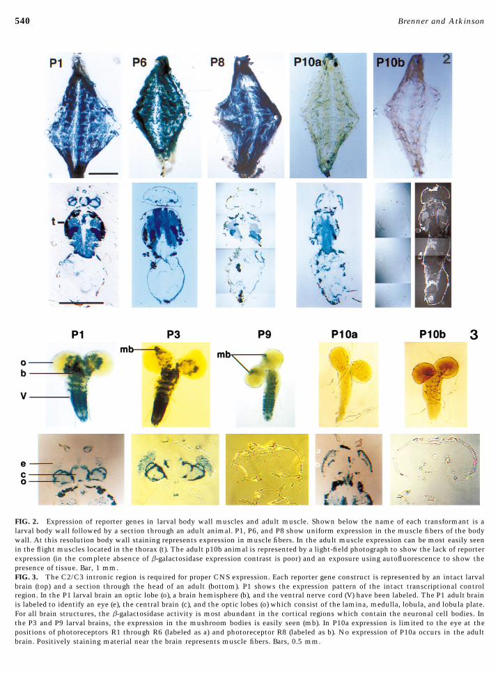
body expression in the central brain is typically visible asThe involvement of the C2/C3 region in muscle expres-sion is shown by comparison of the P1, P6, P8, and P10a shown in Fig. 3. In adults P9 expression is undetectable in
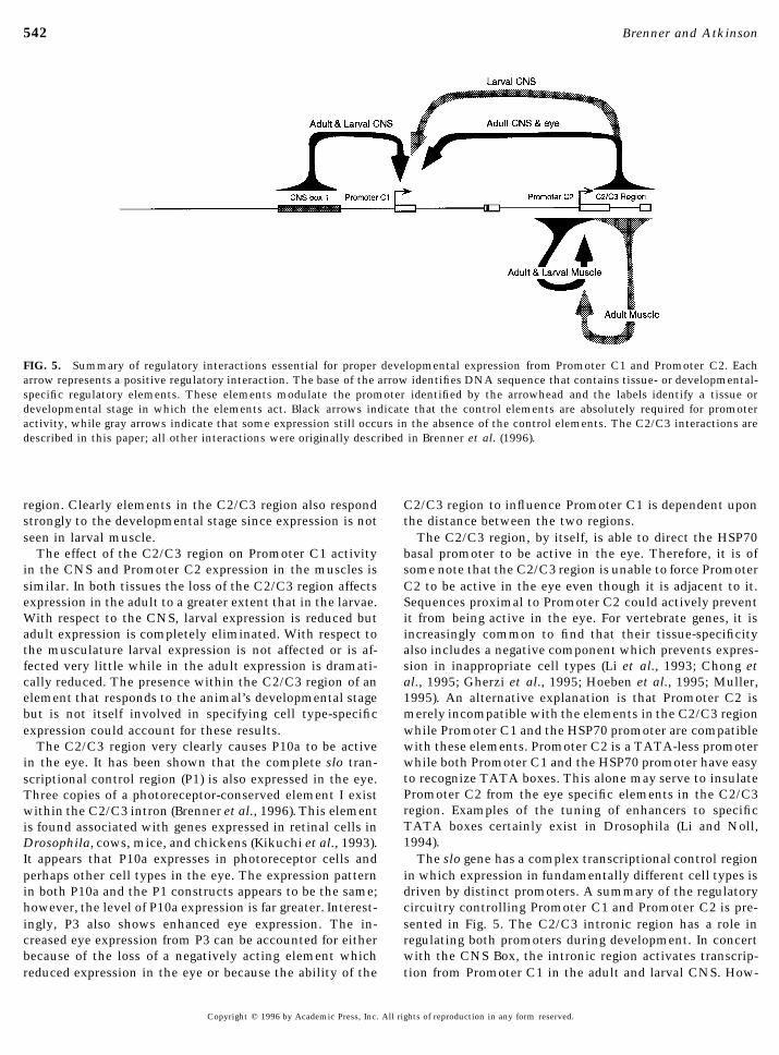
the adult central brain, mushroom bodies, optic lobes, andtransformants. P6 is expressed in adult and larval muscleat the same level as the full-length P1 construct (Fig. 2). in the thoracic ganglion (Fig. 3).
The P10a construct demonstrates that the C2/C3 ele-Of the four promoters, P6 contains only Promoter C2. P8contains all sequences found in P6 (and additional 5* se- ments are, by themselves, not sufficient to activate the
HSP70 promoter in the larval or adult brain, ventral nervequence) but is missing the C2/C3 region (Fig. 1B). In larvalmuscles the levels of expression of P1, P6, and P8 are compa- cord, or thoracic ganglion. P10a is not expressed in any of
these tissues. Therefore, transcription elements within therable. However, in adult muscle, P8 expresses at muchlower levels than P1. Therefore, the C2/C3 region must C2/C3 region, while necessary for proper expression in the
CNS, are not sufficient in the absence of other slo-associ-contain information important for expression in adult mus-cle but not larval muscle. ated elements. P10a, however, is transcriptionally active in
other tissues (discussed below).This was further tested by examining the expression ofP10a in both larval and adult muscles. In P10a, the BglII/ApaI fragment that contains all of the C2/C3 region is adja- Eye Expressioncent to a HSP70 minimal heat shock promoter (Fig. 1B). Byitself, this promoter is essentially silent in flies. However, Both the endogenous slo gene and the P1 construct have
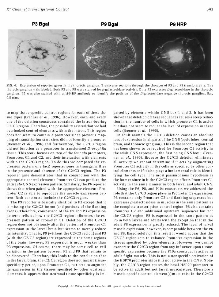
been shown to be expressed in the Drosophila eye (Beckerwhen an enhancer element is placed adjacent to it, b-galac-tosidase is expressed in the tissue-specific pattern dictated et al., 1995). As demonstrated in the P9 construct, removal
of this region causes complete loss of expression in the eyeby the enhancer. Others have used this promoter as a toolfor detecting enhancers and documenting their tissue-speci- (Fig. 3). This indicates that the C2/C3 region contains ele-
ments that activate Promoter C1 in the eye. Interestingly,ficity (Hiromi and Gehring, 1987; Bowtell et al., 1989). Wehave used it to characterize the transcription elements con- in P3, in which the C2/C3 region is much closer to Promoter
C1, eye expression is more robust than with the full-lengthtained in the C2/C3 region. P10a is not active in larvalmuscle; however, it is expressed in the adult musculature P1 construct.
Eye expression is also observed in the P10a construct.(Fig. 2). Therefore, region C2/C3 appears to contain tran-scription elements that activate the minimal HSP70 pro- This indicates that elements within the C2/C3 region are
sufficient to activate a minimal HSP70 promoter in the eye.moter within the adult but not larval musculature.Conversely, the P6 construct is not expressed in the eye,even though it contains both a promoter, Promoter C2, andRegion C2/C3 Is Required for Adult Brain the C2/C3 intron. Apparently, the region’s eye-specific ele-
Expression ments are incapable of causing Promoter C2 to be active inthe eye. P1, P3, and P10a are expressed to different levelsThe P3 construct is expressed in neuronal tissue but not
in muscles, trachea, or midgut. In the CNS, P3 is expressed in the eye. P1 is very weakly expressed, P3 is much moreabundant, and P10a is strongly expressed. In the eye, expres-in essentially the same pattern and at the same abundance
as the full-length P1 construct (Brenner et al., 1996). P3 is sion is seen at the level of photoreceptors R1 through R6,R7, and R8.expressed in the larval central brain, mushroom bodies, and
ventral nerve cord but not in the immature larval opticlobes. In adults, P1 and P3 are expressed in the central brain,including mushroom bodies, optic lobes, and thoracic gan- DISCUSSIONglion (Figs. 3 and 4). P3 is a modified version of P1 in whichall the material between exons C1 and C2 has been deleted. The slo Ca2/-activated K/ channel gene has a complex
transcriptional control region. It contains at least four pro-P3 contains Promoter C1, CNS box 1, and CNS box 2, re-gions previously shown to be required for CNS expression moters and has been shown to be transcriptionally active
in neurons, skeletal and smooth muscle, tracheal cells, and(Brenner et al., 1996). It does not contain any of the otherpromoters. cells of the midgut. Previously, deletion analysis was used
Copyright q 1996 by Academic Press, Inc. All rights of reproduction in any form reserved.
AID DB 8273 / 6x10$$$$41 06-25-96 10:14:24 dba AP: Dev Bio
540 Brenner and Atkinson
FIG. 2. Expression of reporter genes in larval body wall muscles and adult muscle. Shown below the name of each transformant is alarval body wall followed by a section through an adult animal. P1, P6, and P8 show uniform expression in the muscle fibers of the bodywall. At this resolution body wall staining represents expression in muscle fibers. In the adult muscle expression can be most easily seenin the flight muscles located in the thorax (t). The adult p10b animal is represented by a light-field photograph to show the lack of reporterexpression (in the complete absence of b-galactosidase expression contrast is poor) and an exposure using autofluorescence to show thepresence of tissue. Bar, 1 mm.FIG. 3. The C2/C3 intronic region is required for proper CNS expression. Each reporter gene construct is represented by an intact larvalbrain (top) and a section through the head of an adult (bottom). P1 shows the expression pattern of the intact transcriptional controlregion. In the P1 larval brain an optic lobe (o), a brain hemisphere (b), and the ventral nerve cord (V) have been labeled. The P1 adult brainis labeled to identify an eye (e), the central brain (c), and the optic lobes (o) which consist of the lamina, medulla, lobula, and lobula plate.For all brain structures, the b-galactosidase activity is most abundant in the cortical regions which contain the neuronal cell bodies. Inthe P3 and P9 larval brains, the expression in the mushroom bodies is easily seen (mb). In P10a expression is limited to the eye at thepositions of photoreceptors R1 through R6 (labeled as a) and photoreceptor R8 (labeled as b). No expression of P10a occurs in the adultbrain. Positively staining material near the brain represents muscle fibers. Bars, 0.5 mm.
06-25-96 10:14:24 dba AP: Dev Bio
541K/ Channel Transcriptional Control
FIG. 4. Expression of reporter genes in the thoracic ganglion. Transverse sections through the thoraces of P3 and P9 transformants. Thethoracic ganglion (t) is labeled. Both P3 and P9 were stained for b-galactosidase activity. Only P3 expresses b-galactosidase in the thoracicganglion. P9 was also stained with anti-HRP antibody to identify the position of the b-galactosidase negative thoracic ganglion. Bar,0.5 mm.
to map tissue-specific control regions for each of these tis- parted by elements within CNS box 1 and 2. It has beenshown that deletion of these sequences causes a steep reduc-sue types (Brenner et al., 1996). However, each and every
one of the deletion constructs contained the intron-bearing tion in the number of cells in which promoter C1 is activebut does not seem to reduce the level of expression in theseC2/C3 region. Therefore, the possibility existed that we had
overlooked control elements within the intron. This region cells (Brenner et al., 1996).In adult animals the C2/C3 deletion causes an absolutedoes not seem to contain a promoter since previous map-
ping of transcription start sites did not identify a promoter loss of expression in all parts of the CNS (optic lobes, centralbrain, and thoracic ganglion). This is the second region that(Brenner et al., 1996) and furthermore, the C2/C3 region
did not function as a promoter in transformed Drosophila has been shown to be required for Promoter C1 activity inthe adult CNS expression, the first being CNS box 1 (Bren-tissue. This work focuses on two of the four slo promoters,
Promoters C1 and C2, and their interaction with elements ner et al., 1996). Because the C2/C3 deletion eliminatesall activity we cannot determine if it acts by augmentingwithin the C2/C3 region. To do this we compared the ex-
pression pattern of a lacZ reporter gene in transgenic flies Promoter C1 activity in the cells as specified by other con-trol elements or if it also plays a fundamental role in identi-in the presence and absence of the C2/C3 region. The P3
reporter gene demonstrates that in conjunction with the fying the cell type. The most parsimonious hypothesis isthe former since in it the C2/C3 region affects Promoter C1appropriate control elements Promoter C1 can produce the
entire slo CNS expression pattern. Similarly, the P6 reporter activity in the same manner in both larval and adult CNS.Using the P6, P8, and P10a constructs we addressed theshows that when paired with the appropriate elements Pro-
moter C2 is able to reproduce the muscle expression pat- role that the C2/C3 region plays in Promoter C2 expression.P6 contains only Promoter C2 and flanking sequences buttern. Both constructs include the C2/C3 region.
The P9 reporter is basically identical to P3 except that it expresses b-galactosidase in muscles in the same pattern asthe complete transcription control region. P8 also containsis missing the C2/C3 intron (and portions of the flanking
exons). Therefore, comparison of the P9 and P3 expression Promoter C2 and additional upstream sequences but notthe C2/C3 region. P8 is expressed in the same pattern aspatterns tells us how the C2/C3 region influences the ex-
pression pattern of Promoter C1. Deletion of the C2/C3 P6 in both larvae and adults with the exception that in theadult P8 expression is greatly reduced. The level of larvalregion does not obviously alter the pattern of Promoter C1
expression in the larval brain but seems to merely reduce muscle expression, however, is comparable between the P6and P8. Based solely on this result it would appear that theits intensity. That is, P9 (without the C2/C3 region) and P3
(with the C2/C3 region) are expressed in the same regions C2/C3 region acts to enhance Promoter expression in thetissues specified by other elements. However, we cannotof the brain; however, P9 expression is much weaker than
P3 expression. Of course, there may be some cell to cell exonerate the C2/C3 region from any influence upon tissue-specific expression because the P10a construct is active invariation in the pattern between P3 and P9 that remain to
be discovered. Therefore, this leads to the conclusion that adult flight muscle. This is not a nonspecific activation ofthe HSP70 promoter since it is not active in the CNS. Nota-in the larval brain, the C2/C3 region does not impart tissue-
specificity per se, upon Promoter C1, but acts to augment bly, the C2/C3 region causes the basal HSP70 promoter tobe active in adult but not larval musculature. Therefore aits expression in the tissues specified by other upstream
elements. It appears that neuronal tissue-specificity is im- muscle-specific control element(s) must exist in the C2/C3
Copyright q 1996 by Academic Press, Inc. All rights of reproduction in any form reserved.
AID DB 8273 / 6x10$$$$41 06-25-96 10:14:24 dba AP: Dev Bio
542 Brenner and Atkinson
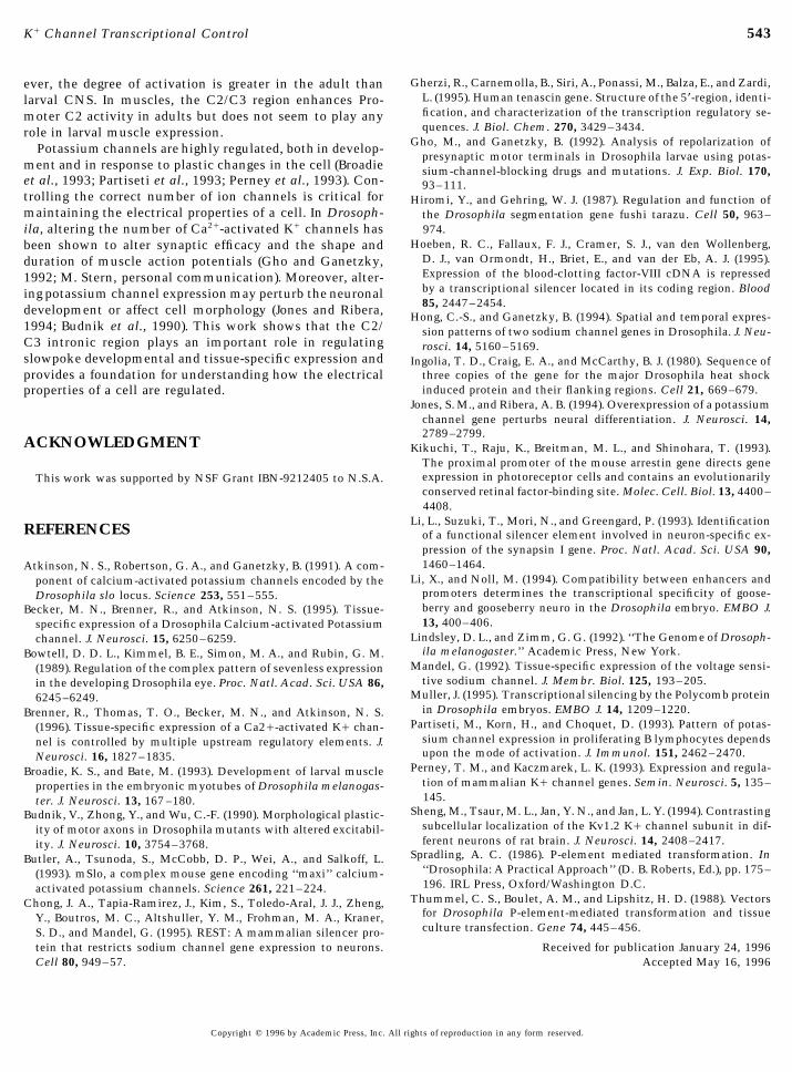
FIG. 5. Summary of regulatory interactions essential for proper developmental expression from Promoter C1 and Promoter C2. Eacharrow represents a positive regulatory interaction. The base of the arrow identifies DNA sequence that contains tissue- or developmental-specific regulatory elements. These elements modulate the promoter identified by the arrowhead and the labels identify a tissue ordevelopmental stage in which the elements act. Black arrows indicate that the control elements are absolutely required for promoteractivity, while gray arrows indicate that some expression still occurs in the absence of the control elements. The C2/C3 interactions aredescribed in this paper; all other interactions were originally described in Brenner et al. (1996).
region. Clearly elements in the C2/C3 region also respond C2/C3 region to influence Promoter C1 is dependent uponthe distance between the two regions.strongly to the developmental stage since expression is not
seen in larval muscle. The C2/C3 region, by itself, is able to direct the HSP70basal promoter to be active in the eye. Therefore, it is ofThe effect of the C2/C3 region on Promoter C1 activity
in the CNS and Promoter C2 expression in the muscles is some note that the C2/C3 region is unable to force PromoterC2 to be active in the eye even though it is adjacent to it.similar. In both tissues the loss of the C2/C3 region affects
expression in the adult to a greater extent that in the larvae. Sequences proximal to Promoter C2 could actively preventit from being active in the eye. For vertebrate genes, it isWith respect to the CNS, larval expression is reduced but
adult expression is completely eliminated. With respect to increasingly common to find that their tissue-specificityalso includes a negative component which prevents expres-the musculature larval expression is not affected or is af-
fected very little while in the adult expression is dramati- sion in inappropriate cell types (Li et al., 1993; Chong etal., 1995; Gherzi et al., 1995; Hoeben et al., 1995; Muller,cally reduced. The presence within the C2/C3 region of an
element that responds to the animal’s developmental stage 1995). An alternative explanation is that Promoter C2 ismerely incompatible with the elements in the C2/C3 regionbut is not itself involved in specifying cell type-specific
expression could account for these results. while Promoter C1 and the HSP70 promoter are compatiblewith these elements. Promoter C2 is a TATA-less promoterThe C2/C3 region very clearly causes P10a to be activewhile both Promoter C1 and the HSP70 promoter have easyin the eye. It has been shown that the complete slo tran-to recognize TATA boxes. This alone may serve to insulatescriptional control region (P1) is also expressed in the eye.Promoter C2 from the eye specific elements in the C2/C3Three copies of a photoreceptor-conserved element I existregion. Examples of the tuning of enhancers to specificwithin the C2/C3 intron (Brenner et al., 1996). This elementTATA boxes certainly exist in Drosophila (Li and Noll,is found associated with genes expressed in retinal cells in1994).Drosophila, cows, mice, and chickens (Kikuchi et al., 1993).
It appears that P10a expresses in photoreceptor cells and The slo gene has a complex transcriptional control regionin which expression in fundamentally different cell types isperhaps other cell types in the eye. The expression pattern
in both P10a and the P1 constructs appears to be the same; driven by distinct promoters. A summary of the regulatorycircuitry controlling Promoter C1 and Promoter C2 is pre-however, the level of P10a expression is far greater. Interest-
ingly, P3 also shows enhanced eye expression. The in- sented in Fig. 5. The C2/C3 intronic region has a role inregulating both promoters during development. In concertcreased eye expression from P3 can be accounted for either
because of the loss of a negatively acting element which with the CNS Box, the intronic region activates transcrip-tion from Promoter C1 in the adult and larval CNS. How-reduced expression in the eye or because the ability of the
Copyright q 1996 by Academic Press, Inc. All rights of reproduction in any form reserved.
AID DB 8273 / 6x10$$$$41 06-25-96 10:14:24 dba AP: Dev Bio
543K/ Channel Transcriptional Control
Gherzi, R., Carnemolla, B., Siri, A., Ponassi, M., Balza, E., and Zardi,ever, the degree of activation is greater in the adult thanL. (1995). Human tenascin gene. Structure of the 5*-region, identi-larval CNS. In muscles, the C2/C3 region enhances Pro-fication, and characterization of the transcription regulatory se-moter C2 activity in adults but does not seem to play anyquences. J. Biol. Chem. 270, 3429–3434.role in larval muscle expression.
Gho, M., and Ganetzky, B. (1992). Analysis of repolarization ofPotassium channels are highly regulated, both in develop-presynaptic motor terminals in Drosophila larvae using potas-
ment and in response to plastic changes in the cell (Broadie sium-channel-blocking drugs and mutations. J. Exp. Biol. 170,et al., 1993; Partiseti et al., 1993; Perney et al., 1993). Con- 93–111.trolling the correct number of ion channels is critical for Hiromi, Y., and Gehring, W. J. (1987). Regulation and function ofmaintaining the electrical properties of a cell. In Drosoph- the Drosophila segmentation gene fushi tarazu. Cell 50, 963–
974.ila, altering the number of Ca2/-activated K/ channels hasHoeben, R. C., Fallaux, F. J., Cramer, S. J., van den Wollenberg,been shown to alter synaptic efficacy and the shape and
D. J., van Ormondt, H., Briet, E., and van der Eb, A. J. (1995).duration of muscle action potentials (Gho and Ganetzky,Expression of the blood-clotting factor-VIII cDNA is repressed1992; M. Stern, personal communication). Moreover, alter-by a transcriptional silencer located in its coding region. Blooding potassium channel expression may perturb the neuronal85, 2447–2454.development or affect cell morphology (Jones and Ribera, Hong, C.-S., and Ganetzky, B. (1994). Spatial and temporal expres-
1994; Budnik et al., 1990). This work shows that the C2/ sion patterns of two sodium channel genes in Drosophila. J. Neu-C3 intronic region plays an important role in regulating rosci. 14, 5160–5169.slowpoke developmental and tissue-specific expression and Ingolia, T. D., Craig, E. A., and McCarthy, B. J. (1980). Sequence ofprovides a foundation for understanding how the electrical three copies of the gene for the major Drosophila heat shock
induced protein and their flanking regions. Cell 21, 669–679.properties of a cell are regulated.Jones, S. M., and Ribera, A. B. (1994). Overexpression of a potassium
channel gene perturbs neural differentiation. J. Neurosci. 14,2789–2799.
ACKNOWLEDGMENT Kikuchi, T., Raju, K., Breitman, M. L., and Shinohara, T. (1993).The proximal promoter of the mouse arrestin gene directs geneexpression in photoreceptor cells and contains an evolutionarilyThis work was supported by NSF Grant IBN-9212405 to N.S.A.conserved retinal factor-binding site. Molec. Cell. Biol. 13, 4400–4408.
Li, L., Suzuki, T., Mori, N., and Greengard, P. (1993). IdentificationREFERENCES of a functional silencer element involved in neuron-specific ex-pression of the synapsin I gene. Proc. Natl. Acad. Sci. USA 90,1460–1464.Atkinson, N. S., Robertson, G. A., and Ganetzky, B. (1991). A com-
Li, X., and Noll, M. (1994). Compatibility between enhancers andponent of calcium-activated potassium channels encoded by thepromoters determines the transcriptional specificity of goose-Drosophila slo locus. Science 253, 551–555.berry and gooseberry neuro in the Drosophila embryo. EMBO J.Becker, M. N., Brenner, R., and Atkinson, N. S. (1995). Tissue-13, 400–406.specific expression of a Drosophila Calcium-activated Potassium
Lindsley, D. L., and Zimm, G. G. (1992). ‘‘The Genome of Drosoph-channel. J. Neurosci. 15, 6250–6259.ila melanogaster.’’ Academic Press, New York.Bowtell, D. D. L., Kimmel, B. E., Simon, M. A., and Rubin, G. M.
Mandel, G. (1992). Tissue-specific expression of the voltage sensi-(1989). Regulation of the complex pattern of sevenless expressiontive sodium channel. J. Membr. Biol. 125, 193–205.in the developing Drosophila eye. Proc. Natl. Acad. Sci. USA 86,
Muller, J. (1995). Transcriptional silencing by the Polycomb protein6245–6249.in Drosophila embryos. EMBO J. 14, 1209–1220.Brenner, R., Thomas, T. O., Becker, M. N., and Atkinson, N. S.
Partiseti, M., Korn, H., and Choquet, D. (1993). Pattern of potas-(1996). Tissue-specific expression of a Ca2/-activated K/ chan-sium channel expression in proliferating B lymphocytes dependsnel is controlled by multiple upstream regulatory elements. J.upon the mode of activation. J. Immunol. 151, 2462–2470.Neurosci. 16, 1827–1835.
Perney, T. M., and Kaczmarek, L. K. (1993). Expression and regula-Broadie, K. S., and Bate, M. (1993). Development of larval muscletion of mammalian K/ channel genes. Semin. Neurosci. 5, 135–properties in the embryonic myotubes of Drosophila melanogas-145.ter. J. Neurosci. 13, 167 –180.
Sheng, M., Tsaur, M. L., Jan, Y. N., and Jan, L. Y. (1994). ContrastingBudnik, V., Zhong, Y., and Wu, C.-F. (1990). Morphological plastic-subcellular localization of the Kv1.2 K/ channel subunit in dif-ity of motor axons in Drosophila mutants with altered excitabil-ferent neurons of rat brain. J. Neurosci. 14, 2408–2417.ity. J. Neurosci. 10, 3754–3768.
Spradling, A. C. (1986). P-element mediated transformation. InButler, A., Tsunoda, S., McCobb, D. P., Wei, A., and Salkoff, L.‘‘Drosophila: A Practical Approach’’ (D. B. Roberts, Ed.), pp. 175–(1993). mSlo, a complex mouse gene encoding ‘‘maxi’’ calcium-196. IRL Press, Oxford/Washington D.C.activated potassium channels. Science 261, 221–224.
Thummel, C. S., Boulet, A. M., and Lipshitz, H. D. (1988). VectorsChong, J. A., Tapia-Ramirez, J., Kim, S., Toledo-Aral, J. J., Zheng,for Drosophila P-element-mediated transformation and tissueY., Boutros, M. C., Altshuller, Y. M., Frohman, M. A., Kraner,culture transfection. Gene 74, 445–456.S. D., and Mandel, G. (1995). REST: A mammalian silencer pro-
tein that restricts sodium channel gene expression to neurons. Received for publication January 24, 1996Accepted May 16, 1996Cell 80, 949–57.
Copyright q 1996 by Academic Press, Inc. All rights of reproduction in any form reserved.
AID DB 8273 / 6x10$$$$41 06-25-96 10:14:24 dba AP: Dev Bio