diabetus mellitus: etiology, pathogenesis, … · · 2013-03-06clinical presentation the classic...
TRANSCRIPT

DIABETUS MELLITUS:
ETIOLOGY, PATHOGENESIS,
CLASSIFICATION, DIAGNOSTIC
CRITERIA
Martynyuk L.P.
Epidemiology • About 2 to 4 % of the world population is affected
with DM
• The disease is more common:
- in persons after age 45
- in obese individuals
- in certain ethnic groups
- in those with a positive family history of DM
• In patients with type 1 DM, complications from end
stage renal disease are major cause of death,
whereas patients with type 2 diabetes are more likely
to have macrovascular diseases leading to
myocardial infarction and stroke as main causes of
the death

Diabetes mellitus (DM) -
is endocrine – metabolic disease, which
develops due to absolute or relative insulin
insufficiency and characterized by chronic
hyperglycemia, changes of different
systems and organs of patient

The term DM
• refers to the excretion of large quantities of
sweet urine. Diabetes is an old word (from
Greece “diabaino”) for siphon and means
“dieresis”, mellitus (from Latina “mell”) means
honey or “sweet” taste of a urine.
Banting
1891 - 1941
Best
1899 - 1978

• The clinical syndrome known as DM
comprises a wide variety of symptoms,
physical findings and laboratory
abnormalities, in which multiple etiologic
factors are involved, the pathophysiology
is partly understood and treatment is
unsatisfactory.
• The hallmark of DM is hyperglycemia.
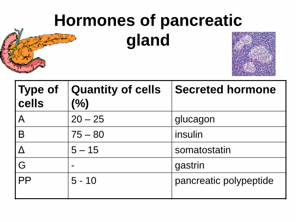
Hormones of pancreatic
gland
Type of
cells
Quantity of cells
(%)
Secreted hormone
Α 20 – 25 glucagon
Β 75 – 80 insulin
Γ 5 – 15 somatostatin
G - gastrin
РР 5 - 10 pancreatic polypeptide
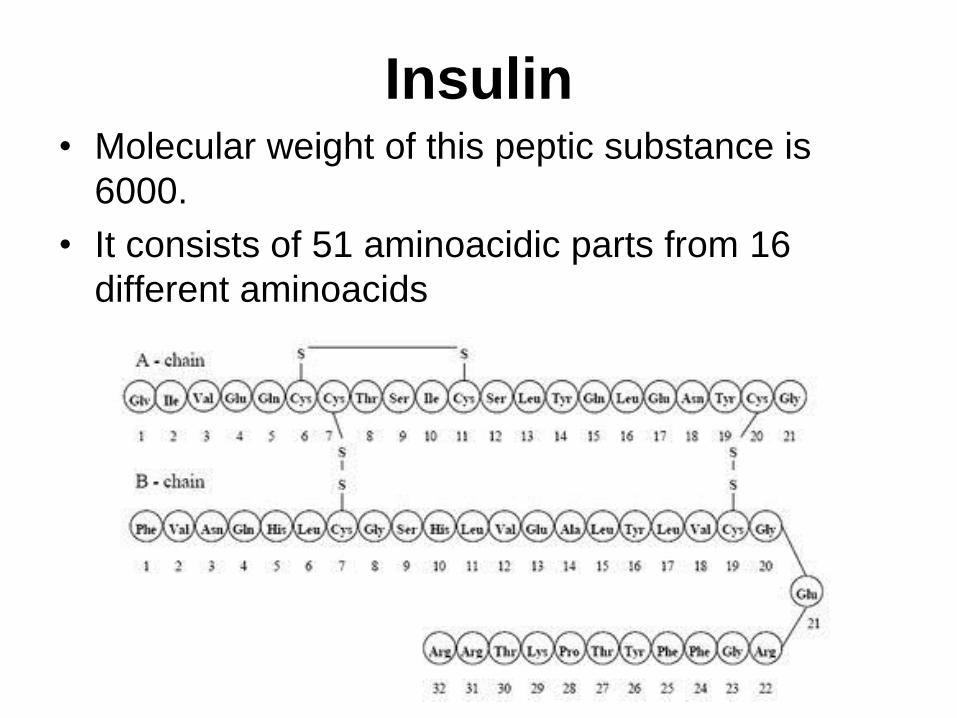
Insulin • Molecular weight of this peptic substance is
6000.
• It consists of 51 aminoacidic parts from 16
different aminoacids

Insulin
• The most important biologic stimulator of
insulin secretion is glucose

The action of insulin
• Insulin is an anabolic hormone (promotes the
synthesis of carbohydrates, proteins, lipids
and nucleic acids). The most important
target organs for insulin action are:
• the liver, muscles and adipocytes.
• The brain (nervous tissue), retina, lens and
blood cells are unresponsive to insulin.
• It has no direct influence on kidney also

The effects of insulin on
carbohydrate metabolism
• stimulation of glucose transport across muscle and
adipose cell membranes;
• regulation of hepatic glycogen synthesis;
• inhibition of glucose formation – from glycogen
(glycogenolysis) and – from amino-acid precursors
(glyconeogenesis).
• The result of these
actions is a reduction in blood
glucose concentration.

Protein metabolism:
• the transfer of amino acids across plasma
membranes;
• stimulation of protein synthesis;
• inhibition of proteolysis.
Lipid metabolism:
• incorporation of fatty acids from circulating
triglyceride into adipose triglyceride;
• stimulation of lipid synthesis;
• inhibition of lipolisis.

Nucleic acids metabolism:
• stimulation of nucleic acid synthesis by stimulating the formation of adenosine triphosphate (ATP), DNA and RNF.
Other effects: • stimulation of the intracellular flew of
potassium, phosphate and magnesium in the heart;
• inhibition of inotropic and chronoropic action (unrelated to hypoglycemia).

Insulin insufficiency
• Absolute
1. Genetic disorders
2. Autoimmune damaging of
β-cells
3. Damaged caused by
virusessuch as mumps,
or Coxsackie B4
4. Toxic influence on β-cells
5. Diseases of pancreatic
gland
• Relative
- β-cells
- Insulin transport
- Receptors (tissue
insensitivity)

Etiologic classification of DM
(1999) I. Type 1 of DM (destruction of β-cells which mostly
leads to absolute insulin insufficiency): • autoimmune;
• idiopathic.
II. Type 2 of DM (resistance to insulin and relative insulin insufficiency or defect of insulin secretion with or without resistance to insulin).
III. Other specific types: • genetic defects of β-cells function;
• genetic defects of insulin action;
• pancreatic diseases (chronic pancreatitis; trauma, pancreatectomy; tumor of pancreatic gland; fibrocalculosis; hemochromatosis);
• endocrine disease (acromegaly, thyrotoxicosis, Cushing’s syndrome);
• drug exposures ;
• infections and others.
IV. Gestation diabetes.

Stages of DM development
• I. Prediabetes (risk factors or predispose
factors).
• II. Impaired glucose tolerance (latent DM).
• III. Clinical manifestation of DM.

Prediabetes (risk factors or predispose
factors) - obesity;
- positive family history of DM;
- persons which were born with weight more than 4,0 kg;
- women who had =children with weight more than 4,0 kg; =abortions and =dead child in anamnesis;
- persons with: = atherosclerosis, hypertension; = auto-immune diseases; = furunculosis; = rubella, mumps, Coxsackie virus, infectious hepatitis, cytomegalovirus, infection mononucleosis;
- endocrine disorders

Glucose tolerance test (GTT)
Fasting serum
glucose, mmol/l
2 hours after
glucose loading,
mmol/l
Capillary blood
Health 3,3 – 5,5 <7,8
Impaired glucose
tolerance
5,6 – 6,1 7,8 – 11,1
Diabetes mellitus > 6,1 > 11,1
Impaired fast
glucose tolerance
5,6 – 6,1 < 7,8

Degrees of severity of DM Mild Moderate Severe
Fast serum
glucose, mmol/l
< 8 8 - 14 > 14
Glucosuria, gr./l < 20
(< 2 %)
20 – 40
(2 – 4 %)
> 40
(4 %)
Compensation
can be achieved
by
diet oral
hypoglycemic
agents or
insulin
oral
hypoglycemic
agents or
insulin
Chronic and acute
complications
only
functional
stages
not last stages
ketosis can
occur
last stages of
chronic
complications
are present,
ketosis is
common

Stages of compensation
• Criteria of compensative stage. 1.Patient hasn’t new complains. 2.Fast serum glucose level is normal. 3.Glucose in urine is absent. 4.Glucose level fluctuation is under 4.4-5.5 mmol/l during the day . 5. HbA1c is 6,0 – 7,0 for the 1 type of DM,
6,0-6,5 for the 2 type of DM
6.Comatose and precomatose status are absent.

Criteria of decompensative stage:
• 1. Postprandial glycemia is >9,0 mmol/l.
• 2. HbA1c is higher then 7,5 for the 1 type of DM,
7,0 for the 2 type of DM
• 3. Comatose or precomatose status are
present.
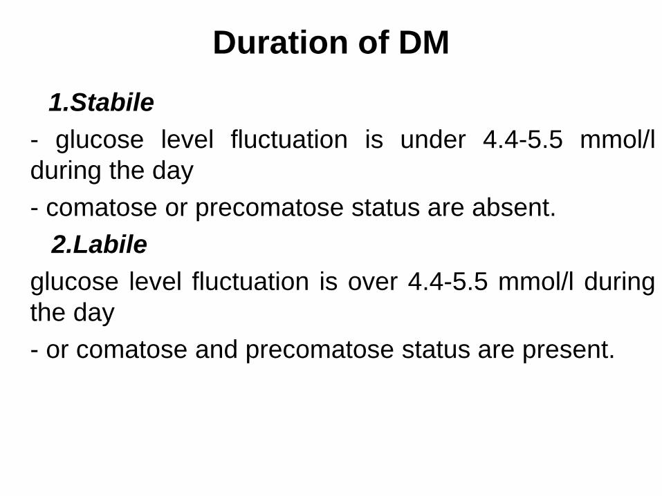
Duration of DM
1.Stabile
- glucose level fluctuation is under 4.4-5.5 mmol/l
during the day
- comatose or precomatose status are absent.
2.Labile
glucose level fluctuation is over 4.4-5.5 mmol/l during
the day
- or comatose and precomatose status are present.

Type I, or insulin-dependent diabetes mellitus (IDDM)
• is characterized by pancreatic islet beta
cell destruction and absolute
insulinopenia.
• This individuals are ketosis prone under
basal conditions. The onset of the disease
is generally in youth, but it can occur at
any age. Patients have dependence on
daily insulin administration for survival.

Pathogenesis of type I DM includes the following:
I. Current formulation of the genetic predisposition, conferred by diabetogenic genes on the short arm of chromosome 6, either as part of it or in close proximity to the major histocompatibility complex (MMHC) region (more than 95 % of type I diabetes individuals are HLA DR3, DR4 or DR3/DR4; on the other hand, HLA DR2 confers protection against the development of type I DM);
II. Putative environmental triggers (possibly viral infections (Coxsackie B, rubella) or chemical toxins (nitrosourea compounds)) that in genetically susceptible individuals might play a role in initiating the disease process.
III. An immune mechanism gone awry, either initiation of immune destruction or loss of tolerance, IV. leading to slow, progressive loss of pancreatic islet β-cells (50%) and
V. eventual clinical onset of type I diabetes. VI. Total destruction of β-cells


Type II, or noninsulin-dependent diabetes mellitus (NIDDM)
• Type 2 is the most common form of diabetes,
accounting for 95 – 90 % of the diabetic population.
Most investigators agree that genetic factors underlie
Type 2 DM, but it is probably not caused by defects at
a single gene locus.
- Obesity,
- diet,
- physical activity,
- intrauterine environment,
- stress
are among the most important environmental factors
which play a role in the development of the disease.

Pathogenetic and clinical difference
of type I and type II DM
N Signs Type 1 Type 2
1 Age Young (under 35) Old, middle
2 Beginning of
disease
Acute Gradual
3 Duration Labile Stable
4 Ketosis,
ketoacidosis
Often develops Rarely develops
5 Body weight Decreased or
normal
Obesity in 80-90 % of
patients

Pathogenetic and clinical difference
of type I and type II DM
N Signs Type 1 Type 2
6 Treatment Insulin, diet Diet, drugs,
insulin
7 Degrees of severity Middle, hard Mild, middle,
hard
8 Season of disease
beginning Frequently autumn-
winter period
Absent
9 Connection with
HBA-system Present Absent
10 Level of insulin and
C-peptide Decreased
or
absent
Frequently
normal level

Pathogenetic and clinical difference
of type I and type II DM
N Signs Type 1 Type 2
11. Antibodies
to β-cells
Present in 80-90 %
of patients on first
week, month
Absent
12. Late
complications
Microangiopathies Macroangiopathies
13. Mortality Less than 10% More than 20%
14. Spreading 10-20% 80-90%
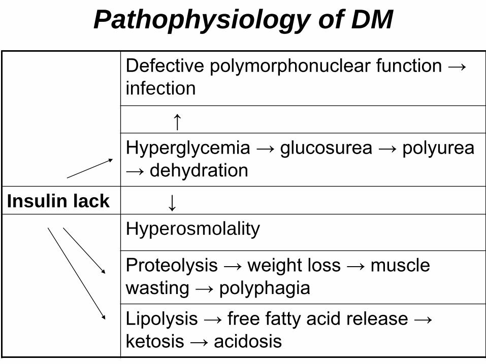
Pathophysiology of DM
Defective polymorphonuclear function →
infection
↑
Hyperglycemia → glucosurea → polyurea
→ dehydration
Insulin lack ↓
Hyperosmolality
Proteolysis → weight loss → muscle
wasting → polyphagia
Lipolysis → free fatty acid release →
ketosis → acidosis

Clinical presentation The classic manifestation of type 1 DM include:
• polyurea ( when the level of the blood glucose is
more then 9 mmol/l, glucosurea arises).
• polidipsia (as more water is excreted,
the body requires more water intake);
• polyphagia (as a result of lack of energy);
• loss of weight (energy (calories) is lost as glucose in the urine.
Loss of water itself also contributes to weight loss. Increased
proteolysis with mobilization of aminoacids leads to progression of
protein catabolism and loss of weight, mostly in muscle mass);
• fatigue and weakness (probably occur as a result of decre-ased
glucose utilization and electrolyte abnormalities);
• acidosis (develops due to increased lipolysis which cause the
release of free fatty acids, which are metabolized to ketones by
the liver)

Presenting signs and symptoms of type 2
DM include:
• polyurea,
• polydipsia,
• polyphagia;
but they are not prominent
The majority of patients (80 – 85 %) are
obese, but it can also occur in thin
persons.

Classification of chronic (long-term)
complications of DM. I. Diabetic angiopathy:
1. Microangiopathy:
1) nephropathy;
2) retinopathy;
3) angiopathy of lower extremitas.
2. Macroangiopathy:
1) heart (ischemic heart disease);
2) brain
3) angiopathy of lower extremities.
II. Diabetic neuropathy:
1) central (encephalopathy);
2) peripheral;
3) visceral (dysfunction of inner organs).

• The long-term degenerative changes in the
blood, vessels, the heart, the kidneys, the
nervous system, and the eyes as
responsible for the most of the morbidity
and mortality of DM.

Skin
• The skin is dry and itch
• Infections of the skin by bacteria and fungi,
candidiasis of the external female genitalia,
hyperkeratosis, nail disorders are common in the
patients with DM but nothing is specific with regard to
their development.

• The most common skin lesion is
diabetic dermopathy (it is
characterized by brown, atrophic,
well-demarcated areas in the
pretibial region which resemble
sears), besides patients sometimes
have xanthoma diabeticorum,
which is usually located on the
buttocks, elbows and knees, look
like eruptions (but is not really
diabeticorum since it occurs in the
patients with lipoprotein
abnormalities, particularly
hyperchylomicronemia, whether or
not patient has DM)


Subcutaneous adipose tissue
• The abdomen type of obesity is common
in patients with type II DM.
• Sometimes generalized subcutaneous
adipose tissue atrophy can be observed in
diabetics.
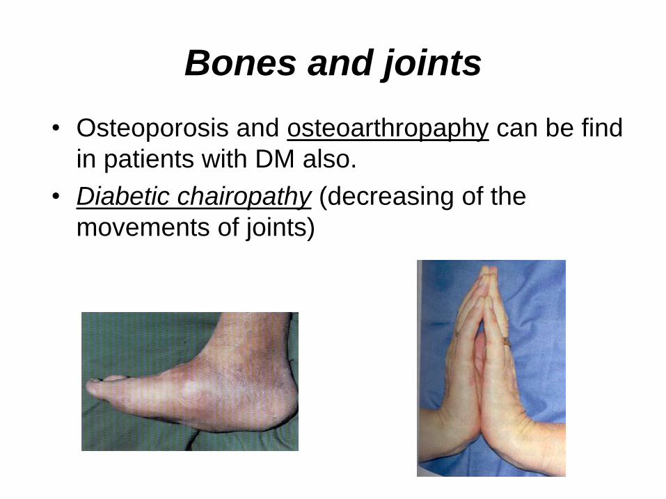
Bones and joints
• Osteoporosis and osteoarthropaphy can be find
in patients with DM also.
• Diabetic chairopathy (decreasing of the
movements of joints)

Gastrointestinal tract
• Paradontosis, gastritis with decreased secretion ability, gastroduodenitis, hepatosis are common in patients with DM.
• Visceral dysfunction gastrointestinal tract:
esophageal neuropathy (disturbances of peristalsis in the body of the esophagus.)
diabetic gastroparesis (irregular food absorption and is characterized by nausea, vomiting, early satiety, bloating and abdomen pain.);
diabetic enteropathy (diarrhea (mostly at night time, postural diarrhea), constipation, malabsorption and fecal incontinence)

Cardiovascular system (CVS).
• Diabetic autonomic cardiopathy:
- orthostatic hypotension (is characterized by dizziness, vertigo, faintness, and syncope upon assumption of the upright posture and is caused by failure of peripheral arteriolar constriction.);
- tachicardia (but it does not occur in response to hypotension because of sympathetic involvement).
• Dismetabolic cardiomyopathy
(IHD, rhythm disturbances)

Ischemic heart disease
1. Cardiovascular changes tend to occur earlier in patients with DM when compared with individuals of the same age.
2. Frequency of myocardial infarction (MI) and mortality is higher in diabetics than that in nondiabetis of the same age.
3. The prognosis is even worse if ketoacidosis,
or other complications of DM are present.
4. Diabetic patients have more complications of MI (arrhythmias, cardiogenic shock and others) than nondiabetic ones.
5. Often can observe atypical forms (without pain).
6. Male : female = 1 : 1 (nondiabetics = 10 : 1).

Respiratory system
• Mucomycosis of the nasopharinx, sinusitis,
bronchitis, pneumonia (prolonged duration, slow
reccurency), tuberculosis are more common
in patients with diabetes than in
nondiabetics.

Kidneys and urinary tract. • Inflammation processes (10 – 30 %): pyelitis,
pyelonephritis, cystitis
• Diabetic cystopathy or neurogenic vesicle dysfunction (enlargement of the volume of the cyst bladder, insidious onset and progression of bladder paralysis with urinary retention, decreasing of quantity of urinations)
• Diabetic nephropathy
Sexual disorders:
• retrograde ejaculation (which is caused by dysfunction of the pelvic autonomic nervous system);
• impotence, and sometimes decreased libido;

Diabetic nephropathy (by Mogensen)
I. Hyperfunction of kidneys
- increased renal blood circulation;
- increased glomerular filtration rate (GFR) (> 140 ml/min);
- hypertrophy of kidneys;
-normoalbuminuria (<30 mg/day).
II. Stage of initial changes of kidney structure.
-mesangial changes due to accumulation of immunoglobulins (IgG, IgM), complement and other nonimmunologic proteins (lipoproteins, fibrin);
- high GFR;
- normoalbuminuria
III. Initial nephropathy.
- microalbuminuria (30 to 300 mg/day);
- high or normal GFR;
- periods of blood hypertension

•
• I IV. Nephropathy or nephrotic
stage.
• - persistent proteinurea (>500 mg/day);
• - normal or decreased GFR;
• - persistent blood hypertension.
•
V. Chronic renal failure or uremia.
- decreased GFR;
- blood hypertension;
- increased serum creatinine
- signs of intoxication.

Eyes • Ceratities, retinatis, chorioretinatis, cataracts,
glaucoma
• Diabetic retinopathy
- Evidence of retinopathy, rarely present at diagnosis in type I DM, is present in up to 20 % of type II DM patients at diagnosis. About 85 % of all diabetics eventually develop some degree of retinopathy
- the initial retinal changes (seen on the ophthalmoloscopic examination) does not significantly alter vision, but it can lead to processes that cause blindness

Diabetic retinopathy (is classified according to the changes seen at
background during ophthalmoscopic examination)
I stage. Background retinopathy
II stage. Maculopathy or preproliferative
retinopathy
III stage. Proliferative retinopathy

Diabetic retinopathy
I stage. Background retinopathy is usually
the earliest sigh and consists of retinal
microaneurysms, hard and soft exudates.

Diabetic retinopathy
II stage. Maculopathy or preproliferative retinopathy is characterized by macular edema and/or hemorrhages.
III stage. The hallmark of
proliferative retinopathy is neovascularization, i.e., growth of new vessels in areas of hypoperfusion. Adhesion of the vessels to the vitreous leads to retinal detachment, vitreous hemorrhage and others. The prognosis is extremely poor. 5 years after recognition of this complication 50 % of the patients are blind.

Diabetic angiopathy of lower
extremities.
Atherosclerosis of large vessels
(macroangiopathy) leads to intermittent
claudication, cold extremities and other
symptoms which can be also find while
arteriols and capillaries are affected
(microangiopathy).

Classification of lower extremities’
angiopathy.
I. Nonclinic stage. (Changes could be find only
during instrumental examination.)
II. Functional stage. (It is characterized by cold
extremities, numbness, tingling, pain during
physical examination.)
III. Organic stage. (It is characterized by
trophyc changes: dry skin, hypo- or atrophy
of muscles, ulcers, gangrene.)

Neuropathic arthropathy (Charcot’s joints)
• is characterized by painless swelling of the feet without edema or signs of infection. The foot becomes shorter and wider, eversion, external rotation, and flattening of the longitudinal arch. This arthropathy is associated with sensory involvelvement, particularly impairment of afferent pain proprioceptive impulses.

Peripheral neuropathy
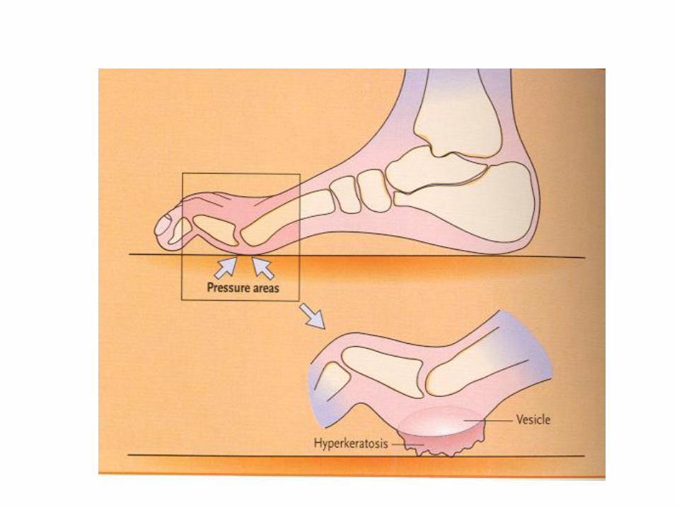

Diabetic foot
Appearance of diabetic foot is caused by a combination of vascular insufficiency, neuropathy, and infection.
Diabetic foot is divided on:
• ischemic;
• neuropathy;
• mixed.

Sign Ischemic Neuropatic
Temperature of the
skin Decreased normal
Color of the skin pallor or cyanotic normal or pink
Pulsation on
peripheral vessels decreased or
absent
normal
Edema Absent can be
Sensibility partly decreased
or normal
decreased or
absent
Ulcers peripheral
(distant)
under the
pressure
Gangrene Dry moist

The diagnosis of DM include:
1. Clinical manifestations of DM.
2. Laboratory findings. • fasting serum glucose (if the value is over 6,1 mmol/l (120 mg/dl)
on two or more separate days, the patient probably has DM);
• the glucose tolerance test (GTT)
• glycohemoglobin >6,5 % (this test is an indicator of blood sugar control during the previous 2-to-3-month period);
• islet cell antibody levels will be positive prior to any insulin administration in 60 – 80 % of patients with type I DM;
• C-peptide (it is not affected by antibodies to exogenous insulin and is used to distinguish type I and II DM if there is still a need after clinical determination);
• glucose level in urine;
• acetonurea;
• blood lipids and others.
3. Instrumental investigations usually are used to diagnose chronic complications of DM.

• Conditions for performing an oral GTT have been standardized:
• no special dietary preparation is required for an oral GTT unless the patient has been ingesting <150 gm/day of carbohydrate. Then give 150 – 200 gm carbohydrate daily for 3 days prior to test;
• unrestricted physical activity should proceed the test;
• test is performed in the morning, following overnight fast of 10 to 16 hours;
• subjects should remain seated, without prior coffee or smoking;
• blood for glucose determination is obtained from an antercubital vein before glucose ingestion and every 30 minutes far 2 hours after ingestion ;
• the amount of glucose given is 75 g for adults (100 g pregnant women, and 1,75 g/kg of ideal body weight for children). Patient have to drink glucose dissolved in 250 ml of water;