diagnosing malaria cases referred to the malaria … · diagnosing malaria cases referred to the...
TRANSCRIPT
Iran J Parasitol: Vol. 10, No. 4, Oct -Dec 2015, pp.547-553
547 Available at: http://ijpa.tums.ac.ir
Original Article
Diagnosing Malaria Cases Referred to the Malaria Reference Laboratory in Tehran University of Medical Science, Iran
Mehdi NATEGHPOUR, Gholamhossein EDRISSIAN, *Afsaneh MOTEVALLI HAGHI, Leila FARIVAR, Elham KAZEMI-RAD
Dept. of Medical Parasitology and Mycology, School of Public Health, Tehran University of Medical Sciences, Tehran, Iran
Received 06 Apr 2015 Accepted 21 Oct 2015
Abstract Background: The number of malaria cases is declining worldwide; however, it remains as a serious health problem. Diagnosing unusual cases is the most im-portant issue to manage the problem. This study designed to describe the number of falciparum and vivax malaria infected patients referred to Malaria Reference La-boratory in Tehran University of Medical Science from 2000 to 2012.
Methods: A retrospective study was conducted based on the collected question-naires from each patient referred to the laboratory. Diagnosing results and demo-graphic information for positive cases were analyzed using SPSS software. Prob-lematic cases were evaluated for any difficulties in diagnosis or in clinical signs. Scanning and molecular methods were performed whenever there was an atypical case referred to the laboratory. Some of the samples had various difficulties for diagnosing such as presence of fussed gametocytes and schizonts of Plasmodium falciparum in peripheral blood and CCHF like hemoragic disorders.
Results: Plasmodium vivax caused a large proportion of the cases (76.1%) in con-
trast with P. falciparum that included smaller proportion (22.8%) and the rest (1.1) belonged to mixed infection. Most of the positive cases (69.6%) were belonged to Afghani people. Men (94.6%) showed more infection than women (5.4%), moreo-ver the most infection (44.5%) was seen at a range of 21-30 yr.
Conclusion: In the case of existing atypical issues to diagnose, it is needed to per-
form more precise microscopical examination beyond the current standard condi-tions. Sometimes molecular method is required to verify the exact agent of the dis-ease.
Keywords: Malaria, Plasmodium, Diagnosis, Iran
*Correspondence [email protected]
Iranian Society of Parasitology
http:// isp.tums.ac.ir
Iran J Parasitol
Open access Journal at
http:// ijpa.tums.ac.ir
Tehran University of Medical
Sciences Publication
http:// tums.ac.ir
Nateghpour et al.: Diagnosing Malaria Cases Referred to the Malaria Reference Laboratory …
Available at: http://ijpa.tums.ac.ir 548
Introduction
n spite of decreasing the number of ma-laria cases in the world it still remains as a serious disease in tropical and sub-
tropical areas. Approximately half of popula-tion in the world is living under the risk of malaria (1, 2). According to the report of WHO released in 2013, about 207 million new cases and 627000 deaths due to malaria oc-curred annually in the world mostly among the children under 5 years old (3). In some parts of the world involve with the malaria elimination program, quick diagnosis and prompt treat-ment with suitable antimalarial drugs are the most important recommendations of WHO.
Malaria microscopy remains as a gold stand-ard method to diagnose the disease, but in some unusual cases, examinations that are more precise are needed. On the other hand, emergence of drug resistance in Plasmodium falciparum to artemisinin combination therapy (ACTs) and P. vivax to chloroquine increases complications of diagnosis and treatment of the infections (4-7). Therefore, more attention would be hinted about diagnosis of the infec-tions where malaria elimination has been es-tablished (8).
Iran is a malarious country with low burden of the infection that has long been involved with malaria control program (9). Due to these control measures, the number of malaria cases reduced significantly in recent years. Preva-lence of malaria in the country was reported 0.21 cases in 10,000 of population in the en-demic area at 2012 (10). The most reported cases in the country were from south & south-
eastern Iran . Although, authorities should al-ways be vigilant about the increasing malaria in Iran. Therefore, control and prevention of the disease is crucial for proper management in Iran, which is in elimination phase. Under these conditions, it is important to identify the exact species causing diseases, especially when there is any atypical matter in the sample. Many of referred cases to the laboratory were
from endemic areas in the country or import-ed cases from neighboring countries where malaria is endemic. The most refugees to Iran were from Afghanistan and Pakistan, the rest of them were from some African countries, India, Ecuador and Malaysia. Therefore, it is clear that in this situation, there are many dif-ferent strains with specific clinical signs or mi-croscopic features appeared in the country. These complexity may result in wrong diag-nose, so parasite maintenance may become more durable in community and then trans-mission conditions will occur easily. Ac-cordingly, early diagnosis and prompt treat-ment of malaria cases are two essential sub-jects (11). Due to the complexity of features to diagnose, these cases referred to the Malaria Reference Laboratory in Tehran University of Medical Science to detect the disease.
This study was aimed to classify, discuss and describe in details considering these compli-cated cases, also analyze the demographic sta-tus including age, gender, nationality and Plas-modia species of those malaria patients who were referred to Malaria Reference Laboratory, School of Public Health Tehran University of Medical Sciences (MRL-TUMS), with empha-sizing on the problematic cases.
Materials and Methods
Sample collection From April 2002 to December 2012, overall, 521 patients were referred to MRL-TUMS by provincial malaria laboratories or academic hospitals. These patients were suspected to have malaria with some various problems that interfered for proper detection of the parasites. After registering a questionnaire form was filled and demographic data of the samples were collected.
Microscopic examination Thick and thin blood films were prepared
using finger-pricked technique. The films were
I
Iran J Parasitol: Vol. 10, No. 4, Oct -Dec 2015, pp.547-553
549 Available at: http://ijpa.tums.ac.ir
stained with Giemsa according to WHO ma-laria microscopy guidebook (12). Some of the samples had various difficulties for diagnosing, in these cases finding agents of the infection required long time. Therefore, microscopic examination was done beyond the standard conditions and sometimes even more than 1 h for examination just one case. Moreover, in such cases molecular examinations, PCR, were used to determine the exact agent of the dis-ease. Finally, based on the existence of any abnormalities, these tricky samples were di-vided into different groups. When some ab-normalities were observed in the feature of parasites, a photography process was em-ployed for the infected blood cells using an optical microscope (Olympus BX41, with Olysia software; Olympus Corporation, Tokyo, Japan) with 100/1.40 oil objective.
Molecular identification Molecular methods were used for those sam-ples with having some problems to diagnose. DNA extraction, primer designing and PCR amplification were conducted in the same manner that has been described in the previ-ous published paper (13).
Results
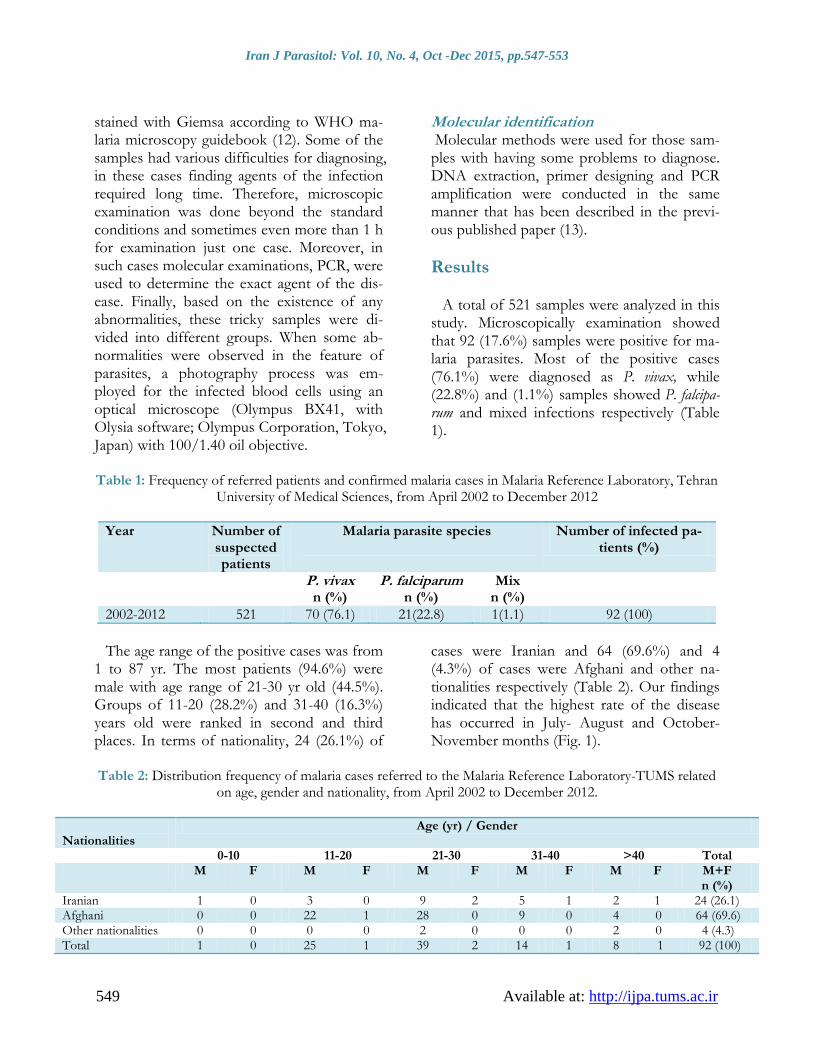
A total of 521 samples were analyzed in this study. Microscopically examination showed that 92 (17.6%) samples were positive for ma-laria parasites. Most of the positive cases (76.1%) were diagnosed as P. vivax, while (22.8%) and (1.1%) samples showed P. falcipa-rum and mixed infections respectively (Table 1).
Table 1: Frequency of referred patients and confirmed malaria cases in Malaria Reference Laboratory, Tehran University of Medical Sciences, from April 2002 to December 2012
Year Number of suspected patients
Malaria parasite species
Number of infected pa-tients (%)
P. vivax n (%)
P. falciparum n (%)
Mix n (%)
2002-2012 521 70 (76.1) 21(22.8) 1(1.1) 92 (100)
The age range of the positive cases was from
1 to 87 yr. The most patients (94.6%) were male with age range of 21-30 yr old (44.5%). Groups of 11-20 (28.2%) and 31-40 (16.3%) years old were ranked in second and third places. In terms of nationality, 24 (26.1%) of
cases were Iranian and 64 (69.6%) and 4 (4.3%) of cases were Afghani and other na-tionalities respectively (Table 2). Our findings indicated that the highest rate of the disease has occurred in July- August and October-November months (Fig. 1).
Table 2: Distribution frequency of malaria cases referred to the Malaria Reference Laboratory-TUMS related
on age, gender and nationality, from April 2002 to December 2012.
Nationalities
Age (yr) / Gender
0-10 11-20 21-30 31-40 >40 Total M F M F M F M F M F M+F
n (%) Iranian 1 0 3 0 9 2 5 1 2 1 24 (26.1) Afghani 0 0 22 1 28 0 9 0 4 0 64 (69.6) Other nationalities 0 0 0 0 2 0 0 0 2 0 4 (4.3) Total 1 0 25 1 39 2 14 1 8 1 92 (100)

Nateghpour et al.: Diagnosing Malaria Cases Referred to the Malaria Reference Laboratory …
Available at: http://ijpa.tums.ac.ir 550
Detection of parasites in low-density sam-
ples with problems required long time to solve the matters. These problematic cases were cat-egorized into two groups; the first was about those samples which had difficulties in para-site diagnosis that means existence abnor-malities in parasite appearance or were clini-cally suspected patients that parasite may not found in the standard microscopy method. The second was about those patients with spe-
cific sign except usual symptom in malaria dis-ease that were led to misdiagnosis by physi-cians. In a few cases, diagnosing process took more time than standard test and results were confirmed with molecular approaches (for two samples) as mentioned above. Cases with ab-normalities in morphology of parasites such as round gametocytes instead of banana shape and schizont in peripheral blood in P. falcipa-rum infection were illustrated in Fig 2, 3.
Fig. 1: Monthly distribution of malaria positive cases during 2002 to December 2012 referred to MRL-TUMS
Fig. 2: A shizont in peripheral blood of a patient infected with Plasmodium falciparum (Original)
Fig. 3: A round shape of Plasmodium falciparum gametocyte (Original)
Iran J Parasitol: Vol. 10, No. 4, Oct -Dec 2015, pp.547-553
551 Available at: http://ijpa.tums.ac.ir
Discussion
Despite of performing malaria control pro-gram in Iran, the disease is still a public health problem in southeastern Iran. Imported cases from eastern neighboring countries Afghani-stan and Pakistan are a major problem against the elimination of malaria in Iran. Although, Tehran as capital of Iran is not a malaria en-demic area however since economic, social and political importance of the city includes thousands of different workers and travelers from the malaria endemic areas.
According to the recorded data in the TUMS Malaria Reference Laboratory, most of the positive cases were Afghani and Pakistani travelers and those Iranian travelers who came back from south Asia and African countries. More cases were observed among men than women due to more entrance of men in this country for finding a job. The results obtained in this study are in agreement with Khalili’s findings who reported that 4257(95%) cases were male and 225 (5%) were female from the total of 4482 confirmed malaria positive cases obtained from Yazd province during 1985 to 2006 (2). Sargolizaie and colleagues have done an extensive study in Sistan & Baluchistan Province during 2008 to 2011, and found that 77.3% of the positive cases were Iranian and 22.7% from foreign nationalities, in contrast to our study that showed 74% and 26.1% were foreign nationalities and Iranian respec-tively (14). This significant difference is be-cause our study was performed in non-en-demic area, but Sargolizaie et al. conducted their study in an endemic area in Iran. Moreo-ver, they reported that 88.6% of the cases were P. vivax and 11.4% P. falciparum (14). Prevalence of Plasmodia species pattern in the country more or less is the same. In the pre-sent study, most of the positive cases that were mostly from malaria endemic area, were P. vivax (76.1%) while 22.8% and 1.1% of the cases were P. falciparum and mixed infections respectively. Khalili et al. found approximately
the same results as Sargolizaie and colleagues (2, 14), such results also have been reported by the other authors from different malarious areas (15-18), as well as by the Center for Dis-ease Control in Iran (10).
Based on the demographic data of this study the number of referred cases were higher in July to August and October to November than other months, comparable to those result reported from Khorasan Province (18). The greatest number of positive cases lay on the age group of 21-30 years. It is clear that most of the infections have occurred in working age people who came from the neighboring coun-tries to Iran. Similar results obtained by other researchers from Iran (15-18).
Some of the referred cases to the laboratory possessed numbers of diagnosing difficulties, such as low parasitemia, mixed infections and abnormalities in morphology of parasites or existence of parasites in non- standard sam-ples (for example bone marrow), also some of them had clinically symptoms with specific signs except common malarious symptoms, like hemorrhagic fever. As it was mentioned above, these abnormalities were classified into two groups, first: complexity in parasite ap-pearance in blood smears and second compli-cations in symptoms of the disease.
In the case of complexity in blood smears, there were several important points that should be considered for example; mixed in-fections either over growth with P. vivax or P. falciparum that diagnosing process takes more time than usual standard microscopy examina-tion. Another issue was probability of seques-tration phenomenon in complicated falciparum malaria that may induce to hidden parasites. In this case, coma likely happens whereas para-sites might not see in the peripheral blood of patients. However, it is advisable that such patient would be hospitalized due to symp-toms while treatment of malaria is being con-sidered for him/her as a supposition treat-ment.
Occurrence of unusual symptoms or atypical sign in a patient may lead to a miss under-
Nateghpour et al.: Diagnosing Malaria Cases Referred to the Malaria Reference Laboratory …
Available at: http://ijpa.tums.ac.ir 552
standing and diagnosing the disease by physi-cian. One of the highly controversial cases referred to the laboratory was from an area where kala-azar disease had been common there, a slide was taken of bone marrow of a patient who suspected to kala-azar, but some P. falciparum rings were seen in RBCs when a microscopist was looking for Leishman body in the BM slide. Recovery happened when the patient was treated with administering anti- falciparum malaria drugs. The other case was about a patient who had severe bleeding. He was hospitalized because of suspected to Cri-mean–Congo Hemorrhagic Fever (CCHF), but blood smears indicated infection of P. vi-vax .He recovered after taking antimalarial drugs. The shape of parasites was normal in both of the above-mentioned cases, but atypi-cal symptoms caused misleading by physicians.
Finding parasite in patients with low para-sitemia (less than 500 parasites/ml) took more time than usual standard time, similar to mixed infections. In patients infected with P. falciparum observing a few number of schi-zonts in stained blood films may lead micros-copist to misdiagnose. Obviously, we can con-sider the presence of P. falciparum shizonts in peripheral blood in some specific strains or when the patient enters in to hyper parasitem-ia phase (Fig. 2) (19).
Another unusual shapes that may be seen in the peripheral blood is P. falciparum round ga-metocyte (Fig. 3). Although it is expected to see banana shape gametocyte for P. falciparum, in some cases round shape gametocytes may confuse a professional microscopist.
Conclusion
The referred cases to MRL-TUMS mim-
icked those cases detected in the field in inci-dence, but in the case of abnormal forms that may be seen in the peripheral blood, micros-copists should be trained further and have enough experience.
Acknowledgements
The authors declare that there is no conflict of interests.
References
1. Fairhurst RM, Wellems TE. Malaria In: Text-
book of infectious Disease Principles and Prac-tice of Infectious Diseases. Mandell: Mandell, Douglas and Bennett, Churchill Livigstone, An Imprint of Elsevier 7thed, 2009; 3437-3463.
2. Khalili MB, Anvari Tafti MH. Sadeh M. Epi-demiological Pattern of Malarial Disease in the Province of Yazd, Iran (Since 1986-2006). Word J Med Sci. 2009; 4 (1): 41-45.
3. World Malaria Report 2013. Available from: http://www.who.int/malaria/publications/world _malaria_report_2013/en/
4. Bhattacharyya N, Mukherjee H, Bose D, Roy S, Das S, Tripathy S, Hati AK. Clinical Case of Artesunate Resistant Plasmodium falciparum Ma-laria in Kolkata: A First Report. J Trop Dis. 2014; 2:1.
5. Mita T, Tanabe K. Evolution of Plasmodium falciparum drug resistance: implications for the development and containment of artemisinin resistance. Jpn J Infect Dis. 2012;65:465-75.
6. Anvikar A R, Sharma B, Sharma SK, Ghosh SK, Bhatt RM, Kumar A, Mohanty SS, Pillai CR, Dash AP, Valecha N. In vitro assessment of drug resistance in Plasmodium falciparum in five States of India. Indian J Med Res. 2012; 135: 494-499.
7. Rieckmann H, Davis DR, Hutton DC. Plasmo-dium vivax resistance to Chloroquine? Lancet. 1989; 2 (8673):1183-4.
8. Roll Back Malaria Final repot of the external evaluation of Roll Back Malaria. Achieving Im-pact: Roll Back Malaria in the Next Phase, No-vember; 2002.
9. Rezaei Hemami M. Akbari Sari A, Raeisi A, Vatandoost H, Majdzadeh R. Malaria Elimina-tion in Iran, Importance and Challenges. Int J Prev Med .2013; 4(1): 88–94.
10. Central of Disease Control. Ministry of Health and Medical Education. Annual report of ma-laria control department. Tehran. 2012; p. 50.
11. Bloland PB. Drug resistance in malaria. World Health Organization. Department of Communi-
Iran J Parasitol: Vol. 10, No. 4, Oct -Dec 2015, pp.547-553
553 Available at: http://ijpa.tums.ac.ir
cable Disease Surveillance and Response. 2001. Available from: WHO/CDS/CSR/DRS/2001.4
12. World Health Organization. Basic Malaria Mi-croscopy Part1. Learner’s guide. 2nd edition. 2010.
13. Nateghpour M, Abed Khojasteh H, Keshavarz H, Hajjaran H, Edrissian Gh, Rahimi A, Go-bakhloo N. Comparison of microscopical ex-amination and semi-nested multiplex polymer-ase chain reaction in diagnosis of Plasmodium fal-ciparum and P. vivax. EMHJ. 2011; 17 (1): 51-55.
14. Sargolzaie N, Salehi M, Kiani M, Sakeni M, Hassanzehi A . Malaria Epidemiology in Sistan and Balouchestan Province during April 2008-March 2011; Iran. Zahedan Journal of Re-search in Medical Sciences. 2013; 16 (4): 41-43.
15. Youssefi MR, Rahimi MT. Prevalence of ma-laria infection in Sarbaz, Sistan and Baluchistan
province. Asian Pac J Trop Biomed. 2011;1 (6):491-492.
16. Ghaffari S, Mahdavi SA, Moulana Z, Mouodi S, Karimi-Nia H, Bayani M, Kalantari N. Malaria in Mazandaran, Northern Iran: Passive case Finding During 1997-2012. Iran J Parasitol. 2012;7(3):82-88.
17. Saghafipour A, Noorozi M, Karami-jooshin M, Poudat A. Epidemiological Features of Malaria in Qom province from 2001 to 2011. Zahedan J Res Med Sci. 2012;14 (8):70-73.
18. Karimi-Zarchi A, Mahmudzadeh A, Vatani H, Shirbazu SH. Epidemiological Evaluation of malaria and vectors in Sarakhs boundary vil-lages. Persian. Iran J Infect Trop Dis. 2004;8 (20):47-50.
19. Schmidt GD, Roberts′ LS. Foundations of para-sitology. Florida International University. In-ternational 8th ed. USA. 2006.