diagnosis and management of idiopathic pulmonary fibrosis...
TRANSCRIPT
Diagnosis and management of idiopathicpulmonary fibrosis: Frenchpractical guidelines
Vincent Cottin, Bruno Crestani, Dominique Valeyre, Benoit Wallaert,Jacques Cadranel, Jean-Charles Dalphin, Philippe Delaval,Dominique Israel-Biet, Romain Kessler, Martine Reynaud-Gaubert,Bernard Aguilaniu, Benoit Bouquillon, Philippe Carre, Claire Danel,Jean-Baptiste Faivre, Gilbert Ferretti, Nicolas Just, Serge Kouzan,Francois Lebargy, Sylvain Marchand-Adam, Bruno Philippe, Gregoire Prevot,Bruno Stach, Francoise Thivolet-Bejui, Jean-Francois Cordier and the FrenchNational Reference Centre and the Network of Competence Centres for RareLung Diseases
Affiliations: For a full list of the authors affiliations see the Acknowledgements section.
Correspondence: Vincent Cottin, Hopital Louis Pradel, Claude Bernard Lyon 1 University, 28 Avenue du DoyenLepine, 69677 Lyon, France. E-mail: [email protected]
ABSTRACT Idiopathic pulmonary fibrosis (IPF) is the most frequent chronic idiopathic interstitial
pneumonia in adults. The management of rare diseases in France has been organised by a national plan for
rare diseases, which endorsed a network of expert centres for rare diseases throughout France. This article is
an overview of the executive summary of the French guidelines for the management of IPF, an initiative that
emanated from the French National Reference Centre and the Network of Regional Competence Centres for
Rare Lung Diseases. This review aims at providing pulmonologists with a document that: 1) combines the
current available evidence; 2) reviews practical modalities of diagnosis and management of IPF; and 3) is
adapted to everyday medical practice. The French practical guidelines result from the combined efforts
of a coordination committee, a writing committee and a multidisciplinary review panel, following
recommendations from the Haute Autorite de Sante. All recommendations included in this article received
at least 90% agreement by the reviewing panel. Herein, we summarise the main conclusions and practical
recommendations of the French guidelines.
@ERSpublications
Practical guidelines for idiopathic pulmonary fibrosis are now available http://ow.ly/uUhKh
IntroductionIdiopathic pulmonary fibrosis (IPF) is a fibroproliferative, irreversible disease of unknown cause. The
evolution of IPF is usually progressive, primarily occurring from 60 years of age and is limited to the lungs.
It is the most frequent type of chronic idiopathic interstitial pneumonia in adults.
Once considered an orphan disease because no specific treatment with proven efficacy was available, IPF is a
rare disease with an estimated prevalence in the USA of between 14 and 28 cases per 100 000 population [1].
Received: March 04 2014 | Accepted after revision: March 17 2014
Support statement: We received financial support from Fondation partenariale Lyon 1 (Villeurbanne, France).
Conflict of interest: Disclosures can be found alongside the online version of this article at err.ersjournals.com
Provenance: Submitted article, peer reviewed.
Copyright �ERS 2014. ERR articles are open access and distributed under the terms of the Creative CommonsAttribution Non-Commercial Licence 4.0.
REVIEWIDIOPATHIC PULMONARY FIBROSIS
Eur Respir Rev 2014; 23: 193–214 | DOI: 10.1183/09059180.00001814 193

The annual estimated incidence in the USA is between 6.8 and 8.8 cases per 100 000 population [1]. No
estimate of the epidemiology of IPF has been published for France.
In France, one national reference centre and nine regional competence centres for rare lung diseases have
been endorsed to organise the diagnosis and management of IPF within the general framework of two
national plans for rare diseases (2008–2011 and 2013–2016).
Since the publication of international guidelines for the diagnosis and management of IPF in 2011 [2], new
data have been published regarding, in particular, the efficacy and tolerance of several new treatments
proposed to modify the evolution of the disease or alleviate symptoms. The current aim, coordinated by the
reference and competence centres, is to provide pulmonologists with a synopsis of the currently available
data and to define as clearly as possible, using terms adapted to real-life daily practice, the modalities of
diagnosis and patient-centred management of IPF [3].
The present document is an English version of the executive summary and outlines the main conclusions of
the French guidelines for the diagnosis and management of IPF [4].
Missions of the committeesThis article was written by French IPF specialists as a practical overview of the international
recommendations for the diagnosis and management of IPF published in 2011 [2], in combination with
a critical review of the literature published in this field since 2011, including therapeutic trials. This article
was produced by a Coordination committee, a Writing committee and a Reviewing committee.
The committees adopted rules that were applicable to the development of good clinical practice based on
the method issued by the Haute Autorite de Sante [5].
Coordination committeeThe Coordination committee submitted the process and validation protocol to the Societe de Pneumologie
de Langue Francaise (SPLF), conducted a systematic review of the literature, prepared a first draft of the
document intended for the Writing committee, organised the process and validation protocol and
monitored its application, and submitted the recommendations validated by the Writing committee and the
Reviewing committee to the SPLF.
Writing committeeThe Writing committee assessed the first version of the document prepared by the Coordination committee.
Using a three-point scale (I agree, I hesitate or I do not agree) they identified the points to be reviewed,
made suggestions about the form and contents of the document, and validated the document to be
submitted to the Reviewing committee.
Reviewing committeeThe Reviewing committee comprised three pulmonologists working in a university hospital (excluding the
competence centres), three pulmonologists working in a general hospital, three pulmonologists working in
private practice, two radiologists with expertise in interstitial lung disease (ILD), and two pathologists who
were specialised in thoracic pathology. The committee assessed all the themes and the corresponding
recommendations using a scale ranging from 1 (total disagreement) to 9 (total agreement). The vote was
conducted electronically and the results were anonymised before analysis. All recommendations submitted
to the scrutiny of the Reviewing committee had been approved first by at least 80% of the Writing
committee members [5]. The rating of each statement was based on the synthesis of data published in the
literature (which was provided together with the questionnaire) and the experience of the reader in the
corresponding field. The members of the Reviewing committee could only answer questions for which they
felt competent.
Guidelines development processThe development of the recommendations comprised the following steps. 1) Critical review of the literature
published since 2010 in the field of IPF by the Coordination committee. 2) First version written by the
Coordination committee. 3) Review of the first version by the Writing committee. 4) Production of
a revised version by the Coordination committee. 5) Review and votes by the Writing committee.
6) Rewriting of issues generating an insufficient consensus level by the Coordination committee. 7) Review
and votes by the Reviewing committee. 8) Production of a revised version by the Coordination committee.
9) Review by the SPLF Scientific Council. 10) Production of a revised version by the Coordination
committee. 11) Submission of the manuscript.
IDIOPATHIC PULMONARY FIBROSIS | V. COTTIN ET AL.
DOI: 10.1183/09059180.00001814194
The SPLF Scientific Council gave advice on the relevance, writing and applicability of the recommendations.
In order to be validated, recommendations had to be approved by at least 90% of the Reviewing committee
members [5].
The recommendations were formulated as follows. 1) ‘‘It is recommended’’ means that the option described
is relevant in the majority of patients (e.g. treatment with established efficacy). 2) ‘‘It is proposed’’ means
that the option described may be relevant in some patients (e.g. treatment with highly probable efficacy).
3) ‘‘It is possible’’ means that the option described may be relevant in some patients but that available
data do not allow a stronger recommendation (e.g. treatment with uncertain efficacy). 4) ‘‘It is not
recommended’’ means that the option described is not relevant in the majority of patients (e.g. treatment
shown to be inefficient). 5) ‘‘It is recommended not to’’ means that the option described should be avoided
(e.g. treatment with harmful effects).
The SPLF Scientific Council approved the methods used to develop these recommendations on January 10,
2013, and approved the written recommendations on June 12, 2013. The recommendations were published
in December 2013 [4]. Herein, we summarise the main conclusions and practical recommendations of the
French guidelines.
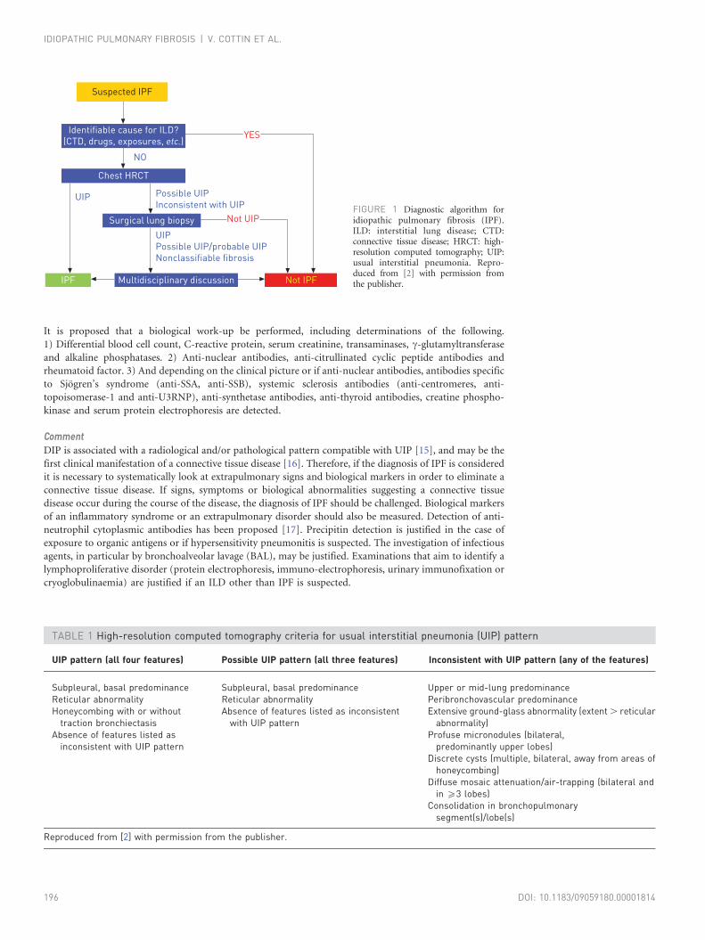
Diagnosis of IPFIPF is a fibroproliferative disease of unknown cause, associated with the histopathological and/or high-
resolution computed tomography (HRCT) pattern of usual interstitial pneumonia (UIP) [2]. The presence
of a radiological and/or histological UIP pattern is required to establish the diagnosis of IPF. In patients not
subjected to video-assisted surgical lung biopsy (SLB), the diagnosis may be made when an ILD is present
(with neither extrapulmonary manifestations nor aetiological context) if HRCT shows a (definite) UIP
pattern (fig. 1 and table 1). In patients subjected to video-assisted SLB, the diagnosis is established in the
presence of specific combinations of HRCT and SLB aspects showing a UIP pattern (table 2). In all cases,
exclusion of other known causes of ILD (in particular those linked to the environment, notably
occupational exposure, drug toxicity or systemic disease) is required to establish the diagnosis of IPF.
IPF primarily occurs between 60 and 70 years of age, and is slightly more predominant in males [6–9].
There are no specific clinical signs of IPF, which explains why the diagnosis is often established (too) late.
The initial clinical presentation consists of progressive exertional dyspnoea combined with dry cough;
bibasilar inspiratory crackles (velcro crackles) are constant and appear early in the disease [2, 10–13]. Finger
clubbing is present in ,50% of cases. Weight loss and alteration of the general status are uncommon. Cyanosis
and signs of right ventricular failure only occur in the advanced stages with respiratory insufficiency.
The disease progresses towards chronic restrictive respiratory failure and death. Precapillary pulmonary
hypertension is often present in advanced stages, in particular if emphysema is associated with IPF.
Question 1: What main causes of diffuse interstitial pneumonia should be clinicallyinvestigated in patients considered to potentially have IPF?RecommendationIt is recommended that a cause of ILD, including exposure to pharmaceutical agents, an inhaled organic
antigen or mineral particles, or connective tissue disease and cancer, be clinically investigated in patients for
whom a diagnosis of IPF is considered.
CommentThe diagnosis of IPF requires the exclusion of other forms of ILD [14], including interstitial pneumonias
either 1) with an identified cause: hypersensitivity pneumonitis due to inhalation of organic antigen(s),
toxicity of pharmaceutical agents, pneumoconiosis caused by a mineral agent (silica and asbestos among
others), primary or secondary cancer, or traumatic or haemodynamic pulmonary oedema; or 2) with no
identified cause but occurring in a specific context of connective tissue disease (especially rheumatoid
arthritis, Sjogren’s syndrome and systemic sclerosis), sarcoidosis, a well-defined infiltrative lung disease
such as lymphangioleiomyomatosis, pulmonary Langerhans’ cell granulomatosis or idiopathic chronic
eosinophilic pneumonia, or any other well identified ILD.
Question 2: What biological work-up should be performed in patients considered tohave IPF?RecommendationIt is recommended that biological signs of connective tissue disease be investigated if the diagnosis of IPF
is considered.
IDIOPATHIC PULMONARY FIBROSIS | V. COTTIN ET AL.
DOI: 10.1183/09059180.00001814 195
It is proposed that a biological work-up be performed, including determinations of the following.
1) Differential blood cell count, C-reactive protein, serum creatinine, transaminases, c-glutamyltransferase
and alkaline phosphatases. 2) Anti-nuclear antibodies, anti-citrullinated cyclic peptide antibodies and
rheumatoid factor. 3) And depending on the clinical picture or if anti-nuclear antibodies, antibodies specific
to Sjogren’s syndrome (anti-SSA, anti-SSB), systemic sclerosis antibodies (anti-centromeres, anti-
topoisomerase-1 and anti-U3RNP), anti-synthetase antibodies, anti-thyroid antibodies, creatine phospho-
kinase and serum protein electrophoresis are detected.
CommentDIP is associated with a radiological and/or pathological pattern compatible with UIP [15], and may be the
first clinical manifestation of a connective tissue disease [16]. Therefore, if the diagnosis of IPF is considered
it is necessary to systematically look at extrapulmonary signs and biological markers in order to eliminate a
connective tissue disease. If signs, symptoms or biological abnormalities suggesting a connective tissue
disease occur during the course of the disease, the diagnosis of IPF should be challenged. Biological markers
of an inflammatory syndrome or an extrapulmonary disorder should also be measured. Detection of anti-
neutrophil cytoplasmic antibodies has been proposed [17]. Precipitin detection is justified in the case of
exposure to organic antigens or if hypersensitivity pneumonitis is suspected. The investigation of infectious
agents, in particular by bronchoalveolar lavage (BAL), may be justified. Examinations that aim to identify a
lymphoproliferative disorder (protein electrophoresis, immuno-electrophoresis, urinary immunofixation or
cryoglobulinaemia) are justified if an ILD other than IPF is suspected.
Suspected IPF
Identifiable cause for ILD?
(CTD, drugs, exposures, etc.)
Chest HRCT
NO
UIP Possible UIP
Inconsistent with UIP
UIP
Possible UIP/probable UIP
Nonclassifiable fibrosis
Surgical lung biopsy
Multidisciplinary discussionIPF Not IPF
YES
Not UIP
TABLE 1 High-resolution computed tomography criteria for usual interstitial pneumonia (UIP) pattern
UIP pattern (all four features) Possible UIP pattern (all three features) Inconsistent with UIP pattern (any of the features)
Subpleural, basal predominance Subpleural, basal predominance Upper or mid-lung predominanceReticular abnormality Reticular abnormality Peribronchovascular predominanceHoneycombing with or without
traction bronchiectasisAbsence of features listed as inconsistent
with UIP patternExtensive ground-glass abnormality (extent . reticular
abnormality)Absence of features listed as
inconsistent with UIP patternProfuse micronodules (bilateral,
predominantly upper lobes)Discrete cysts (multiple, bilateral, away from areas of
honeycombing)Diffuse mosaic attenuation/air-trapping (bilateral and
in o3 lobes)Consolidation in bronchopulmonary
segment(s)/lobe(s)
Reproduced from [2] with permission from the publisher.
FIGURE 1 Diagnostic algorithm foridiopathic pulmonary fibrosis (IPF).ILD: interstitial lung disease; CTD:connective tissue disease; HRCT: high-resolution computed tomography; UIP:usual interstitial pneumonia. Repro-duced from [2] with permission fromthe publisher.
IDIOPATHIC PULMONARY FIBROSIS | V. COTTIN ET AL.
DOI: 10.1183/09059180.00001814196
Question 3: Should BAL be performed in patients suspected of having IPF?RecommendationIt is proposed that BAL be performed if the diagnosis of IPF is considered, especially if HRCT does not show
a definite UIP pattern.
CommentIn IPF patients, BAL shows hypercellularity with an increased number of neutrophils and, frequently, an
accompanying lower increase of the number of eosinophils. An increased number of lymphocytes evoke a
diagnosis other than IPF [18], including chronic hypersensitivity pneumonitis [19], nonspecific interstitial
pneumonia (suggesting connective tissue disease) or sarcoidosis. BAL contributes to the diagnosis primarily
if the radiological examination does not show a typical UIP pattern, in particular if chronic hypersensitivity
pneumonitis is suspected [2]. A predominance of lymphocytes .30% does not favour IPF [19].
Question 4: When should genetic testing be performed in patients suspected of havingIPF?RecommendationIf a diagnosis of IPF is suspected, it is recommended that tests are systematically performed during the
medical interview to identify the presence of other causes of ILD within the family, and to search for clinical
and biological signs suggesting a genetic cause (hepatic, cutaneous, mucosal and haematological
abnormalities).
It is proposed that patients presenting with IPF in a familial context be referred to an outpatient clinic
specialising in genetics to establish a pedigree and propose genetic molecular analysis primarily targeting,
with the currently available of knowledge, the telomerase complex genes and the surfactant protein-C genes.
CommentFamilial forms of IPF affecting ,5% of patients have been reported [20–23]. The probability of a genetic
cause seems to be higher in younger individuals (in particular those aged ,50 years).
Most frequently, the mode of genetic transmission of IPF in those familial cases is an autosomal dominant
pattern of inheritance with variable penetrance [21, 22, 24–26]. A congenital form of dyskeratosis,
characterised by a mutation of the telomerase complex genes (TERT and TERC), may be suggested by
clinical and biological abnormalities, including macrocytosis, refractory anaemia due to erythroblastopenia,
cryptogenic hepatic cirrhosis, abnormal cutaneous pigmentation, mucosal abnormalities such as
leukoplakia of the tongue margin, or leukotrichia (premature greying of hair) [27].
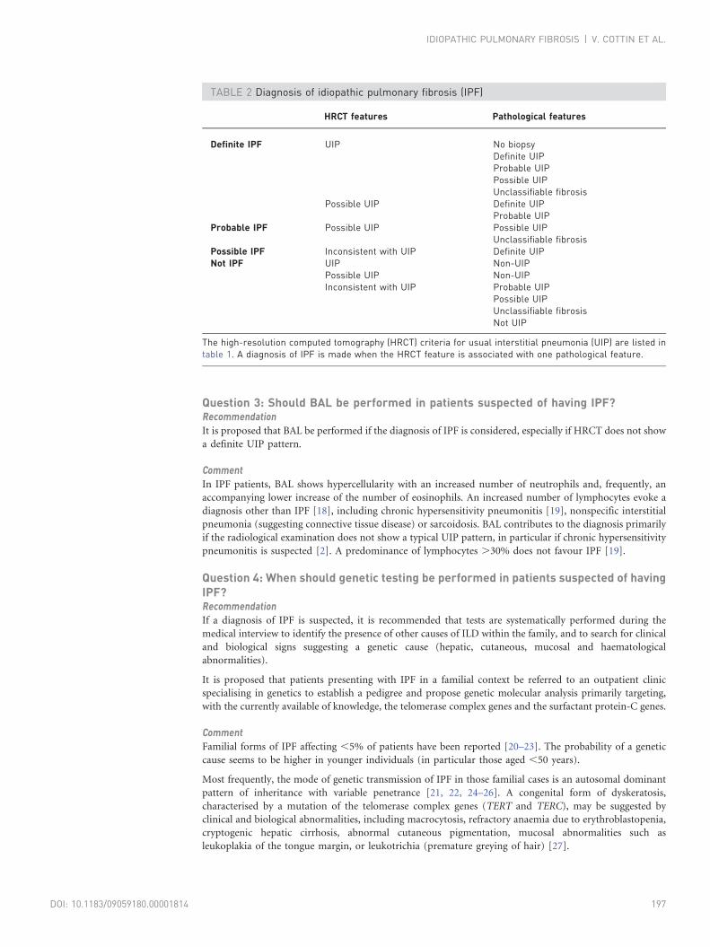
TABLE 2 Diagnosis of idiopathic pulmonary fibrosis (IPF)
HRCT features Pathological features
Definite IPF UIP No biopsyDefinite UIPProbable UIPPossible UIPUnclassifiable fibrosis
Possible UIP Definite UIPProbable UIP
Probable IPF Possible UIP Possible UIPUnclassifiable fibrosis
Possible IPF Inconsistent with UIP Definite UIPNot IPF UIP Non-UIP
Possible UIP Non-UIPInconsistent with UIP Probable UIP
Possible UIPUnclassifiable fibrosisNot UIP
The high-resolution computed tomography (HRCT) criteria for usual interstitial pneumonia (UIP) are listed intable 1. A diagnosis of IPF is made when the HRCT feature is associated with one pathological feature.
IDIOPATHIC PULMONARY FIBROSIS | V. COTTIN ET AL.
DOI: 10.1183/09059180.00001814 197

Question 5: What pulmonary function tests should be performed at the time of IPFdiagnosis?RecommendationIt is recommended that forced vital capacity (FVC) and diffusing capacity of the lung for carbon monoxide
(DLCO) should be assessed in patients being tested for IPF. It is proposed that total lung capacity, resting
arterial blood gas at room air, 6-min walk test (6MWT) distance and percutaneous oxygen saturation
should also be assessed.
CommentPulmonary function tests performed at rest in IPF patients show several abnormalities: 1) pulmonary
function restrictive pattern (decreased FVC and total lung capacity); 2) early decrease of DLCO and transfer
coefficient of the lung for carbon monoxide; and 3) usually, normal arterial blood gas values at rest or
hypocapnia (increased alveolar–arterial oxygen tension difference).
In addition, pulmonary function tests performed at exercise show a reduced exercise capacity, which may be
assessed by measuring: exercise hypoxaemia (even if not present at rest); the distance walked during a
6MWT; the decrease of percutaneous oxygen saturation during exercise, notably during the 6MWT; and the
decrease of maximal consumption of oxygen uptake and maximal power workload during exercise.
Question 6: When is HRCT sufficient for the diagnosis of IPF in patients not subjectedto video-assisted SLB?RecommendationIt is recommended that HRCT findings showing a UIP pattern, in particular a honeycombing pattern
(table 2), be considered sufficient to establish the diagnosis of IPF if other causes of ILD have been ruled out.
CommentHRCT of the chest is mandatory for the diagnosis of IPF. In ,50% of cases, HRCT shows a definite UIP
pattern with honeycombing (fig. 2) [2, 28], which is sufficient for the diagnosis of IPF if the analysis is
performed by a team of pulmonologists experienced in the field of IPF in a compatible clinical context. In
other cases, the imaging features are not characteristic (fig. 3) and video-assisted SLB is required to establish
the diagnosis. In some cases, HRCT findings may be inconsistent with a UIP pattern (fig. 4). Table 2
indicates the criteria used to define HRCT findings as definite or possible UIP. Honeycombing is required
to consider a definite UIP pattern on HRCT. Table 3 indicates how HRCT should be performed.
Question 7: In what patients should SLB be considered to establish the diagnosis ofIPF?RecommendationIt is recommended that video-assisted SLB be considered in patients for whom the diagnosis of IPF is
suspected, if a definite UIP pattern is not present on HRCT. The decision to perform a biopsy is taken
during a multidisciplinary team discussion after careful evaluation of the operative risk. The potential risk
FIGURE 2 High-resolution computedtomography scan demonstrating atypical example of usual interstitialpneumonia pattern with honeycombingchange and traction bronchiectasis, aswell as subpleural and basal reticulation.No features are seen suggesting analternative diagnosis.
IDIOPATHIC PULMONARY FIBROSIS | V. COTTIN ET AL.
DOI: 10.1183/09059180.00001814198

associated with biopsy must be taken into account, especially due to the age of the patient, the presence of
comorbidities, and the severity and progression of interstitial pneumonia.
CommentIf the imaging findings are not typical of a UIP pattern, a definite diagnosis of IPF requires a histological
UIP pattern to be demonstrated on video-assisted SLB (tables 4 and 5) and is based on the combination of
radiological and histological findings [2].
In some cases, HRCT only shows a possible UIP pattern but video-assisted SLB is not performed (because of
contraindications or possible risk, or biopsy is not proposed or is declined by the patient). It is then
impossible to establish the diagnosis of IPF according to the international recommendation criteria [2].
Question 8: What is the role of multidisciplinary team discussion in the diagnosis ofIPF and how is it conducted?RecommendationDuring a multidisciplinary discussion involving pulmonologists, radiologists and pathologists experienced
in the field of interstitial pneumonias, it is recommended that a definite diagnosis of IPF integrates clinical
assessment, computed tomography features and, if available, pathological features (table 1).
It is recommended that complex cases be referred to expert centres (reference or competence centres), or to
pulmonology departments experienced in ILDs.
CommentThe diagnosis of IPF results from the combination of clinical, radiological, biological, respiratory function
and, if available, pathological features and is established during multidisciplinary discussion. Ideally, the
multidisciplinary discussion takes place in an expert or specialised centre, i.e. the reference centre or one of
the competence centres for rare lung diseases, or a pulmonology department experienced in ILDs.
Question 9: How should prognosis be assessed in IPF patients?RecommendationIt is recommended that prognosis be assessed in IPF patients at the time of diagnosis based on: severity of
dyspnoea; results of pulmonary function tests (FVC and DLCO); percutaneous oxygen saturation at the end
of the 6MWT; extent of honeycombing on HRCT; signs of pulmonary hypertension at echocardiography;
and using a score. Prognosis should also be assessed in IPF patients during follow-up based on: progression
of symptoms; FVC and DLCO; fibrosis on HRCT; and, where applicable, on signs of pulmonary
hypertension at echocardiography.
CommentThe following factors have been associated with an increased risk of death in IPF patients [29]. 1) Older age
and male sex. 2) Early signs and symptoms: including severity of dyspnoea, DLCO ,35–40% predicted,
percutaneous oxygen saturation ,88% during a 6MWT at room air, extent of honeycombing on HRCT,
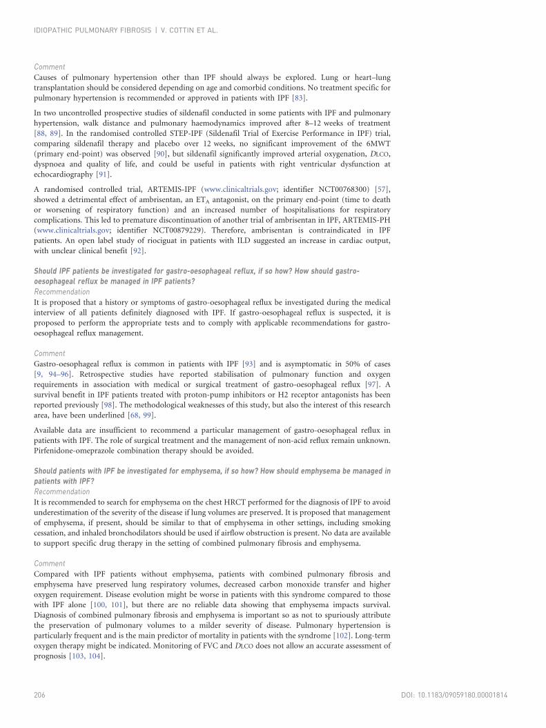
[R] [L] [R] [L]
a) b)
FIGURE 3 a, b) Representative examples of the lung biopsies. High-resolution computed tomography scansdemonstrating possible usual interstitial pneumonia pattern with subpleural and basal reticulation, and tractionbronchiectasis. No features are seen suggesting an alternative diagnosis. Video-assisted surgical lung biopsy demonstrateddefinite usual interstitial pneumonia pattern.
IDIOPATHIC PULMONARY FIBROSIS | V. COTTIN ET AL.
DOI: 10.1183/09059180.00001814 199
and precapillary pulmonary hypertension. 3) Signs and symptoms occurring during the clinical course:
including worsening of dyspnoea, .5% (absolute value) or 10% (absolute or relative value) decrease in
FVC over 6 months [30], .15% (absolute or relative value) decrease in DLCO over 6 months, .50 m
decrease in distance walked during the 6MWT [31], and worsening of fibrosis on HRCT.
The survival estimates at 1, 2 and 3 years can be predicted using the GAP (gender, age, physiology) score
based on age, sex, FVC and DLCO (www.acponline.org/journals/annals/extras/gap/) [32, 33].
Treatment of IPFWhat medical treatments have been proposed to treat IPF?Prednisone, azathioprine and N-acetylcysteine triple therapyRecommendationIt is recommended not to initiate a triple therapy with prednisone, azathioprine and N-acetylcysteine
(NAC) in patients definitely diagnosed with IPF.
CommentIn the randomised, placebo-controlled IFIGENIA(Idiopathic Pulmonary Fibrosis International Group
Exploring N-Acetylcysteine I Annual) trial [34], an antioxidant dose (1.8 g?day-1) of NAC reduced the
decrease of FVC and DLCO compared to placebo in a population of IPF patients who were also receiving a
combination of prednisone and azathioprine. This trial was not designed to assess the benefits of triple
therapy compared to placebo.
[R] [L]
[R] [L]
a) b)
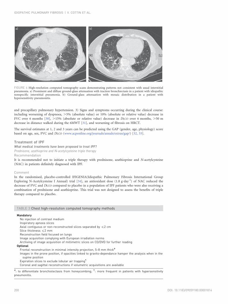
FIGURE 4 High-resolution computed tomography scans demonstrating patterns not consistent with usual interstitialpneumonia. a) Prominent and diffuse ground-glass attenuation with traction bronchiectasis in a patient with idiopathicnonspecific interstitial pneumonia. b) Ground-glass attenuation with mosaic distribution in a patient withhypersensitivity pneumonitis.
TABLE 3 Chest high-resolution computed tomography methods
MandatoryNo injection of contrast mediumInspiratory apnoea slicesAxial contiguous or non-reconstructed slices separated by f2 cmSlice thickness f2 mmReconstruction field focused on lungsImage acquisition complying with European irradiation normsArchiving of image acquisition of millimetric slices on CD/DVD for further reading
OptionalFrontal reconstruction in minimal intensity projection, 5–8 mm thick#
Images in the prone position, if opacities linked to gravito-dependance hamper the analysis when in thesupine position
Expiration slices to exclude lobular air trapping"
Coronal and sagittal reconstructions if volumetric acquisitions are available
#: to differentiate bronchiolectasis from honeycombing; ": more frequent in patients with hypersensitivitypneumonitis.
IDIOPATHIC PULMONARY FIBROSIS | V. COTTIN ET AL.
DOI: 10.1183/09059180.00001814200
The PANTHER (Prednisolone, Azathioprine and NAC: a study that evaluates response in IPF) trial assessed
the efficacy of NAC antioxidant treatment in mild-to-moderate IPF [35]. The trial initially comprised three
arms with similar sample sizes (triple therapy: azathioprine + prednisone + NAC versus NAC versus
placebo), the main outcome being decrease in FVC at 60 weeks. The triple therapy arm was prematurely
discontinued at 6 months after inclusion of 236 patients because of increased risks of all-cause mortality
(p50.01) and unscheduled hospitalisation (p,0.001) compared to placebo.
NAC monotherapyRecommendationIt is possible to prescribe NAC treatment to some patients with a definite diagnosis of IPF, taking into
account the benefit/risk ratio and the patient’s preferences, if treatment with an approved drug is not
indicated after having considered participation in a therapeutic clinical trial.
CommentThe IFIGENIA trial [34] did not assess the benefits of NAC monotherapy compared to placebo. Results of
the ongoing PANTHER trial [35], which currently comprises the NAC and placebo arms, will be released in
2014. Pending the results of ongoing clinical trials, monotherapy treatment with NAC may be considered on
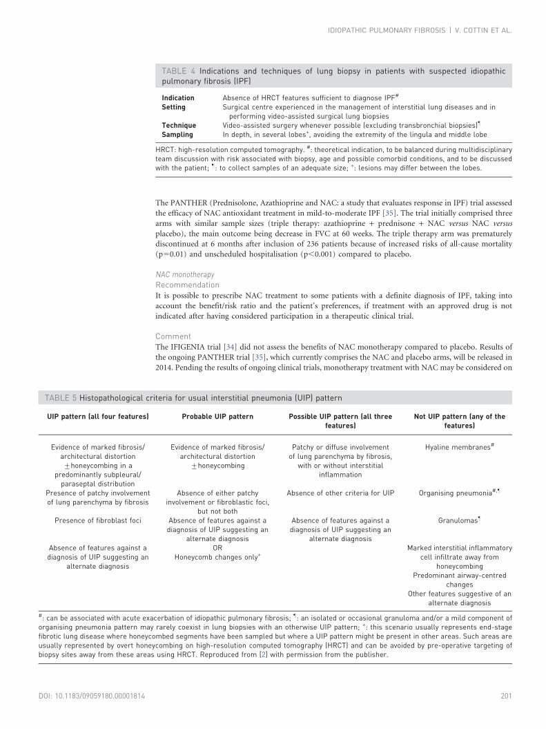
TABLE 4 Indications and techniques of lung biopsy in patients with suspected idiopathicpulmonary fibrosis (IPF)
Indication Absence of HRCT features sufficient to diagnose IPF#
Setting Surgical centre experienced in the management of interstitial lung diseases and inperforming video-assisted surgical lung biopsies
Technique Video-assisted surgery whenever possible (excluding transbronchial biopsies)"
Sampling In depth, in several lobes+, avoiding the extremity of the lingula and middle lobe
HRCT: high-resolution computed tomography. #: theoretical indication, to be balanced during multidisciplinaryteam discussion with risk associated with biopsy, age and possible comorbid conditions, and to be discussedwith the patient; ": to collect samples of an adequate size; +: lesions may differ between the lobes.
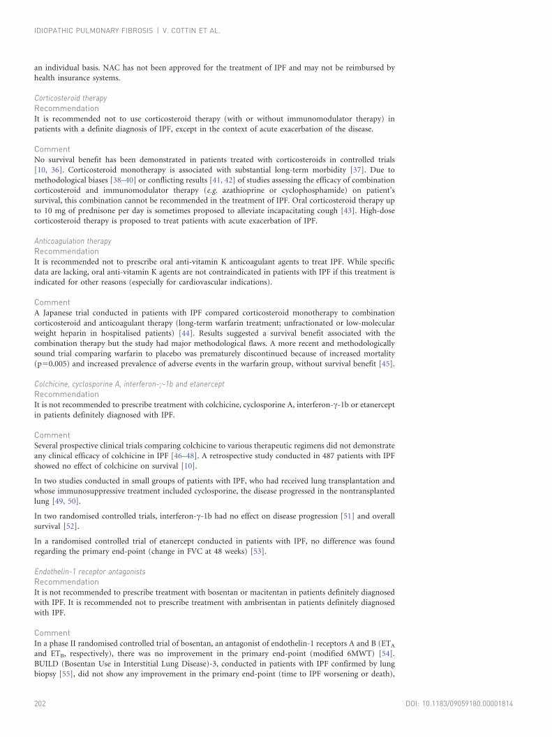
TABLE 5 Histopathological criteria for usual interstitial pneumonia (UIP) pattern
UIP pattern (all four features) Probable UIP pattern Possible UIP pattern (all threefeatures)
Not UIP pattern (any of thefeatures)
Evidence of marked fibrosis/architectural distortion¡honeycombing in a
predominantly subpleural/paraseptal distribution
Evidence of marked fibrosis/architectural distortion
¡honeycombing
Patchy or diffuse involvementof lung parenchyma by fibrosis,
with or without interstitialinflammation
Hyaline membranes#
Presence of patchy involvementof lung parenchyma by fibrosis
Absence of either patchyinvolvement or fibroblastic foci,
but not both
Absence of other criteria for UIP Organising pneumonia#,"
Presence of fibroblast foci Absence of features against adiagnosis of UIP suggesting an
alternate diagnosis
Absence of features against adiagnosis of UIP suggesting an
alternate diagnosis
Granulomas"
Absence of features against adiagnosis of UIP suggesting an
alternate diagnosis
ORHoneycomb changes only+
Marked interstitial inflammatorycell infiltrate away from
honeycombingPredominant airway-centred
changesOther features suggestive of an
alternate diagnosis
#: can be associated with acute exacerbation of idiopathic pulmonary fibrosis; ": an isolated or occasional granuloma and/or a mild component oforganising pneumonia pattern may rarely coexist in lung biopsies with an otherwise UIP pattern; +: this scenario usually represents end-stagefibrotic lung disease where honeycombed segments have been sampled but where a UIP pattern might be present in other areas. Such areas areusually represented by overt honeycombing on high-resolution computed tomography (HRCT) and can be avoided by pre-operative targeting ofbiopsy sites away from these areas using HRCT. Reproduced from [2] with permission from the publisher.
IDIOPATHIC PULMONARY FIBROSIS | V. COTTIN ET AL.
DOI: 10.1183/09059180.00001814 201
an individual basis. NAC has not been approved for the treatment of IPF and may not be reimbursed by
health insurance systems.
Corticosteroid therapyRecommendationIt is recommended not to use corticosteroid therapy (with or without immunomodulator therapy) in
patients with a definite diagnosis of IPF, except in the context of acute exacerbation of the disease.
CommentNo survival benefit has been demonstrated in patients treated with corticosteroids in controlled trials
[10, 36]. Corticosteroid monotherapy is associated with substantial long-term morbidity [37]. Due to
methodological biases [38–40] or conflicting results [41, 42] of studies assessing the efficacy of combination
corticosteroid and immunomodulator therapy (e.g. azathioprine or cyclophosphamide) on patient’s
survival, this combination cannot be recommended in the treatment of IPF. Oral corticosteroid therapy up
to 10 mg of prednisone per day is sometimes proposed to alleviate incapacitating cough [43]. High-dose
corticosteroid therapy is proposed to treat patients with acute exacerbation of IPF.
Anticoagulation therapyRecommendationIt is recommended not to prescribe oral anti-vitamin K anticoagulant agents to treat IPF. While specific
data are lacking, oral anti-vitamin K agents are not contraindicated in patients with IPF if this treatment is
indicated for other reasons (especially for cardiovascular indications).
CommentA Japanese trial conducted in patients with IPF compared corticosteroid monotherapy to combination
corticosteroid and anticoagulant therapy (long-term warfarin treatment; unfractionated or low-molecular
weight heparin in hospitalised patients) [44]. Results suggested a survival benefit associated with the
combination therapy but the study had major methodological flaws. A more recent and methodologically
sound trial comparing warfarin to placebo was prematurely discontinued because of increased mortality
(p50.005) and increased prevalence of adverse events in the warfarin group, without survival benefit [45].
Colchicine, cyclosporine A, interferon-c-1b and etanerceptRecommendationIt is not recommended to prescribe treatment with colchicine, cyclosporine A, interferon-c-1b or etanercept
in patients definitely diagnosed with IPF.
CommentSeveral prospective clinical trials comparing colchicine to various therapeutic regimens did not demonstrate
any clinical efficacy of colchicine in IPF [46–48]. A retrospective study conducted in 487 patients with IPF
showed no effect of colchicine on survival [10].
In two studies conducted in small groups of patients with IPF, who had received lung transplantation and
whose immunosuppressive treatment included cyclosporine, the disease progressed in the nontransplanted
lung [49, 50].
In two randomised controlled trials, interferon-c-1b had no effect on disease progression [51] and overall
survival [52].
In a randomised controlled trial of etanercept conducted in patients with IPF, no difference was found
regarding the primary end-point (change in FVC at 48 weeks) [53].
Endothelin-1 receptor antagonistsRecommendationIt is not recommended to prescribe treatment with bosentan or macitentan in patients definitely diagnosed
with IPF. It is recommended not to prescribe treatment with ambrisentan in patients definitely diagnosed
with IPF.
CommentIn a phase II randomised controlled trial of bosentan, an antagonist of endothelin-1 receptors A and B (ETA
and ETB, respectively), there was no improvement in the primary end-point (modified 6MWT) [54].
BUILD (Bosentan Use in Interstitial Lung Disease)-3, conducted in patients with IPF confirmed by lung
biopsy [55], did not show any improvement in the primary end-point (time to IPF worsening or death),
IDIOPATHIC PULMONARY FIBROSIS | V. COTTIN ET AL.
DOI: 10.1183/09059180.00001814202

or in quality of life and dyspnoea in the bosentan group. The primary end-point (change in FVC) was also
not reached in the MUSIC (Macitentan Use in an Idiopathic Pulmonary Fibrosis Clinical) trial that
evaluated macitentan [56], another antagonist of ETA and ETB. In ARTEMIS-IPF [57], ambrisentan, an ETA
antagonist, had detrimental effects on the primary end-point (time to death or worsening of pulmonary
function) and was associated with an increased rate of hospitalisations for respiratory complications.
Ambrisentan is currently contraindicated in IPF, including IPF with severe pulmonary hypertension.
EtanerceptRecommendationIt is not recommended to prescribe treatment with etanercept in patients definitely diagnosed with IPF.
PirfenidoneRecommendationIt is currently recommended to treat patients definitely diagnosed with mild-to-moderate IPF (defined as
FVC o50% pred and DLCO o35% pred) with pirfenidone. This treatment should be initiated and
supervised by a physician experienced in the diagnosis and management of IPF, and requires regular
monitoring of clinical tolerance and liver enzymes. The patient must not smoke during pirfenidone
treatment and patients should be warned against UV exposure.
CommentA pooled analysis of two phase III studies (006/Capacity 1 and 004/Capacity 2) conducted in patients with
mild-to-moderate IPF showed a significant reduction in FVC decline at week 72 in patients treated with
pirfenidone [58]. Disease progression (defined as a o10% decrease in FVC absolute value, a o15%
decrease in DLCO, or death) and decline in 6MWT performance were also reduced. In a phase III study
conducted in Japan [59], pirfenidone (1800 mg?day-1) significantly reduced FVC decline compared to
placebo. Another phase III trial is ongoing in the USA (www.clinicaltrials.gov; identifier NCT01366209).
Pirfenidone was granted European marketing authorisation in 2011, and was marketed in France in 2012
for the treatment of patients with a clinically and radiologically confirmed diagnosis of mild-to-moderate
IPF (FVC o50% pred and DLCO o35% pred). Pirfenidone should be initiated and supervised by specialist
physicians experienced in the diagnosis and management of IPF.
Pirfenidone should not be administered to patients treated with fluvoxamine and patients with severe
hepatic or renal impairment. The most commonly reported adverse reactions with pirfenidone were nausea,
rash, fatigue, diarrhoea, dyspepsia, photosensitivity reaction and weight loss [58]. Pirfenidone may also
induce elevations in liver enzymes. Liver function tests should be performed prior to the initiation of
treatment with pirfenidone, and subsequently at monthly intervals for the first 6 months and every
3 months thereafter.
Smoking has the potential to increase the activity of enzymes involved in pirfenidone metabolism and
should be discontinued prior to and during treatment with pirfenidone. Concomitant use of omeprazole
may theoretically result in changes in pirfenidone pharmacokinetics and should be avoided.
Should IPF patients be vaccinated against influenza and pneumococcal infections?RecommendationIt is recommended that annual influenza vaccination and anti-pneumococcal vaccination be performed in
patients definitely diagnosed with IPF.
CommentAlthough no specific studies of these vaccinations have been conducted in the context of IPF, it is very likely
that, like other patients suffering from chronic respiratory diseases, IPF patients are exposed to a high risk if
they develop a pneumococcal infection or influenza. Anti-pneumococcal vaccinations may be performed
using the polysaccharide pneumococcal vaccine.
Should IPF patients receive supplemental oxygen therapy?RecommendationIt is recommended to use long-term oxygen therapy in patients definitely diagnosed with IPF and with
severe hypoxaemia at rest (severe chronic respiratory failure).
CommentIndirect evidence of benefits of oxygen therapy has been suggested by studies conducted in patients with
other lung diseases and hypoxaemia. Two randomised controlled trials have reported a survival benefit in
IDIOPATHIC PULMONARY FIBROSIS | V. COTTIN ET AL.
DOI: 10.1183/09059180.00001814 203

patients with chronic obstructive lung disease receiving long-term oxygen therapy [60, 61]. Supplemental
oxygen therapy is usually indicated in severe chronic respiratory failure (arterial oxygen tension (PaO2)
f55 mmHg (7.3 kPa)), i.e. arterial oxygen saturation f88% measured at rest in stable conditions on two
separate occasions or PaO2 55–60 mmHg (7.3–8.0 kPa) if at least one of the following criterion is present:
haematocrit .55%, signs of pulmonary hypertension and documented right heart failure. Long-term
oxygen therapy is commonly used in patients with PaO2 f55–60 mmHg (7.3–8.0 kPa) measured at rest in
stable conditions on two separate occasions.
Should IPF patients receive respiratory rehabilitation?RecommendationIt is proposed that a respiratory rehabilitation programme should be initiated in patients definitely
diagnosed with IPF in whom limitation of exercise capacity results in significant impairment.
CommentSeveral studies have reported improvement in walking distance, symptoms or quality of life in IPF patients
following a respiratory rehabilitation programme [62, 63]. Respiratory rehabilitation programmes may
include exercise training, smoking cessation, psychosocial assistance and supportive care. Rehabilitation
may not be feasible in patients with advanced disease.
What is the indication for lung transplantation in IPF patients?RecommendationIt is recommended to consider lung transplantation in all patients definitely diagnosed with IPF aged
,65 years if the disease is severe or worsening. It is recommended that the patient is given information
about lung transplantation early in the course of the disease. Early assessment of the patient in a lung
transplantation centre is advised.
CommentLung transplantation improves survival in patients with advanced stage IPF [64–66]. International
recommendations specify that transplantation should be considered in patients aged ,65 years if DLCO is
,39% pred and FVC has decreased by o10% over 6 months of follow-up [67]. The (physiological) age limit
of ,65 years is relative and depends on local practice; comorbid conditions should be taken into account.
What are the diagnostic criteria of acute exacerbation of IPF?RecommendationIt is recommended to diagnose acute exacerbation of IPF in the case of recent worsening of dyspnoea
(,30 days) associated with additional lung opacities on imaging, after other possible causes of respiratory
function worsening have been ruled out (i.e. infection, pulmonary embolism and left heart failure).
CommentAcute exacerbation of IPF is characterised by acute (,30 days) worsening of dyspnoea with no identified
cause (e.g. infection, pulmonary embolism, left heart failure or cardiac arrhythmia) in a patient definitely
diagnosed with IPF. HRCT shows new opacities in addition to pre-existing abnormalities, in particular
ground-glass opacities. Worsening of hypoxaemia is common (o10 mmHg decrease of PaO2). Video-
assisted SLB is usually considered too hazardous and is not performed in this context.
Which treatments can be suggested to patients with acute exacerbation of IPF?RecommendationIt is proposed to use high-dose corticosteroids to treat acute exacerbations of IPF. It is possible to use
intravenous cyclophosphamide to treat acute exacerbations of IPF. There are insufficient data regarding the
use of low-molecular weight heparin to treat acute exacerbations of IPF.
CommentAlthough no controlled trials have assessed their efficacy in this indication, high-dose corticosteroids are
commonly prescribed to treat exacerbations of IPF [68]. Some observations have suggested that
immunosuppressive agents might be beneficial. Cyclosporine A has also been used, but without convincing
results. A benefit of intravenous cyclophosphamide has been suggested previously [69].
Low-molecular weight heparins have not demonstrated any benefits in acute exacerbations of IPF, but this
treatment is sometimes used. As a trial comparing warfarin to placebo was prematurely discontinued
IDIOPATHIC PULMONARY FIBROSIS | V. COTTIN ET AL.
DOI: 10.1183/09059180.00001814204

because of increased mortality and increased prevalence of adverse events in the warfarin group, without
apparent survival benefit [45], long-term oral anticoagulant therapy is not recommended in IPF (see
previous section). Wide-spectrum antibiotics may be used when infection has not been definitely ruled out.
Anticoagulant therapy may be prescribed in the case of acute worsening of symptoms or if thromboembolic
venous disease is suspected.
What is the role of invasive and noninvasive ventilation in the management of IPF?RecommendationIt is not recommended to use invasive ventilation in patients definitely diagnosed with IPF and acute or
chronic respiratory failure. It is possible to use invasive or noninvasive ventilation in a minority of patients
with IPF and acute respiratory failure; in particular, if criteria for emergency lung transplantation are met, if
exacerbation is the first manifestation of IPF, or in case of acute infection or reversible cause.
CommentGiven the high mortality associated with mechanical ventilation in IPF [70–79], this therapy should only be
used after discussion with the patients and their caregivers (ideally, during a previous visit) regarding goals
of care, in particular reduction of unnecessary suffering. Intensive care is justified as a bridge to lung
transplantation or if a reversible cause of worsening has been identified [80].
Data on noninvasive ventilation in IPF are scarce. A retrospective study in which some patients with acute
exacerbation of IPF were included suggests that, in this context, noninvasive ventilation may be preferred to
invasive ventilation without increasing mortality [81].
Should IPF patients be investigated for pulmonary hypertension, if so how?RecommendationIt is proposed that echocardiography be performed during the IPF diagnostic procedure and then annually
to detect pulmonary hypertension in patients with IPF. It is proposed that right heart catheterisation be
performed to diagnose pulmonary hypertension in patients definitely diagnosed with IPF: 1) prior to lung
transplantation; 2) in cases of clinical deterioration, limitation of exercise capacity, decrease of DLCO
(especially if DLCO is ,40% pred) and/or hypoxaemia disproportionate with regard to the restrictive
ventilatory defect (in particular if emphysema is present); 3) if an accurate evaluation of prognosis is
deemed essential; 4) if severe precapillary pulmonary hypertension is suspected at echocardiography
(tricuspid regurgitation flow .3.5 m?s-1) and to discuss possible off-label treatment of pulmonary
hypertension; and 5) if left ventricular disease with preserved systolic function is suspected.
CommentPrecapillary pulmonary hypertension (mean pulmonary artery pressure (Ppa) o25 mmHg and pulmonary
artery wedge pressure f15 mmHg) is present in ,10% of patients with IPF at the time of diagnosis, and
30–45% during evaluation prior to lung transplantation [82]. Severe pulmonary hypertension (mean Ppa
o35–40 mmHg) [83] is present in 2–9% of patients with IPF [84, 85]. Precapillary pulmonary
hypertension is associated with increased mortality [85], dyspnoea and hypoxaemia, decreased exercise
capacity and DLCO, and risk of acute exacerbation [86]. Doppler echocardiography is the first-line
noninvasive examination but its positive and negative predictive values for the diagnosis of pulmonary
hypertension are low [87]. Whether right heart catheterisation is indicated in an IPF patient should be
decided in expert centres [82].
How should pulmonary hypertension be managed if present in a patient with IPF?RecommendationIt is recommended that patients with IPF and pulmonary hypertension are investigated for resting
hypoxaemia, thromboembolic venous disease and left heart failure, and that patients receive appropriate
treatment if needed. Lung transplantation should also be considered.
In patients with IPF and moderate precapillary pulmonary hypertension (mean Ppa f35–40 mmHg at
rest), it is not recommended to prescribe any specific treatment for pulmonary hypertension.
In patients with IPF and severe precapillary pulmonary hypertension (mean Ppa o35–40 mmHg at rest), it
is possible to prescribe a specific treatment for pulmonary hypertension, preferably with sildenafil in a
specialised centre, if pulmonary hypertension is held responsible for worsening of symptoms.
In patients with IPF and severe precapillary pulmonary hypertension (mean Ppa o35–40 mmHg at rest), it
is recommended not to prescribe ambrisentan.
IDIOPATHIC PULMONARY FIBROSIS | V. COTTIN ET AL.
DOI: 10.1183/09059180.00001814 205
CommentCauses of pulmonary hypertension other than IPF should always be explored. Lung or heart–lung
transplantation should be considered depending on age and comorbid conditions. No treatment specific for
pulmonary hypertension is recommended or approved in patients with IPF [83].
In two uncontrolled prospective studies of sildenafil conducted in some patients with IPF and pulmonary
hypertension, walk distance and pulmonary haemodynamics improved after 8–12 weeks of treatment
[88, 89]. In the randomised controlled STEP-IPF (Sildenafil Trial of Exercise Performance in IPF) trial,
comparing sildenafil therapy and placebo over 12 weeks, no significant improvement of the 6MWT
(primary end-point) was observed [90], but sildenafil significantly improved arterial oxygenation, DLCO,
dyspnoea and quality of life, and could be useful in patients with right ventricular dysfunction at
echocardiography [91].
A randomised controlled trial, ARTEMIS-IPF (www.clinicaltrials.gov; identifier NCT00768300) [57],
showed a detrimental effect of ambrisentan, an ETA antagonist, on the primary end-point (time to death
or worsening of respiratory function) and an increased number of hospitalisations for respiratory
complications. This led to premature discontinuation of another trial of ambrisentan in IPF, ARTEMIS-PH
(www.clinicaltrials.gov; identifier NCT00879229). Therefore, ambrisentan is contraindicated in IPF
patients. An open label study of riociguat in patients with ILD suggested an increase in cardiac output,
with unclear clinical benefit [92].
Should IPF patients be investigated for gastro-oesophageal reflux, if so how? How should gastro-oesophageal reflux be managed in IPF patients?RecommendationIt is proposed that a history or symptoms of gastro-oesophageal reflux be investigated during the medical
interview of all patients definitely diagnosed with IPF. If gastro-oesophageal reflux is suspected, it is
proposed to perform the appropriate tests and to comply with applicable recommendations for gastro-
oesophageal reflux management.
CommentGastro-oesophageal reflux is common in patients with IPF [93] and is asymptomatic in 50% of cases
[9, 94–96]. Retrospective studies have reported stabilisation of pulmonary function and oxygen
requirements in association with medical or surgical treatment of gastro-oesophageal reflux [97]. A
survival benefit in IPF patients treated with proton-pump inhibitors or H2 receptor antagonists has been
reported previously [98]. The methodological weaknesses of this study, but also the interest of this research
area, have been underlined [68, 99].
Available data are insufficient to recommend a particular management of gastro-oesophageal reflux in
patients with IPF. The role of surgical treatment and the management of non-acid reflux remain unknown.
Pirfenidone-omeprazole combination therapy should be avoided.
Should patients with IPF be investigated for emphysema, if so how? How should emphysema be managed inpatients with IPF?RecommendationIt is recommended to search for emphysema on the chest HRCT performed for the diagnosis of IPF to avoid
underestimation of the severity of the disease if lung volumes are preserved. It is proposed that management
of emphysema, if present, should be similar to that of emphysema in other settings, including smoking
cessation, and inhaled bronchodilators should be used if airflow obstruction is present. No data are available
to support specific drug therapy in the setting of combined pulmonary fibrosis and emphysema.
CommentCompared with IPF patients without emphysema, patients with combined pulmonary fibrosis and
emphysema have preserved lung respiratory volumes, decreased carbon monoxide transfer and higher
oxygen requirement. Disease evolution might be worse in patients with this syndrome compared to those
with IPF alone [100, 101], but there are no reliable data showing that emphysema impacts survival.
Diagnosis of combined pulmonary fibrosis and emphysema is important so as not to spuriously attribute
the preservation of pulmonary volumes to a milder severity of disease. Pulmonary hypertension is
particularly frequent and is the main predictor of mortality in patients with the syndrome [102]. Long-term
oxygen therapy might be indicated. Monitoring of FVC and DLCO does not allow an accurate assessment of
prognosis [103, 104].
IDIOPATHIC PULMONARY FIBROSIS | V. COTTIN ET AL.
DOI: 10.1183/09059180.00001814206
No data are available to support recommendation of either specific management of emphysema in IPF
patients, or specific management of fibrosis in patients presenting with combined pulmonary fibrosis and
emphysema. Possible treatment (pirfenidone and NAC in monotherapy) must be assessed individually,
taking into account side-effects, the absence of data on the potential benefit of this treatment in this
indication, and the difficulty of evaluating disease evolution (little change in FVC).
Should IPF patients be explored for obstructive sleep apnoea syndrome, if so how?RecommendationIt is recommended that ventilatory polygraphy be performed in patients definitely diagnosed with IPF if
clinical signs suggest obstructive sleep apnoea syndrome.
CommentSeveral studies have shown a high rate of obstructive sleep apnoea syndrome in patients with IPF [105–107],
up to 88% in a series of 50 patients [108]. Compared to polysomnography, medical interview has a much
lower sensitivity to establish the diagnosis. Obesity is not always present. The clinical importance of
detecting obstructive sleep apnoea syndrome is not demonstrated in IPF, especially if the syndrome is
asymptomatic.
How should obstructive sleep apnoea syndrome be managed in patients with IPF?RecommendationNo specific data on the management of sleep apnoea in patients with IPF are available. It is proposed that
when present in a patient definitely diagnosed with IPF, obstructive sleep apnoea syndrome should be
managed with the usual recommendations applicable in patients without IPF.
CommentAvailable data are insufficient to recommend a specific management of obstructive sleep apnoea syndrome
in patients with IPF [109].
How should symptoms be managed in patients with IPF?RecommendationIt is possible to prescribe a transient, low-dose oral corticosteroid therapy in patients with IPF presenting
with incapacitating dry cough not alleviated by codeine. Efficacy and tolerance of this therapy should
be monitored.
It is possible to prescribe ambulatory supplemental oxygen therapy in patients definitely diagnosed with IPF
presenting with major dyspnoea at exercise and oxygen desaturation at exercise (percutaneous oxygen
saturation ,88% during daily life activities or standardised exercise, such as the 6MWT).
It is possible to prescribe low-dose morphine derivatives in patients with IPF presenting with major
dyspnoea, in the absence of hypercapnia, while monitoring the efficacy and tolerance of this treatment.
CommentLimited data suggest that oral corticosteroid therapy and thalidomide may alleviate chronic cough
associated with IPF. High-dose corticosteroids or thalidomide are poorly tolerated and inadvisable. Low-
dose morphine derivatives may be used in cases of major dyspnoea but side-effects of this treatment should
be closely monitored [110]. Low-dose morphine derivatives (f30 mg oral morphine equivalents a day) are
not associated with increased mortality in patients with chronic obstructive pulmonary disease and starting
long-term oxygen therapy [111]. No similar data are available in patients with IPF.
A small study has suggested that exercise capacity might be improved in patients with IPF and hypoxaemia
at rest who were treated with oxygen therapy [112]. Two retrospective studies have shown that ambulatory
oxygen therapy may significantly improve 6MWT performance and dyspnoea in patients with IPF
[113, 114]. In those two studies, oxygen flow was increased stepwise until percutaneous oxygen saturation
was o88% in one study and 90% in the other.
What examinations should be performed and at what frequency during follow-up of an IPF patient?RecommendationIt is recommended that patients definitely diagnosed with IPF should have a clinical visit and undergo
pulmonary function tests, including the measurement of FVC, every 3–6 months. It is proposed that chest
computed tomography be performed: if acute exacerbation of IPF is suspected; if unexplained clinical
changes occur; if lung cancer is suspected; and prior to lung transplantation.
IDIOPATHIC PULMONARY FIBROSIS | V. COTTIN ET AL.
DOI: 10.1183/09059180.00001814 207
CommentIf no other cause can be identified, the following changes suggest worsening of IPF [29, 115]: 1) progressive
increase of the level of dyspnoea; 2) progressive decrease of FVC (especially by o10% of the relative
or absolute value); 3) progressive decrease of DLCO (especially by o15% of the relative or absolute
value); 4) worsening of signs of pulmonary fibrosis on computed tomography; 5) acute exacerbation
of the disease.
Visits may be repeated every 3–6 months (table 6). Visits to a specialised centre (reference centre,
competence centre or hospital department experienced in ILDs) must take place at least annually or more
frequently if deterioration occurs. 3 monthly visits may take place in the vicinity of the patient’s home,
alternating with visits to a specialised centre, preferably in the context of a formal or informal care network
involving the treating general practitioner and the treating pulmonologist.
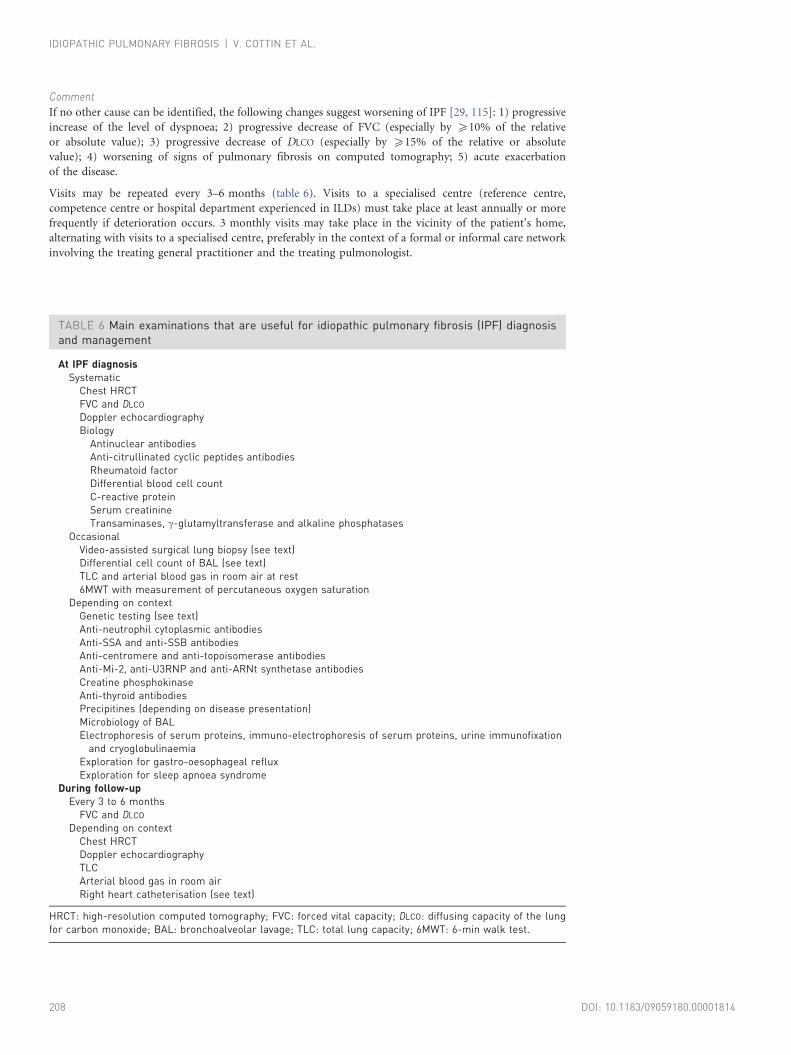
TABLE 6 Main examinations that are useful for idiopathic pulmonary fibrosis (IPF) diagnosisand management
At IPF diagnosisSystematic
Chest HRCTFVC and DLCO
Doppler echocardiographyBiology
Antinuclear antibodiesAnti-citrullinated cyclic peptides antibodiesRheumatoid factorDifferential blood cell countC-reactive proteinSerum creatinineTransaminases, c-glutamyltransferase and alkaline phosphatases
OccasionalVideo-assisted surgical lung biopsy (see text)Differential cell count of BAL (see text)TLC and arterial blood gas in room air at rest6MWT with measurement of percutaneous oxygen saturation
Depending on contextGenetic testing (see text)Anti-neutrophil cytoplasmic antibodiesAnti-SSA and anti-SSB antibodiesAnti-centromere and anti-topoisomerase antibodiesAnti-Mi-2, anti-U3RNP and anti-ARNt synthetase antibodiesCreatine phosphokinaseAnti-thyroid antibodiesPrecipitines (depending on disease presentation)Microbiology of BALElectrophoresis of serum proteins, immuno-electrophoresis of serum proteins, urine immunofixation
and cryoglobulinaemiaExploration for gastro-oesophageal refluxExploration for sleep apnoea syndrome
During follow-upEvery 3 to 6 months
FVC and DLCO
Depending on contextChest HRCTDoppler echocardiographyTLCArterial blood gas in room airRight heart catheterisation (see text)
HRCT: high-resolution computed tomography; FVC: forced vital capacity; DLCO: diffusing capacity of the lungfor carbon monoxide; BAL: bronchoalveolar lavage; TLC: total lung capacity; 6MWT: 6-min walk test.
IDIOPATHIC PULMONARY FIBROSIS | V. COTTIN ET AL.
DOI: 10.1183/09059180.00001814208
Is the risk of lung cancer increased during follow-up in a patient with IPF?RecommendationThe risk of lung cancer is increased in patients with IPF. It is proposed that the physician in charge of
follow-up be made aware of the frequent occurrence of lung cancer in patients definitely diagnosed with
IPF. It is recommended that smokers are urged to quit smoking and are informed about smoking cessation
assistance. In patients definitely diagnosed with IPF and lung cancer, it is recommended that IPF is taken
into account for treatment decisions.
CommentCompared with the risk of lung cancer in the general population, the relative risk of lung cancer in patients
with IPF is increased by approximately seven-fold [116]. Lung cancer prevalence in the IPF population is
between 4.4% and 9.8% [116–118], and a retrospective study reported a 55% 10-year risk of developing
lung cancer [119]. Neither specific screening nor specific management of lung cancer in IPF patients can
be recommended.
Lung cancer management is made more difficult by IPF and by the risk of acute respiratory failure and/or
acute exacerbation associated with treatments of cancer (surgical resection, radiation therapy and
chemotherapy) [120].
Should patients with IPF be investigated for other comorbid conditions?RecommendationIt is proposed that comorbid conditions (cardiovascular disease, thromboembolic venous disease, diabetes
and depression) be evaluated in patients definitely diagnosed with IPF, in order to inform the physician who
is in charge of follow-up.
CommentComorbid conditions, in particular those linked to smoking including cardiovascular disease,
thromboembolic venous disease, diabetes and depression, are frequently present in patients with IPF
[120]. Available data are insufficient to recommend systematic screening of such comorbid conditions, but
informing the physician in charge of follow-up (general practitioner or pulmonologist responsible for the
organisation of care) of the presence of a comorbid condition is important.
ConclusionSince the ATS/ERS/JRS/ALAT guidelines on the diagnosis and management of IPF were published in 2011,
new data from major clinical studies has prompted IPF experts from several European countries to release
updated recommendations in the form of national consensus statements or opinions [121, 122].
The need to incorporate recent evidence into recommendations for the diagnosis and management of IPF
was the basis of the present document. Our main objective was to provide French pulmonologists with an
updated, practical, decision-making tool that could be used in the daily clinical care of patients with
suspected or definitely diagnosed IPF.
This document complied with the specific national method and validation processes defined by the Haute
Autorite de Sante as good clinical practice for the development of guidelines. The organisation of the three
committees with specific missions ensured the involvement of prominent pulmonologists, radiologists and
pathologists experienced in the diagnosis and/or management of IPF.
In France, the network of the reference centre and the competence centres for rare lung diseases is
responsible for organising the care of patients with rare diseases, including IPF, in collaboration with local
pulmonologists, general physicians and other specialists involved in diagnosis and management. These
expert centres are geographically spread across all regions of France. The reference centre and the
competence centres for rare lung diseases coordinated the entire process of preparation of the present
guidelines, in order to standardise and optimise care, but the present recommendations accurately reflect
the collective thinking of all the experts involved.
Another feature of the present document is that it gives clinicians guidance even when the compelling
evidence for (or against) an examination or treatment is lacking. Formulations such as ‘‘It is proposed to’’
or ‘‘It is possible to’’ accompanied by a summary of the literature justifying these formulations should help
the reader to exert informed clinical judgment and decide whether the related diagnostic or therapeutic
intervention is appropriate in a given clinical situation.
The main differences between our recommendations and the 2011 guidelines originate from recently
emerged evidence. 1) Triple therapy with a prednisone, azathioprine and NAC should not be initiated in
IDIOPATHIC PULMONARY FIBROSIS | V. COTTIN ET AL.
DOI: 10.1183/09059180.00001814 209

patients with IPF as it has been shown to increase mortality. 2) Corticosteroid therapy is associated with
substantial morbidity and, in combination with immunomodulator therapy, might increase mortality.
Corticosteroid therapy is no longer an option for the treatment of IPF, except to alleviate incapacitating
cough or to treat acute exacerbation of IPF. 3) Ambrisentan is now contraindicated in the treatment in IPF.
4) To date, pirfenidone is the only pharmacological treatment with demonstrated benefits in IPF patients,
and the only treatment that has been approved and granted marketing authorisation for this indication.
We are confident that the present recommendations will help clinicians caring for IPF patients to base their
practice on currently available evidence before the much needed updated international guidelines are released.
AcknowledgementsThe author’s affiliations are as follows. V. Cottin: Hospices Civils de Lyon, Hopital Louis Pradel, Service de pneumologie– Centre de reference national des maladies pulmonaires rares, Lyon, and Universite de Lyon, Universite Claude BernardLyon 1, INRA, UMR754 IFR 128, Lyon, France. B. Crestani: Service de Pneumologie, CHU Bichat, Competence Centrefor Rare Lung Diseases, Paris, France. D. Valeyre: Dept of Pneumology, Hopital Universitaire Avicenne, CHU of Bobigny,Competence Centre for Rare Lung Diseases, Bobigny, France. B. Wallaert: CHU of Lille, Competence Centre for RareLung Diseases, Lille, France. J. Cadranel: CHU Tenon, Competence Centre for Rare Lung Diseases, Paris, France.J-C. Dalphin: CHU of Besancon, Competence Centre for Rare Lung Diseases, Besancon, France. P. Delaval: CHU ofRennes, Competence Centre for Rare Lung Diseases, Rennes, France. D. Israel-Biet: CHU Georges Pompidou,Competence Centre for Rare Lung Diseases, Paris, France. R. Kessler: CHU of Strasbourg, Competence Centre for RareLung Diseases, Strasbourg, France. M. Reynaud-Gaubert: CHU of Marseille, Competence Centre for Rare Lung Diseases,Marseille, France. B. Aguilaniu: Clinique du Mail, Grenoble, France. B. Bouquillon: Opened Mind Health, Roubaix,France. P. Carre: Polyclinique Montreal, Carcassone, France. C. Danel: CHU Bichat, Paris, France. J-B. Faivre: HopitalAlbert Calmette, Lille, France. G. Ferretti: Hopital Albert Michallon, Grenoble, France. N. Just: Centre Hospitalier VictorProvo, Roubaix, France. S. Kouzan: Centre Hospitalier Intercommunal sud Leman, Saint-Julien-en-Genevois, France.F. Lebargy: Hopital Maison Blanche, Reims, France. S. Marchand-Adam: CHU de Tours, Service de Pneumologie, CEPR,Inserm U1100, Tours, France. B. Philippe: Centre Hospitalier Rene Dubos, Pontoise, France. G. Prevot: Hopital Larrey,Toulouse, France. B. Stach: Cabinet medical, Valenciennes, France. F. Thivolet-Bejui: Centre de biologie et pathogie est,Hopital Louis Pradel, Lyon, France. J-F. Cordier: Hospices Civils de Lyon, Hopital Louis Pradel, Service de pneumologie– Centre de reference national des maladies pulmonaires rares, Lyon, France.
The committee members are as follows. Coordination committee: V. Cottin, B. Crestani, D. Valeyre, B. Wallaert andJ-F Cordier. Writing committee: V. Cottin, B. Crestani, D. Valeyre, B. Wallaert, J-F. Cordier, J. Cadranel, J-C. Dalphin,P. Delaval, D. Israel-Biet, R. Kessler and M. Reynaud-Gaubert. Reviewing committee: the Writing committee memberswith the addition of the following. Private practice pulmonologists: P. Carre, B. Stach and B. Aguilaniu. General hospitalpulmonologists: N. Just, S. Kouzan and B. Philippe. University hospital pulmonologists: F. Lebargy, S. Marchand-Adamand G. Prevot. Radiologists: G. Ferretti and J-B. Faivre. Pathologists: C. Danel and F. Thivolet-Bejui.
We received support from Opened Mind Health (Roubaix, France) and Michel Bordier (Clamart, France).
References1 Nalysnyk L, Cid-Ruzafa J, Rotella P, et al. Incidence and prevalence of idiopathic pulmonary fibrosis: review of the
literature. Eur Respir Rev 2012; 21: 355–361.2 Raghu G, Collard HR, Egan JJ, et al. An Official ATS/ERS/JRS/ALAT statement: idiopathic pulmonary fibrosis:
evidence-based guidelines for diagnosis and management. Am J Respir Crit Care Med 2011; 183: 788–824.3 Wuyts WA, Peccatori FA, Russell A-M. Patient-centred management in idiopathic pulmonary fibrosis: similar
themes in three communication models. Eur Respir Rev 2014; 23: 231–238.4 Cottin V, Crestani B, Valeyre D, et al. Recommandations pratiques pour le diagnostic et la prise en charge de la
fibrose pulmonaire idiopathique. Elaborees par le Centre national de reference et les Centres de competence pourles maladies pulmonaires rares sous l’egide de la Societe de pneumologie de langue francaise [French practicalguidelines for the diagnosis and management of idiopathic pulmonary fibrosis. From the National Reference andthe Competence centers for rare diseases and the Societe de Pneumologie de Langue Francaise]. Rev Mal Respir2013; 30: 879–902.
5 Haute Autorite de Sante. Elaboration de recommandations de bonne pratique. Methode. Recommandations pourla pratique clinique. HAS, 2010.
6 Gribbin J, Hubbard RB, Le Jeune I, et al. Incidence and mortality of idiopathic pulmonary fibrosis and sarcoidosisin the UK. Thorax 2006; 61: 980–985.
7 Scott J, Johnston I, Britton J. What causes cryptogenic fibrosing alveolitis? A case-control study of environmentalexposure to dust. Br Med J 1990; 301: 1015–1017.
8 Mannino DM, Etzel RA, Parrish RG. Pulmonary fibrosis deaths in the United States, 1979–1991. An analysis ofmultiple-cause mortality data. Am J Respir Crit Care Med 1996; 153: 1548–1552.
9 Raghu G, Freudenberger TD, Yang S, et al. High prevalence of abnormal acid gastro-oesophageal reflux inidiopathic pulmonary fibrosis. Eur Respir J 2006; 27: 136–142.
10 Douglas WW, Ryu JH, Schroeder DR. Idiopathic pulmonary fibrosis: impact of oxygen and colchicine, prednisone,or no therapy on survival. Am J Respir Crit Care Med 2000; 161: 1172–1178.
11 King TE Jr, Tooze JA, Schwarz MI, et al. Predicting survival in idiopathic pulmonary fibrosis: scoring system andsurvival model. Am J Respir Crit Care Med 2001; 164: 1171–1181.
12 Cottin V, Cordier JF. Velcro crackles: the key for early diagnosis of idiopathic pulmonary fibrosis?Eur Respir J 2012;40: 519–521.
13 Bohadana A, Izbicki G, Kraman SS. Fundamentals of lung auscultation. N Engl J Med 2014; 370: 744–751.
IDIOPATHIC PULMONARY FIBROSIS | V. COTTIN ET AL.
DOI: 10.1183/09059180.00001814210
14 American Thoracic Society, European Respiratory Society. American Thoracic Society/European RespiratorySociety International Multidisciplinary Consensus Classification of the Idiopathic Interstitial Pneumonias. Am JRespir Crit Care Med 2002; 165: 277–304.
15 Park JH, Kim DS, Park IN, et al. Prognosis of fibrotic interstitial pneumonia: idiopathic versus collagen vasculardisease-related subtypes. Am J Respir Crit Care Med 2007; 175: 705–711.
16 Cottin V. Interstitial lung disease: are we missing formes frustes of connective tissue disease? Eur Respir J 2006; 28:893–896.
17 Foulon G, Delaval P, Valeyre D, et al. ANCA-associated lung fibrosis: analysis of 17 patients. Respir Med 2008; 102:1392–1398.
18 Meyer KC, Raghu G, Baughman RP, et al. An official American Thoracic Society clinical practice guideline: theclinical utility of bronchoalveolar lavage cellular analysis in interstitial lung disease. Am J Respir Crit Care Med 2012;185: 1004–1014.
19 Ohshimo S, Bonella F, Cui A, et al. Significance of bronchoalveolar lavage for the diagnosis of idiopathicpulmonary fibrosis. Am J Respir Crit Care Med 2009; 179: 1043–1047.
20 Bitterman PB, Rennard SI, Keogh BA, et al. Familial idiopathic pulmonary fibrosis. Evidence of lung inflammationin unaffected family members. N Engl J Med 1986; 314: 1343–1347.
21 Marshall RP, Puddicombe A, Cookson WO, et al. Adult familial cryptogenic fibrosing alveolitis in the UnitedKingdom. Thorax 2000; 55: 143–146.
22 Hodgson U, Laitinen T, Tukiainen P. Nationwide prevalence of sporadic and familial idiopathic pulmonaryfibrosis: evidence of founder effect among multiplex families in Finland. Thorax 2002; 57: 338–342.
23 Lee HL, Ryu JH, Wittmer MH, et al. Familial idiopathic pulmonary fibrosis: clinical features and outcome. Chest2005; 127: 2034–2041.
24 Steele MP, Speer MC, Loyd JE, et al. Clinical and pathologic features of familial interstitial pneumonia. Am J RespirCrit Care Med 2005; 172: 1146–1152.
25 Rosas IO, Ren P, Avila NA, et al. Early interstitial lung disease in familial pulmonary fibrosis. Am J Respir Crit CareMed 2007; 176: 698–705.
26 Musk AW, Zilko PJ, Manners P, et al. Genetic studies in familial fibrosing alveolitis. Possible linkage withimmunoglobulin allotypes (Gm). Chest 1986; 89: 206–210.
27 Young NS. Telomere biology and telomere diseases: implications for practice and research. Hematology Am SocHematol Educ Program 2010; 2010: 30–35.
28 Cottin V, Capron F, Grenier P, et al. Pneumopathies interstitielles diffuses idiopathiques. Classification deConsensus International Multidisciplinaire de l’American Thoracic Society et de l’European Respiratory Society,principales entites anatomo-cliniques, et conduite du diagnostic [Diffuse idiopathic interstitial pneumonias.International multidisciplinary consensus classification by the American Thoracic Society and the EuropeanRespiratory Society, principal clinico-pathological entities, and diagnosis]. Rev Mal Respir 2004; 21: 299–318.
29 Ley B, Collard HR, King TE Jr. Clinical course and prediction of survival in idiopathic pulmonary fibrosis. Am JRespir Crit Care Med 2011; 183: 431–440.
30 du Bois RM, Weycker D, Albera C, et al. Forced vital capacity in patients with idiopathic pulmonary fibrosis: testproperties and minimal clinically important difference. Am J Respir Crit Care Med 2011; 184: 1382–1389.
31 du Bois RM, Weycker D, Albera C, et al. Six-minute-walk test in idiopathic pulmonary fibrosis: test validation andminimal clinically important difference. Am J Respir Crit Care Med 2011; 183: 1231–1237.
32 du Bois RM, Weycker D, Albera C, et al. Ascertainment of individual risk of mortality for patients with idiopathicpulmonary fibrosis. Am J Respir Crit Care Med 2011; 184: 459–466.
33 Ley B, Ryerson CJ, Vittinghoff E, et al. A multidimensional index and staging system for idiopathic pulmonaryfibrosis. Ann Intern Med 2012; 156: 684–691.
34 Demedts M, Behr J, Buhl R, et al. IFIGENIA: effects of N-acetylcysteine (NAC) on primary end points VC andDL,CO. Eur Respir J 2004; 24: Suppl. 48s, 668s.
35 Raghu G, Anstrom KJ, King TE Jr, et al. Prednisone, azathioprine, and N-acetylcysteine for pulmonary fibrosis.N Engl J Med 2012; 366: 1968–1977.
36 Nagai S, Kitaichi M, Hamada K, et al. Hospital-based historical cohort study of 234 histologically proven Japanesepatients with IPF. Sarcoidosis Vasc Diffuse Lung Dis 1999; 16: 209–214.
37 Gay SE, Kazerooni EA, Toews GB, et al. Idiopathic pulmonary fibrosis: predicting response to therapy and survival.Am J Respir Crit Care Med 1998; 157: 1063–1072.
38 Winterbauer RH, Hammar SP, Hallman KO, et al. Diffuse interstitial pneumonitis. Clinicopathologic correlationsin 20 patients treated with prednisone/azathioprine. Am J Med 1978; 65: 661–672.
39 Raghu G, Depaso WJ, Cain K, et al. Azathioprine combined with prednisone in the treatment of idiopathicpulmonary fibrosis: a prospective double-blind, randomized, placebo- controlled clinical trial. Am Rev Respir Dis1991; 144: 291–296.
40 Johnson MA, Kwan S, Snell NJ, et al. Randomised controlled trial comparing prednisolone alone withcyclophosphamide and low dose prednisolone in combination in cryptogenic fibrosing alveolitis. Thorax 1989; 44:280–288.
41 Collard HR, Ryu JH, Douglas WW, et al. Combined corticosteroid and cyclophosphamide therapy does not altersurvival in idiopathic pulmonary fibrosis. Chest 2004; 125: 2169–2174.
42 Pereira CA, Malheiros T, Coletta EM, et al. Survival in idiopathic pulmonary fibrosis-cytotoxic agents compared tocorticosteroids. Respir Med 2006; 100: 340–347.
43 Hope-Gill BD, Hilldrup S, Davies C, et al. A study of the cough reflex in idiopathic pulmonary fibrosis. Am J RespirCrit Care Med 2003; 168: 995–1002.
44 Kubo H, Nakayama K, Yanai M, et al. Anticoagulant therapy for idiopathic pulmonary fibrosis. Chest 2005; 128:1475–1482.
45 Noth I, Anstrom KJ, Calvert SB, et al. A placebo-controlled randomized trial of warfarin in idiopathic pulmonaryfibrosis. Am J Respir Crit Care Med 2012; 186: 88–95.
46 Fiorucci E, Lucantoni G, Paone G, et al. Colchicine, cyclophosphamide and prednisone in the treatment of mild-moderate idiopathic pulmonary fibrosis: comparison of three currently available therapeutic regimens. Eur RevMed Pharmacol Sci 2008; 12: 105–111.
IDIOPATHIC PULMONARY FIBROSIS | V. COTTIN ET AL.
DOI: 10.1183/09059180.00001814 211
47 Antoniou KM, Nicholson AG, Dimadi M, et al. Long-term clinical effects of interferon gamma-1b and colchicine inidiopathic pulmonary fibrosis. Eur Respir J 2006; 28: 496–504.
48 Douglas WW, Ryu JH, Swensen SJ, et al. Colchicine versus prednisone in the treatment of idiopathic pulmonaryfibrosis. A randomized prospective study. Members of the Lung Study Group. Am J Respir Crit Care Med 1998; 158:220–225.
49 Grgic A, Lausberg H, Heinrich M, et al. Progression of fibrosis in usual interstitial pneumonia: serial evaluation ofthe native lung after single lung transplantation. Respiration 2008; 76: 139–145.
50 Wahidi MM, Ravenel J, Palmer SM, et al. Progression of idiopathic pulmonary fibrosis in native lungs after singlelung transplantation. Chest 2002; 121: 2072–2076.
51 Raghu G, Brown KK, Bradford WZ, et al. A placebo-controlled trial of interferon-c-1b in patients with idiopathicpulmonary fibrosis. N Engl J Med 2004; 350: 125–133.
52 King TE Jr, Albera C, Bradford WZ, et al. Effect of interferon-c-1b on survival in patients with idiopathicpulmonary fibrosis (INSPIRE): a multicentre, randomised, placebo-controlled trial. Lancet 2009; 374: 222–228.
53 Raghu G, Brown KK, Costabel U, et al. Treatment of idiopathic pulmonary fibrosis with etanercept: an exploratory,placebo-controlled trial. Am J Respir Crit Care Med 2008; 178: 948–955.
54 King TE Jr, Behr J, Brown KK, et al. BUILD-1: a randomized placebo-controlled trial of bosentan in idiopathicpulmonary fibrosis. Am J Respir Crit Care Med 2008; 177: 75–81.
55 King TE Jr, Brown KK, Raghu G, et al. BUILD-3: a randomized, controlled trial of bosentan in idiopathicpulmonary fibrosis. Am J Respir Crit Care Med 2011; 184: 92–99.
56 Raghu G, Million-Rousseau R, Morganti A, et al. Efficacy and safety of macitentan in idiopathic pulmonaryfibrosis: results of a prospective, randomized, double-blind, placebo-controlled trial. Am J Respir Crit Care Med2012; 185: A3631.
57 Raghu G, Behr J, Brown KK, et al. ARTEMIS-IPF: a placebo-controlled trial of ambrisentan in idiopathicpulmonary fibrosis. Am J Respir Crit Care Med 2012; 185: A3632.
58 Noble PW, Albera C, Bradford WZ, et al. Pirfenidone in patients with idiopathic pulmonary fibrosis (CAPACITY):two randomised trials. Lancet 2011; 377: 1760–1769.
59 Taniguchi H, Ebina M, Kondoh Y, et al. Pirfenidone in idiopathic pulmonary fibrosis. Eur Respir J 2010; 35:821–829.
60 Continuous or nocturnal oxygen therapy in hypoxemic chronic obstructive lung disease: a clinical trial. NocturnalOxygen Therapy Trial Group. Ann Intern Med 1980; 93: 391–398.
61 Long term domiciliary oxygen therapy in chronic hypoxic cor pulmonale complicating chronic bronchitis andemphysema. Report of the Medical Research Council Working Party. Lancet 1981; 1: 681–686.
62 Holland AE, Hill CJ, Conron M, et al. Short term improvement in exercise capacity and symptoms followingexercise training in interstitial lung disease. Thorax 2008; 63: 549–554.
63 Nishiyama O, Kondoh Y, Kimura T, et al. Effects of pulmonary rehabilitation in patients with idiopathicpulmonary fibrosis. Respirology 2008; 13: 394–399.
64 Mason DP, Brizzio ME, Alster JM, et al. Lung transplantation for idiopathic pulmonary fibrosis. Ann Thorac Surg2007; 84: 1121–1128.
65 Keating D, Levvey B, Kotsimbos T, et al. Lung transplantation in pulmonary fibrosis: challenging early outcomescounterbalanced by surprisingly good outcomes beyond 15 years. Transplant Proc 2009; 41: 289–291.
66 Thabut G, Mal H, Castier Y, et al. Survival benefit of lung transplantation for patients with idiopathic pulmonaryfibrosis. J Thorac Cardiovasc Surg 2003; 126: 469–475.
67 Orens JB, Estenne M, Arcasoy S, et al. International guidelines for the selection of lung transplant candidates: 2006update – a consensus report from the Pulmonary Scientific Council of the International Society for Heart and LungTransplantation. J Heart Lung Transplant 2006; 25: 745–755.
68 Raghu G. Idiopathic pulmonary fibrosis: increased survival with ‘‘gastroesophageal reflux therapy’’: fact or fallacy?Am J Respir Crit Care Med 2011; 184: 1330–1332.
69 Morawiec E, Tillie-Leblond I, Pansini V, et al. Exacerbations of idiopathic pulmonary fibrosis treated withcorticosteroids and cyclophosphamide pulses. Eur Respir J 2011; 38: 1487–1489.
70 Blivet S, Philit F, Sab JM, et al. Outcome of patients with idiopathic pulmonary fibrosis admitted to the ICU forrespiratory failure. Chest 2001; 120: 209–212.
71 Kim DS, Park JH, Park BK, et al. Acute exacerbation of idiopathic pulmonary fibrosis: frequency and clinicalfeatures. Eur Respir J 2006; 27: 143–150.
72 Molina-Molina M, Badia JR, Marin-Arguedas A, et al. Caracterısticas clınicas y pronostico de los pacientes confibrosis pulmonar que ingresan en cuidados intensivos por insuficiencia respiratoria. Analisis de 20 casos[Outcomes and clinical characteristics of patients with pulmonary fibrosis and respiratory failure admitted to anintensive care unit. A study of 20 cases]. Med Clin (Barc) 2003; 121: 63–67.
73 Saydain G, Islam A, Afessa B, et al. Outcome of patients with idiopathic pulmonary fibrosis admitted to theintensive care unit. Am J Respir Crit Care Med 2002; 166: 839–842.
74 Nava S, Rubini F. Lung and chest wall mechanics in ventilated patients with end stage idiopathic pulmonaryfibrosis. Thorax 1999; 54: 390–395.
75 Stern JB, Mal H, Groussard O, et al. Prognosis of patients with advanced idiopathic pulmonary fibrosis requiringmechanical ventilation for acute respiratory failure. Chest 2001; 120: 213–219.
76 Al-Hameed FM, Sharma S. Outcome of patients admitted to the intensive care unit for acute exacerbation ofidiopathic pulmonary fibrosis. Can Respir J 2004; 11: 117–122.
77 Fumeaux T, Rothmeier C, Jolliet P. Outcome of mechanical ventilation for acute respiratory failure in patients withpulmonary fibrosis. Intensive Care Med 2001; 27: 1868–1874.
78 Mollica C, Paone G, Conti V, et al. Mechanical ventilation in patients with end-stage idiopathic pulmonary fibrosis.Respiration 2010; 79: 209–215.
79 Rangappa P, Moran JL. Outcomes of patients admitted to the intensive care unit with idiopathic pulmonaryfibrosis. Crit Care Resusc 2009; 11: 102–109.
80 Mullier A, Cottin V, Richard J-C, et al. Fibrose pulmonaire: dans quels cas et sur quels criteres envisager uneadmission en reanimation? [Pulmonary fibrosis: which patient should be admitted to the ICU and based on whichcritera?]. Reanimation 2013; 22: 45–53.
IDIOPATHIC PULMONARY FIBROSIS | V. COTTIN ET AL.
DOI: 10.1183/09059180.00001814212
81 Yokoyama T, Kondoh Y, Taniguchi H, et al. Noninvasive ventilation in acute exacerbation of idiopathic pulmonaryfibrosis. Intern Med 2010; 49: 1509–1514.
82 Nathan SD, Cottin V. Pulmonary hypertension in patients with idiopathic pulmonary fibrosis. In: Hoeper MH,Humbert M, eds. Pulmonary Hypertension. ERS Monogr 2012; 57: 148–160.
83 Seeger W, Adir Y, Barbera JA, et al. Pulmonary hypertension in chronic lung diseases. J Am Coll Cardiol 2013; 62:Suppl. 25, D109–D116.
84 Shorr AF, Wainright JL, Cors CS, et al. Pulmonary hypertension in patients with pulmonary fibrosis awaiting lungtransplant. Eur Respir J 2007; 30: 715–721.
85 Lettieri CJ, Nathan SD, Barnett SD, et al. Prevalence and outcomes of pulmonary arterial hypertension in advancedidiopathic pulmonary fibrosis. Chest 2006; 129: 746–752.
86 Judge EP, Fabre A, Adamali HI, et al. Acute exacerbations and pulmonary hypertension in advanced idiopathicpulmonary fibrosis. Eur Respir J 2012; 40: 93–100.
87 Arcasoy SM, Christie JD, Ferrari VA, et al. Echocardiographic assessment of pulmonary hypertension in patientswith advanced lung disease. Am J Respir Crit Care Med 2003; 167: 735–740.
88 Madden BP, Allenby M, Loke TK, et al. A potential role for sildenafil in the management of pulmonaryhypertension in patients with parenchymal lung disease. Vascul Pharmacol 2006; 44: 372–376.
89 Collard HR, Anstrom KJ, Schwarz MI, et al. Sildenafil improves walk distance in idiopathic pulmonary fibrosis.Chest 2007; 131: 897–899.
90 Zisman DA, Schwarz M, Anstrom KJ, et al. A controlled trial of sildenafil in advanced idiopathic pulmonaryfibrosis. N Engl J Med 2010; 363: 620–628.
91 Han MK, Bach DS, Hagan P, et al. Sildenafil preserves exercise capacity in IPF patients with right ventriculardysfunction. Chest 2013; 143: 1699–1708.
92 Hoeper MM, Halank M, Wilkens H, et al. Riociguat for interstitial lung disease and pulmonary hypertension: apilot trial. Eur Respir J 2013; 41: 853–860.
93 Noth I, Zangan SM, Soares RV, et al. Prevalence of hiatal hernia by blinded multidetector CT in patients withidiopathic pulmonary fibrosis. Eur Respir J 2012; 39: 344–351.
94 Tobin RW, Pope CE 2nd, Pellegrini CA, et al. Increased prevalence of gastroesophageal reflux in patients withidiopathic pulmonary fibrosis. Am J Respir Care Med 1998; 158: 1804–1808.
95 Sweet MP, Patti MG, Leard LE, et al. Gastroesophageal reflux in patients with idiopathic pulmonary fibrosisreferred for lung transplantation. J Thorac Cardiovasc Surg 2007; 133: 1078–1084.
96 Tcherakian C, Cottin V, Brillet PY, et al. Progression of idiopathic pulmonary fibrosis: lessons from asymmetricaldisease. Thorax 2011; 66: 226–231.
97 Linden PA, Gilbert RJ, Yeap BY, et al. Laparoscopic fundoplication in patients with end-stage lung disease awaitingtransplantation. J Thorac Cardiovasc Surg 2006; 131: 438–446.
98 Lee JS, Ryu JH, Elicker BM, et al. Gastroesophageal reflux therapy is associated with longer survival in patients withidiopathic pulmonary fibrosis. Am J Respir Crit Care Med 2011; 184: 1390–1394.
99 Raghu G, Meyer KC. Silent gastro-oesophageal reflux and microaspiration in IPF: mounting evidence for anti-reflux therapy? Eur Respir J 2012; 39: 242–245.
100 Cottin V, Nunes H, Brillet PY, et al. Combined pulmonary fibrosis and emphysema: a distinct under recognisedentity. Eur Respir J 2005; 26: 586–593.
101 Mejia M, Carrillo G, Rojas-Serrano J, et al. Idiopathic pulmonary fibrosis and emphysema: decreased survivalassociated with severe pulmonary arterial hypertension. Chest 2009; 136: 10–15.
102 Cottin V, Le Pavec J, Prevot G, et al. Pulmonary hypertension in patients with combined pulmonary fibrosis andemphysema syndrome. Eur Respir J 2010; 35: 105–111.
103 Schmidt SL, Nambiar AM, Tayob N, et al. Pulmonary function measures predict mortality differently in IPF versuscombined pulmonary fibrosis and emphysema. Eur Respir J 2011; 38: 176–183.
104 Akagi T, Matsumoto T, Harada T, et al. Coexistent emphysema delays the decrease of vital capacity in idiopathicpulmonary fibrosis. Respir Med 2009; 103: 1209–1215.
105 Clark M, Cooper B, Singh S, et al. A survey of nocturnal hypoxaemia and health related quality of life in patientswith cryptogenic fibrosing alveolitis. Thorax 2001; 56: 482–486.
106 Aydogdu M, Ciftci B, Firat Guven S, et al. Interstisyel akciger hastalarinda polisomnografi ile uyku ozelliklerinindegerlendirilmesi [Assessment of sleep with polysomnography in patients with interstitial lung disease]. TuberkToraks 2006; 54: 213–221.
107 Mermigkis C, Chapman J, Golish J, et al. Sleep-related breathing disorders in patients with idiopathic pulmonaryfibrosis. Lung 2007; 185: 173–178.
108 Lancaster LH, Mason WR, Parnell JA, et al. Obstructive sleep apnea is common in idiopathic pulmonary fibrosis.Chest 2009; 136: 772–778.
109 Recommandations pour la Pratique Clinique Syndrome d’apnees hyperpnees obstructives du sommeil de l’adulte[Recommendations for clinical practice. Obstructive sleep apnea hypopnea syndrome in adults]. Rev Mal Respir2010; 27: 806–833.
110 Allen S, Raut S, Woollard J, et al. Low dose diamorphine reduces breathlessness without causing a fall in oxygensaturation in elderly patients with end-stage idiopathic pulmonary fibrosis. Palliat Med 2005; 19: 128–130.
111 Ekstrom MP, Bornefalk-Hermansson A, Abernethy AP, et al. Safety of benzodiazepines and opioids in very severerespiratory disease: national prospective study. BMJ 2014; 348: g445.
112 Morrison DA, Stovall JR. Increased exercise capacity in hypoxemic patients after long-term oxygen therapy. Chest1992; 102: 542–550.
113 Visca D, Montgomery A, de Lauretis A, et al. Ambulatory oxygen in interstitial lung disease. Eur Respir J 2011; 38:987–990.
114 Frank RC, Hicks S, Duck AM, et al. Ambulatory oxygen in idiopathic pulmonary fibrosis: of what benefit? EurRespir J 2012; 40: 269–270.
115 Richeldi L, Ryerson CJ, Lee JS, et al. Relative versus absolute change in forced vital capacity in idiopathic pulmonaryfibrosis. Thorax 2012; 67: 407–411.
116 Hubbard R, Venn A, Lewis S, et al. Lung cancer and cryptogenic fibrosing alveolitis. A population-based cohortstudy. Am J Respir Crit Care Med 2000; 161: 5–8.
IDIOPATHIC PULMONARY FIBROSIS | V. COTTIN ET AL.
DOI: 10.1183/09059180.00001814 213

117 Turner-Warwick ME, Burrows B, Johnson A. Cryptogenic fibrosing alveolitis: clinical features and their influenceon survival. Thorax 1980; 35: 171–180.
118 Harris JM, Johnston ID, Rudd R, et al. Cryptogenic fibrosing alveolitis and lung cancer: the BTS study. Thorax2010; 65: 70–76.
119 Ozawa Y, Suda T, Naito T, et al. Cumulative incidence of and predictive factors for lung cancer in IPF. Respirology2009; 14: 723–728.
120 Fell CD. Idiopathic pulmonary fibrosis: phenotypes and comorbidities. Clin Chest Med 2012; 33: 51–57.121 Behr J. Evidence-based treatment strategies in idiopathic pulmonary fibrosis. Eur Respir Rev 2013; 22: 163–168.122 Xaubet A, Behr J, Bendstrup E, et al. Review of IPF diagnosis and management recommendations in Europe.
Sarcoidosis Vasc Diffuse Lung Dis 2013; 30: 249–261.
IDIOPATHIC PULMONARY FIBROSIS | V. COTTIN ET AL.
DOI: 10.1183/09059180.00001814214