diagnosis and treating slow growing non-hodgkin lymphomas · diagnosing and treating slow growing...
TRANSCRIPT
2/21/17
1
Welcome & Introductions
1
Diagnosing and Treating Slow Growing Non-Hodgkin Lymphomas
Dr. Hagemeister’s slides are available for download at
www.LLS.org/programs
Diagnosis and Treating Slow Growing Non-Hodgkin Lymphomas
Fredrick Hagemeister, MDProfessor of MedicineDepartment of Lymphoma/MyelomaThe University of Texas MD Anderson Cancer CenterHouston, TX
2Tuesday, February 21, 2017

2/21/17
2
Lymphoma: A Model for Basic Science and Clinical Research
• “Staging”– does the extent of disease make a difference?
• Combinations of drugs - better than “single-agent” therapies?
• Cytogenetics – a tool for better classification and basic science?
• Radiotherapy – a useful treatment?
• Prognostic factors – determining outcome?
• Antibody therapy – new targeted treatment?
• Gene Microarray Studies – understanding the basic cause of cancer?
• Molecular Studies-testing minor variations that make a difference?
Follicular lymphoma (22%)Follicular lymphoma (22%)
Diffuse largeB-cell (31%)
Diffuse largeB-cell (31%)
Armitage et al. J Clin Oncol. 1998;16:2780–2795.
Mantle cell (6%)Mantle cell (6%)
Peripheral T-cell (6%)Peripheral T-cell (6%)
Other subtypes with a frequency 2% (9%)Other subtypes with a frequency 2% (9%)
Frequency of Lymphoma Subtypes in Adults
Composite lymphomas (13%)
Composite lymphomas (13%)
Small lymphocytic lymphoma (6%)Small lymphocytic lymphoma (6%)
Marginal zone B-cell lymphomaMALT type (5%)
Marginal zone B-cell lymphomaMALT type (5%)
Marginal zone B-cell lymphomanodal type (1%)
Marginal zone B-cell lymphomanodal type (1%)
Lymphoplasmacytic lymphoma (1%)Lymphoplasmacytic lymphoma (1%)

2/21/17
3
Diagnosing and Treating Slow-Growing Non-Hodgkin Lymphomas
• Diagnosis
• Possible Causes
• Pathology
• Clinical Evaluation
• Therapy
• Follicular Lymphomas
• Small Lymphocytic Lymphoma and Chronic Lymphocytic Leukemia
• Mantle Cell Lymphomas
• Marginal Zone Lymphomas
• T Cell Lymphomas
Possible Causes of Lymphomas• Aging
• Immunodeficiency/ Immunosuppression– Congenital - Ataxia telangiectasia, Wiskott-Aldrich, SCID– Acquired – HIV infection, organ transplant, aging, autoimmune
disease– Drug induced – Immunosuppressants, organ or allogeneic SC
transplantation
• Environmental/Toxic Exposure– Agent orange, dioxins, PCBs, pesticides, herbicides, solvents
• Radiation– Atomic bomb exposure, Nuclear reactor accidents, Therapeutic RT
• Chemotherapy– Methotrexate and other immunosuppressive drugs suspected
• Viruses– EBV, HIV, HTLV-1, Hepatitis C, Human Herpesvirus 8
• Bacteria– H. Pylori, B. burgdorferi, C. jejuni, C. psittaci

2/21/17
4
Primary Immunodeficiency Disorders Associated with NHL
• Wiskott-Aldrich Syndrome
• Ataxia Telangiectasia
• Common Variable Immunodeficiency
• X-Linked Immunoproliferative Syndrome
• SCIDS – “Bubble Boy”
• Autoimmune Lymphoproliferative Syndrome (ALPS)
• Job’s Syndrome (subcutaneous abscesses)
Autoimmune Disorders Associated with Development of Lymphomas
• Hashimoto’s Thyroiditis• Sjogren’s Syndrome• Rheumatoid Arthritis• Systemic Lupus Erythematosis• Sprue, Inflammatory Bowel Disease• Autoimmune Hemolytic Anemia and
Immunopathic Thrombocytopenic Purpura• Dermatitis Herpetiformis

2/21/17
5
Models for Increased Risk of NHL in Patients with Autoimmune Disorders
• Chronic Immune Stimulation by Self Antigens
– Defective apoptosis of B-cells
– Impaired T-Cell function
– Secondary inflammation
• Genetic Factors
– Defects in inherited self-tolerance genes (TNF and IL-10 polymorphisms) with increased TNF, and increased NF-KB
– Other polymorphisms possibly associated (IL-7, IL-12, IL-13, and Interferon-gamma)
• Environmental Factors
– Dietary antigens (as in gluten, intestinal inflammation, and lymphoma)
– Abnormal response to viral or other infectious agents.
Relative Risks of NHL for Patients with Selected Autoimmune Diseases
Disorder DLBCL CLL T-Cell MCL MZL LPL
RA 1.8* 1.4 1.9 1.2 1.4 2.5*
SS 11* -- UD UD 28* --
SLE 6.2* -- UD UD -- --
Celiac Dz 2.8* 0.5 17* 3.3 UD 3.4
DM (Type1) 1.3 3.6* UD 5.0* 2.8 3.9
Autoimmune Diseases for which there are cases, but there either no cases in the “Control Group” or the Relative Risk of NHL is not statistically significant include: Crohn’s disease, Ulcerative Colitis, Sarcoidosis, and Psoriasis
* P < 0.05; UD: No cases in the control group; --: Too few cases in the AD or the control group
Smedby et al. JNCI 98: 51-60, 2006.

2/21/17
6
Clinical Features of 126 Patients with RA and Risk of Lymphoma (2905 Controls)
Feature RRisk of NHL
Male : Female 0.8 : 9.2
Duration of Disease <5 : >5 yrs 2.4 : 1.4
Family history Autoimmune Disorders 1.1
ESR > 45 2.8
Severe Small : Severe Large Joint Damage 10.5 : 29.3
Steroids/NSAIDS Therapy 1.5
NSAIDS > 10 yrs 1.9
Immunosuppressant Therapy 3.5
Immunosuppressants > 10 yrs 5.8
Dias and Isenberg. Nature Reviews/Rheumatology 3: 361-368, 2011.
Drugs Associated with Development of Lymphoproliferative Disorders
• TNF-Blockers (Used for other inflammatory disorders besides those listed)
• Eternacept: Approved for RA, psoriasis, ankylosing spondilitis
• Associated with NHL in RA (one study), maybe other solid tumors
• Infliximab: For RA, Crohn’s, amylosing spondylitis, psoriasis, UC
• Combined with azathioprine or 6-MP, associated with hepatosplenic T-cell NHL
• Adalimumab: Same as eternacept
• Alemtuzumab
• In combination With CHOP for aggressive T-cell NHL
• 3/20 developed EBV+ lymphoproliferative disorders
• Methotrexate in rheumatoid arthritis patients
• Reports of regression following discontinuation
• WHO I.atrogenic Immunodeficiency-associated LPD
Hoshida et al. J Rheum 34: 3222-331, 2007. Callen. Sem Cutan Med Surg 26: 8-14, 2007.
2/21/17
7
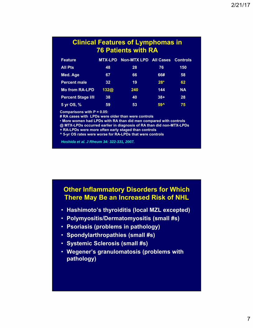
Clinical Features of Lymphomas in 76 Patients with RA
Feature MTX-LPD Non-MTX LPD All Cases Controls
All Pts 48 28 76 150
Med. Age 67 66 66# 58
Percent male 32 19 28* 62
Mo from RA-LPD 132@ 240 144 NA
Percent Stage I/II 38 40 38+ 28
5 yr OS, % 59 53 59^ 75
Comparisons with P = 0.05:# RA cases with LPDs were older than were controls• More women had LPDs with RA than did men compared with controls@ MTX-LPDs occurred earlier in diagnosis of RA than did non-MTX-LPDs+ RA-LPDs were more often early staged than controls^ 5-yr OS rates were worse for RA-LPDs that were controls
Hoshida et al. J Rheum 34: 322-331, 2007.
Other Inflammatory Disorders for Which There May Be an Increased Risk of NHL
• Hashimoto’s thyroiditis (local MZL excepted)
• Polymyositis/Dermatomyositis (small #s)
• Psoriasis (problems in pathology)
• Spondylarthropathies (small #s)
• Systemic Sclerosis (small #s)
• Wegener’s granulomatosis (problems with pathology)
2/21/17
8
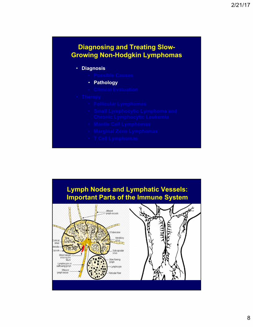
Diagnosing and Treating Slow-Growing Non-Hodgkin Lymphomas
• Diagnosis
• Possible Causes
• Pathology
• Clinical Evaluation
• Therapy
• Follicular Lymphomas
• Small Lymphocytic Lymphoma and Chronic Lymphocytic Leukemia
• Mantle Cell Lymphomas
• Marginal Zone Lymphomas
• T Cell Lymphomas
Lymph Nodes and Lymphatic Vessels: Important Parts of the Immune System

2/21/17
9
A Normal Lymph Node Under the Microscope
1. Capsule; 2 Subcapsular sinus; 3 Follicle (Germinal Center); 4. Lymphoid Nodule; 5. Trabeculae.
Some Lymphomas Get Their Names from Where the Normal Cell
Counterpart Is Found
Follicle Center
Marginal Zone
Mantle Zone

2/21/17
10
Examples of Various Proteins on the Surface of B Cell Lymphomas
DR
Surface Immunoglobulin
CD19
CD22
CD23
CD40CD80
CD20 Antibody that attaches to
these proteins can find them
under the microscope
“Markers” That Make a Difference in Diagnosis of Indolent Lymphomas
Marker FL SLL/CLL MCL MZL T Cell
CD20 Pos Pos Pos Pos Neg
CD10 Pos Neg Neg Neg Neg
CD5 Neg Pos Pos Neg Pos
CD23 Neg Pos Pos Neg Neg
Cyclin D1 Neg Neg Pos Neg Neg
Cytogenetics t(14;18) Various t(11;14) Various Various
• “Markers” are sugar/protein complexes that are produced by cells• They can be produced by both cancer cells and normal cells• These can be studied under the microscope to identify certain types of
lymphomas
• CD: Cluster of Differentiation• Not all are absolute: There are often variations in positivity/negativity• Note: The genetics are only in the cancer cells

2/21/17
11
One of the best known cytogenetic abnormalities
in cancer management
CD20 Expression in B-Cell Malignancies
0
Burkitt’s lymphoma
CLL
CLL/PLL
Follicular small cell
Hairy cell
Large cell
LP/Waldenström’s
Mantle cell
Marginal zone
Small cleaved
Adapted with permission from D. Maloney.
Mean channel fluorescence
100 200 300 400 500
Important because it may suggest how
well Anti-CD20 antibodies work in therapy of these specific diseases
2/21/17
12
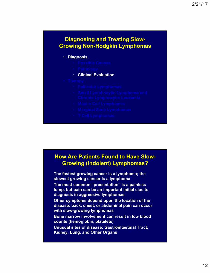
Diagnosing and Treating Slow-Growing Non-Hodgkin Lymphomas
• Diagnosis
• Possible Causes
• Pathology
• Clinical Evaluation
• Therapy
• Follicular Lymphomas
• Small Lymphocytic Lymphoma and Chronic Lymphocytic Leukemia
• Mantle Cell Lymphomas
• Marginal Zone Lymphomas
• T Cell Lymphomas
How Are Patients Found to Have Slow-Growing (Indolent) Lymphomas?
The fastest growing cancer is a lymphoma; the slowest growing cancer is a lymphoma
The most common “presentation” is a painless lump, but pain can be an important initial clue to diagnosis in aggressive lymphomas
Other symptoms depend upon the location of the disease: back, chest, or abdominal pain can occur with slow-growing lymphomas
Bone marrow involvement can result in low blood counts (hemoglobin, platelets)
Unusual sites of disease: Gastrointestinal Tract, Kidney, Lung, and Other Organs
2/21/17
13
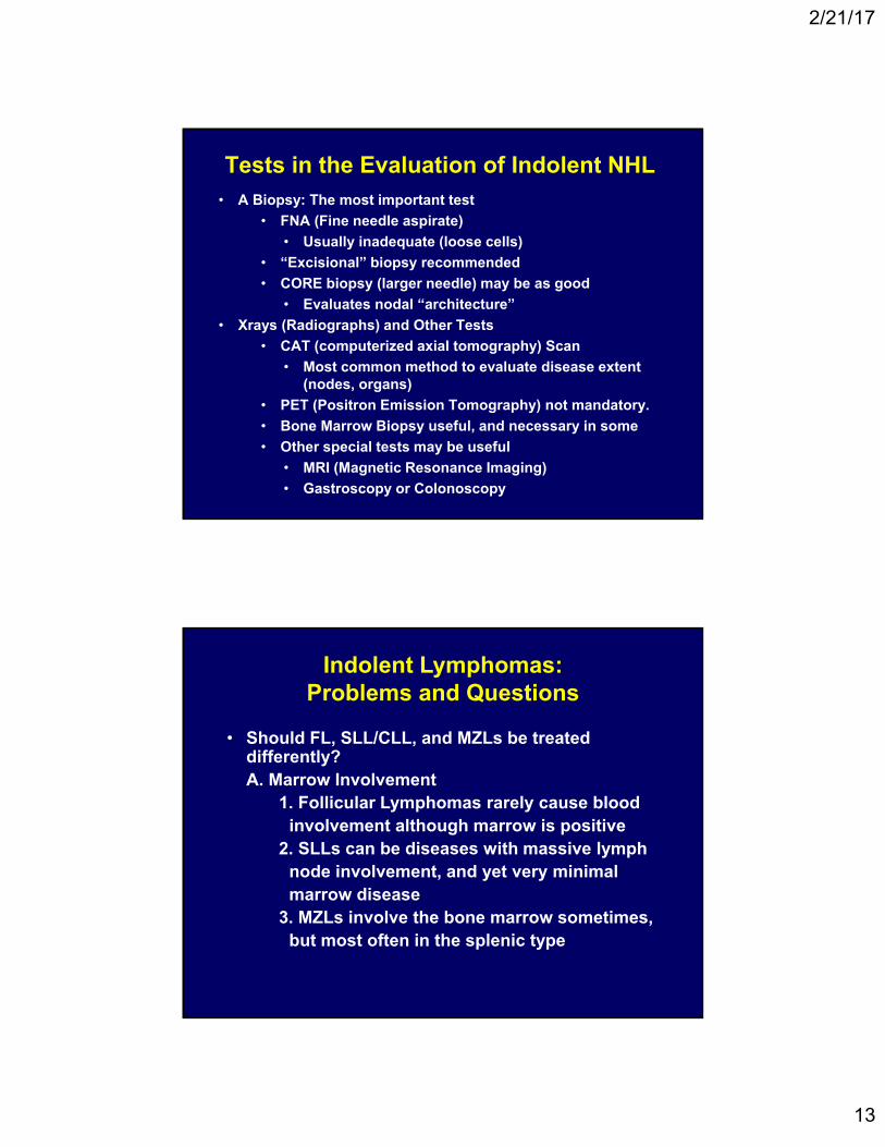
Tests in the Evaluation of Indolent NHL• A Biopsy: The most important test
• FNA (Fine needle aspirate)
• Usually inadequate (loose cells)
• “Excisional” biopsy recommended
• CORE biopsy (larger needle) may be as good
• Evaluates nodal “architecture”
• Xrays (Radiographs) and Other Tests
• CAT (computerized axial tomography) Scan
• Most common method to evaluate disease extent (nodes, organs)
• PET (Positron Emission Tomography) not mandatory.
• Bone Marrow Biopsy useful, and necessary in some
• Other special tests may be useful
• MRI (Magnetic Resonance Imaging)
• Gastroscopy or Colonoscopy
Indolent Lymphomas: Problems and Questions
• Should FL, SLL/CLL, and MZLs be treated differently?A. Marrow Involvement
1. Follicular Lymphomas rarely cause blood involvement although marrow is positive
2. SLLs can be diseases with massive lymph node involvement, and yet very minimal marrow disease
3. MZLs involve the bone marrow sometimes, but most often in the splenic type

2/21/17
14
Indolent Lymphomas:Problems and Questions
B. Extranodal Disease1. FLs rarely present with disease outside of lymph nodes, esp. Gastrointestinal sites, until transformation2. SLLs can be indistinguishable from MZLs when disease is present outside of nodes3. MZLs often have disease outside of the lymph nodes, but the nodal form is poorly
defined
Indolent Lymphomas: Problems and Questions
C. Risk of Transformation1. FLs have the perhaps the highest risk, but when and how the diagnosis is made can be difficult: Bulkiness, Pure DLCL, CT type?2. Grading of FLs is very subjective: FLCL? 3. Transformation may not be such a bad thing at initial diagnosis4. SLL/CLLs transform infrequently and may be a very poor risk feature: Richter’s Syndrome5. MZLs transform at an unknown rate, despite classic involvement outside of lymph nodes

2/21/17
15
Indolent Lymphoma: Treatment Choice Considerations
• Efficacy
• Patient’s age
• Prior therapies
• Safety profile
• The FLIPI (Follicular Lymphoma International Prognostic Index)
• Patient Choice
• Future therapies
• AE management
• QOL
• Treatment goals and expectations
• Even with advanced disease, Observation is an option
Involvement of 3 nodal sites, each with a diameter of ≥ 3 cm
Any nodal or extranodal tumor mass with a diameter of ≥ 7 cm
B symptoms
Splenomegaly
Pleural effusions or peritoneal ascites
Cytopenias (leukocytes < 1.0 × 10⁹/L and/or platelets < 100 × 109/L)
Leukemia (> 5.0 × 10⁹/L malignant cells)
Indications for Treatment by GELF Criteria
GELF = Groupe d’Etude des Lymphomes Folliculaires.Solal-Celigny et al. JCO 16: 2332-2338, 1998.

2/21/17
16
Standard Regimens for Therapy of Indolent Lymphomas
• Initial Therapy
• Single-Agent Rituximab
• Bendamustine + Rituximab
• R-CHOP
• Fludarabine-like Regimens
• Relapsed Disease
• Any of the above
• Lenalidomide + Rituximab
• Regimens not often used
• Platinum-, Gemcitabine-, Etoposide-Based Regimens
Novel Therapies in Treatment of Lymphomas
• Induce expression of costimulatory molecules and tumor immunity in melanoma, Hodgkin lymphoma, and NHLs
BCL2 Inhibitors
• Lead drug is lenalidomide, which has efficacy in multiple NHL subtypes (ie, MCL, FL, DLBCL, T‐cell lymphoma)
IMiDs
• Anti‐CD20 (obinutuzumab and ofatumumab), as well as other antigens on the cell surface (eg, CD19, CD22)
moAbs
• Significant activity, especially in aggressive lymphomas and leukemias
CAR T‐Cell Therapy
• Have effects in CLL and subtypes of aggressive and indolent lymphomas
PI3K and BTK Inhibitors
• Effective in Hodgkin’s and other lymphomasPD‐1 moAbs
Btk: Bruton's tyrosine kinase; CAR: chimeric antigen receptor; CLL: chronic lymphocytic leukemia; IMiD: immunomodulatory drug; mAbs: monoclonal antibodies; MCL: mantle cell lymphoma; NHL: non‐Hodgkin lymphoma; PI3k: phosphoinositide‐3‐kinase;; PD‐1: programmed death‐1

2/21/17
17
Diagnosing and Treating Slow-Growing Non-Hodgkin Lymphomas
• Diagnosis
• Possible Causes
• Pathology
• Clinical Evaluation
• Therapy
• Follicular Lymphomas
• Small Lymphocytic Lymphoma and Chronic Lymphocytic Leukemia
• Mantle Cell Lymphomas
• Marginal Zone Lymphomas
• T Cell Lymphomas
WHO Histologic Grading of Follicular Lymphoma
Grade Histology Clinical Behavior
1 0-5 centroblasts/HPF Indolent
2 6-15 centroblasts/HPF Indolent
3a >15 centroblasts/HPF, centrocytes present
Indolent-Aggressive
3b >15 centroblasts/HPF, centrocytes absent;
centroblasts in large sheets
Aggressive (similar to
LCL)
Nathwani et al. Intnl Agency Res Ca Press, Lyon 162-168, 2001.Nathwani et al. Intnl Agency Res Ca Press, Lyon 162-168, 2001.

2/21/17
18
Mortality According to FLIPI Index Using “NoLASH”
Risk GroupNumberFactors
% Patients(n = 1795)
5-year OS (%)
Good
10-year OS (%) RR
Intermediate
Poor
0-1
2
≥ 3
36
37
27
91
78
53
71
51
36
1
2.3
4.3
No = 5 or more Nodal Sites of Involvement
L = Elevated LDH A = Age Greater than 60
S = Stage III – IV H = Hemoglobin 12 or Less
Solal-Celigny et al. Blood 104: 1258-1265, 2004.Solal-Celigny et al. Blood 104: 1258-1265, 2004.
Prognosis of FL: OS Related to Duration of EFS Following Initial Therapy
• EFS: absence of progression, relapse, retreatment, or death
Patients with EFS > 1 Yr
Similar OS rate to that of the general US population
Patients with EFS < 1 Yr
Significantly inferior OSrate compared with the general US population
Years
Per
cen
t A
live
0
020406080
100
2 4 6 8 10 12
P = .20
FL survivalExpected OS (US)
Years
Per
cen
t A
live
00
20406080
100
2 4 6 8 10 12
P = 4.8 × 10⁻¹⁹
FL survivalExpected OS (US)
Maurer et al. Blood 124: 2014 (Abst 1664).
2/21/17
19
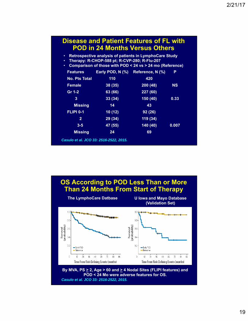
Disease and Patient Features of FL with POD in 24 Months Versus Others
Features Early POD, N (%) Reference, N (%) P
No. Pts Total 110 420
Female 38 (35) 200 (48) NS
Gr 1-2 63 (66) 227 (60)
3 33 (34) 150 (40) 0.33
Missing 14 43
FLIPI 0-1 10 (12) 92 (26)
2 29 (34) 119 (34)
3-5 47 (55) 140 (40) 0.007
Missing 24 69
Casulo et al. JCO 33: 2516-2522, 2015.
• Retrospective analysis of patients in LymphoCare Study• Therapy: R-CHOP-588 pt; R-CVP-280; R-Flu-207• Comparison of those with POD < 24 vs > 24 mo (Reference)
OS According to POD Less Than or More Than 24 Months From Start of Therapy
• Rom Start of TherapyFrom T
Casulo et al. JCO 33: 2516-2522, 2015.
The LymphoCare Datbase U Iowa and Mayo Database (Validation Set)
By MVA, PS > 2, Age > 60 and > 4 Nodal Sites (FLIPI features) and POD < 24 Mo were adverse features for OS.

2/21/17
20
Transformed FL Outcomes from the LymphoCare Database
• PFS and OS rates were not statistically different for those with suspected (S) versus confirmed (biopsy-proven)T-FL
• Clinical features of ST-FL (Vancouver): LDH >2XNL, rapid single node growth, new ENS, new B SX, hypercalcemia (not collected in this study)
The M7-FLIPI: A Prognostic Model for Prediction of POD24
• Evaluation of 74 genes from 151 pts with FL who received R-CHOP and interferon maintenance.
• Selected genes that appeared mutated in more than 5 patients
• Calculated FFS models using high Risk FLIPI and other clinical and lab features
• Generated models that incorporated molecular features of 7 genes providing best FFS discrimination
• Validated: BCCA Cohort receiving R-CVP and MR.
Pastore et al. Lancet Oncol 16: 2015.

2/21/17
21
Foll05 Trial: R-CVP vs R-CHOP vs R-FN: 3 Year TTF and OS Results
R-CHOP TTF-64% OS-95%
R-CVP TTF-46% OS-98%
R-FN TTF-61% OS- 93%
0.007
0.0210.969
Federico et al. ASCO 2012 (abst 8006).
Follicular Lymphoma Mantle Cell Lymphoma
Marginal Zone
BR vs R-CHOP for Indolent Lymphomas
Waldenstrom’s
Rummel et al. Lancet 381: 1203-1210, 2013.
2/21/17
22
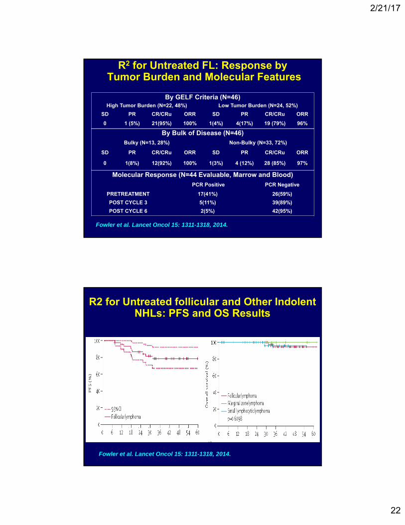
R2 for Untreated FL: Response byTumor Burden and Molecular Features
By GELF Criteria (N=46)High Tumor Burden (N=22, 48%) Low Tumor Burden (N=24, 52%)
SD PR CR/CRu ORR SD PR CR/CRu ORR
0 1 (5%) 21(95%) 100% 1(4%) 4(17%) 19 (79%) 96%
By Bulk of Disease (N=46) Bulky (N=13, 28%) Non-Bulky (N=33, 72%)
SD PR CR/CRu ORR SD PR CR/CRu ORR
0 1(8%) 12(92%) 100% 1(3%) 4 (12%) 28 (85%) 97%
Fowler et al. Lancet Oncol 15: 1311-1318, 2014.
Molecular Response (N=44 Evaluable, Marrow and Blood)
PCR Positive PCR Negative
PRETREATMENT 17(41%) 26(59%)
POST CYCLE 3 5(11%) 39(89%)
POST CYCLE 6 2(5%) 42(95%)
R2 for Untreated follicular and Other Indolent NHLs: PFS and OS Results
Fowler et al. Lancet Oncol 15: 1311-1318, 2014.

2/21/17
23
Obinutuzumab-CIT vs Rituximab-CIT for Untreated FL: The Gallium Study
Marcus et al. ASH 2016, abstract 6.
PFS OS
PFSPFS
Results for other iNHLs pending.
Phase 3 Obinutuzumab/Bendamustine vs Bendamustine for R-Refractory FL
Cheson et al. ASH 2016, abstract 615.
PFS iNHL p<0.001
PFS FL p<0.001
OS FL p=0.006
OS iNHL p=0.03
2/21/17
24
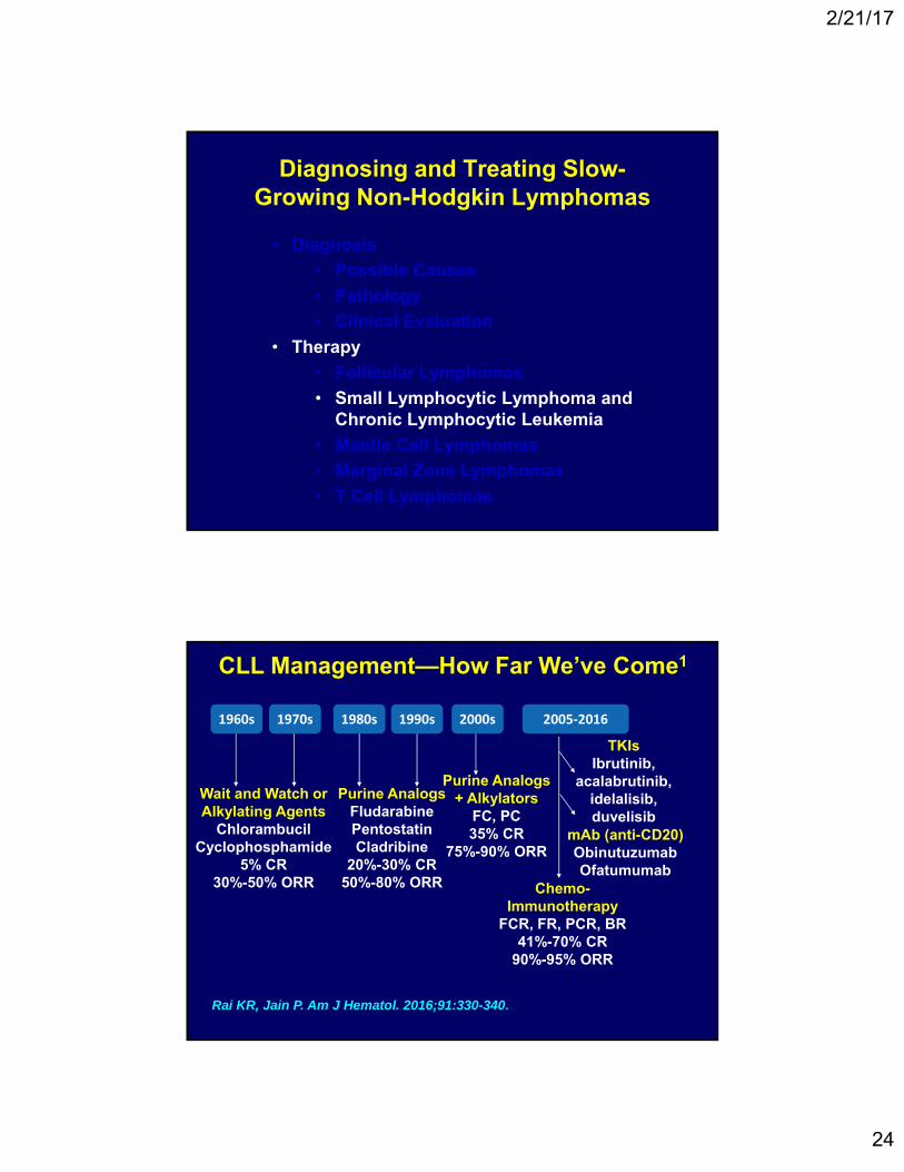
Diagnosing and Treating Slow-Growing Non-Hodgkin Lymphomas
• Diagnosis
• Possible Causes
• Pathology
• Clinical Evaluation
• Therapy
• Follicular Lymphomas
• Small Lymphocytic Lymphoma and Chronic Lymphocytic Leukemia
• Mantle Cell Lymphomas
• Marginal Zone Lymphomas
• T Cell Lymphomas
CLL Management—How Far We’ve Come1
1960s 1970s 1980s 1990s 2000s 2005‐2016
Wait and Watch or Alkylating Agents
ChlorambucilCyclophosphamide
5% CR30%-50% ORR
Purine AnalogsFludarabinePentostatinCladribine
20%-30% CR50%-80% ORR
Purine Analogs + Alkylators
FC, PC35% CR
75%-90% ORR
TKIsIbrutinib,
acalabrutinib, idelalisib, duvelisib
mAb (anti-CD20)ObinutuzumabOfatumumab
Chemo-Immunotherapy
FCR, FR, PCR, BR41%-70% CR
90%-95% ORR
Rai KR, Jain P. Am J Hematol. 2016;91:330-340.

2/21/17
25
Traditional and Newer PFs associated With Inferior OS in CLL
• Traditional PFs
1. Advanced stage
at diagnosis
2. Short lymphocyte
doubling time
3. Diffuse pattern of
bone marrow disease
4. Advanced age / male
5. -2 microglobulin or
circulating CD23
6. prolymphs (PLL)
• Newer PFs
1. FISH cytogenetics
– 17p del: agg dz
– 11q del: agg dz
– 13q del: indolent dz
2. Unmutated IgVH
(<2% homology with
germline)
3. ZAP70 ( 20% positive)
4. CD38 ( 30% positive)
1.0
0.8
0.6
0.4
0.2
0
FCR vs BR in Pts With Advanced CLL: PFS
Eichhorst B, et al. ASH 2014. Abstract 19
Cu
mu
lati
ve S
urv
ival
ITT PFS = Primary EndpointPFS in IGHV-Matched Population
(n = 398; FCR = 201; BR = 197)
0 12 24 36 48 60
Median PFSFCR: 55.2 mosBR: 41.7 mos
P < .001HR: 1.626
Cu
mu
lati
ve S
urv
ival
1.0
0.8
0.6
0.4
0.2
00 12 24 36 48 60
Median PFSFCR: NRBR: 43.1 mos
P < .005HR: 1.565

2/21/17
26
MR After FCR for Untreated CLL: The French FILO CLL 2007 Trial
Maintenance R (202) Observation (207)
Median PFS (mo) 59.3 49
3 Yr PFS (%)* 83 64.2
3 Yr OS (%) 92.6 87.2
Secondary Cancer 15.3 11.1
Heme SAEs* 6.9 1.9
Infectious SAEs* 18.8 10.1
Dartigeas et al. ASCO 2016 (abst 7505).
• 409 pts with untreated CLL, >65 yrs, in CR/PR. No del(17p).• Therapy: 4 cycles of FCR, followed by MR (500 mg/m2 q 2 mo for
2 yr) vs Observation• CR/CRi = 38%. Stratified by del(11q), CR/PR, and IGHV status.
• PFS also better with MR for those with/without del(11q) or unmutated IGHv
* P < 0.05.
After a median follow-up of 19.1 months
van Oers et al. Lancet Oncol 2015;16:1370-79
Time since randomization (mo)
Maintenance Ofatumumab vs Observation for 2nd or 3rd CR/PR: The PROLONG study
PFS OS
Approved by FDA

2/21/17
27
Maintenance Len After Initial Therapy for “High –Risk” Disease: CLL M1 Study• Median observation time of 17.7 months
Median PFSPlacebo group: 14.6 mosLenalidomide group: NRHR 0.198 [95% CI: 0.083‐0.475]
CLL11 Results: OS in Older Patients
No statistically significant difference
in OS is noted vs rituximab-
chlorambucila
a OS results are not yet mature.
Obinutuzumab-chlorambucil is associated with
significant OS benefit vs chlorambucila
1. Goede V et al. Leukemia. 2015;29:1602‐1604.

2/21/17
28
Ibrutinib (BTK Inhibitor) for High-Risk CLL: RESONATE-2 Survival Outcomes
Tedeschi A et al. ASH 2015. Abstract 495.
Barr et al: ASH 2016. Abstract 234.
CR rate improved from 11% to 18% with longer f/u: at 29 mo: 88% reduction in risk of PD or death with ibrutinib vs CHL
Ib + BR vs PL + BR for Rel CLL: HELIOS
Investigator-assessed HR for ib + BR vs pl + BR: 0.201 (CI: 0.145-0.278)
No Richter’s observed on the ib and 3 on the pl arms
Chanan-Khan et al. ASCO 2015; LBA 7005
2/21/17
29
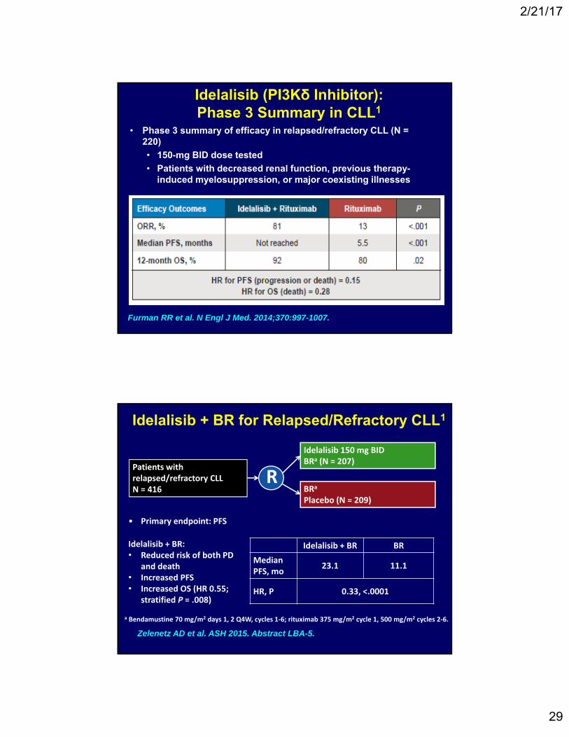
Idelalisib (PI3Kδ Inhibitor): Phase 3 Summary in CLL1
• Phase 3 summary of efficacy in relapsed/refractory CLL (N = 220)
• 150-mg BID dose tested
• Patients with decreased renal function, previous therapy-induced myelosuppression, or major coexisting illnesses
Furman RR et al. N Engl J Med. 2014;370:997-1007.
Idelalisib + BR for Relapsed/Refractory CLL1
Idelalisib + BR:• Reduced risk of both PD
and death• Increased PFS • Increased OS (HR 0.55;
stratified P = .008)
Patients with relapsed/refractory CLLN = 416
R
Idelalisib 150 mg BIDBRa (N = 207)
BRa
Placebo (N = 209)
Idelalisib + BR BR
Median PFS, mo
23.1 11.1
HR, P 0.33, <.0001
a Bendamustine 70 mg/m2 days 1, 2 Q4W, cycles 1‐6; rituximab 375 mg/m2 cycle 1, 500 mg/m2 cycles 2‐6.
• Primary endpoint: PFS
Zelenetz AD et al. ASH 2015. Abstract LBA-5.

2/21/17
30
Emerging Strategies for Therapy of Lymphomas: Bcl-2 Inhibition1-3
• Venetoclax: orally bioavailable, selective Bcl‐2 inhibitor that induces apoptosis in CLL cells independent of p53
– 79% ORR in early clinical studies in relapsed/refractory CLL1
Roberts et al. NEJM 2016;374:311-322. Stilgenbauer et al. ASH 2015. Abstract LBA-6. Konopleva et al. ASH 2014. Abstract 118.
Response, n (%) IRC Investigator
ORR 85 (79.4) 79 (73.8)
CR or CRi 8 (7.5) 17 (15.9)
nPR 3 (2.8) 4 (3.7)
PR 74 (69.2) 58 (54.2)
Response and Main Safety Findings
Safety Summary
• 40% grade 3/4 neutropenia; 22.4% baseline neutropenia (any grade)
• Infections in 72% of patients (20% grade ≥3)
• Laboratory TLS in 5 patients during the ramp‐up period; no clinical TLS
• Most common SAEs: pyrexia (7%), AIHA (7%), pneumonia (6%), FN (5%)
Venetoclax Monotherapy: Phase 2 Study in Relapsed/Refractory del(17p) CLL (N = 107)1
Stilgenbauer et al. ASH 2015. Abstract LBA-6.

2/21/17
31
Diagnosing and Treating Slow-Growing Non-Hodgkin Lymphomas
• Diagnosis
• Possible Causes
• Pathology
• Clinical Evaluation
• Therapy
• Follicular Lymphomas
• Small Lymphocytic Lymphoma and Chronic Lymphocytic Leukemia
• Mantle Cell Lymphomas
• Marginal Zone Lymphomas
• T Cell Lymphomas
Diagnosis of Mantle Cell Lymphoma
• 5%-10% of B-cell NHL, with moderately aggressive course
• 74% male, median age 63 years
• >80% stage III/IV including marrow involvement
• Extranodal sites common: lymphomatous polyposis, gastrointestinal, soft tissue, or leukemic phase
• Classic translocation: >70% t(11;14); overexpression of cyclin D1 (bcl-1)
• CD19+, 20+, 5+, 23–, FMC7+, SOX11+
• In the past, prognosis was poor: chemoresponsive, but median survival 30 months with CHOP-type chemotherapy
Fisher et al. Hematology, 221: 2004.

2/21/17
32
MCL Histologic Subtypes
Mantle Zone Nodular
Diffuse Blastoid
Indolent Mantle Cell Lymphoma
• Characterization
– Mantle Zone Lymphoma
• With this pathology, only the Mantle Zone is involved by the disease
– Any MCL (except Blastoid variant) with Ki-67 < 10%
– Mantle Cell Lymphoma involving the spleen and marrow only (usually do not have colon involvement)
– Low MIPI and small tumor burden
• None of these have been studied in prospective trials

2/21/17
33
A Prognostic Index (M-IPI) for Patients With Advanced-Stage MCL
– Age (↑ 10 y, HR 1.42; P=0.0002)
– Sex
– ECOG PS (>1, HR 2.01; P=0.0088)
– Ann Arbor stage
– B-symptoms
– Number of ENS
– Number of involved nodal areas
– Tumor size
– Serum LDH (2 × ↑ LDH, HR 1.51; P<0.0059)
– WBC (10 × ↑ WBC, HR 2.56; P<0.0001)
– Platelet count
– Hemoglobin
– Albumin
– β2-microglobulin
– Ki-67
Hoster et al. ASH, 2006. Abstract 814.
Patients with advanced MCL
treated with first-line from 3 GLSG trials
N=455
Analysis based on a list of prognostic factors*
Romaguera et al. Brit Jnl Hematol 150::200-208, 2010.
Initial R-HyperCVAD had 97 pts
Median age 61 (range 41-80); 1/3 pts > 65 y
Pts > 65y worse: med PFS just over 2y (delays / dose reduction / toxicity)
120
FFS
Months
100806040200
1.0
0.9
0.8
0.7
0.6
0.5
0.4
0.3
0.2
0.1
0
P = .004
Pro
po
rtio
n F
ailu
re-F
ree
≤ 65 yrs > 65 yrs
R-HyperCVAD for MCL: FFS by Age

2/21/17
34
VcR-CVAD and MR for Untreated MCL
Chang et al. ASH 2016, abstract 149.
PFS
OS
Tolerance better with Bor 1.3 and Vin 1
BCR Inhibition in Relapsed MCL: Ibrutinib
Wang ML et al. N Engl J Med. 2013;369:507-516.

2/21/17
35
CR 73%, ongoing. Better in those with high Ki-67%, but not affected by MIPI or degree of rash.
Wang et al. ASH 2016, abstract 147.
Ibrutinib + Rituximab for Untreated MCL < 66: Responses by Features
Diagnosing and Treating Slow-Growing Non-Hodgkin Lymphomas
• Diagnosis
• Possible Causes
• Pathology
• Clinical Evaluation
• Therapy
• Follicular Lymphomas
• Small Lymphocytic Lymphoma and Chronic Lymphocytic Leukemia
• Mantle Cell Lymphomas
• Marginal Zone Lymphomas
• T Cell Lymphomas

2/21/17
36
Outcome in Treatment Subsets of Stage IE Gastric MALT NHL: OS and EFS
Treatment n CR PR NC 5 Yr OS 5 Yr EFS
Antibiotics 45 67% 9% 24% 94% 75%
Local tx* 14 100% 0 0 92% 80%
Chemo 8 50% 12% 38% 75% 49%
CMT† 5 100% 0 0 80% 80%
Total 72 74% 7% 19% 89% 72%
Pinotti G et al. Leuk Lymphoma. 1997;26:527-537.
* Surgery alone (n = 11), surgery and XRT (n = 2), or XRT alone (n = 1)† Surgery and adjuvant chemotherapy
C
Primary Site Percent (%)
Head and neck 30
Ocular adnexa 24
Lung 12
Skin 12
Intestinal tract/GU 8/1
Thyroid/Breast 7/2From International Extranodal Lymphoma Study Group (IELSG), others
Nongastric MALT Lymphoma: Presenting Sites
Cavalli F et al. Hematology. 2001:241-258.

2/21/17
37
Splenic Marginal-Zone Lymphoma: Clinical Presentation
• Typical presentation:
– Splenomegaly
– Circulating lymphoma cells
– BM involvement
– No enlarged nodes
• Rare lymphoma (<1% of all NHL)
• Also called splenic lymphoma with or without villous lymphocytes
• Was confused with Hairy Cell Leukemia
Nodal Marginal-Zone Lymphoma: Clinical Features
• B symptoms (14%)
• Stages I/II (29%) and III/IV (71%)
• Elevated LDH (36%)
• Bone marrow involvement (28%)
• 5-year survival (56%)
Nathwani BN et al. J Clin Oncol. 1999;17:2486-2492.

2/21/17
38
Therapy for Marginal Zone Lymphomas
• Few randomized trials
– Good survival rates, even with active disease
– Many different therapies work
– Treatment depends on site of disease, and patient features
• Individualized Choices
– Observation (no immediate therapy)
– Radiation Therapy (often low dose)
– Single-Agent Rituximab
– Bendamustine/Rituximab
– B-cell pathway drugs (Ibrutinib, Idelalisib)
– Other novel agents being studied
Diagnosing and Treating Slow-Growing Non-Hodgkin Lymphomas
• Diagnosis
• Possible Causes
• Pathology
• Clinical Evaluation
• Therapy
• Follicular Lymphomas
• Small Lymphocytic Lymphoma and Chronic Lymphocytic Leukemia
• Mantle Cell Lymphomas
• Marginal Zone Lymphomas
• T Cell Lymphomas

2/21/17
39
The WHO Classification of PTCL• PTCL Leukemic
Adult T cell leukemia/lymphoma (HTLV-1+)
• PTCL, Predominantly ExtranodalExtranodal NK/T cell lymphoma, nasal typeEnteropathy-type T cell lymphomaHepatosplenic T cell lymphoma (gamma/delta)Subcutaneous panniculitis-type T cell lymphoma
Indolent: Mycosis fungoides/Sezary syndrome
Primary cutaneous ALCL
• PTCL, Predominantly NodalPeripheral T cell lymphoma, NOSAngioimmunoblastic T cell lymphoma, AILD-likeAnaplastic large cell lymphoma (T and Null cell)
Initial Therapy of PTCL
• Relative rarity and heterogeneity of subtypes has limited clinical trials for these entities– Cell size does not correlate well with
prognosis• Most series indicate a higher relapse rate and
poorer survival for PTCL, NOS vs DLBCL– Important to recognize ALCL, ALK+, containing
the t(2;5) translocation high curability with CHOP alone
• CHOP therapy has been the most commonly utilized front-line therapy– ORR ~ 60-70%, CR ~ 40-60%– Relapse @ 2 years > 70-80% in most series

2/21/17
40
Initial Therapy of PTCL
• HyperCVAD often used for ATCL and other highly aggressive variants: PTCL trial at MDACC demonstrated no real benefit
• ACVBP may be better than CHOP in GELA trials• EPOCH has activity in both front-line and
relapsed settings• Nucleoside analogues (fludarabine, cladribine,
pentostatin) are more often used in MF or PTCL with cutaneous involvement– Inhibit adenosine deaminase, high
concentrations in T-cells– ORR 20-70%, CR 3-25%, DR often < 6 months
• Improved therapies are needed !
Q&A SessionAsk a question by phone:
• Press star (*) then the number 1 on your keypad.
Ask a question by web:• Click “Ask a question”• Type your question • Click “Submit”
Due to time constraints, we can only take one question per person. Once you’ve asked your question, the operator will transfer you back into the audience line.
80
Diagnosing and Treating Slow Growing Non-Hodgkin Lymphomas

2/21/17
41
• Online Chats: Online moderated chat forums: www.LLS.org/chat
• Questions to ask your treatment team: www.LLS.org/whattoask
• Free education materials: www.LLS.org/booklets
• Past NHL education programs: www.LLS.org/programs
• Additional information on NHL: www.LLS.org/NHL
• Information Resource Center: Speak one-on-one with an Information Specialist who can assist you through cancer treatment, financial, and social challenges.
EMAIL: [email protected]
TOLL-FREE PHONE: (800) 955- 4572
81
Diagnosing and Treating Slow Growing Non-Hodgkin Lymphomas