diagnosis and treatment of … and treatment...for tinea unguium (onychomycosis) and tinea capitis,...
TRANSCRIPT

et al., IJSIT, 2018, 7(3), 665-678 Dr. Simant Ankit
IJSIT (www.ijsit.com), Volume 7, Issue 3, May-June 2018
665
DIAGNOSIS AND TREATMENT OF DERMATOPHYTES INFECTIONS
Dr. Simant Ankit1* and Prof. Dr. Tongxiang Zeng1
1Department of Dermatology & Venereology, Jingzhou Central Hospital, Yangtze University, Jingzhou, Hubei,
China
ABSTRACT
Dermatophytes are fungi that require keratin for growth. These fungi can cause superficial infections
of the skin, hair, and nails. Treatment of dermatophyte infection involves primarily oral and/or topical
formulations of azoles or allylamines, particularly itraconazole and terbinafine. Topical medications applied
once or twice daily are the primary treatment indicated for tinea corporis/cruris, and tinea pedis/manuum.
Use of oral antifungals may be practical where the tinea involvement is extensive or chronic, or where
application of a topical is not feasible. For tinea unguium (onychomycosis) and tinea capitis, oral therapies
are the primary treatments provided. Recently, topical amorolfine and ciclopirox formulations have been
approved for use in milder onychomycosis cases, and their role in the treatment of the different clinical forms
of onychomycosis is currently being defined. Relapse of infection remains a problem, particularly with tinea
pedis/unguium. This article reviews will update readers on the diagnosis and treatment of common
dermatophyte infections.
Keywords: Dermatophytosis, superficial fungal infections, tinea corporis, tinea cruris, tinea pedis, tinea
barbae, tinea mamuum.

et al., IJSIT, 2018, 7(3), 665-678 Dr. Simant Ankit
IJSIT (www.ijsit.com), Volume 7, Issue 3, May-June 2018
666
INTRODUCTION
Dermatophytes are referred to as “tinea” infections. Dermatophytes require keratin for growth, they
are restricted to hair, nails, and superficial skin. Thus, these fungi do not infect mucosal surfaces[1]. The
dryness of the skin’s outer layer discourages colonization by microorganisms, and the shedding of epidermal
cells keeps many microbes from establishing residence[2]. However, the skin’s mechanisms of protection may
fail because of trauma, irritation, or maceration. Furthermore, occlusion of the skin with nonporous materials
can interfere with the skin’s barrier function by increasing local temperature and hydration[3] With
inhibition or failure of the skin’s protective mechanisms, cutaneous infection may occur. Some dermatophytes
are spread directly from one person to another (anthropophilic organisms). Others live in and are
transmitted to humans from soil (geophilic organisms), and still others spread to humans from animal hosts
(zoophilic organisms). Transmission of dermatophytes also can occur indirectly from fomites (e.g.,
upholstery, hairbrushes, hats). Anthropophilic organisms are responsible for most fungal skin infections[4].
Transmission can occur by direct contact or from exposure to desquamated cells. Direct inoculation through
breaks in the skin occurs more often in persons with depressed cell-mediated immunity. Once fungi enter the
skin, they germinate and invade the superficial skin layers. In patients with dermatophytoses, physical
examination may reveal a characteristic pattern of inflammation, termed an “active” border. The
inflammatory response is usually characterized by a greater degree of redness and scaling at the edge of the
lesion or, occasionally, blister formation. Central clearing of the lesion may be present and distinguishes
dermatophytoses from other papulosquamous eruptions such as psoriasis or lichen planus, in which the
inflammatory response tends to be uniform over the lesion. The location of the lesions also can help identify
the pathogen. A dermatophytosis can most likely be ruled out if a patient has mucosal involvement with an
adjacent red, scaly skin rash. In this situation, the more probable diagnosis is a candidal infection such as
perlèche (if single or multiple fissures are present in the corners of the mouth) or vulvo vaginitis or balanitis
(if lesions are present in the genital mucosa). They are also named for the body site involved. Microsporum,
Trichophyton, and Epider- mophyton species are the most common pathogens in skin infections. Less
frequently, superficial skin infections are caused by nondermatophyte fungi (e.g., Malassezia furfur in tinea
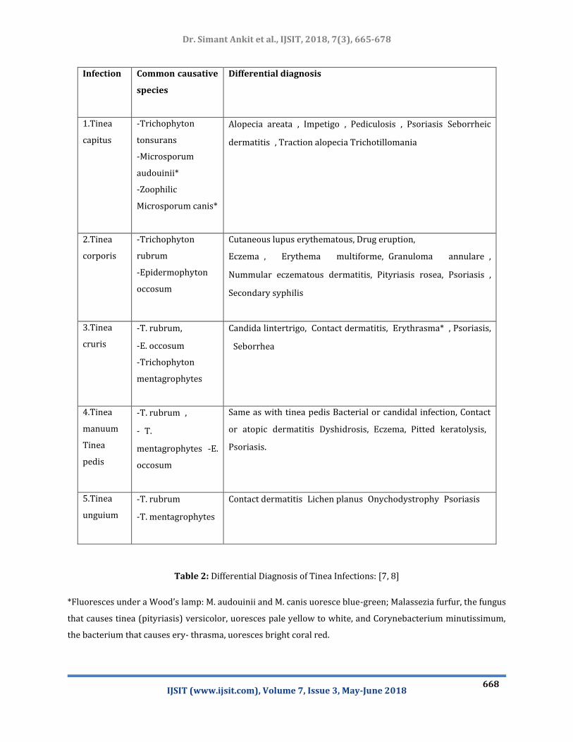
[pityriasis] versicolor) and Candida species. Differential diagnosis of tinea infections is given in the table 2.

et al., IJSIT, 2018, 7(3), 665-678 Dr. Simant Ankit
IJSIT (www.ijsit.com), Volume 7, Issue 3, May-June 2018
667
Table1: Diagnostic Methods for Dermatophyte Infections:[5, 6]
1.Potassium hydroxide (KOH) microscopy
Value: aids in visualizing hyphae and confirming the diagnosis of dermatophyte infection.
2.Wood’s lamp examination (ultraviolet light)
Value: generally, of limited usefulness, because most dermatophytes currently seen in the United States do
not fluoresce; may have value in the following situations:For diagnosing a brown, scaly rash in the scrotum
or axilla: erythrasma, caused by the bacterium Corynebacterium minutissimum, fluoresces a brilliant coral red,
whereas tinea cruris or cutaneous candidal infections do not fluoresce.
For diagnosing tinea (pityriasis) versicolor, which fluoresces pale yellow to white
For diagnosing tinea capitis caused by two zoophilic Microsporum species that fluoresce blue-green.
3.Fungal culture
Value: slow and expensive, but useful to confirm the diagnosis of onychomycosis when long-term oral therapy
is being considered.
4.Skin or nail biopsy
Value: may guide treatment decisions when the diagnosis is difficult to establish, a dermatophyte infection
has not responded to previous treatment, or KOH microscopy is negative in a patient with dystrophic nails.
et al., IJSIT, 2018, 7(3), 665-678 Dr. Simant Ankit
IJSIT (www.ijsit.com), Volume 7, Issue 3, May-June 2018
668
Infection
Common causative
species
Differential diagnosis
1.Tinea
capitus
-Trichophyton
tonsurans
-Microsporum
audouinii*
-Zoophilic
Microsporum canis*
Alopecia areata, Impetigo, Pediculosis, PsoriasisSeborrheic
dermatitis, Traction alopecia Trichotillomania
2.Tinea
corporis
-Trichophyton
rubrum
-Epidermophyton
occosum
Cutaneous lupus erythematous, Drug eruption,
Eczema, Erythema multiforme,Granuloma annulare,
Nummular eczematous dermatitis, Pityriasis rosea,Psoriasis,
Secondary syphilis
3.Tinea
cruris
-T. rubrum,
-E. occosum
-Trichophyton
mentagrophytes
Candida lintertrigo,Contact dermatitis,Erythrasma*, Psoriasis,
Seborrhea
4.Tinea
manuum
Tinea
pedis
-T. rubrum,
-T.
mentagrophytes -E.
occosum
Same as with tinea pedis Bacterial or candidal infection, Contact
or atopic dermatitis Dyshidrosis,Eczema,Pitted keratolysis,
Psoriasis.
5.Tinea
unguium
-T. rubrum
-T. mentagrophytes
Contact dermatitisLichen planusOnychodystrophyPsoriasis
Table 2: Differential Diagnosis of Tinea Infections: [7, 8]
*Fluoresces under a Wood’s lamp: M. audouinii and M. canis uoresce blue-green; Malassezia furfur, the fungus
that causes tinea (pityriasis) versicolor, uoresces pale yellow to white, and Corynebacterium minutissimum,
the bacterium that causes ery- thrasma, uoresces bright coral red.

et al., IJSIT, 2018, 7(3), 665-678 Dr. Simant Ankit
IJSIT (www.ijsit.com), Volume 7, Issue 3, May-June 2018
669
Tinea Capitis:
Tinea capitis, the most common dermatophytosis in children, is an infection of the scalp and hair
shafts[9]. Transmission is fostered by poor hygiene and overcrowding, and can occur through contaminated
hats, brushes, pillowcases, and other inanimate objects. After being shed, affected hairs can harbor viable
organisms for more than one year. Tinea capitis is characterized by irregular or well-demarcated alopecia
and scaling. When swollen hairs fracture a few millimeters from the scalp, “black dot” alopecia is
produced[10]. Tinea scalp infection also may result in a cell- mediated immune response termed a “kerion,”
which is a boggy , sterile, inflammatory scalp mass[11]. Cervical and occipital lymphadenopathy may be
prominent. Today, about 90 to 95 percent of tinea scalp infections in adults and children are caused by
Trichophyton tonsurans, which does not fluoresce. Therefore, Wood’s lamp examination has become a less
useful diagnostic test for tinea capitis[12]. Tinea capitis is generally identified by the presence of branching
hyphae and spores on KOH microscopy. If hyphae and spores are not visualized, Wood’s lamp examination
can be performed. If KOH microscopy and Wood’s lamp examinations are negative, fungal culture may be
considered when tinea capitis is strongly suspected. Alternatively, clinical features can point to the diagnosis.
In one study[13], tinea capitis was confirmed by culture in 92 percent of children who had at least three of
the following clinical features: scalp scaling, scalp pruritus, occipital adenopathy, and diffuse, patchy, or
discrete alopecia. When scaling and inflammation are prominent, other diagnoses to consider include
seborrheic dermatitis (no hair loss), atopic dermatitis (lesions in flexural folds of the neck, arms, or legs), and
psoriasis (nail changes and silvery scales on the knees or elbows). When alopecia is prominent, diagnoses to
rule out include alopecia areata (complete, rather than patchy, hair loss), traction alopecia (history of tight
hair braiding), and trichotillomania (hairs of differing lengths and a history of obsessive hair manipulation).
Topical treatment is not effective for tinea capitis. Systemic antifungal therapy is required to
penetrate the hair follicles. Griseofulvin is the only agent that the U.S. Food and Drug Administration (FDA)
has labeled for the treatment of tinea capitis[14]. Although griseofulvin remains the gold standard, it is a less
than ideal agent for several reasons[15],resistant organisms require dosage increases to affect a cure;
treatment must be continued for six to 12 weeks; relapse rates are high because of rapid clearance of the drug
from the skin with the cessation of therapy; and the liquid form for young children is a bit- ter-tasting
solution. Compared with griseofulvin, ketoconazole is no more effective and has the potential for adverse
hepatic effects and drug interactions[16]. In one study involving a small number of children, treatment with
itraconazole, in a dosage of 3 to 5 mg per kg per day for four weeks, resulted in clinical and mycologic cure
rates of 90 to 100 percent[17] [Evidence level B, nonrandomized clinical trial] .Fluconazole and terbinafine
are promising agents; randomized, comparative studies with griseofulvin should clarify their role in the
treatment of tinea capitis[18].One randomized trial[19]in patients with tinea capitis caused by Trichophyton
species showed that treatment with terbinafine, fluconazole, or itraconazole for two weeks was as effective as

et al., IJSIT, 2018, 7(3), 665-678 Dr. Simant Ankit
IJSIT (www.ijsit.com), Volume 7, Issue 3, May-June 2018
670
six weeks of griseofulvin therapy.
Adjunctive topical therapy with selenium sulfide (e.g., Exsel), ketoconazole, or povidone iodine
(Betadine) lotion or shampoo (applied for five minutes twice weekly) is useful to decrease shedding of viable
fungi and spores[20]; over-the-counter 1 percent selenium sulfide shampoo works as well as the prescription
2.5 percent strength[21].
Tinea Corporis:
Tinea corporis or ringworm[22], typically appears as single or multiple, annular, scaly lesions with
central clearing, a slightly elevated, reddened edge, and sharp margination on the trunk, extremities or face.
The border of the lesion may contain pustules or follicular papules. Itching is variable. The diagnosis of tinea
corporis is based on clinical appearance and KOH examination of skin [23]scrapings from the active edge. The
differential diagnosis [24]includes nummular eczema, pityriasis rosea, Lyme disease, tinea versicolor, contact
dermatitis, granuloma annulare, and psoriasis. Previous topical corticosteroid use can alter the appearance of
the lesions, so that raised edges with central clearing are not present. Corticosteroid use may also be a factor
in the development of Majocchi’s granuloma, a deep follicular tinea infection that usually involves the legs
and is more common in women[22]. Treatment of tinea corporis usually consists of measures to decrease
excessive skin moisture and the use of topical antifungal creams[25]as shown in table 3. Rarely, widespread
infections may require systemic therapy.
Tinea Barba:
Tinea barbae involves the skin and coarse hairs of the beard and mustache area[26]. This
dermatophyte infection occurs in adult men and hirsute women[26]. Because the usual cause is a zoophilic
organism, farm workers are most often affected. Tinea barbae may cause scaling, follicular pustules, and
erythema. The differential diagnosis[27] includes bacterial folliculitis, perioral dermatitis, pseudo folliculitis
barbae, contact dermatitis, and herpes simplex. One clue to the diagnosis is that hair removal is painless in
tinea barbae but painful in bacterial infections[28]. Like tinea capitis, tinea barbae is treated with oral
antifungal therapy as shown in table 3. Treatment is continued for two to three weeks after resolution of the
skin lesions.
Tinea Faciei:
Tinea faciei tends to occur in the non- bearded area of the face. The patient may complain of itching
and burning, which become worse after sunlight exposure[29]. Some round or annular red patches are
present. Often, however, red areas may be indistinct, especially on darkly pigmented skin, and lesions may
have little or no scaling or raised edges. Because of the subtle appearance, this dermatophytosis is sometimes

et al., IJSIT, 2018, 7(3), 665-678 Dr. Simant Ankit
IJSIT (www.ijsit.com), Volume 7, Issue 3, May-June 2018
671
known as “tinea incognito”[30]. The differential diagnosis includes seborrheic dermatitis, rosacea, discoid
lupus erythematosus, and contact dermatitis[31]. A high index of suspicion, along with a KOH microscopy of
scrapings from the leading edge of the skin change, may help in establishing the diagnosis[32]. Treatment is
similar to that for tinea corporis as shown in table 3.
Tinea Manuum:
Tinea manuum is a fungal infection of one or, occasionally, both hands. It often occurs in patients
with tinea pedis[33]. The palmar surface is diffusely dry and hyperkeratotic. When the fingernails are
involved, vesicles and scant scaling may be present, and the condition resembles dyshidrotic eczema. The
differential diagnosis[34] includes contact dermatitis, psoriasis, and callus formation. Topical antifungal
therapy and the application of emollients containing lactic acid are effective[35]. Relapses may be frequent if
onychomycosis or tinea pedis is not resolved[35].
Tinea Cruris:
Tinea cruris, frequently called “jock itch,” is a dermatophyte infection of the groin[36]. This
dermatophytosis is more common in men than in women and is frequently associated with tinea pedis[23].
Tinea cruris occurs when ambient temperature and humidity are high. Occlusion from wet or tight-fitting
clothing provides an optimal environment for infection. Tinea cruris affects the proximal medial thighs and
may extend to the buttocks and abdomen. The scrotum tends to be spared. Patients with this
dermatophytosis frequently complain of burning and pruritus[27]. Pustules and vesicles at the active edge of
the infected area, along with maceration, are present on a background of red, scaling lesions with raised
borders. The feet should be evaluated as a source of the infection. Adjunctive treatment can include a low-
dose corticosteroid (e.g., 2.5 percent hydrocortisone ointment for the first few days. Rarely, systemic
antifungal therapy is needed for refractory tinea cruris. Patient education on avoiding prolonged exposure to
moisture and keeping the affected area dry is important.
Tinea Pedis:
Tinea pedis, or athlete’s foot, has three common presentations[37]. The interdigital form of tinea
pedis is most common. It is characterized by fissuring, maceration, and scaling in the interdigital spaces of the
fourth and fifth toes[38]. Patients with this infection complain of itching or burning. A second form, usually
caused by Trichophyton rubrum[39], has a moccasin-like distribution pattern in which the plantar skin
becomes chronically scaly and thickened, with hyperkeratosis and erythema of the soles, heels, and sides of
the feet. The vesiculobullous form of tinea pedis is characterized by the development of vesi- cles, pustules,
and sometimes bullae in an inflammatory pattern, usually on the soles[38]. The differential diagnosis includes
contact dermatitis, eczema, and pustular psoriasis[27]. Streptococcal cellulitis is a potential complication of

et al., IJSIT, 2018, 7(3), 665-678 Dr. Simant Ankit
IJSIT (www.ijsit.com), Volume 7, Issue 3, May-June 2018
672
all three forms of tinea pedis. Streptococcal infection of normal skin is unlikely. However, the presence of
fungal maceration and fissuring permits streptococci to colonize the web spaces between the toes in patients
with tinea pedis. The clinical features of symptomatic athlete’s foot are a result of the inter- action of fungi
and bacteria. Treatment of tinea pedis involves application of an antifungal cream to the web spaces and
other infected areas[40]. Infrequently, systemic therapy is used for refractory infections. In several studies,
twice-daily application of the allylamine terbinafine resulted in a higher cure rate than twice-daily application
of the imidazole clotrimazole (Lotrimin; 97 percent versus 84 percent), and at a quicker rate (one week for
terbinafine versus four weeks for clotrimazole)[41]. A pharmacoeconomic analysis of tinea treatments found
topical terbinafine to be more cost-effective than imidazole or ciclopirox cream . When marked inflammation
and vesicle formation occur and signs of early cellulitis are present, the addition of a systemic or topical
antibiotic with streptococcal coverage is warranted. Reinfection is common, especially if onychomycosis is
present[6]. Nail infections should be treated. In addition, footwear should be disinfected, and patients with
tinea pedis should avoid walking barefoot in public areas such as locker rooms. Other measures to reduce
recurrence include controlling hyper- hidrosis with powders and wearing absorbent socks and nonocclusive
shoes[42].
Tinea Unguium:
Tinea unguium, a dermatophyte infection of the nail, is a subset of onychomycosis, which also may be
caused by yeast and non-dermatophyte molds[43]. Risk factors for this infection include aging, diabetes,
poorly fit- ting shoes, and the presence of tinea pedis. Onychomycosis accounts for about 40 to 50 percent of
nail dystrophies[44] The differential diagnosis includes trauma, lichen planus, psoriasis, nail-bed tumor,
peripheral vascular disease, atopic dermatitis, contact dermatitis, and yellow nail syndrome. Because
onychomycosis requires expensive, prolonged therapy (three to four months for fingernail infections and four
to six months for toenail infections), the diagnosis should be confirmed before treatment is initiated[7].
Periodic acid-Schiff staining with histologic examination of the clipped, distal free edge of the nail and
attached subungual debris is the most sensitive diagnostic method and is painless for patients[45]. Tinea
unguium, especially of the toenails, is difficult to eradicate. Topical agents have low efficacy. Mycologic cure
rates for ciclopirox nail lacquer, applied daily for up to 48 weeks, have ranged from 29 to 47 percent[45]. Oral
treatment with griseofulvin must be continued for 12 to 24 months, and ketoconazole carries a risk of
hepatotoxicity. Fluconazole has not been studied extensively in the treatment of onychomycosis and is not
labeled by the FDA for this indication. Mycologic and clinical cure rates are similar for 12 weeks of treatment
with itraconazole in a dosage of 200 mg per day and terbinafine in a dosage of 250 mg per day as shown in
table 3[40]. Itraconazole costs more for the same regimen. Continuous terbinafine therapy has a better
mycologic cure rate than intermittent or “pulse” terbinafine therapy, in which 500 mg of terbinafine is given
once daily for seven days of each of four months (94 percent versus 80 percent); how- ever, continuous
treatment is more expensive. Intermittent itraconazole therapy, in a dosage of 400 mg per day for seven days

et al., IJSIT, 2018, 7(3), 665-678 Dr. Simant Ankit
IJSIT (www.ijsit.com), Volume 7, Issue 3, May-June 2018
673
of each of four months, and intermittent terbinafine therapy are similarly effective.
Newer Topical Antifungals:
Luliconazole, an azole antifungal has fungicidal action against Trichophyton species similar to or
more than that of terbinafine. Available in 1% cream formulation, it is effective as once daily application for
1–2 weeks for dematophytic infection. Approved by the US Food and Drug Administration for the treatment
of interdigital tinea pedis, tinea cruris, and tinea corporis, it has a favorable safety profile[46]. Econazole
nitrate foam preparation has also shown its efficacy over foam vehicle for tinea pedis[47]. However, these
newer drugs are costlier which in turn may lead to issues of adherence to treatment in resource-poor
settings, and may predispose to development of resistance.
Newer Oral Antifungal Agents:
There is lack of any recent literature regarding systemic antifungals in the treatment of tinea cruris
and corporis. Although few newer systemic antifungals have been approved in last two decades but most of
them are reserved for more severe life-threatening invasive systemic mycoses with paucity of evidence on
efficacy in superficial mycoses. Recently, posoconazole was found to be effective in a patient with extensive
dermatophytic skin and nail infection with underlying CARD9[48].
New and Potential Therapies:
Other than the antifungals already mentioned, few plant extract (Chinese herbals) are also found to
be effective against common dermatophytic infection. One of them is macrocarpal C, an active ingredient
obtained from the fresh leaves of Eucalyptus globulus Labill with antifungal action against T.
mentagrophytes and T. rubrum[49].Demicidin, an antimicrobial peptide has antifungal action at a
concentration normally present in sweat providing an insight to newer therapeutic target for dermatophytic
infection[50].

et al., IJSIT, 2018, 7(3), 665-678 Dr. Simant Ankit
IJSIT (www.ijsit.com), Volume 7, Issue 3, May-June 2018
674
Table 3: Antifungal Agent for Treatment for Tinea Infestions [13, 23, 27]
CONCLUSION
Dermatophytes are fungi that invade and multiply within keratinized tissues (skin, hair, and nails)
causing infection. Based upon their genera, dermatophytes can be classified into three
groups: Trichophyton (which causes infections on skin, hair, and nails), epidermophyton (which causes
Drug Doses
1.Tinea unguium
-Terbinafine
-Itraconazole
250 mg orally every day for 12 weeks† or
500 mg orally every day during the rst
week of each month for four month
200 mg orally every day for 12 weeks
400 mg orally every day during the rst
week of each month for four months
2.Tinea capitis, barbae
-Griseofulvin, micronized (Grifulvin)
-Terbinafine
20 mg per kg per day for eight weeks
62.5 mg per day for four weeks
3.Tinea corporis, pedis, cruris, faciei
-Butenafine (Mentax)
-Terbinafine
-Miconazole (Micatin)
-Clotrimazole (Lotrimin AF)
Applied to the lesion and a 2-cm area
surrounding the lesion once daily for
approximately 14 days
Applied to the lesion and a 2-cm area
surrounding the lesion twice daily for
approximately 14 days
Applied to the lesion and a 2-cm area
surrounding the lesion twice daily for
approximately 14 days
Applied to the lesion and a 2-cm area
surrounding the lesion twice daily for
approximately 14 days
et al., IJSIT, 2018, 7(3), 665-678 Dr. Simant Ankit
IJSIT (www.ijsit.com), Volume 7, Issue 3, May-June 2018
675
infections on skin and nails), and Microsporum (which causes infections on skin and hair). Based upon mode
of transmission, these have been classified as anthropophillic, zoophilic, and geophilic. Finally, based upon the
affected site, these have been classified clinically into tinea capitis (head), tinea faciei (face), tinea barbae
(beard), tinea corporis (body), tinea manus (hand), tinea cruris (groin), tinea pedis (foot), and tinea unguium
(nail). Dermatophyte infections can be readily diagnosed based on the history, physical examination, and
potassium hydroxide (KOH) microscopy. Diagnosis occasionally requires Wood’s lamp examination and
fungal culture or histologic examination. Topical therapy is used for most dermatophyte infections. Cure rates
are higher and treatment courses are shorter with topical fungicidal allylamines than with fungistatic azoles.
Oral therapy is preferred for tinea capitis, tinea barbae, and onychomycosis. Orally administered griseofulvin
remains the standard treatment for tinea capitis. Topical treatment of onychomycosis with ciclopirox nail
lacquer has a low cure rate. For ony- chomycosis, “pulse” oral therapy with the newer imidazoles
(itraconazole or fluconazole) or allylamines (terbinafine) is considerably less expensive. Although there is
sufficient evidence to demonstrate the efficacy of topical antifungals in limited disease yet, there is scarce
data on the frequency of relapse once topical monotherapy is discontinued and need more research in the
future for more improvement. Appropriate follow-up duration and education of patients on proper foot
hygiene are also important components in providing effective therapy.
Conflict Of Interest:
There are no conflicts of interest.
Acknowledgement:
This research is supported by the National Natural Science Foundation of China (31700736), Hubei
Province Natural Science Foundation of China (2016CFB180) and Hubei Province Health and Family Planning
Scientific Research Project (WJ2016Y07).
REFERENCES
1. Segal, E. and M. Frenkel, Dermatophyte infections in environmental contexts. Res Microbiol, 2015. 166(7):
p. 564-9.
2. Bamba, S., et al., Burden of Severe Fungal Infections in Burkina Faso. J Fungi (Basel), 2018. 4(1).
3. Lecha, M., et al., Treatment options--development of consensus guidelines. J Eur Acad Dermatol Venereol,
2005. 19 Suppl 1: p. 25-33.
4. Nenoff, P., et al., Mycology - an update. Part 1: Dermatomycoses: causative agents, epidemiology and
pathogenesis. J Dtsch Dermatol Ges, 2014. 12(3): p. 188-209; quiz 210, 188-211; quiz 212.
5. Kelly, B.P., Superficial fungal infections. Pediatr Rev, 2012. 33(4): p. e22-37.
6. Effendy, I., et al., Epidemiology and clinical classification of onychomycosis. J Eur Acad Dermatol Venereol,

et al., IJSIT, 2018, 7(3), 665-678 Dr. Simant Ankit
IJSIT (www.ijsit.com), Volume 7, Issue 3, May-June 2018
676
2005. 19 Suppl 1: p. 8-12.
7. Ely, J.W., S. Rosenfeld, and M. Seabury Stone, Diagnosis and management of tinea infections. Am Fam
Physician, 2014. 90(10): p. 702-10.
8. Moriarty, B., R. Hay, and R. Morris-Jones, The diagnosis and management of tinea. Bmj, 2012. 345: p.
e4380.
9. Gupta, A.K., et al., Tinea capitis in children: a systematic review of management. J Eur Acad Dermatol
Venereol, 2018.
10. Elghblawi, E., Tinea Capitis in Children and Trichoscopic Criteria. Int J Trichology, 2017. 9(2): p. 47-49.
11. Fremerey, C. and P. Nenoff, Tinea Capitis in a Newborn. N Engl J Med, 2018. 378(21): p. 2022.
12. Farokhipor, S., et al., Characterizing the clinical isolates of dermatophytes in Hamadan city, Central west of
Iran, using PCR-RLFP method. J Mycol Med, 2018. 28(1): p. 101-105.
13. Chen, X., et al., Systemic antifungal therapy for tinea capitis in children. Cochrane Database Syst Rev,
2016(5): p. Cd004685.
14. Meadows-Oliver, M., Tinea capitis: diagnostic criteria and treatment options. Dermatol Nurs, 2009. 21(5):
p. 281-6.
15. Tan, C.W., et al., A Review of Tinea Capitis in a Cohort of Asian Children. Ann Acad Med Singapore, 2018.
47(4): p. 156-158.
16. Bennett, M.L., et al., Oral griseofulvin remains the treatment of choice for tinea capitis in children. Pediatr
Dermatol, 2000. 17(4): p. 304-9.
17. Elewski, B.E., et al., Terbinafine hydrochloride oral granules versus oral griseofulvin suspension in children
with tinea capitis: results of two randomized, investigator-blinded, multicenter, international, controlled
trials. J Am Acad Dermatol, 2008. 59(1): p. 41-54.
18. Chan, Y.C. and S.F. Friedlander, New treatments for tinea capitis. Curr Opin Infect Dis, 2004. 17(2): p. 97-
103.
19. Bhanusali, D., et al., Treatment outcomes for tinea capitis in a skin of color population. J Drugs Dermatol,
2012. 11(7): p. 852-6.
20. Higgins, E.M., L.C. Fuller, and C.H. Smith, Guidelines for the management of tinea capitis. British Association
of Dermatologists. Br J Dermatol, 2000. 143(1): p. 53-8.
21. Givens, T.G., M.M. Murray, and R.C. Baker, Comparison of 1% and 2.5% selenium sulfide in the treatment of
tinea capitis. Arch Pediatr Adolesc Med, 1995. 149(7): p. 808-11.
22. Sonthalia, S., A. Singal, and S. Das, Tinea cruris and tinea corporis masquerading as tinea indecisiva: case
report and review of the literature. J Cutan Med Surg, 2015. 19(2): p. 171-6.
23. El-Gohary, M., et al., Topical antifungal treatments for tinea cruris and tinea corporis. Cochrane Database
Syst Rev, 2014(8): p. Cd009992.
24. van Zuuren, E.J., Z. Fedorowicz, and M. El-Gohary, Evidence-based topical treatments for tinea cruris and
tinea corporis: a summary of a Cochrane systematic review. Br J Dermatol, 2015. 172(3): p. 616-41.

et al., IJSIT, 2018, 7(3), 665-678 Dr. Simant Ankit
IJSIT (www.ijsit.com), Volume 7, Issue 3, May-June 2018
677
25. Sardana, K., et al., Is Antifungal Resistance a Cause for Treatment Failure in Dermatophytosis: A Study
Focused on Tinea Corporis and Cruris from a Tertiary Centre? Indian Dermatol Online J, 2018. 9(2): p. 90-
95.
26. Singh, S., et al., Tinea barbae presenting as kerion. Indian J Dermatol Venereol Leprol, 2017. 83(6): p. 741.
27. Kaushik, N., G.G. Pujalte, and S.T. Reese, Superficial Fungal Infections. Prim Care, 2015. 42(4): p. 501-16.
28. Sidwell, R.U., et al., Trichophyton erinacei kerion barbae from a hedgehog with direct osculatory transfer to
another person. Clin Exp Dermatol, 2014. 39(1): p. 38-40.
29. Gioseffi, M.L., et al., [Tinea faciei]. Arch Argent Pediatr, 2013. 111(1): p. 74-6.
30. Lin, R.L., J.C. Szepietowski, and R.A. Schwartz, Tinea faciei, an often deceptive facial eruption. Int J
Dermatol, 2004. 43(6): p. 437-40.
31. Bertelsen, T. and K.A.U. Pallesen, [Tinea faciei]. Ugeskr Laeger, 2017. 179(29).
32. Dev, T., H. Saginatham, and G. Sethuraman, Tinea Faciei: Challenges in the Diagnosis. J Pediatr, 2017. 187:
p. 331.
33. Perrier, P. and M. Monod, Tinea manuum caused by Trichophyton erinacei: first report in Switzerland. Int J
Dermatol, 2015. 54(8): p. 959-60.
34. Zhan, P., et al., The epidemiology of tinea manuum in Nanchang area, South China. Mycopathologia, 2013.
176(1-2): p. 83-8.
35. Kiraz, N., et al., The prevalence of tinea pedis and tinea manuum in adults in rural areas in Turkey. Int J
Environ Health Res, 2010. 20(5): p. 379-86.
36. Sahoo, A.K. and R. Mahajan, Management of tinea corporis, tinea cruris, and tinea pedis: A comprehensive
review. Indian Dermatology Online Journal, 2016. 7(2): p. 77-86.
37. Ilkit, M. and M. Durdu, Tinea pedis: the etiology and global epidemiology of a common fungal infection. Crit
Rev Microbiol, 2015. 41(3): p. 374-88.
38. Canavan, T.N. and B.E. Elewski, Identifying Signs of Tinea Pedis: A Key to Understanding Clinical Variables. J
Drugs Dermatol, 2015. 14(10 Suppl): p. s42-7.
39. Kawai, M., et al., A retrospective cohort study of tinea pedis and tinea unguium in inpatients in a psychiatric
hospital. Med Mycol J, 2014. 55(2): p. E35-41.
40. Badri, T., Comment on "Prevalence, Etiology, and Risk Factors of Tinea Pedis and Tinea Unguium in Tunisia".
Can J Infect Dis Med Microbiol, 2018. 2018: p. 4859514.
41. Wang, R., et al., Skin microbiome changes in patients with interdigital tinea pedis. Br J Dermatol, 2018.
42. Hoffman, L.K., I. Raymond, and L. Kircik, Treatment of Signs and Symptoms (Pruritus) of Interdigital Tinea
Pedis With Econazole Nitrate Foam, 1. J Drugs Dermatol, 2018. 17(2): p. 229-232.
43. Miura, Y., et al., Screening for tinea unguium by thermography in older adults with subungual
hyperkeratosis. Geriatr Gerontol Int, 2015. 15(8): p. 991-6.
44. Harada, T., [Tinea unguium]. Med Mycol J, 2011. 52(2): p. 77-95.
45. Tolstrup, J., et al., [Diagnosing and treating of onychomycosis]. Ugeskr Laeger, 2018. 180(20).

et al., IJSIT, 2018, 7(3), 665-678 Dr. Simant Ankit
IJSIT (www.ijsit.com), Volume 7, Issue 3, May-June 2018
678
46. Khanna, D. and S. Bharti, Luliconazole for the treatment of fungal infections: an evidence-based review. Core
Evid, 2014. 9: p. 113-24.
47. Elewski, B.E. and T.C. Vlahovic, Econazole nitrate foam 1% for the treatment of tinea pedis: results from
two double-blind, vehicle-controlled, phase 3 clinical trials. J Drugs Dermatol, 2014. 13(7): p. 803-8.
48. Jachiet, M., et al., Posaconazole treatment of extensive skin and nail dermatophytosis due to autosomal
recessive deficiency of CARD9. JAMA Dermatol, 2015. 151(2): p. 192-4.
49. Wong, J.H., et al., Antifungal mode of action of macrocarpal C extracted from Eucalyptus globulus Labill
(Lan An) towards the dermatophyte Trichophyton mentagrophytes. Chin Med, 2015. 10: p. 34.
50. Arai, S., et al., Mycostatic effect of recombinant dermcidin against Trichophyton rubrum and reduced
dermcidin expression in the sweat of tinea pedis patients. J Dermatol, 2015. 42(1): p. 70-6.