diagnosis and treatment of aspergillosis in an ostrich...
TRANSCRIPT

Kafkas Univ Vet Fak Derg17 (4): 671-674, 2011DOI:10.9775/kvfd.2011.3594
CASE REPORT
SummaryThis study consists of the clinical, microbiological and pathological findings, and the results of Amphoterisin B and Biostarter for
supported treatment, of focal aspergillosis in a flock of ostriches. The clinical signs were listlessness, anorexia, diarrhoea, increased respiration, dyspnoea, and mucoid discharge from the nostrils. At post-mortem examination caseous nodules were observed in various organs. Histopathological examination of the lungs, air sacs and the pleural membrane showed in different sizes in different parts of necrosis in the center of the surrounding foreign body giant cells, epitheloid macrophages, lymphocytes and granulomas surrounded by a fibrous connective tissue. In treatment, Amphotericin B and Biostarter was given orally as a supported treatment. There were no sick birds after the treatment. As a conclusion, aspergillosis could be treated with amphotericin B and as a supported treatment Biostarter, especially in the early stages of the disease.
Keywords: Ostrich, Struthio camelus, Aspergillosis, Amphotericin B, Biostarter
Bir Deve Kuşu Sürüsünde Aspergillozis’in Teşhis ve Tedavisi
ÖzetBu çalışma, deve kuşu sürüsünde gözlenen aspergillosisin klinik, mikrobiyolojik, histopatolojik bulguları ile Amphotericin B ve
destekleyici amaçla Biomaya ile sağaltım sonuçlarını içermektedir. Klinik muayenede ilgisizlik, iştahsızlık, ishal, solunum sayısı artışı, dispnea ve mukoid bir burun akıntısı tespit edildi. Nekropside çeşitli iç organlarda kazeöz-nodüler odaklar görüldü. Histopatolojik muayene de ise akciğerde, hava kesesi membranlarında ve pleuranın degisik kesimlerinde farklı büyüklüklerde, merkezinde kazeifikasyon nekrozu bulunan çevresi yabancı cisim dev hücreleri, epiteiloid makrofajlar, lenfositler ve fibröz bir bağ doku ile çevrili granulomlar görüldü. Sağaltımda Amphotericin B ve destekleyici amacıyla Biomaya verildi. Sağaltımdan sonra hayvanların hiç birinde hastalık görülmedi. Sonuç olarak özelikle hastalığın ilk döneminde Amphoterisin B ve destekleyici olarak Biomaya kullanımının tedavide başarılı olabileceği kanaatine varıldı.
Anahtar sözcükler: Deve kuşu,Struthio camelus, Aspergillozis, Amfoterisin B, Biomaya
Diagnosis and Treatment of Aspergillosis in An Ostrich Flock
Hasan İÇEN * Nuretin IŞIK ** Simten YEŞİLMEN *** Mehmet TUZCU **** Servet SEKİN *
***
*******
Department of Internal Medicine, Faculty of Veterinary Medicine, Dicle University, TR-21280 Diyarbakır - TURKEYLaboratuary of Research, Diagnosis and Control of Animal Diseases, microbiology, TR-21010 Diyarbakır - TURKEYDepartment of Microbiology Faculty of Veterinary Medicine, Dicle University, TR-21280 Diyarbakır - TURKEYVeterinary Control and Research Institute, Pathology, TR-01170 Adana - TURKEY
Makale Kodu (Article Code): KVFD-2011-3594
In recent years, increasing attention has been paid to ostrich breeding. However, there is little information concerning the diseases of ostriches in Turkey 1. Aspergillosis is a debilitating disease and affects a variety of avian species including turkeys, chickens, waterfowl pigeons, quails, ostriches, and penguins 2-7. The clinical features of a fungal infection may be influenced by stress factors, mouldy straw, or long-term antibiotic treatment 8.
The disease is characterized by lesions of the internal organs, eyes and, in certain cases, the brain in poultry 9-11.
The disease may be chronic and insidious, or it may cause peracute death. Chronic aspergillosis is divided into focal -nasal, tracheal, cutaneous, and ophthalmic- and generalised forms 4. Lower respiratory infections such as aspergillosis may be evident as harsh lung sounds when the bird is ausculated 12. The most common clinical signs are lethargy, anorexia, depression, feed consumption decreases, and loss of weight. Mortality in young ostriches is 5-20 percent, but may be as high as 50 percent. Mortality in mature birds is usually less than 5 percent 13,14.
INTRODUCTION
İletişim (Correspondence) +90 412 2488020/8620 [email protected]

672Diagnosis and Treatment of...
A high prevalence of aspergillosis has been reported among ostriches in Israel, Iran, Turkey, and Saudi Arabia 2,15-17. The disease is usually diagnosed at post- mortem examination, often based on the observation of white caseous nodules in the lungs or air sacs of affected birds 18. The diagnosis can be confirmed by laboratory culture of lung nodule portions with Aspergillus sp. isolation or by histopathological analysis. However, in animals clinically suspected of aspergillosis, X-rays, broncho- scopy, tracheal swabs and serology may be used as diagnostic methods 18. The target tissues are lungs and air sacs; gross pathologic lesions include caseous nodules or plaques, and massive granulomas with necrotic cores can be seen on histopathology 14.
Nistatin, amfoterisin-B, flusitosine, ketoconazol, itra- conazol, fluconazol and enilconazol are used for the treatment of aspergillosis. Focal aspergillosis shows a better response to treatment, while in the generalised form, the treatment is prolonged, usually ineffective, and the prognosis is poor 17,18.
This study presents the clinical, microbiological, and histopathological findings and treatment of an aspergillosis in ostrich flock.
CASE HISTORY
This study was performed in a flock of ostrich with focal aspergillosis. Eight young ostriches had died, and the two dead birds were brought to the Veterinary Faculty of Dicle University in December 2008. At the anamnesis, before feeding with corn silage, the ostrich were fed with clean water and feed, comprised of barley, maize, and wheat mixture, ad libitum. When the ostriches were fed with rotten corn silage, the disease was seen five days later; eight ostriches died and six were severely ill. All the ostriches prior to referral to the Veterinary Faculty had been treated with oxytetracycline, gentamycine, enro-floxacin, and clostridal vaccination by the local veterinary surgeon, but no clinical response was observed. At the clinical examinations made by researcher, listlessness, anorexia, diarrhoea, increased respiration, dyspnoea, open mouthed breathing, and mucoid discharge from the nostrils were seen.
Mycological Examinations
Tracheal and pharyngeal swabs and samples of the lungs and air sacs were collected from the dead ostriches onto sterile glass petri plates. The tissue samples were inoculated on Sabouraud dextrose agar with chloramphenicol (0.05 mg/mL) and incubated at 25°C and 37°C under aerobic conditions for 3-5 days. The samples of litter and feed (corn silage) were suspended separately in 10 ml of Sabouraud liquid medium and incubated for 30 min at 25°C. Afterwards, the samples were shaken; the supernatant was collected
after 3-5 min, filtered through aseptic gauze and finally sown at a volume of 0.2 ml on to Sabouraud dextrose agar with chloramphenicol (0.05 mg/mL) and incubated at 25°C and 37°C under aerobic conditions for 3-5 days.
Histopathological Examinations
The samples of the lungs, trachea, pharynx, and thoracic air sacs were fixed in 10% neutral buffered formalin, routinely processed and embedded in paraffin blocks. The 5 µm sections were stained with haematoxylin and eosin (HE) by the periodic acid-Schiff (PAS) method. The morphological evaluation of the samples was performed under a light microscope.
Treatment Protocol
Treatment consisted of Amphotericin B (Abalecet 100 mg) 1 mg/kg intravenously once daily for 3 days in 5% Dextrose 100 ml and Biostarter (Akpe Biyomaya ingredient (SCC 3.5x10 CFU, Active Clinoptilolite 500.000 mg, Biotin 1.3 (U/g), Thiamine 60-100 (U/g), Riboflavin 35-50 (U/g), Pantothenate, 70 (U/g), Folic acid 5-13 (U/g), Choline 4.000 (U/g), Niacin 300-500 (U/g) ) orally for a week.
The ostriches showed signs of listlessness, anorexia, dyspnoea, open mouthed breathing, increased respiration, mucoid discharge from nostrils, and watery diarrhoea. Eight cases of death were noted and six ostriches were severely ill. The mycological examinations demonstrated the presence of A. fumigatus cells in swabs from the lungs and air sacs examined post-mortem. The culture examinations revealed an intensive growth of A. fumigatus in all cases. The mycological examinations of litter and rotten corn silage showed evident contamination.
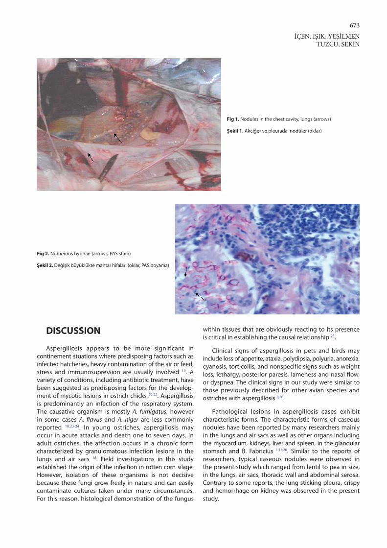
At post-mortem examination of the dead ostriches, caseous nodules were observed in the chest cavity, lungs, and also in the thoracic air sacs (Fig. 1). Some of these nodular structures on the surface were of a gray-white color, and some were blue-green. Similar nodules were also observed on the surface of the sternum, on bone, and on the heart. Nodules observed in the lungs and air sacs corresponded to acute aspergillosis lesions. Moreover, the lung sticking pleura and crispy and haemorrhage on the kidneys was observed. Histopathological analysis revealed caseous necrosis of different sizes in different parts of the lungs, air sacs and the pleural membranes in the centre of the surrounding foreign body giant cells, epithelioid macrophages, lymphocytes and granulomas surrounded by a fibrous connective tissue were seen. In the areas of necrosis in this granuloma numerous hyphae, mycelia and conidiospores were also seen in the middle of these lesions using H.E. and PAS stain (Fig. 2). Hyperemia in the liver and vacuolar degeneration of hepatocytes was also determined. Intertubular bleeding was seen in the kidneys. Two of the ill birds died during the treatment but four recovered and began eating. There were no sick birds after the treatment.
673
İÇEN, IŞIK, YEŞİLMENTUZCU, SEKİN
DISCUSSION
Aspergillosis appears to be more significant in continement stuations where predisposing factors such as infected hatcheries, heavy contamination of the air or feed, stress and immunosupression are usually involved 19. A variety of conditions, including antibiotic treatment, have been suggested as predisposing factors for the develop-ment of mycotic lesions in ostrich chicks 20-22. Aspergillosis is predominantly an infection of the respiratory system. The causative organism is mostly A. fumigatus, however in some cases A. flavus and A. niger are less commonly reported 18,23-24. In young ostriches, aspergillosis may occur in acute attacks and death one to seven days. In adult ostriches, the affection occurs in a chronic form characterized by granulomatous infection lesions in the lungs and air sacs 18. Field investigations in this study established the origin of the infection in rotten corn silage. However, isolation of these organisms is not decisive because these fungi grow freely in nature and can easily contaminate cultures taken under many circumstances. For this reason, histological demon stration of the fungus
within tissues that are obviously reacting to its presence is critical in establishing the causal relationship 25.
Clinical signs of aspergillosis in pets and birds may include loss of appetite, ataxia, polydipsia, polyuria, anorexia, cyanosis, torticollis, and nonspecific signs such as weight loss, lethargy, posterior paresis, lameness and nasal flow, or dyspnea. The clinical signs in our study were similar to those previously described for other avian species and ostriches with aspergillosis 8,26.
Pathological lesions in aspergillosis cases exhibit characteristic forms. The characteristic forms of caseous nodules have been reported by many researchers mainly in the lungs and air sacs as well as other organs including the myocardium, kidneys, liver and spleen, in the glandular stomach and B. Fabricius 1,13,26. Similar to the reports of researchers, typical caseous nodules were observed in the present study which ranged from lentil to pea in size, in the lungs, air sacs, thoracic wall and abdominal serosa. Contrary to some reports, the lung sticking pleura, crispy and hemorrhage on kidney was observed in the present study.
Fig 1. Nodules in the chest cavity, lungs (arrows)
Şekil 1. Akciğer ve pleurada nodüler (oklar)
Fig 2. Numerous hyphae (arrows, PAS stain)
Şekil 2. Değişik büyüklükte mantar hifaları (oklar, PAS boyama)

674Diagnosis and Treatment of...
Histopatholgy provides the best opportunity for definitive diagnosis of aspergillosis, since both the fungal organisms and associated granulomatous inflammation can be observed, thus confirming disease 1,3,8-10,13. The researchers have also observed the presence of Aspergillus hyphae and spores in caseous necrotic masses, as well as cell infiltrations consisting of giant cells, heterophils and macrophages together with necrotic granulomatous foci surrounded with an outer layer of connective tissue. Microscopic and mycological findings deter mined in the ostriches in this study were found to be similar to lesions detected in various poultry species diagnosed with aspergillosis 3,12,27,28.
Treatment of Aspergillosis generally involves administration of antifungal medications either loccaly or systematically. There are two main classes of antifungal drugs currently employed in the treatment of avian aspergillosis, the polyenes, such amphotericin B, and azoles including itraconazole, ketoconazole, fluconazole, enilconazole, nistatin 17,29,30. Amphotericin B is an amphoteric polyene macrolide and has poor absorption across the gastrointestinal mucosa. Thus must be administered intravenously or topically via nebulization. Because of this Amphotericin B was given by intravenously once daily for three days, and Biostarter orally for a week, in this study. Amphotericin B used as an antifungal and Biostarter were used as mineral based, toxin binding has been combined with clinoptiloite, and retains the balance in stomach intestinal microfauna, microflora and cellulose improves digestion, and as a result of the toxin is adsorbed by reducing the redox potential, prevents the production of aerobic pathogens. Enhance antibody production by increasing the activity of lymphocytes and regulate immunity. It was shown that Amphoterisin B were successful for treatment and Biostarter applications were helped as a supported treatment. The rotten corn silage was taken away and no more sick and dead ostriches were observed after the treatment.
In conclusion, treatment with amphotericin B were successful in aspergillosis and biostarter as supported treatment. Recognition and elimination of the infection source by disinfection with specific antifungal preparations proves to play a crucial role in the control of aspergillosis.
REFERENCES
1. Atasever A, Gumussoy KS: Pathological, clinical and mycological findings in experimental aspergillosis infections of starlings. J Vet Med A, 51, 19-22, 2004.
2. Khosravi AR, Shokri H, Ziglari T, Naeini AR, Mousavi Z, Hashemi H: Outbreak of severe disseminated aspergillosis in a flock of ostrich (Struthio camelus). Mycoses, 51, 557-559, 2008.
3. Akan M, Atasever A, Yardımcı H: Bir bıldırcın sürüsünde Aspergillus fumigatus infeksiyonu. Ankara Üniv Vet Fak Derg, 43, 147-150, 1996.
4. Tokarzewski S, Ziolkowska G, Lopuszynski W, Plotnicki ZN: Aspergillus fumigatus infection in a pigeon flock. Bull Vet Inst Pulawy, 51, 563-567, 2007.
5. Asan A: Aspergillus, penicillium, and related species reported from Turkey. Mycotaxon, 89 (1): 155-157, 2004.6. Beyaz L, Gümüşsoy KS, Çam Y, Abay S, Atasever A: Kayseri hayvanat bahçesinde bulunan bazi yabani kanatli türlerinde rastlanan sistemik aspergillozis. Ankara Üniv Vet Fak Derg, 55, 31-35, 2008.7. Cacciuttolo E, Rossi G, Nardoni S, Legrottalie R, Mani P: Anatomo-pathological aspects of avian aspergillosis. Vet Res Commun, 33, 521-527, 2009.8. Reissig EC, Uzal FA Schettino A, Robles CA: Pulmonary aspergillosis in a great rhea (Rhea americana), Avian Dis, 46, 754-756, 2002.9. Nardoni S, Ceccherelli R, Giacomo R, Manciati F: Aspergillosis in larus cachinnans miscaellis: Survey of eight cases. Mycopathologia, 161, 317-321, 2006.10. Bauck L: Mycoses In, Ritchie BW, Harrison GJ, Harrison LR (Eds): Avian Medicine: Principles and Application. pp. 997-1006, Wingers Publishing, Lake Worth, FL, 1994.11. Tell LA: Aspergillosis in mammals and birds: Impact on veterinary medicine. Medical Mycology Supplement, 43, 71-73, 2005.12. Cooper RG: Bacterial, Fungal and parasitic infections in the ostrich (Struthio camelus var. domesticus), Anim Sci J, 76, 97-106, 2005.13. Akkoç A, Yılmaz R, Cangül İT, Özyiğit MÖ: Pulmoner aspergillosis and amyloid accumulation in an ostrich. Turk J Vet Anim Sci, 33 (2): 157-160, 2009.14. Femenia F, Fontaine JJ, Lair-Fulleringer S, Berkova N, Huet D, Towanou N, Rakotovao F, Granet OI, Le Loch G, Arne P, Guillot J: Clinical, mycological and pathological findings in turkeys experimentally infected by Aspergillus fumigatus. Avian Pathol, 36 (3): 213-219, 2007.15. Elfaki MG, Abas B, Mahmoud OM, Harun EM, Abdel Magied EM: Septicaemic pasteurellosis in ostrich (Struthio camelus) in central Saudi Arabia. Vet J, 163, 218-221, 2002.16. Pérez J, García PM, Méndez A, Astorga R, Luque I, Tarradas C: Outbreak of aspergillosis in a flock of adult ostriches (Struthio camelus). Vet Rec, 153 (4): 124-125, 2003.17. Keskin O, Çimtay I: Devekuşlarında aspergillozisin tedavisinde keto-konazolun etkinliğinin araştırılması. Vet Mikrobiyol Derg, 2, 27-30, 2002.18. Rochette F, Engelen M, Bossche HV: Antifungal agents of use in animal health -practical applications. J Vet Pharmacol, 26, 31-53, 2003.19. Copetti MV, Segabinazi SD, Flores ML, Alves SH, Santurio JM: Pulmonary aspergillosis outbreak in rhea americana in southern Brazil. Mycopathologia, 157, 269-271, 2004.20. Sajid MA, Khan IA, Rauf U: Aspergillus fumigatus in commercial poultry flocks, a serious threat to poultry industry in Pakistan. J Anim Pl Sci, 16 (3-4): 79-81, 2006.21. Sancak AA: Aspergillosis and gastric impaction in an ostrich. Turk J Vet Anim Sci, 29, 933-935, 2005.22. Sennazlı G, Turan N, Gurel A, Yılmaz H: Bir deve kuşu çiftliğinde saptanan aspergillozis. İstanbul Üniv Vet Fak Derg, 27 (2): 459-467, 2001.23. Ozmen O, Sahinduran S, Haligur M, Albay MK: Deve kuşlarında pulmoner aspergilloziste patolojik bulgular ve tedavi. Erciyes Üniv Vet Fak Derg, 4, 1,1-4, 2007.24. Para PNS, Santos R de L: Aspergillosis in an ostriches (Struthio camelus) in Brazil. Ciencia Rural, Santa Maria, 34 (2): 573-576, 2004.25. Dahlhausen RD: Implications of Mycoses in Clinical Disorders. In, Harrison GJ, Lightfoot TL (Eds): Clinical Avian Medicine. pp. 691-704, Spix Publishing, Inc. Palm Beach, FL, 2006.26. Gulbahar MY, Agaoglu Z, Biyik H, Yuksek N: Zygomotic pro-ventriculitis and ventriculitis in ostriches (Struthio camelus) with impaction. Aust Vet J, 78 (4): 247-249, 2000.27. Perelman B, Kuttin E: Zygomycosis in ostriches. Avian Pathol, 21 (4): 675-680, 1992.28. Yokota T, Shibahara T, Wada Y, Hiraki R, Ishikawa Y, Kadota K: Aspergillus fumigatus infection in an ostrich (Struthio camelus), Avian Pathol, 66, 201-204, 2004.29. Plotnick AN: Lipid-based formulations of amphotericin B. JAVMA, 216, 838-841, 2000.30. Burges JL, Birchall R: Nephrotoxicity of amphotericin B, with emphasis on changes in tubular function. Am J Med, 53, 77-84, 1972.