diagnosis and treatment of learning objectives ocular ... · pdf fileblepharitis and dry eye...
TRANSCRIPT
Diagnosis and Treatment of Ocular Surface Conditions:
Focus on Blepharitis and Dry Eye
C. Lisa Prokopich, OD, MSc, FAAOUniversity of Waterloo, School of Optometry and Vision Science
COPE Course Code ID: 31662-SD Expires: 04/01/2014 Qualified Credit: 2 hours
Thank you also to Drs. Karpecki, Melton, Thomas, Bartlettand Michaud for their contributions.
Learning Objectives
After completing this lesson, optometrists will be able to:
Understand the epidemiology and etiology of blepharitis and dry eye
Diagnose blepharitis and dry eye
Manage and recommend treatment for these conditions
Counsel patients for better self-management
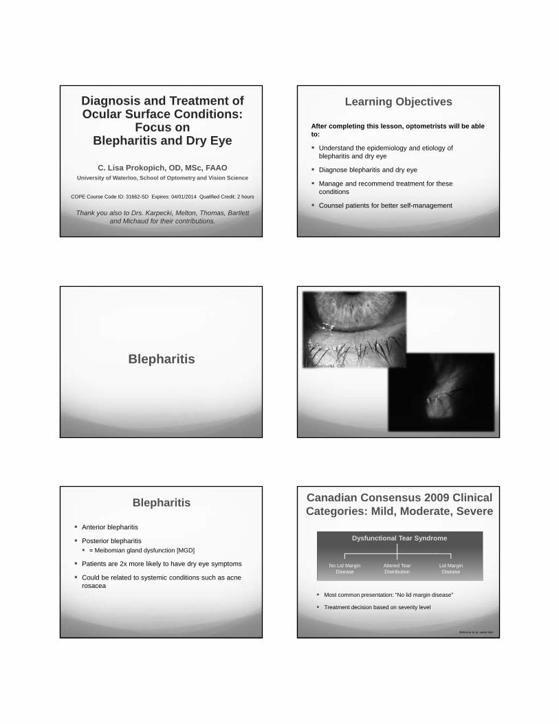
Blepharitis
Blepharitis
Anterior blepharitis
Posterior blepharitis= Meibomian gland dysfunction [MGD]
Patients are 2x more likely to have dry eye symptoms
Could be related to systemic conditions such as acne rosacea
Canadian Consensus 2009 Clinical Categories: Mild, Moderate, Severe
Most common presentation: “No lid margin disease”
Treatment decision based on severity level
Dysfunctional Tear Syndrome
Lid Margin Disease
Altered Tear Distribution
No Lid Margin Disease
Behrens et al, subm tted
Treatment Algorithm:Blepharitis (Anterior)
LEVEL 1 - Warm compresses (BID-QID then 2x / wk) + massage
- Lid scrub (to remove the crusts) with commercial eyelid cleaner or tea-tree oil (50%) if Demodex is present
IMP: educate the patient on the chronicity of the disease and the need of maintaining long term lid hygiene and treatment
- Topical antibiotics (if bacterial or parasitic) erythromycin, bacitracin, azithromycin,fusidic acid in gel, 1-4x/day x 2 wks then HS x 8 wks
- Order culture for severe cases or f resistance
If no improvement add level 2 treatments
If no improvement add level 3 treatments
LEVEL 3 - Steroids (ointment) - Can be prescribed in combo drugs (ant bio-steroids)- Dexamethasone with polymyxin B (Maxitrol®)- Dosage according to the severity of the case (BID to QID)- Monitor side effects - Metronidazole gel 0.2% with eyelid scrub - Off-label use to treat Demodex infestation
Lid Scrubs
Recommended productsLid-Care® (Novartis)I-Lid-N’ Lash® (I-MED Pharma)
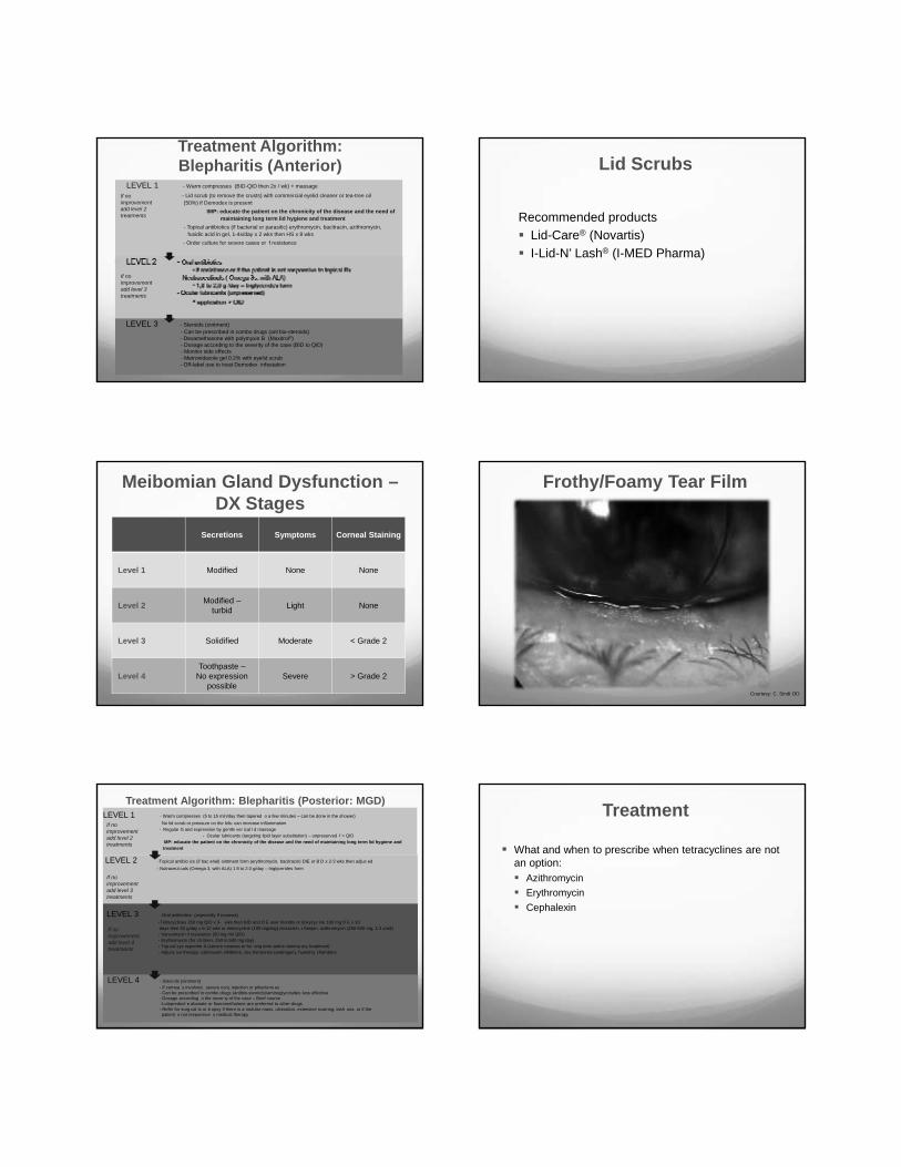
Meibomian Gland Dysfunction –DX Stages
Secretions Symptoms Corneal Staining
Level 1 Modified None None
Level 2 Modified –turbid Light None
Level 3 Solidified Moderate < Grade 2
Level 4Toothpaste –
No expression possible
Severe > Grade 2
Frothy/Foamy Tear Film
Courtesy: C. Sindt OD
Treatment Algorithm: Blepharitis (Posterior: MGD)LEVEL 1 - Warm compresses (5 to 15 min/day then tapered o a few minutes – can be done in the shower)
No lid scrub or pressure on the lids: can increase inflammation- Regular G and expression by gentle ver ical l d massage
- Ocular lubricants (targeting lipid layer substitution) – unpreserved f > QID MP: educate the patient on the chronicity of the disease and the need of maintaining long term lid hygiene and treatment
If no improvement add level 2 treatments
LEVEL 2 -Topical antibio ics (if bac erial) ointment form (erythromycin, bacitracin) DIE or B D x 2-3 wks then adjus ed- Nutraceut cals (Omega 3, with ALA) 1 8 to 2.0 g/day – triglycerides form
LEVEL 3 - Oral antibiotics (especially if rosacea) -Tetracyclines 250 mg QID x 3- wks then BID and D E over months or doxycyc ine 100 mg D E x 10days then 50 g/day x 6-12 wks or minocycline (100 mg/day),cloxacil in, r fampin, azithromycin (250-500 mg, 1-3 x/wk)- Vancomycin if resistance (50 mg /ml QID)- Erythromycin (for ch ldren, 250 to 500 mg day)- Top cal cyc osporine A (severe rosacea or for ong term anti-in lamma ory treatment)- Adjunc ive therapy: calcineurin inhibitors, sex hormones (androgen), humid ty chambers
LEVEL 4 - Stero ds (ointment) - If cornea s involved, severe conj. injection or phlyctenu es- Can be prescribed in combo drugs (antibio-steroids)/aminoglycosides less effective- Dosage according o the sever ty of the case – Brief course -Loteprednol e abonate or fluorometholone are preferred to other drugs - Refer for surg cal tx or b opsy if there is a nodular mass, ulceration, extensive scarring, lash oss, or if thepatient s not responsive o medical therapy
If no improvement add level 4 treatments
If no improvement add level 3 treatments
What and when to prescribe when tetracyclines are not an option:
AzithromycinErythromycinCephalexin
Treatment
Pregnant, nursing or child-bearing age
Children (pre-pubescent)
Contraindication:Oral Tetracycline
Photosensitivity
Chelates with dairy products, antacids etc.
Minocycline may cause vestibular toxicity
Pseudotumor cerebri
Number one drop-out reason?GI problems or esophagitis
Cautions
Tetracyclines
Use for rosacea/blepharitis
Antibiotics inhibit bacterialprotein synthesis by binding 30S ribosome
Anti-inflammatory propertiesDecreases L-1, TNF-αDecreases NO productionDecreases HLA Class II antigen expressionDecreases metalloproteinase production and activation
Decrease symptoms and joint destruction in RA
1. Do not take the second pill (b.i.d.) before going to bed
2. Do not take pills with acidic beverages
3. Take pills with food (except a high-dairy meal)
4. Prescribe the lowest dose available
How to Minimize Stomach Problems with Tetracycline
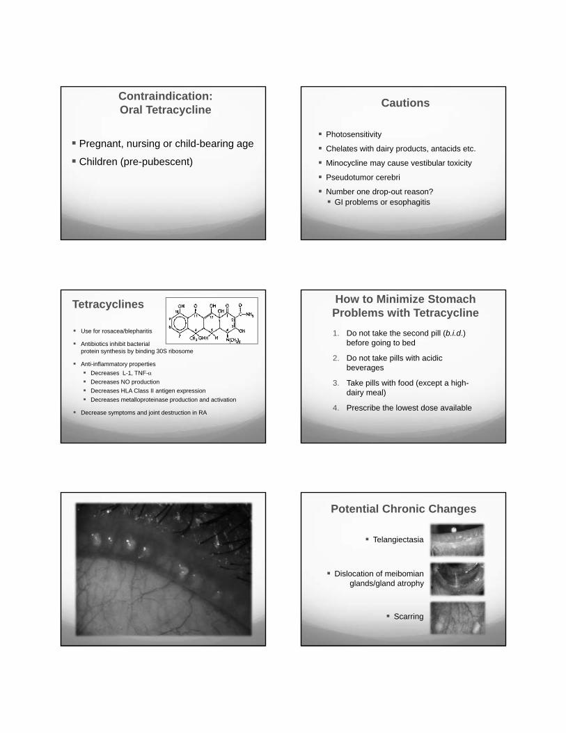
Potential Chronic Changes
Telangiectasia
Scarring
Dislocation of meibomianglands/gland atrophy
Educate patients about initial treatment and chronic treatments:
Lipid-based artificial tearsRefresh® UltraLiposic®
Systane® Balance
LubricationPatients with MGD may benefit most from a lipid-based tear
e.g., Liposic® , Refresh® Ultra, Systane®Balance
Patients with aqueous deficient dry eye may benefit most from aqueous ATs
e.g., Systane®, Refresh® Plus, GenTeal® or Blink®Tears
Patients with severe dry eye may benefit most from preservative-free artificial tears
e.g. preservative-free Blink® Tears or Gel or Refresh®
Tears PF, Tears Naturale FREE®, Bion® Tears, TheraTears®
Tear vs. Diagnosis
Liposic® Drops and Gel Liposic® Facts
To treat symptomatic dryness related to lipid layer deficiency
q.i.d. for most of the conditionsUse Celluvisc® (non-preserved) if >q.i.d.
Product ingredients – CMC(Carboxymethylcellulose)
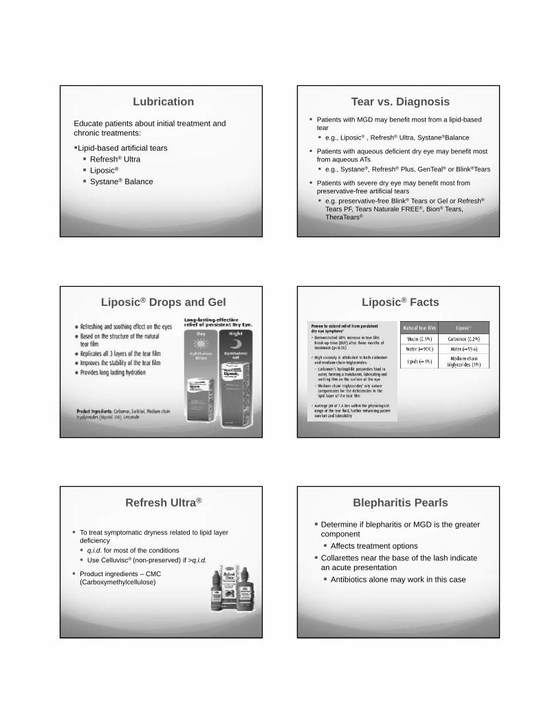
Refresh Ultra®
Determine if blepharitis or MGD is the greater component
Affects treatment optionsCollarettes near the base of the lash indicate an acute presentation
Antibiotics alone may work in this case
Blepharitis Pearls
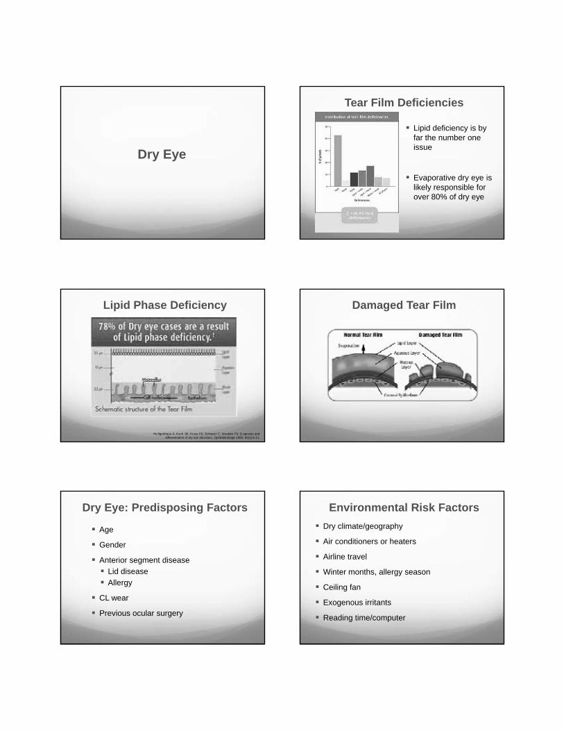
Dry Eye
Lipid deficiency is by far the number one issue
Evaporative dry eye is likely responsible for over 80% of dry eye
Tear Film Deficiencies
He ligenhaus A, Koch JM, Kruse FE, Schwarz C, Waubke TN. D agnosis and differentiation of dry eye disorders. Ophthalmologe 1995; 92(1):6-11.
Lipid Phase Deficiency Damaged Tear Film
Age
Gender
Anterior segment diseaseLid diseaseAllergy
CL wear
Previous ocular surgery
Dry Eye: Predisposing FactorsDry climate/geography
Air conditioners or heaters
Airline travel
Winter months, allergy season
Ceiling fan
Exogenous irritants
Reading time/computer
Environmental Risk Factors
Top 4 causesSmoking
Caffeine (more than moderate)
Diet
Alcohol
Systemic medications are also top intake causes of dry eye
Risk Factors: Behavioural
Antihistamines
Diuretics
Antihypertensives
Anticholinergics
Antidepressants
Cardiac antiarrhythmic
Oral contraceptives
Hormone replacement therapy
Isotretinoin e.g., Accutane®
Some chemotherapy drugs
Risk Factors: Systemic Medications
Diabetes (most common; cell damage, medications, metabolic issues)
Rheumatoid arthritisSjögren’s syndrome
Lupus (SLE)
Thyroid eye disease
Rosacea & psoriasis
Risk Factors: Systemic Disease
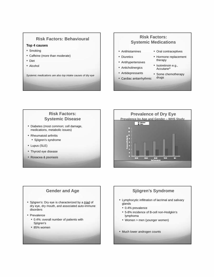
Prevalence of Dry EyePrevalence by Age and Gender – WHS Study
Sjögren’s: Dry eye is characterized by a triad of dry eye, dry mouth, and associated auto-immune disorders
Prevalence0.4%: overall number of patients with Sjögren’s85% women
Gender and Age
Lymphocytic infiltration of lacrimal and salivary glands
0.4% prevalence5-8% incidence of B-cell non-Hodgkin’s lymphomaWomen > men (younger women)
Much lower androgen counts
Sjögren’s Syndrome
Treat underlying immune disorderCo-manage with patient’s physician
SecretagoguesSalagen® 5 mg (Pilocarpine tablets)
Avoid in asthma patients, GI ulcer, acute iritis or narrow anglesMonitor carefully Side effects:
Scalp and back sweatsCevimeline e.g., Evoxac® 30 mg t.i.d.– saliva stimulating drug
Very effective with a lot less side effectsNot yet available in Canada
Sjögren’s Syndrome
Burning
Stinging
Transient blur
Dryness
Photophobia
Epiphora
Asthenopia
Symptoms of Dry Eye
Contact lens intolerence
Injection
Increased blink rate
Foreign body sensation
Grittiness
Transient blurred vision
Visual degradation
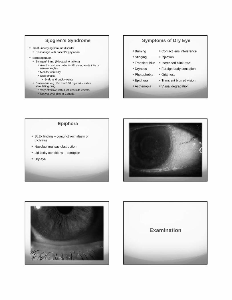
SLEx finding – conjunctivochalasis or trichiasis
Nasolacrimal sac obstruction
Lid laxity conditions – ectropion
Dry eye
Epiphora
Examination
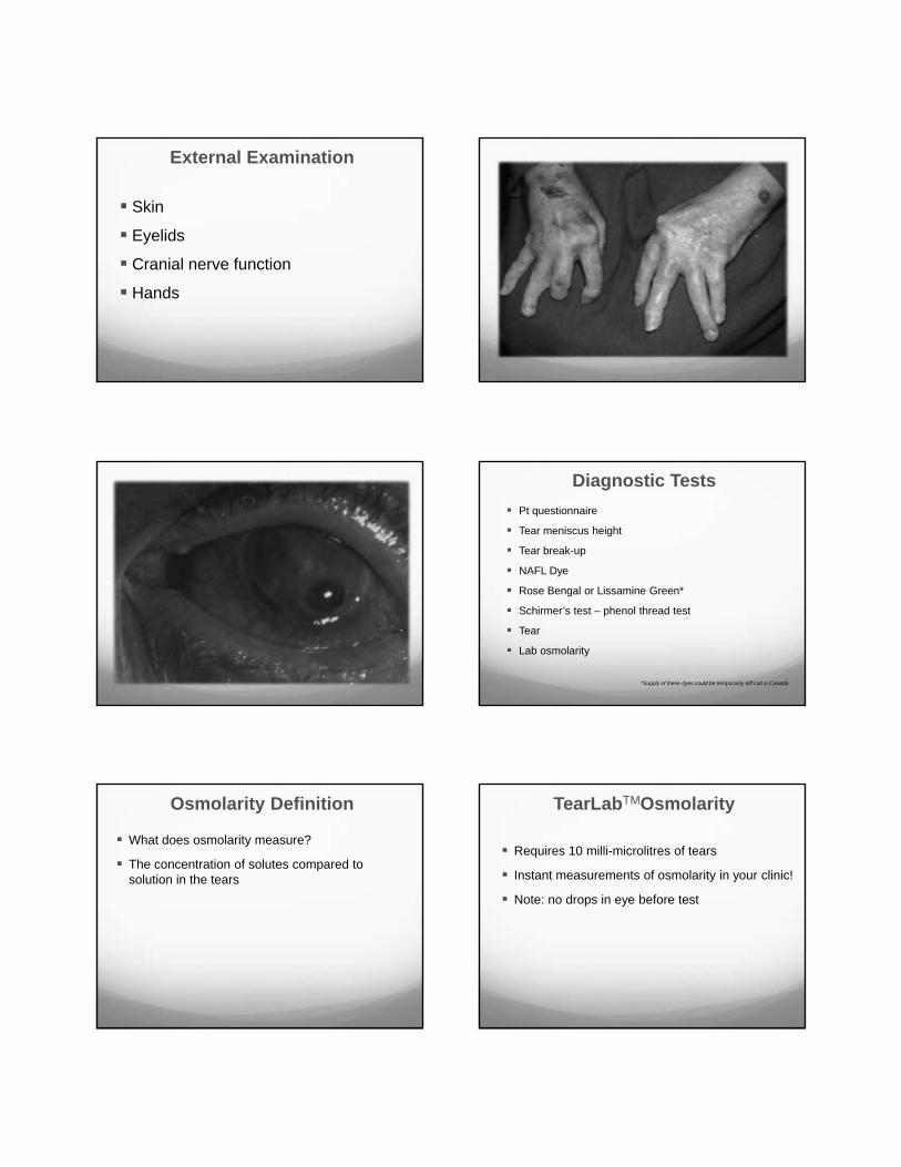
Skin
Eyelids
Cranial nerve function
Hands
External Examination
Pt questionnaire
Tear meniscus height
Tear break-up
NAFL Dye
Rose Bengal or Lissamine Green*
Schirmer’s test – phenol thread test
Tear
Lab osmolarity
*Supply of these dyes could be temporarily diff cult in Canada
Diagnostic Tests
What does osmolarity measure?
The concentration of solutes compared to solution in the tears
Osmolarity Definition
Requires 10 milli-microlitres of tears
Instant measurements of osmolarity in your clinic!
Note: no drops in eye before test
TearLabTMOsmolarity
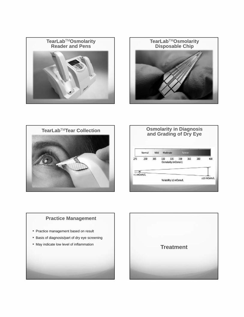
TearLabTMOsmolarityReader and Pens
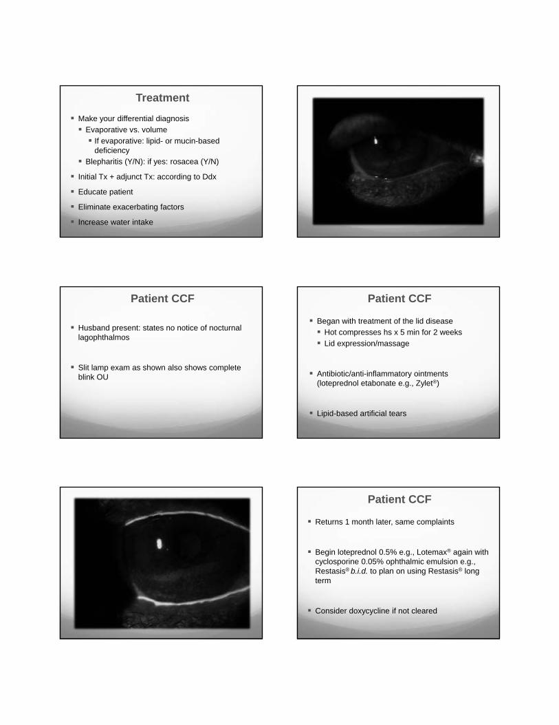
TearLabTMOsmolarityDisposable Chip
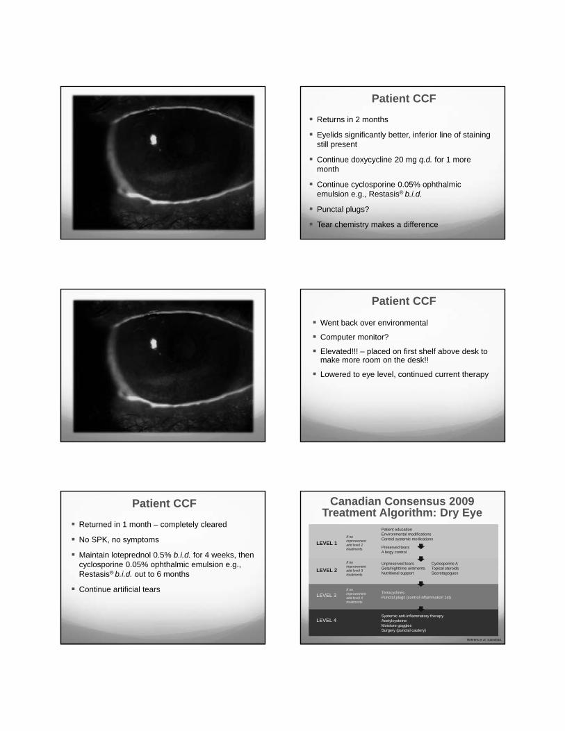
TearLabTMTear Collection Osmolarity in Diagnosisand Grading of Dry Eye
Practice management based on result
Basis of diagnosis/part of dry eye screening
May indicate low level of inflammation
Practice Management
Treatment
Make your differential diagnosis Evaporative vs. volume
If evaporative: lipid- or mucin-based deficiency
Blepharitis (Y/N): if yes: rosacea (Y/N)
Initial Tx + adjunct Tx: according to Ddx
Educate patient
Eliminate exacerbating factors
Increase water intake
Treatment
Husband present: states no notice of nocturnal lagophthalmos
Slit lamp exam as shown also shows complete blink OU
Patient CCF
Began with treatment of the lid diseaseHot compresses hs x 5 min for 2 weeksLid expression/massage
Antibiotic/anti-inflammatory ointments (loteprednol etabonate e.g., Zylet®)
Lipid-based artificial tears
Patient CCF
Returns 1 month later, same complaints
Begin loteprednol 0.5% e.g., Lotemax® again with cyclosporine 0.05% ophthalmic emulsion e.g., Restasis® b.i.d. to plan on using Restasis® long term
Consider doxycycline if not cleared
Patient CCF
Returns in 2 months
Eyelids significantly better, inferior line of staining still present
Continue doxycycline 20 mg q.d. for 1 more month
Continue cyclosporine 0.05% ophthalmic emulsion e.g., Restasis® b.i.d.
Punctal plugs?
Tear chemistry makes a difference
Patient CCF
Went back over environmental
Computer monitor?
Elevated!!! – placed on first shelf above desk to make more room on the desk!!
Lowered to eye level, continued current therapy
Patient CCF
Returned in 1 month – completely cleared
No SPK, no symptoms
Maintain loteprednol 0.5% b.i.d. for 4 weeks, then cyclosporine 0.05% ophthalmic emulsion e.g., Restasis® b.i.d. out to 6 months
Continue artificial tears
Patient CCF Canadian Consensus 2009 Treatment Algorithm: Dry Eye
Behrens et al, submitted.
LEVEL 1
LEVEL 2
LEVEL 3
LEVEL 4
Patient educationEnvironmental modificationsControl systemic medications
Preserved tearsA lergy control
Cyclosporine ATopical steroidsSecretagogues
Unpreserved tearsGels/nighttime ointmentsNutritional support
TetracyclinesPunctal plugs (control inflammation 1st)
Systemic anti-inflammatory therapyAcetylcysteineMoisture gogglesSurgery (punctal cautery)
If no improvement add level 2 treatments
If no improvement add level 3 treatments
If no improvement add level 4 treatments
Treatment – Artificial Tearsvs. Layer Deficiency
Omega fatty acids shown to help with dry eye disease:
ALA: e.g., flaxseed oil EPA/DHA: e.g., fish oilsGLA: e.g., black currant seed or evening primrose oil
Nutritional Supplements: Essential Fatty Acids
Omegas • Defined by the number of carbons, double links and their respective position
– Ex: LA 18:3 ω-3– Linoleic acid: composed
with 18 carbons, with 3 double links
– The first one being at 3 carbons from the end (this defines an omega 3)
• Long or short chain– (SC): considered
essentials – cannot be metabolised
– Long chain (LC) – can be metabolised from short chains
How an Omega Works… ω-3 and ω-6 = eicosinoids precursors
Hormones with a local action against inflammation 4 families:
Prostaglandins (PG)Prostacyclins (PGIs)Thromboxanes (TXs) Leukotrienes (LTs)
Omegas can influence globaland local health through theirinfluence on these hormones’production
Antagonist Actions(2)
ω-3 vs ω-6Compete each other for the same enzymes that break them downAll omegas have some pro-inflammatory proprieties and all can be anti-inflammatory. However, overall action is:
ω-3: an i-inflammatory. Action on T cell and interleukins. ω-6: pro-inflammatory and anti-platelet aggregationHowever, primrose oil (GLA) posi ively influences DGLA rate and acts against ocular dryness
Table of ωFatty acids (Name) Type Code
Omega-3
Alpha-linolenic acid ALA SC-PUFA 18:3 ω-3
Eicosapentaenoic acid EPA LC-PUFA 20:5 ω-3
Docosahexaenoic acid DHA LC-PUFA 22:6 ω-3
Omega-6
Linoleic acid LA SC-PUFA18:2 ω-6
Gamma-linolenic acid GLA LC-PUFA18:3 ω-6
Dihomo-gamma-linolenic acid DGLA LC-PUFA20:3 ω-6
Arachidonic acid AA LC-PUFA20:4 ω-6
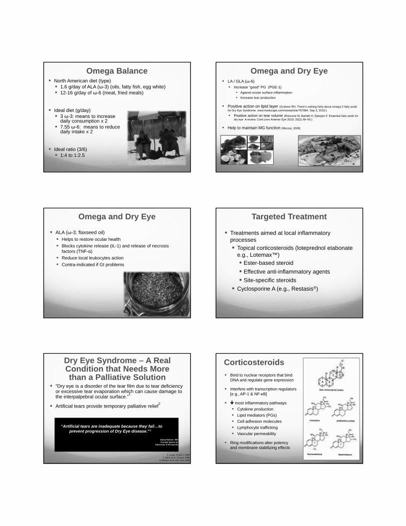
Omega BalanceNorth American diet (type)
1.6 g/day of ALA (ω-3) (oils, fatty fish, egg white)12-16 g/day of ω-6 (meat, fried meals)
Ideal diet (g/day)3 ω-3: means to increasedaily consumption x 2 7.55 ω-6: means to reducedaily intake x 2
Ideal ratio (3/6)1:4 to 1:2.5
LA / GLA (ω-6) Increase “good” PG (PGE-1)
Against ocular surface inflammationIncrease tear production
Positive action on lipid layer (Graham RH. There’s nothing fishy about omega-3 fatty acids for Dry Eye Syndrome. www.medscape.com/viewarticle/707984. Sep 3, 2010.)
Positive action on tear volume (Roncone M, Bartlett H, Eperjesi F. Essential fatty acids for dry eye A review. Cont Lens Anterior Eye 2010; 33(2) 49–54.)
Help to maintain MG function (Macsai, 2008)
Omega and Dry Eye
ALA (ω-3; flaxseed oil) Helps to restore ocular health Blocks cytokine release (IL-1) and release of necrosis factors (TNF-α)Reduce local leukocytes actionContra-indicated if GI problems
Omega and Dry Eye
Treatments aimed at local inflammatory processes
Topical corticosteroids (loteprednol etabonatee.g., Lotemax™)
Ester-based steroidEffective anti-inflammatory agentsSite-specific steroids
Cyclosporine A (e.g., Restasis®)
Targeted Treatment
Dry Eye Syndrome – A Real Condition that Needs More than a Palliative Solution
“Dry eye is a disorder of the tear film due to tear deficiency or excessive tear evaporation which can cause damage to the interpalpebral ocular surface.”1
Artificial tears provide temporary palliative relief2
“Artificial tears are inadequate because they fail…to prevent progression of Dry Eye disease.”3
Daniel Nelson MDCorneal Specia ist
University of M nnesota
1. Lemp. CLAO J 1995.2. Stern et al. Cornea 1998.
3. Nelson et al. Adv Ther 2000
CorticosteroidsBind to nuclear receptors that bindDNA and regulate gene expression
Interfere with transcription regulators [e g., AP-1 & NF-κB]
most inflammatory pathwaysCytokine productionLipid mediators (PGs)Cell adhesion moleculesLymphocyte traffickingVascular permeability
Ring modifications alter potency and membrane stabilizing effects
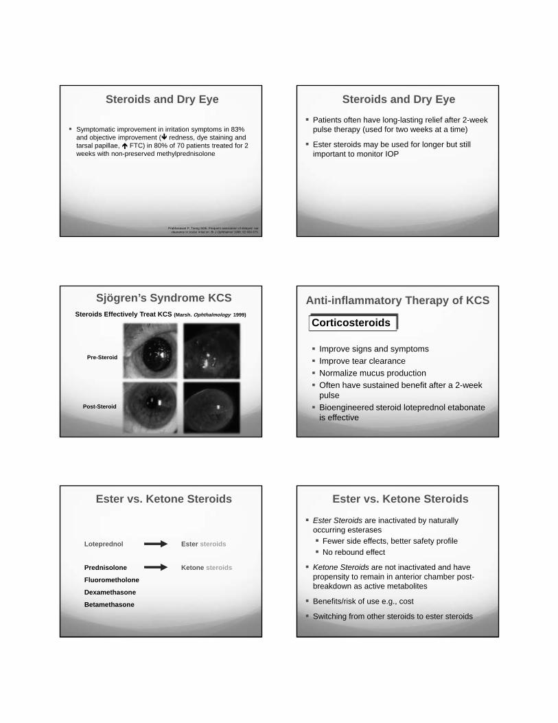
Steroids and Dry Eye
Symptomatic improvement in irritation symptoms in 83% and objective improvement ( redness, dye staining and tarsal papillae, FTC) in 80% of 70 patients treated for 2 weeks with non-preserved methylprednisolone
Prabhasawat P, Tseng SGE. Frequent association of delayed ear clearance in ocular irritat on. Br J Ophthalmol 1998; 82:666-675.
Patients often have long-lasting relief after 2-week pulse therapy (used for two weeks at a time)
Ester steroids may be used for longer but still important to monitor IOP
Steroids and Dry Eye
Pre-Steroid
Post-Steroid
Steroids Effectively Treat KCS (Marsh. Ophthalmology 1999)
Sjögren’s Syndrome KCS
Corticosteroids
Improve signs and symptoms Improve tear clearanceNormalize mucus productionOften have sustained benefit after a 2-week pulseBioengineered steroid loteprednol etabonateis effective
Anti-inflammatory Therapy of KCS
Ester vs. Ketone Steroids
Loteprednol Ester steroids
Prednisolone Ketone steroids
Fluorometholone
Dexamethasone
Betamethasone
Ester Steroids are inactivated by naturally occurring esterases
Fewer side effects, better safety profileNo rebound effect
Ketone Steroids are not inactivated and have propensity to remain in anterior chamber post-breakdown as active metabolites
Benefits/risk of use e.g., cost
Switching from other steroids to ester steroids
Ester vs. Ketone Steroids
Loteprednol 0.2% (Alrex®); 0.5% (Lotemax™)
Fewer side effects (M Abelson, 88 patients 35 days)IOP rise, secondary infection or PSC formation: 0%No reported cases of PCS cataract in over 66 million prescriptions (IMS Health Data)Almost no systemic absorption – so no need to taper
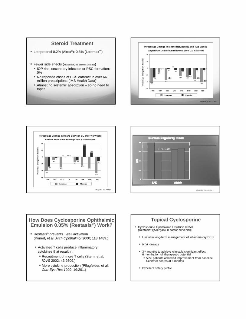
Steroid Treatment
Pflugfelder, et al. AJO 200 .
Percentage Change in Means Between BL and Two WeeksSubjects with Conjunctival Hyperemia Score ≥ 2 at Baseline
42 5
12 8
35 1
0 0
24 0 25 0
32 1
41 0
27 3
0 73 2
6 3 7 7
20 0
4 8 4 8
60
40
20
0
20
40
SUB OBJ CSS LMI ITH BCH NBCH RED
Lotemax Placebo
Perc
enta
ge C
hang
e Fr
om B
asel
ine
Percentage Change in Means Between BL and Two WeeksSubjects with Corneal Staining Score ≥ 10 at Baseline
41 8
9 7
30 6
0 0
15 8
25 0
31 8
47 3
28 4
2 8
0 0 0 0
10 0
33 3
12 5
11 7
60
40
20
0
20
40
SUB OBJ CCS LMI ITH IBH NBH RED
Lotemax Placebo
Perc
enta
ge C
hang
e Fr
om B
asel
ine
Pflugfe der, et al. AJO 200 .
Pflugfelder, et al. AJO 200 .
P < 0.04
How Does Cyclosporine Ophthalmic Emulsion 0.05% (Restasis®) Work?
Restasis® prevents T-cell activation(Kunert, et al. Arch Ophthalmol 2000; 118:1489.)
Activated T cells produce inflammatory cytokines that result in:
Recruitment of more T cells (Stern, et al. IOVS 2002; 43:2609.)More cytokine production (Pflugfelder, et al. Curr Eye Res 1999; 19:201.)
Topical CyclosporineCyclosporine Ophthalmic Emulsion 0.05% (Restasis®)(Allergan) in castor oil vehicle
Useful in long-term management of inflammatory DES
b.i.d. dosage
3-4 months to achieve clinically significant effect,6 months for full therapeutic potential
59% patients achieved improvement from baseline Schirmer scores at 6 months
Excellent safety profile
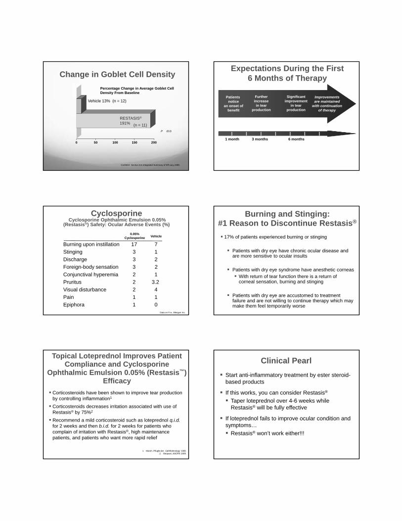
Change in Goblet Cell Density
0 50 100 150 200
Percentage Change in Average Goblet Cell Density From Baseline
Vehicle 13% (n = 12)
P .013
RESTASIS®
191% (n = 11)
CsANDA: Section 8.6 Integrated Summary of Eff cacy,1999.
Expectations During the First 6 Months of Therapy
Patients notice
an onset ofbenefit
Furtherincreasein tear
production
Significantimprovement
in tearproduction
Improvementsare maintained
with continuationof therapy
1 month 3 months 6 months
CyclosporineCyclosporine Ophthalmic Emulsion 0.05%
(Restasis®) Safety: Ocular Adverse Events (%)
0.05%Cyclosporine Vehicle
Data on Fi e, Allergan Inc.
Burning upon instillation 17 7Stinging 3 1Discharge 3 2Foreign-body sensation 3 2Conjunctival hyperemia 2 1Pruritus 2 3.2Visual disturbance 2 4Pain 1 1Epiphora 1 0
Burning and Stinging:#1 Reason to Discontinue Restasis®
17% of patients experienced burning or stinging
Patients with dry eye have chronic ocular disease and are more sensitive to ocular insults
Patients with dry eye syndrome have anesthetic corneasWith return of tear function there is a return of corneal sensation, burning and stinging
Patients with dry eye are accustomed to treatment failure and are not willing to continue therapy which may make them feel temporarily worse
Topical Loteprednol Improves Patient Compliance and Cyclosporine
Ophthalmic Emulsion 0.05% (Restasis™) Efficacy
Corticosteroids have been shown to improve tear production by controlling inflammation1
Corticosteroids decreases irritation associated with use of Restasis® by 75%2
Recommend a mild corticosteroid such as loteprednol q.i.d.for 2 weeks and then b.i.d. for 2 weeks for patients who complain of irritation with Restasis®, high maintenance patients, and patients who want more rapid relief
1. Marsh, Pflugfe der. Ophthalmology 1999.2. Shepard, ASCRS 2005.
Clinical PearlStart anti-inflammatory treatment by ester steroid-based products
If this works, you can consider Restasis®
Taper loteprednol over 4-6 weeks while Restasis® will be fully effective
If loteprednol fails to improve ocular condition and symptoms…
Restasis® won’t work either!!!
Established Safety Profile
Favourable safety profile for Cyclosporine Ophthalmic Emulsion 0.05% (Restasis®)
Safety parameters monitored:Adverse eventsBlood chemistryIntraocular pressure (IOP) Visual acuityBiomicroscopyConjunctival microbiologyCyclosporine blood levels
Please see slides 6 & 7 for important safety information.Small et al. J Ocul PharmTher. 2002.
No Cyclosporine in BloodNo detectable cyclosporine in blood of any cyclosporine ophthalmic emulsion 0.05% (Restasis®)-treated patient
Toxicity associated with systemic or oral cyclosporine was not observed with cyclosporine 0.05% ophthalmic emulsion
Please see slides 6 & 7 for important safety information.
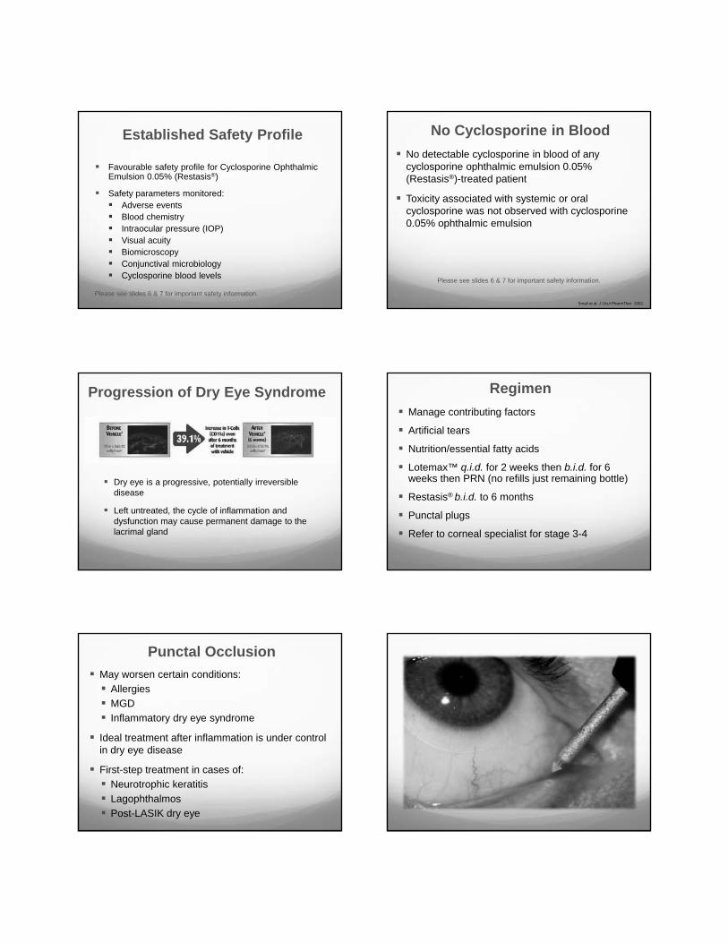
Progression of Dry Eye Syndrome
Dry eye is a progressive, potentially irreversible disease
Left untreated, the cycle of inflammation and dysfunction may cause permanent damage to the lacrimal gland
RegimenManage contributing factors
Artificial tears
Nutrition/essential fatty acids
Lotemax™ q.i.d. for 2 weeks then b.i.d. for 6 weeks then PRN (no refills just remaining bottle)
Restasis® b.i.d. to 6 months
Punctal plugs
Refer to corneal specialist for stage 3-4
Punctal OcclusionMay worsen certain conditions:
AllergiesMGDInflammatory dry eye syndrome
Ideal treatment after inflammation is under control in dry eye disease
First-step treatment in cases of:Neurotrophic keratitisLagophthalmosPost-LASIK dry eye
5/8/2012
18
Options for Treatment Failures
Autologous serum
Surgery
Dry Eye Pearls
Signs and symptoms rarely correlate in dry eye Treat either signs or symptoms
If a systemic disease such as an auto-immune condition is noted in a dry eye patient:
The dry eye will not fully resolve without management of the systemic disease
Sjögren’s SyndromeDiscuss dry mouth treatments as wellNote the high association with non-Hodgkin’s lymphoma and educate the patientFollow the patient more often
Dry Eye PearlsContact lens wear in dry eye patients?
Consider:Daily disposablesCertain SiHy lenses on a daily wear basisIncrease use of ATs or re-wetting dropsUse therapeutics before and after lens insertionChange contact lens solutions
Preservative-free, H2O2Biotrue™ with sodium hyaluronan
Dry Eye Pearls
Ocular Surface Disease: Conclusions
Ocular surface disease conditions are the most common optometry will faceNumerous new management options that are more effective Build a practice where patients will return for all primary eye care needs